Article

Isolation and Characterization of Bacterial Endophytes from Small Nodules of Field-Grown Peanut

by
Md Shakhawat Hossain
1,2,*,†,
Christine Frith
3,
Siddhartha Shankar Bhattacharyya
1,2,
Paul B. DeLaune
4 and
Terry J. Gentry
1,2
1
Department of Soil and Crop Sciences, Texas A&M University, College Station, TX 77843, USA
2
Texas A&M AgriLife Research, College Station, TX 77843, USA
3
Department of Geosciences, Texas A&M University, College Station, TX 77843, USA
4
Texas A&M AgriLife Research, Vernon, TX 76384, USA
*
Author to whom correspondence should be addressed.
Current address: Division of Research, Innovation and Economic Development, Tarleton State University, Stephenville, TX 76402, USA.
Microorganisms 2023, 11(8), 1941; https://doi.org/10.3390/microorganisms11081941
Submission received: 5 July 2023 / Revised: 25 July 2023 / Accepted: 26 July 2023 / Published: 29 July 2023

Abstract

:
It is evident that legume root nodules can accommodate rhizobial and non-rhizobial bacterial endophytes. Our recent nodule microbiome study in peanuts described that small nodules can harbor diverse bacterial endophytes. To understand their functional role, we isolated 87 indigenous endophytes from small nodules of field-grown peanut roots and characterized them at molecular, biochemical, and physiological levels. The amplified 16S rRNA genes and phylogenetic analysis of these isolates revealed a wide variety of microorganisms related to the genera Bacillus, Burkholderia, Enterobacter, Herbaspirillum, Mistsuaria, Pantoea, Pseudomonas, and Rhizobia. It was observed that 37% (100% identity) and 56% (>99% identity) of the isolates matched with the amplified sequence variants (ASVs) from our previous microbiome study. All of these isolates were tested for stress tolerance (high temperature, salinity, acidic pH) and phosphate (P) solubilization along with ammonia (NH3), indole-3-acetic acid (IAA), 1-aminocyclopropane-1-carboxylate deaminase (ACCD), and siderophore production. The majority (78%) of the isolates were found to be halotolerant, thermotolerant, and acidophilic, and a few of them showed a significant positive response to the production of IAA, NH3, siderophore, ACCD, and P-solubilization. To evaluate the plant growth promotion (PGP) activity, plant and nodulation assays were performed in the growth chamber conditions for the selected isolates from both the non-rhizobial and rhizobial groups. However, these isolates appeared to be non-nodulating in the tested conditions. Nonetheless, the isolates 2 (Pantoea), 17 (Burkholderia), 21 (Herbaspirillum), 33o (Pseudomonas), and 77 (Rhizobium sp.) showed significant PGP activity in terms of biomass production. Our findings indicate that these isolates have potential for future biotechnological applications through the development of biologicals for sustainable crop improvement.

1. Introduction

Legume nodules are specialized structures that form on the roots of leguminous plants via the symbiotic relationship with nitrogen-fixing soil bacteria called rhizobia. The main function of these nodules is to facilitate the conversion of atmospheric nitrogen gas (N2) into plant-available forms such as ammonium (NH4+) or nitrate (NO3) through a process known as biological nitrogen fixation (BNF) [1]. Traditionally, it was believed that legume nodules exclusively housed Rhizobium/Bradyrhizobium as a host symbiont, but recent studies have shown that other non-rhizobial bacterial endophytes (NRBEs) coexist with rhizobial strains [2,3,4]. NRBEs can enter host plants by colonizing the root’s inner cells/tissues or by exploiting discontinuities on the plant’s surface. Once inside, they establish a symbiotic relationship with the host and receive a protected and nutrient-rich environment in exchange for improving the plant’s health [3,4]. The coexistence of rhizobia and NRBEs in legume nodules highlights the complexity of the nodule microbiome and the interactions that occur within this environment.
Both rhizobia and NRBEs from nodules have been isolated from various legume species including wild legumes, fenugreek, lupine, groundnut, peanut, etc. [4,5,6,7,8,9]. Several genera of NRBEs other than rhizobia were isolated including Agrobacterium, Bacillus, Enterobacter, Paenibacillus, Erwinia, Aerobacter, Herbaspirillum, Klebsiella, Pantoea, and Pseudomonas [10,11]. However, the functional roles of NRBEs in legumes have not been fully explored [12].
Previous studies have shown that NRBEs can directly or indirectly influence PGP activity by producing indole-3-acetic acid (IAA), siderophores, 1-aminocyclopropane-1-carboxylate deaminase (ACCD), and P-solubilization [5,12]. Tryptophan, an amino acid, serves as a precursor molecule for IAA biosynthesis [13], and IAA is an essential phytohormone for plant growth and development including cell elongation, root formation, etc. Some IAA-producing NRBEs include Enterobacter spp. and Pseudomonas spp. [13], which can improve the seed germination, shoot growth, and root architecture in legumes [14]. Siderophores (molecules) attach to available forms of iron (e.g., Fe3+), allowing for an increased solubilization and accessibility of Fe [15,16]. In addition, the release of the ACCD enzyme leads to the cleavage of ACC, which is a precursor of ethylene, resulting in the production of ammonia (NH3) and α-ketobutyrate. This process reduces the levels of ethylene in plants and enhances their resistance to environmental stress [17].
Again, NRBEs can facilitate nutrient acquisition (e.g., P-solubilization) by producing organic acids [18]. Ferchichi et al. [6] observed that the efficiency of P-solubilization by bacterial endophytes obtained from lupine nodules varied. Among these, Pseudomonas brenneri exhibited the highest P-solubilization efficiency, followed by Bacillus subtillis and Burkholderia gramini, respectively. Recent studies elucidated that some NRBEs are capable of inducing stress tolerance, and some halotolerant NRBEs including Brachybacterium nov., Zhihengliuella sp., Brevibacteriumcasei, Halomonas sp., Vibrio sp., and Pseudomonas spp., have been isolated [19]. In addition, some halotolerant NRBEs from cowpea nodules, especially Pseudomonas Fluorescens, Bacillus endophyticus, Bacillus pumilus, and Paenibacillus polymyxa have shown potential for stimulating plant growth and development [20]. Ruiz-Díez et al. [21] postulated that NRBEs (e.g., Phylobacterium myrsinacearum), having the plant growth promoting ability, can withstand acidic pH. Currently, there is a dearth of knowledge on the presence of bacterial endophytes in the small nodules of peanuts and their potential ability to facilitate plant growth promotion and tolerate biotic and abiotic stresses.
Peanut (Arachis hypogaea L.) is an indigenous South American legume [22] and is widely grown in more than 100 countries [23]. Peanuts can form both effective and ineffective nodules with distinct sizes [24,25]. Large nodules (pink color, >1.5 mm) are effective for BNF, while small nodules (white color, <1.5 mm) are ineffective or exhibit lower nitrogen-fixing activity compared to medium-sized to large nodules [24]. Previously, the amplicon-based (16s rRNA) approach was used to study the peanut rhizosphere and bulk soil microbiome [26,27,28]. However, in our recent study on the peanut nodules’ microbiome [25], we showed that the small nodules (<1.5 mm) had approximately 31% of diverse sets of bacterial endophytes compared to the big nodules. But the function of these diverse NRBEs in the small nodules remains largely unexplored. To date, and to the best of our knowledge, no studies have been conducted to explore NRBEs from the small nodules of peanut roots specifically. Hence, in this study, the main objectives are to isolate and characterize mostly NRBEs together with a few examples of rhizobial isolates from small nodules of peanut roots and evaluate their role in PGP activity for biomass production.

2. Materials and Methods

2.1. Isolation of Bacterial Isolates from Small Nodules of Peanut Roots

To isolate bacterial endophytes, small nodules (<1.5 mm) from field-grown peanut roots (cultivated at the Texas A&M AgriLife Research and Extension Center, Vernon) were used, which were harvested during our previous study [25]. Twenty-four small nodules (four from each plant) from peanut roots were collected and cleaned with water to remove any dirt or foreign particles attached to the nodules. Cleaned nodules were then surface sterilized using 70% ethanol for 1.5 min, washed with sterile water (H2O) three times, and then soaked in 10% commercial bleach for 5 min. After this, nodules were washed at least 5–6 times with sterile H2O to rinse off any trace element of bleach. Then, washed nodules were crushed in 1 mL of 1× sterilized phosphate-buffered saline, and the resulting suspension was streaked on yeast extract mannitol agar (YMA, catalog number M715; https://www.himedialabs.com/us/ (accessed on 27 August 2021)) plates with multiple dilution series. We also streaked 100 µL of the final washed solution from the above to check if any contamination occurred during the sterilization process. The streaked YMA agar plates were incubated at 28 °C for 3 to 5 days, and the selected isolates (pure colonies) were grown in liquid YMA and later archived in glycerol stock.

2.2. Identification and Molecular Characterization of Bacterial Isolates

The genomic DNA was extracted from all the selected isolates (87) using the Promega genomic isolation kit (https://www.promega.com/ (accessed on 15 July 2022)). The 16S rRNA gene was amplified from these isolates using 27F and 1492R primer sets [29] under the following PCR conditions: 1 cycle of 95 °C for 5 min (preheating), 25 cycles of 95 °C for 30 s, 55 °C for 30 s, 72 °C for 1 min, and a final extension period at 72 °C for 5 min. The amplified PCR products (5 µL) were loaded into 2% agarose gel to check the fragment size and then purified from the PCR products using the Promega PCR cleanup kit. The resulting purified PCR products were Sanger sequenced by Eton Bioscience Inc. (https://etonbio.com (accessed on 11 August 2022).
The 16S rRNA gene sequences of the isolates were compared with the 16S rRNA small subunit (SSA) using the SILVA database ACT (alignment, classification, and tree services) tools [30]. The annotated neighbor sequences were downloaded from the SILVA database and multiple sequence alignment was performed together with the peanut small nodule isolate sequences using the MAFFT version 7 program [31]. For phylogenetic relationship analysis, the aligned Clustal data were opened in ClustalX and the Neighbor Joining (NJ) tree was constructed using the Jukes–Cantor model with bootstrap resampling = 1000 and visualized in FigTree. For the identification of isolates matched with peanut small nodule ASVs [25], the NCBI two-way blast was performed.

2.3. Biochemical Analysis of Isolates for Growth-Promoting Traits

2.3.1. Ammonia (NH3) Production Assay

NH3 production assay was determined using the method described by Singh et al. [32] with some modifications. All the isolates were freshly grown in peptone water and incubated at 28 ± 2 °C for 48 to 72 h. After incubation, 0.5 mL of Nessler’s reagent was added to 4 mL of freshly cultured isolates in peptone water and incubated in the dark at room temperature for 15 to 30 min for color changes. An absorbance (450 nm) was then measured using a UV spectrophotometer. The NH3 production was calculated from three independent replications for each isolate and based on a standard curve (0–0.5 mg/mL) ammonium sulfate range (Figure S1A).

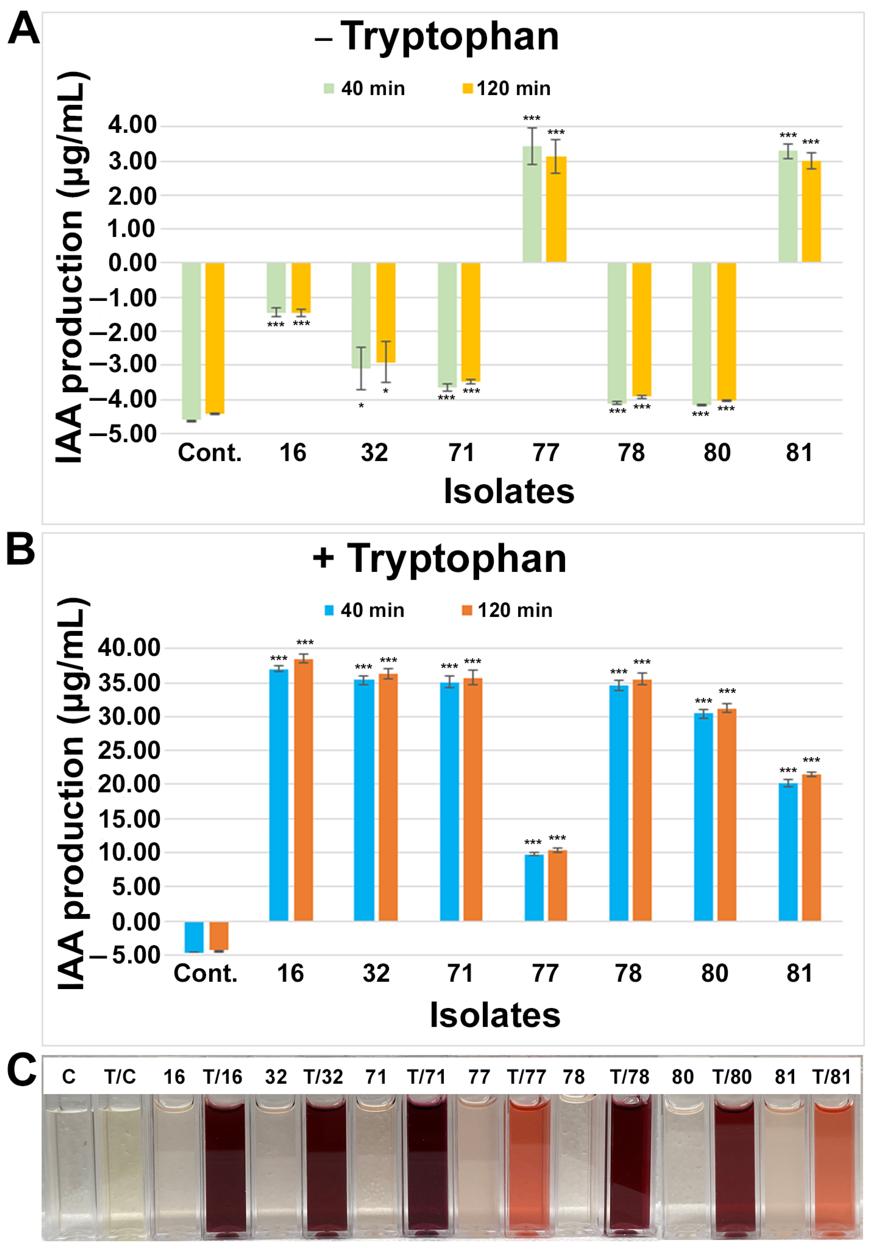
2.3.2. Indole-3-Acetic Acid (IAA) Production Assay

To determine the amounts of IAA produced by each isolate, a colorimetric assay was performed with Van Urk Salkowski reagent using the method described by B. Mohite [33] with some modifications. All the isolates were freshly grown in YMA liquid medium and incubated at 28 °C for 4 days. After incubation, the culture broth was centrifuged, and equal volumes of isolate supernatant and Salkowski reagent (2% 0.5 M FeCl3 in 35% HClO4 solution) were added and incubated in the dark at room temperature. The absorbance (530 nm) was measured at 30 min and 120 min. The IAA amount was estimated from three independent replications for each isolate and compared with (5 mM) or without tryptophan added to the YMA medium. The IAA production was calculated using methods described by Gordon and Weber [34] and based on an IAA standard curve (0–30 µg/mL) concentration range (Figure S1B).

2.3.3. P-Solubilization Assay (Agar Plate Method)

All the isolates were tested for P-solubilization using YMA and Pikovskaya’s (PVK) [35] agar plates containing known amounts of dipotassium phosphate (K2HPO4) and tricalcium phosphate (Ca3(PO4)2). Independent isolates were spotted four times on each plate, representing four replications, and incubated at 37 °C for 72 h. P-solubilization was observed for the development of clear zones surrounding the bacterial colony [36] and the P-solubilization index (PSI) was calculated according to the method by Pande et al. [37].

2.3.4. Siderophore Production Assay

All the isolates were tested for siderophore production using the method described by Virpiranta et al. [38] with some modifications. Isolates were cultured in 3 mL fructose–glucose medium [38] and incubated at 28 °C for 72 h. After incubation, bacterial cultures were centrifuged, and equal volumes of bacterial supernatant and liquid Fe-CAS media [38] were mixed and then incubated in the dark at room temperature for 20 min. The absorbance (620 nm) was measured using a UV spectrophotometer, and siderophores produced by the isolates were calculated as percent siderophore units (PSUs) based on the formula used by Virpiranta et al. [38] from three independent replications for each isolate.

2.3.5. ACCD Activity (Plate Assay)

All the isolates were screened for ACCD activity using the method described by Maheshwari et al. [39] with modified ACC medium containing 0.005% Bromothymol Blue and 0.008% Phenol Red. The isolates were cultured in YMA liquid medium at 28 °C for 48 h and then the bacterial pellets were washed twice with sterilized 0.1 M Tris-HCL (pH 7.5) via centrifugation. After washing, the bacterial cell pellets were spotted on modified Dworkin and Foster (DF) medium [40] with or without nitrogen (NH4SO4), and ACC (3 mM) was used only for the sole nitrogen source. Four independent spots were streaked on these plates for each isolate and incubated at 30 °C for 48 h to observe the color changes. ACCD catalysis was detected by color changes in the medium (due to change in pH), which catalyzes ACC into NH3 and ⍺-ketobutyrate.

2.3.6. Tolerance of Isolates to High Temperature, NaCl, and Low pH

All the isolates were tested for growth at high temperature, elevated NaCl, and low pH on YMA agar plates. Freshly grown isolates were spotted four times as replications for a single isolate on YMA agar plates and incubated at 28 °C for 48 h for low pH (4, 5, 7) and 72 h for various NaCl concentrations (0, 0.15, 0.3, and 0.6 M). For high temperature, isolates were incubated at 37 °C, 40 °C, and 45 °C for 72 h. Bacterial growth was measured based on visible changes in growth and colony size on YMA agar plates. For example, when a colony became smaller in size by the effects of higher or lower dose/concentrations of above conditions, this was considered as slower or decreased growth, and no colony growth on the plate was counted as no growth.

2.4. Evaluation of Plant Growth Promotion (PGP) Ability with the Selected Endophytes

Peanut seeds var. Tamnut 74 (provided by Dr. John Cason, Texas AgriLife Research, Stephenville, TX, USA) were surface sterilized using 70% EtOH for 2 min and were rinsed with sterilized H2O three times before being immersed with 10% commercial bleach solution for 8 min. This was followed by 6–7 vigorous washes with sterilized water to rinse off any trace elements of bleach. Four to five seeds were then germinated in plastic pots containing sterilized coarse vermiculite and perlite (PVP industries, North Bloomfield, OH, USA) mixture (3:1 w/w) and grown on growth racks at 25–26 °C with 16 h light and 8 h dark conditions. The following three treatments were used: (1) negative control without any nitrogen source; (2) positive control supplemented with 0.5 mM NH4NO3; and (3) inoculation with isolates + 0.5 mM NH4NO3. The N source was applied to the plants once a week. The selected isolates were freshly cultured at 28 °C for 48 h, centrifuged, and washed with sterile water before inoculation. The germinated peanut seedlings at the base of tap roots were inoculated with 3–4 mL of freshly prepared inoculum for each pot (concentration adjusted to 0.2, OD 600 nm), and seven to twelve total plants from 2–3 pots were used for data analysis and served as replications for each isolate.

3. Results

3.1. Isolation of Bacterial Endophytes from Small Nodules of Peanut Roots

The bacterial endophytes were isolated from a mix of small nodules of peanut roots. A total of 87 isolates were randomly selected based on the morphology, appearance, shape, diameter, and color of the colonies on the YMA agar plates. No isolates were grown or observed on the YMA agar plates when the sterilized water from the final wash of the nodule surface was used as an inoculum. The isolates were given an ID name (numerical order) for the subsequent molecular identification, biochemical characterization, and plant growth promotion assay.

3.2. Molecular Identification of Small Nodule Endophytes of Peanut Roots

The 16S rRNA gene sequences from all the isolates were analyzed against the 16S rRNA small subunit (SSA) using ACT tools from the SILVA database (see Section 2), which provides sequence alignment, taxonomic classification, and phylogenetic tree services. Based on the percentage of identity in the ACT analysis, all 87 isolates were characterized at the family and genus levels (Figure S2, Table S1). To further understand and identify the sequence similarity of these isolates with previously published microbiome sequences of peanut nodules [25], an NCBI two-way blast analysis was used and it was found that 37% (n = 32) and 56% (n = 49) of isolates were 100% and >99% identical with the ASVs, respectively (Table 1 and Table S1).
The phylogenetic analysis using all the isolates with reference neighboring sequences from the SILVA database shows that the peanut small nodule endophytes were grouped among several branches and nodes with the genera Enterobacter and Pantoea, Pseudomonas, Herbaspirillum and Burkholderia, Rhizobium, Flavobacterium, Streptomyces, and Bacillus (Figure S2). A close analysis of the phylogenetic relationship of each of these branches clearly showed that the small nodule endophytes of peanut plants were unique, made several nodes, and were closely grouped together with the genera Enterobacter and Pantoea, Herbaspirillum and Burkholderia, Pseudomonas, Bacillus, and Rhizobium (Figure 1 and Figure S3).

3.3. Characterization of Growth Promoting Traits of Small Nodule Endophytes from Peanut Roots

After molecular characterization, we lost four isolates due to growth failure during storage. All 83 of the remaining isolates were tested at least two times with 3–4 replications (for a single isolate) for thermotolerance, halotolerance, acid tolerance conditions, and growth promoting traits, i.e., NH3, IAA, and siderophore production; ACCD activity; and P-solubilization.
For the high temperature tolerance tests (37 °C, 40 °C, and 45 °C), all 83 isolates were grown normally at 37 °C (Table S2). With the rise in temperature, the number of isolates and their growth decreased or slowed down (e.g., 33% at 45 °C). For the salinity and low pH tests, all the isolates were grown at 28 °C (Table S2) with various NaCl concentrations (0, 0.15, 0.3, and 0.6 M) and pH levels (4, 5, 7), respectively. Without NaCl or with a low concentration of NaCl (0.15 M), the isolates were grown normally. In contrast, higher concentrations of NaCl (0.3 and 0.6 M) or a low acidic pH (4) seemed to have no significant impact on the endophytes’ growth. Our data suggest that most of the isolates can tolerate a higher salt concentration and a low acidic pH. Moreover, a select few can also tolerate higher temperature conditions.
In terms of NH3 production, 16% (n = 14) of isolates showed significant (p < 0.05) NH3 production (Figure 2) compared to the media control and the negative NH3 producer Burkholderia (isolate 17). NH3 production (300–350 µg/mL) was observed in only three groups of bacteria (Bacillus, Pseudomonas, and Mitsuaria) and remained consistent among most of the isolates except for isolate 25 (Mitsuaria), which had a significant (p < 0.001) amount of NH3 production (around 400 µg/mL).
A Salkowski colorimetric assay was performed (see Materials and Methods; Figure 3C) to test and identify the IAA-producing endophytes. Based on the assay, the IAA production was calculated using the standard curve (Figure S1B), and seven isolates were identified as IAA-producing endophytes (Figure 3). Among them, most of the isolates, including the media control, showed a negative IAA production, while two rhizobial categories of the isolates (77 and 81) significantly (p < 0.001) produced IAA at the level of 3–4 µg/mL without any tryptophan application to the growth media. All seven isolates from the genera Rhizobium (16, 77, 80, 81), Herbaspirillum (78), Mitsuaria (32), and Pantoea (71) showed significant (p < 0.001) levels of IAA production when tryptophan was supplemented with the media, suggesting that these isolates required tryptophan to produce or increase further IAA production. After adding tryptophan to the media, IAA production was increased 3–6× by the isolates 77 and 81, while isolates 16, 32, 71, 78, and 80 produced 7–26× more IAA. Since Salkowski reagent reacts to IAA by changing color within minutes, and the respective intensity and concentration may continue to increase for a short period of time, we observed IAA production after 40 min and 120 min. The IAA production did not significantly change by the extended period with or without tryptophan.
To screen for ACCD-producing endophytes in our collection of small nodule endophytes of peanut roots, we tested all the isolates for ACCD activity. Among the 83 isolates that we tested, only 3 isolates (8, 21, and 28) belonging to the genus Herbaspirillum showed strong growth on the DF medium supplemented with ACC with two separate indicator dyes (bromothymol blue and phenol red) compared with or without a N source (Table S3; Figure S4), suggesting that these isolates were solely utilizing ACC as a N source in the production of NH3 and ⍺-ketobutyrate.
Endophytic bacteria also play a significant role in plant growth promotion by P-solubilization. We investigated all small nodule endophytes of peanut roots to test P-solubilization using two types of media (Figure 4). In the media constituents, YMA had a known amount of dipotassium phosphate, whereas PVK contained tricalcium phosphate. The P-solubilization index (PSI) was seen in distinct groups of isolates based on their media types. Considering PSI > 2.0, only nine isolates were shown to have P-solubilization activity due to the YMA media; however, that number was increased to almost 50% (n = 16 isolates) when using the PVK media. No P-solubilization was observed for the control isolates 11 and 25 for the YMA and PVK media, respectively. The isolates from the Enterobacteriaceae family were mostly abundant for P-solubilization by the YMA media, whereas the isolates from the genera Pseudomonas and Pantoea predominantly solubilized P on the PVK media. Only the bacteria from the Pantoea group were observed to undergo P-solubilization on both the YMA and PVK media, and a single isolate (38) showed the highest amount of PSI activity when using these two media types. In addition to the genera Pseudomonas and Pantoea, a single isolate from Rhizobium (16), Herbaspirillum (27), and Mitsuaria (43) were also observed to have PSI activity due to the PVK media. However, no PSI activity was observed in these groups of bacteria when the YMA media was used.
To investigate siderophore-producing endophytes, we tested all the isolates using liquid Fe-CAS medium. As evidence for siderophore production, microbial isolates normally alter the color from dark blue to yellow/orange in liquid CAS medium (Figure 5). Five endophytic small nodule isolates were found to be strong siderophore producers compared to the media control and the negative siderophore producer Pantoea (isolate 2). It was observed that most of them were from the Herbaspirillum group of bacteria. However, a positive siderophore-producing isolate (71) from the Pantoea group was also observed, suggesting variable siderophore production within the same groups of bacteria.

3.4. Plant Growth Promotion (PGP) Assay

To investigate the endophytes’ roles in peanut growth promotion, isolates 2, 17, 21, 25, 33o, 64, and 77 (Pantoea, Burkholderia, Herbaspirillum, Mitsuaria, Pseudomonas, Bacillus, and Rhizobium sp., respectively), were inoculated from non-rhizobial and rhizobial groups on pre-germinated peanut seedlings along with non-inoculated seedlings with N (+control) and/or without a N (−control) source (Figure 6, Figure 7 and Figure S5). At 24 days post inoculation, isolates 2 (Pantoea), 17 (Burkholderia), 21 (Herbaspirillum), 33o (Pseudomonas), and 77 (Rhizobium sp.) showed healthy plants with visible green shoots compared to the non-inoculated seedlings without N or even compared with the N-fed seedlings (Figure 6A), suggesting shoot biomass improvement via the application of these isolates. Similarly, root biomass induction was observed by these corresponding isolates (Figure 6B) after the soil debris was rinsed off. However, isolates 25 (Mitsuaria) and 64 (Bacillus) did not show noticeable plant biomass induction, although it seemed that isolate 25 might have a slightly increased shoot biomass (Figure S5). The quantitative analysis for the biomass of the shoots and roots via the application of these isolates clearly showed significant (p < 0.05) growth promotion compared to the seedlings without inoculation and N sources (Figure 7). Although all five isolates had significant (p < 0.05) shoot length induction, the quantitative fresh weight analysis revealed that only three of them (21, 33o, and 77) significantly matched with the shoot length (p < 0.05). Although no significant differences were observed for the root length, the root fresh weight was significantly increased by these five isolates (p < 0.05).

4. Discussion

Previously, bacterial endophytes were characterized from field-grown peanut seeds, cotyledons and germs, and the root tips of seedlings [41,42,43]. However, the isolation and characterization of bacterial endophytes from peanut small nodules remain largely unknown. In this study, we isolated and identified 87 indigenous bacterial endophytes from small nodules of field-grown peanut roots. The molecular characterization using the SILVA database and NCBI revealed that diverse bacterial endophytes inhabited the small nodules of peanut roots including Rhizobium and NRBEs. Among these, four isolates were lost during storage due to growth failure in the culture medium. We randomly selected bacterial isolates on the YMA agar plates without selecting any antibiotic or indicator dyes, and a further analysis of the 16S rRNA genes and their taxonomic classifications revealed that Bacillaceae was the predominant group of endophytes, followed by Erwiniaceae, Pseudomonadaceae, Rhizobiaceae, Enterobacteriaceae, Oxalobacteraceae, Comamonadaceae, Burkholderiaceae, Flavobacteriaceae, and Streptomycetaceae. Bacillaceae was also observed as the dominant group in the previous studies where isolates were obtained from peanut seeds, root tips, or from cotyledons and germs [41,42,43]. The phylogenetic relationship analysis revealed that most of the endophytes isolated from the small nodules of peanut plants clustered together and generated unique nodes and branches. This is consistent with our previous study on the peanut nodule microbiome, where we observed a diverse set of endophytic microbes that were abundantly present in the small nodules of peanut roots compared to the big nodules [25]. It was also previously reported that Pseudomonas, Enterobacter, and Klebsiella were also isolated from the nodules of peanut roots [8]. The coexistence of diverse NRBEs in the nodules is not new [42], and NRBEs have been isolated from many legumes [44,45,46,47].
Numerous previous studies have reported that NRBEs can mitigate adverse impacts of climate change and soil stresses such as drought, salinity, acidity, alkalinity, and heavy metal toxicity [12,48,49]. With the aforementioned considerations in mind, we conducted a comprehensive evaluation of our endophyte collections to identify the strains that exhibit resilience under conditions of high temperatures, low pH levels, and elevated concentrations of NaCl (indicative of salt stress). A significant proportion of the isolated endophytes demonstrated tolerance to acidic pH, high salinity, and elevated temperatures. While we have not yet tested these endophytes in real-world conditions, their potential as valuable biological resources for the development of formulations aimed at enhancing crop cultivation and sustainability and mitigating the adverse environmental impacts outlined above is apparent. Furthermore, a biochemical analysis of these endophytes was conducted to assess their capabilities in terms of NH3, IAA, and siderophore production; ACCD activity; and P-solubilization. A subset of endophytes from each category exhibited these traits, indicating their potential to promote plant growth. This is supported by numerous previously published studies [49,50,51,52,53,54].
The PGP activity (especially morphology and biomass) was observed by interacting with several of these endophytes from non-rhizobial and rhizobial groups in the peanut roots under growth chamber conditions. Not all of the tested endophytes (such as 25, 64) showed growth promoting traits when interacting with peanut plants; however, isolates 2, 17, 21, 33o, and 77 from Pantoea, Burkholderia, Herbaspirillum, Pseudomonas, and Rhizobium showed significant growth promotion for biomass production. In this study, we observed that Pantoea sp. performed better growth promotion in peanut; however, Li et al. [43] found that Pantoea dispersa caused peanut plants to wilt, suggesting that species variations can function differently. All of these endophytes appeared to be non-nodulating under the tested growth chamber conditions. Nonetheless, these endophytes consistently performed better biomass production. Overall, these findings shed light on the rich diversity of microorganisms within the small nodules of peanut roots and highlight the potential of non-rhizobial strains to positively influence peanut plant growth and development. The presence of these strains can improve stress tolerance, nutrient acquisition, and growth promotion. Further research in field settings may lead to the development of novel agricultural practices, such as targeted microbial inoculations, to enhance peanut productivity and sustainability. Harnessing the beneficial interactions between these endophytes and peanut plants can have significant implications for sustainable agriculture by reducing the reliance on chemical fertilizers and pesticides while enhancing crop productivity and resilience in the face of changing environmental conditions.

Supplementary Materials

The following supporting information can be downloaded at: https://www.mdpi.com/article/10.3390/microorganisms11081941/s1. Figure S1. Preparation of the standard curve concentrations. (A) NH4SO4 and (B) IAA for the estimation of NH3 and IAA production by spectrophotometric absorbances. Figure S2. Phylogenetic relationship of 87 small nodule isolates of peanut roots along with known neighbor sequences from SILVA database. Phylogenetic tree was made based on 16S rRNA gene sequences. The accession number of neighbor sequences are shown after the genus/species name. Figure S3. Phylogenetic relationship of Bacillus strains from small nodules of peanut roots with neighbor sequences from SILVA database. The dendrogram was made based on 16S rRNA gene sequences. The accession number of neighbor sequences are shown after the genus/species name. Colored line/branches indicate distinct/novel isolates from peanut small nodules. Figure S4: Peanut small nodule isolates 28 show aminocyclopropane-1-carboxylate deaminase (ACCD) activity. Figure S5. Plant growth promotion (PGP) assay using peanut small nodule endophytic isolates 25 (Mitsuaria) and 64 (Bacillus). Table S1. Taxonomic characterization of 87 small nodule isolates of peanut roots that were matched with amplicon sequence variants (ASV’s) using NCBI two-way blast. The blast was conducted using 16S rRNA gene sequences and the gene base pairs, annotated family & genus information are shown based on SILVA database. Table S2. Tolerance of small nodule isolates of peanut roots to high temperature, NaCl and pH on YMA agar plates. Table S3. Small nodule isolates of peanut roots show aminocyclopropane-1-carboxylate deaminase (ACCD) activity.

Author Contributions

M.S.H. designed and planned the experimental procedures, performed all the experimental work, analyzed the data, generated the figures, and wrote the manuscript. C.F. helped with the experiment, S.S.B. helped with writing and revision, and P.B.D. collected and shipped the plants to the laboratory from the field site and revised the manuscript. T.J.G. edited and revised the manuscript. All authors have read and agreed to the published version of the manuscript.

Funding

This research was partly funded by the Texas Peanut Producers Board and Texas A&M AgriLife Research. The APC was funded by Texas A&M AgriLife Research.

Data Availability Statement

Data will be available upon request to the corresponding author.

Acknowledgments

We thank John Cason from Texas AgriLife Research, Stephenville, TX for providing us with the peanut seeds. We also thank Brian Hux for reading the manuscript and providing editorial suggestions.

Conflicts of Interest

The authors declare no conflict of interest.

References

  1. Guo, K.; Yang, J.; Yu, N.; Luo, L.; Wang, E. Biological Nitrogen Fixation in Cereal Crops: Progress, Strategies, and Perspectives. Plant Commun. 2023, 4, 100499. [Google Scholar] [CrossRef] [PubMed]
  2. Peix, A.; Ramírez-Bahena, M.H.; Velázquez, E.; Bedmar, E.J. Bacterial Associations with Legumes. Crit. Rev. Plant Sci. 2015, 34, 17–42. [Google Scholar] [CrossRef]
  3. Lòpez-Fernàndez, S.; Mazzoni, V.; Pedrazzoli, F.; Pertot, I.; Campisano, A. A Phloem-Feeding Insect Transfers Bacterial Endophytic Communities between Grapevine Plants. Front. Microbiol. 2017, 8, 834. [Google Scholar] [CrossRef] [PubMed]
  4. Cardoso, P.; Alves, A.; Silveira, P.; Sá, C.; Fidalgo, C.; Freitas, R.; Figueira, E. Bacteria from Nodules of Wild Legume Species: Phylogenetic Diversity, Plant Growth Promotion Abilities and Osmotolerance. Sci. Total Environ. 2018, 645, 1094–1102. [Google Scholar] [CrossRef]
  5. Rajendran, G.; Patel, M.H.; Joshi, S.J. Isolation and Characterization of Nodule-Associated Exiguobacterium Sp. from the Root Nodules of Fenugreek (Trigonella Foenum-Graecum) and Their Possible Role in Plant Growth Promotion. Int. J. Microbiol. 2012, 2012, 1–8. [Google Scholar] [CrossRef] [Green Version]
  6. Ferchichi, N.; Toukabri, W.; Boularess, M.; Smaoui, A.; Mhamdi, R.; Trabelsi, D. Isolation, Identification and Plant Growth Promotion Ability of Endophytic Bacteria Associated with Lupine Root Nodule Grown in Tunisian Soil. Arch. Microbiol. 2019, 201, 1333–1349. [Google Scholar] [CrossRef]
  7. Preyanga, R.; Anandham, R.; Krishnamoorthy, R.; Senthilkumar, M.; Gopal, N.O.; Vellaikumar, A.; Meena, S. Groundnut (Arachis Hypogaea) Nodule Rhizobium and Passenger Endophytic Bacterial Cultivable Diversity and Their Impact on Plant Growth Promotion. Rhizosphere 2021, 17, 100309. [Google Scholar] [CrossRef]
  8. Ibañez, F.; Arroyo, M.E.; Angelini, J.; Tonelli, M.L.; Muñoz, V.; Ludueña, L.; Valetti, L.; Fabra, A. Non-Rhizobial Peanut Nodule Bacteria Promote Maize (Zea Mays L.) and Peanut (Arachis Hypogaea L.) Growth in a Simulated Crop Rotation System. Appl. Soil Ecol. 2014, 84, 208–212. [Google Scholar] [CrossRef]
  9. Mansour, S.; Elhaloos, B.; Abdel-Lateif, K. Phenotypic and Genetic Diversity of Native Rhizobium Isolated from Root Nodules of Leguminous Plants Grown in Reclaimed Soil, Egypt. SABRAOJBG 2023, 52, 344–357. [Google Scholar] [CrossRef]
  10. Patra, D.; Mandal, S. Non-Rhizobia Are the Alternative Sustainable Solution for Growth and Development of the Nonlegume Plants. Biotechnol. Genet. Eng. Rev. 2022, 2022, 1–30. [Google Scholar] [CrossRef]
  11. Dudeja, S.S.; Giri, R.; Saini, R.; Suneja-Madan, P.; Kothe, E. Interaction of Endophytic Microbes with Legumes. J. Basic Microbiol. 2012, 52, 248–260. [Google Scholar] [CrossRef] [PubMed]
  12. Etesami, H. Root Nodules of Legumes: A Suitable Ecological Niche for Isolating Non-Rhizobial Bacteria with Biotechnological Potential in Agriculture. Curr. Res. Biotechnol. 2022, 4, 78–86. [Google Scholar] [CrossRef]
  13. Ahmad, E.; Sharma, P.K.; Khan, M.S. IAA Biosynthesis in Bacteria and Its Role in Plant-Microbe Interaction for Drought Stress Management. In Plant Stress Mitigators; Vaishnav, A., Arya, S.S., Choudhary, D.K., Eds.; Springer Nature Singapore: Singapore, 2022; ISBN 9789811677588. [Google Scholar]
  14. Bhutani, N.; Maheshwari, R.; Negi, M.; Suneja, P. Optimization of IAA Production by Endophytic Bacillus Spp. from Vigna Radiata for Their Potential Use as Plant Growth Promoters. Isr. J. Plant Sci. 2018, 65, 83–96. [Google Scholar] [CrossRef]
  15. Pandya, M.; Rajput, M.; Rajkumar, S. Exploring Plant Growth Promoting Potential of Non Rhizobial Root Nodules Endophytes of Vigna Radiata. Microbiology 2015, 84, 80–89. [Google Scholar] [CrossRef]
  16. Kramer, J.; Özkaya, Ö.; Kümmerli, R. Bacterial Siderophores in Community and Host Interactions. Nat. Rev. Microbiol. 2020, 18, 152–163. [Google Scholar] [CrossRef]
  17. Saleem, A.R.; Brunetti, C.; Khalid, A.; Della Rocca, G.; Raio, A.; Emiliani, G.; De Carlo, A.; Mahmood, T.; Centritto, M. Drought Response of Mucuna Pruriens (L.) DC. Inoculated with ACC Deaminase and IAA Producing Rhizobacteria. PLoS ONE 2018, 13, e0191218. [Google Scholar] [CrossRef]
  18. Marra, L.M.; de Oliveira-Longatti, S.M.; Soares, C.R.F.S.; Olivares, F.L.; Moreira, F.M.D.S. The Amount of Phosphate Solubilization Depends on the Strain, C-Source, Organic Acids and Type of Phosphate. Geomicrobiol. J. 2019, 36, 232–242. [Google Scholar] [CrossRef]
  19. Jha, B.; Gontia, I.; Hartmann, A. The Roots of the Halophyte Salicornia Brachiata Are a Source of New Halotolerant Diazotrophic Bacteria with Plant Growth-Promoting Potential. Plant Soil 2012, 356, 265–277. [Google Scholar] [CrossRef]
  20. Mukhtar, S.; Hirsch, A.M.; Khan, N.; Malik, K.A.; Humm, E.A.; Pellegrini, M.; Shi, B.; Briscoe, L.; Huntemann, M.; Clum, A.; et al. Impact of Soil Salinity on the Cowpea Nodule-Microbiome and the Isolation of Halotolerant PGPR Strains to Promote Plant Growth under Salinity Stress. Phytobiomes J. 2020, 4, 364–374. [Google Scholar] [CrossRef]
  21. Ruiz-Díez, B.; Fajardo, S.; Puertas-Mejía, M.A.; de Felipe, M.D.R.; Fernández-Pascual, M. Stress Tolerance, Genetic Analysis and Symbiotic Properties of Root-Nodulating Bacteria Isolated from Mediterranean Leguminous Shrubs in Central Spain. Arch Microbiol. 2009, 191, 35–46. [Google Scholar] [CrossRef]
  22. Hammons, R.O.; Herman, D.; Stalker, H.T. Origin and Early History of the Peanut. In Peanuts; Elsevier: Amsterdam, The Netherlands, 2016; ISBN 978-1-63067-038-2. [Google Scholar]
  23. Desmae, H.; Janila, P.; Okori, P.; Pandey, M.K.; Motagi, B.N.; Monyo, E.; Mponda, O.; Okello, D.; Sako, D.; Echeckwu, C.; et al. Genetics, Genomics and Breeding of Groundnut (Arachis Hypogaea L.). Plant Breed 2019, 138, 425–444. [Google Scholar] [CrossRef] [PubMed]
  24. Tajima, R.; Lee, O.N.; Abe, J.; Lux, A.; Morita, S. Nitrogen-Fixing Activity of Root Nodules in Relation to Their Size in Peanut (Arachis Hypogaea L.). Plant Prod. Sci. 2007, 10, 423–429. [Google Scholar] [CrossRef] [Green Version]
  25. Hossain, M.S.; DeLaune, P.B.; Gentry, T.J. Microbiome Analysis Revealed Distinct Microbial Communities Occupying Different Sized Nodules in Field-Grown Peanut. Front. Microbiol. 2023, 14, 1075575. [Google Scholar] [CrossRef] [PubMed]
  26. Haldar, S.; Sengupta, S. Impact of Plant Development on the Rhizobacterial Population of Arachis Hypogaea: A Multifactorial Analysis: Bacterial Community Dynamics in Arachis Rhizosphere. J. Basic Microbiol. 2015, 55, 922–928. [Google Scholar] [CrossRef]
  27. Dai, L.; Zhang, G.; Yu, Z.; Ding, H.; Xu, Y.; Zhang, Z. Effect of Drought Stress and Developmental Stages on Microbial Community Structure and Diversity in Peanut Rhizosphere Soil. IJMS 2019, 20, 2265. [Google Scholar] [CrossRef] [Green Version]
  28. Hinsu, A.T.; Panchal, K.J.; Pandit, R.J.; Koringa, P.G.; Kothari, R.K. Characterizing Rhizosphere Microbiota of Peanut (Arachis Hypogaea L.) from Pre-Sowing to Post-Harvest of Crop under Field Conditions. Sci. Rep. 2021, 11, 17457. [Google Scholar] [CrossRef]
  29. Johnson, J.S.; Spakowicz, D.J.; Hong, B.-Y.; Petersen, L.M.; Demkowicz, P.; Chen, L.; Leopold, S.R.; Hanson, B.M.; Agresta, H.O.; Gerstein, M.; et al. Evaluation of 16S RRNA Gene Sequencing for Species and Strain-Level Microbiome Analysis. Nat. Commun. 2019, 10, 5029. [Google Scholar] [CrossRef] [Green Version]
  30. Pruesse, E.; Quast, C.; Knittel, K.; Fuchs, B.M.; Ludwig, W.; Peplies, J.; Glockner, F.O. SILVA: A Comprehensive Online Resource for Quality Checked and Aligned Ribosomal RNA Sequence Data Compatible with ARB. Nucleic Acids Res. 2007, 35, 7188–7196. [Google Scholar] [CrossRef] [Green Version]
  31. Katoh, K.; Rozewicki, J.; Yamada, K.D. MAFFT Online Service: Multiple Sequence Alignment, Interactive Sequence Choice and Visualization. Brief. Bioinform. 2019, 20, 1160–1166. [Google Scholar] [CrossRef] [Green Version]
  32. Singh, P.; Singh, R.K.; Li, H.-B.; Guo, D.-J.; Sharma, A.; Lakshmanan, P.; Malviya, M.K.; Song, X.-P.; Solanki, M.K.; Verma, K.K.; et al. Diazotrophic Bacteria Pantoea Dispersa and Enterobacter Asburiae Promote Sugarcane Growth by Inducing Nitrogen Uptake and Defense-Related Gene Expression. Front. Microbiol. 2021, 11, 600417. [Google Scholar] [CrossRef]
  33. Mohite, B. Isolation and Characterization of Indole Acetic Acid (IAA) Producing Bacteria from Rhizospheric Soil and Its Effect on Plant Growth. J. Soil Sci. Plant Nutr. 2013, 13, 638–649. [Google Scholar] [CrossRef]
  34. Gordon, S.A.; Weber, R.P. Colorimetric estimation of indoleacetic acid. Plant Physiol. 1951, 26, 192–195. [Google Scholar] [CrossRef] [PubMed] [Green Version]
  35. Chen, J.; Zhao, G.; Wei, Y.; Dong, Y.; Hou, L.; Jiao, R. Isolation and Screening of Multifunctional Phosphate Solubilizing Bacteria and Its Growth-Promoting Effect on Chinese Fir Seedlings. Sci. Rep. 2021, 11, 9081. [Google Scholar] [CrossRef]
  36. Pikovskaya, R.I. Mobilization of Phosphorus in Soil Connection with the Vital Activity of Some Microbial Species. Microbiology 1984, 17, 362–370. [Google Scholar]
  37. Pande, A.; Pandey, P.; Mehra, S.; Singh, M.; Kaushik, S. Phenotypic and Genotypic Characterization of Phosphate Solubilizing Bacteria and Their Efficiency on the Growth of Maize. J. Genet. Eng. Biotechnol. 2017, 15, 379–391. [Google Scholar] [CrossRef] [PubMed]
  38. Virpiranta, H.; Banasik, M.; Taskila, S.; Leiviskä, T.; Halttu, M.; Sotaniemi, V.-H.; Tanskanen, J. Isolation of Efficient Metal-Binding Bacteria from Boreal Peat Soils and Development of Microbial Biosorbents for Improved Nickel Scavenging. Water 2020, 12, 2000. [Google Scholar] [CrossRef]
  39. Maheshwari, R.; Bhutani, N.; Suneja, P. Isolation and Characterization of ACC Deaminase Producing Endophytic Bacillus Mojavensis PRN2 from Pisum Sativum. Iran J. Biotch. 2020, 18, 2308. [Google Scholar] [CrossRef]
  40. Dworkin, M.; Foster, J.W. Experiments with some microorganisms which utilize ethane and hydrogen. J. Bacteriol. 1958, 75, 592–603. [Google Scholar] [CrossRef] [Green Version]
  41. Sobolev, V.S.; Orner, V.A.; Arias, R.S. Distribution of Bacterial Endophytes in Peanut Seeds Obtained from Axenic and Control Plant Material under Field Conditions. Plant Soil 2013, 371, 367–376. [Google Scholar] [CrossRef]
  42. Huang, Y.; Kuang, Z.; Deng, Z.; Zhang, R.; Cao, L. Endophytic Bacterial and Fungal Communities Transmitted from Cotyledons and Germs in Peanut (Arachis Hypogaea L.) Sprouts. Environ. Sci. Pollut. Res. 2017, 24, 16458–16464. [Google Scholar] [CrossRef]
  43. Li, L.; Zhang, Z.; Pan, S.; Li, L.; Li, X. Characterization and Metabolism Effect of Seed Endophytic Bacteria Associated With Peanut Grown in South China. Front. Microbiol. 2019, 10, 2659. [Google Scholar] [CrossRef] [PubMed]
  44. Deng, Z.S.; Zhao, L.F.; Kong, Z.Y.; Yang, W.Q.; Lindström, K.; Wang, E.T.; Wei, G.H. Diversity of Endophytic Bacteria within Nodules of the Sphaerophysa Salsula in Different Regions of Loess Plateau in China: Sphaerophysa Salsula Endophytic Bacteria. FEMS Microbiol. Ecol. 2011, 76, 463–475. [Google Scholar] [CrossRef]
  45. Martínez-Hidalgo, P.; Hirsch, A.M. The Nodule Microbiome: N 2 -Fixing Rhizobia Do Not Live Alone. Phytobiomes J. 2017, 1, 70–82. [Google Scholar] [CrossRef] [Green Version]
  46. Aserse, A.A.; Räsänen, L.A.; Aseffa, F.; Hailemariam, A.; Lindström, K. Diversity of Sporadic Symbionts and Nonsymbiotic Endophytic Bacteria Isolated from Nodules of Woody, Shrub, and Food Legumes in Ethiopia. Appl. Microbiol. Biotechnol. 2013, 97, 10117–10134. [Google Scholar] [CrossRef]
  47. Wigley, K.; Moot, D.; Wakelin, S.A.; Laugraud, A.; Blond, C.; Seth, K.; Ridgway, H. Diverse Bacterial Taxa Inhabit Root Nodules of Lucerne (Medicago Sativa L.) in New Zealand Pastoral Soils. Plant Soil 2017, 420, 253–262. [Google Scholar] [CrossRef]
  48. Omar Ahmed Idris, A.; Alamin Gibreel Noh, E. Legumes and nodule associated bacteria interaction as key factor for abiotic stresses impact mitigation. In Legumes Research—Volume 1; Jimenez-Lopez, C.J., Clemente, A., Eds.; IntechOpen: London, UK, 2022; ISBN 978-1-83969-490-5. [Google Scholar]
  49. Gupta, S.; Pandey, S.; Kotra, V.; Kumar, A. Assessing the Role of ACC Deaminase-Producing Bacteria in Alleviating Salinity Stress and Enhancing Zinc Uptake in Plants by Altering the Root Architecture of French Bean (Phaseolus vulgaris) Plants. Planta 2023, 258, 3. [Google Scholar] [CrossRef] [PubMed]
  50. Ratnaningsih, H.R.; Noviana, Z.; Dewi, T.K.; Loekito, S.; Wiyono, S.; Gafur, A.; Antonius, S. IAA and ACC Deaminase Producing-Bacteria Isolated from the Rhizosphere of Pineapple Plants Grown under Different Abiotic and Biotic Stresses. Heliyon 2023, 9, e16306. [Google Scholar] [CrossRef] [PubMed]
  51. Amy, C.; Avice, J.-C.; Laval, K.; Bressan, M. Are Native Phosphate Solubilizing Bacteria a Relevant Alternative to Mineral Fertilizations for Crops? Part I. When Rhizobacteria Meet Plant P Requirements. Rhizosphere 2022, 21, 100476. [Google Scholar] [CrossRef]
  52. Kumar, A.; Tripti; Maleva, M.; Bruno, L.B.; Rajkumar, M. Synergistic Effect of ACC Deaminase Producing Pseudomonas Sp. TR15a and Siderophore Producing Bacillus Aerophilus TR15c for Enhanced Growth and Copper Accumulation in Helianthus Annuus L. Chemosphere 2021, 276, 130038. [Google Scholar] [CrossRef]
  53. Aeron, A.; Maheshwari, D.K.; Meena, V.S. Endophytic Bacteria Promote Growth of the Medicinal Legume Clitoria Ternatea L. by Chemotactic Activity. Arch. Microbiol. 2020, 202, 1049–1058. [Google Scholar] [CrossRef]
  54. Gupta, S.; Pandey, S. ACC Deaminase Producing Bacteria With Multifarious Plant Growth Promoting Traits Alleviates Salinity Stress in French Bean (Phaseolus vulgaris) Plants. Front. Microbiol. 2019, 10, 1506. [Google Scholar] [CrossRef] [PubMed]
Figure 1. Phylogenetic tree based on 16S rRNA gene sequences of small nodule isolates of peanut roots with known neighbor sequences from SILVA database. The accession numbers of neighbor sequences are shown after the genus/species names. (A) Pseudomonas, (B) Herbaspirillum and Burkholderia, (C) Enterobacter and Pantoea, and (D) Rhizobium clades are shown. Colored branches indicate distinct/novel isolates from small nodules of peanut roots.
Microorganisms 11 01941 g001aMicroorganisms 11 01941 g001b
Figure 2. Screening and quantification of ammonia (NH3) production in small nodule isolates from peanut roots. (A) NH3 production assay performed in peptone water liquid medium and quantified via absorbance after incubation with Nessler’s reagent in dark conditions, and production of NH3 was estimated based on NH4SO4 standard curve (Figure S3A). Isolate names are indicated on the x-axis. The values are the means of three replicates, and the bars indicate ± standard error. The significance levels are indicated by p < 0.05 = *; p < 0.001 = ***. (B) The representative of qualitative colorimetric NH3 assay showing a subset of isolates from small nodule of peanut roots as indicated by the numerical number. ‘Cont.’ in A and ‘C’ in B stands for media control and the numerical number indicates isolate number.
Microorganisms 11 01941 g002
Figure 3. Screening and quantification of indole 3-acetic acid (IAA) production from small nodule isolates of peanut roots without (A) or with (B) tryptophan. Isolate names are indicated on the x-axis. IAA production assay was performed in the liquid medium, quantified absorbance was performed after 40 min and 120 min incubation with Salkowski reagents in dark conditions, and production of IAA was estimated based on IAA standard curve (Figure S3B). The values are the means of three replicates and the bars indicate ± standard error. The significance levels are indicated by p < 0.05 = *; p < 0.001 = ***. ‘Cont.’ in (A,B) stands for media control, and the numerical number indicates isolate number. (C) Qualitative colorimetric IAA assays after 40 min from small nodule isolates of peanut roots shown in (A,B). ‘C’ stands for media control, ‘T’ stands for tryptophan-treated assay, and the numerical number indicates corresponding isolate number in (A,B).
Microorganisms 11 01941 g003
Figure 4. Phosphate solubilization index (PSI) from small nodule isolates of peanut roots using YMA (A) and PVK (B) media. The values are the means of four replicates and the bars indicate ± standard error. ‘Cont.’ in (A,B) stands for negative P-solubilization control for the indicated isolates and the numerical number indicates isolate number. Representative agar plate assay for P-solubilization on YMA (C) and PVK (D) from small nodule isolates of peanut roots.
Microorganisms 11 01941 g004
Figure 5. Quantitative and qualitative analyses of percent siderophore production from small nodule isolates of peanut roots. (A) Average siderophore production was calculated as percent siderophore unit (psu). The values are the means of three replicates and the bars indicate ± standard error. (B) Qualitative colorimetric siderophore assays from small nodule isolates of peanut roots shown in (A). ‘Cont.’ stand for media control and the numerical number indicates corresponding isolates number in A.
Microorganisms 11 01941 g005
Figure 6. Plant growth promotion (PGP) assays using small nodule endophytic isolates from peanut roots as indicated by the numerical number. (A) Peanut plants and shoot growth after 24 days post inoculation (dpi) with (+N) or without (−N) N source. (B) Images of peanut root growth from the corresponding plants in (A).
Microorganisms 11 01941 g006
Figure 7. Effect of the shoot and root growth induction by the small nodule endophytes from peanut roots. The shoot and root length (A) and fresh weight (B) were measured after 24 days post inoculation with or without added N source. The bar indicates ± standard error from 7 to 12 individual plants for shoot and root length and fresh weight. Student’s t-test was conducted based on N-fed plants’ shoots and roots as controls, and the significance levels are indicated by p < 0.05 = *; p < 0.001 = ***.
Microorganisms 11 01941 g007
Table 1. Taxonomic characterization of 32 small nodule isolates of peanut roots that were 100% matched with amplicon sequence variants (ASVs) using the NCBI two-way blast. The blast was conducted using 16S-rRNA gene sequences, and the isolates’ fragment sizes (bp) and annotated taxa (genus) information are shown based on SILVA database.
Isolate ID % Identity with ASVsASV Fragment Size (bp)Isolate Fragment Size (bp)Genus
1100.004451107Pantoea
2100.004451098Pantoea
16100.004201145Allorhizobium-Neorhizobium-Pararhizobium-Rhizobium
17100.004451195Burkholderia-Caballeronia-Paraburkholderia
18100.004451117Mitsuaria
22100.004201165Allorhizobium-Neorhizobium-Pararhizobium-Rhizobium
23100.004451104Herbaspirillum
25100.004451159Mitsuaria
30100.004451113Pseudomonas
32100.004451154Mitsuaria
33100.004201140Allorhizobium-Neorhizobium-Pararhizobium-Rhizobium
39100.004461118Bacillus
42100.004461029Bacillus
43100.004451004Mitsuaria
49100.004461030Bacillus
50100.004461022Bacillus
56100.004461067Bacillus
57100.004461075Bacillus
58100.004461104Bacillus
59100.004461087Bacillus
60100.004461123Bacillus
61100.004461029Bacillus
62100.004461075Bacillus
63100.004461053Bacillus
64100.004461088Bacillus
65100.004461071Bacillus
66100.004461122Bacillus
67100.004461095Bacillus
68100.004461159Bacillus
69100.004461095Bacillus
77100.004201143Rhizobium sp.
84100.004201232Rhizobium sp. CT 6-01
Disclaimer/Publisher’s Note: The statements, opinions and data contained in all publications are solely those of the individual author(s) and contributor(s) and not of MDPI and/or the editor(s). MDPI and/or the editor(s) disclaim responsibility for any injury to people or property resulting from any ideas, methods, instructions or products referred to in the content.

