Monkeypox: A Histopathological and Transmission Electron Microscopy Study
Abstract
1. Introduction
2. Materials and Methods
2.1. Patients
2.2. Histology and Transmission Electron Microscopy
3. Results
3.1. Clinical Features
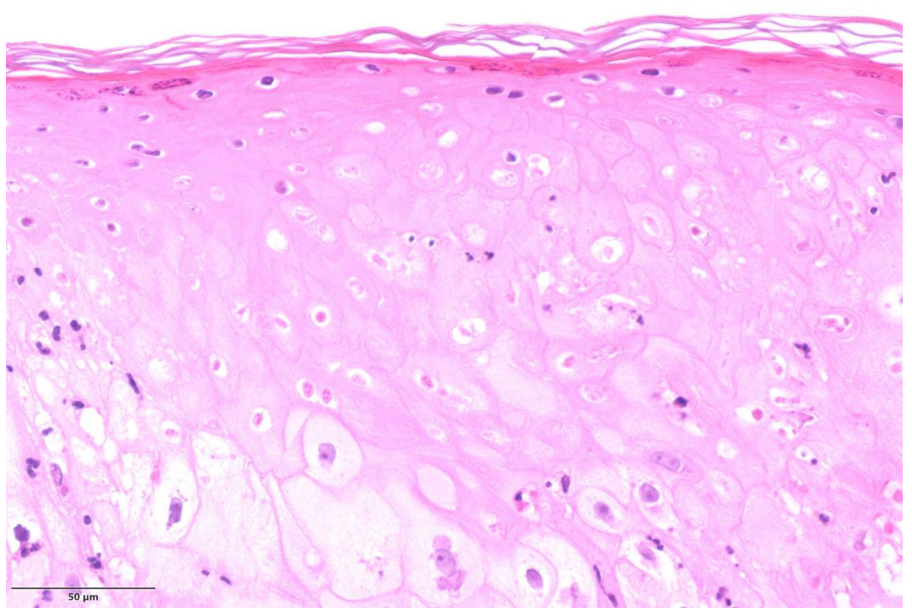
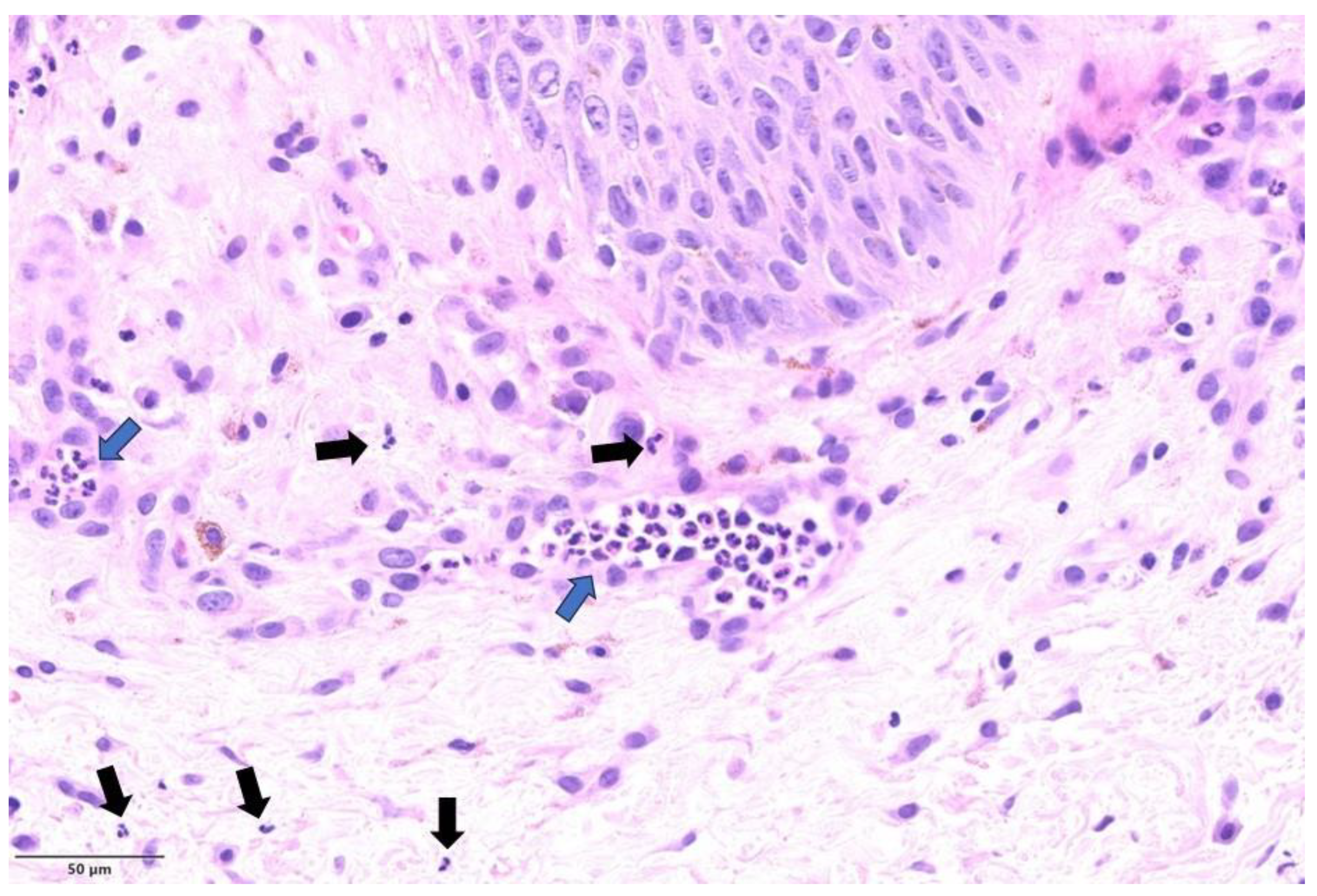
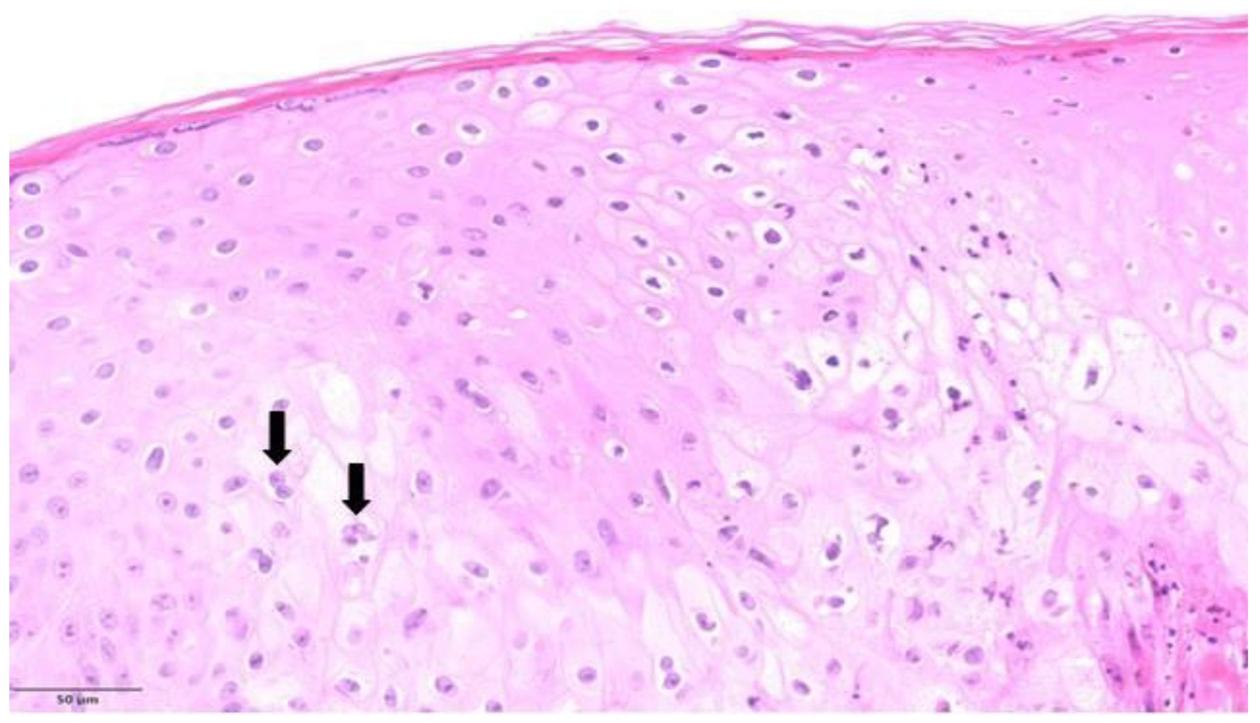
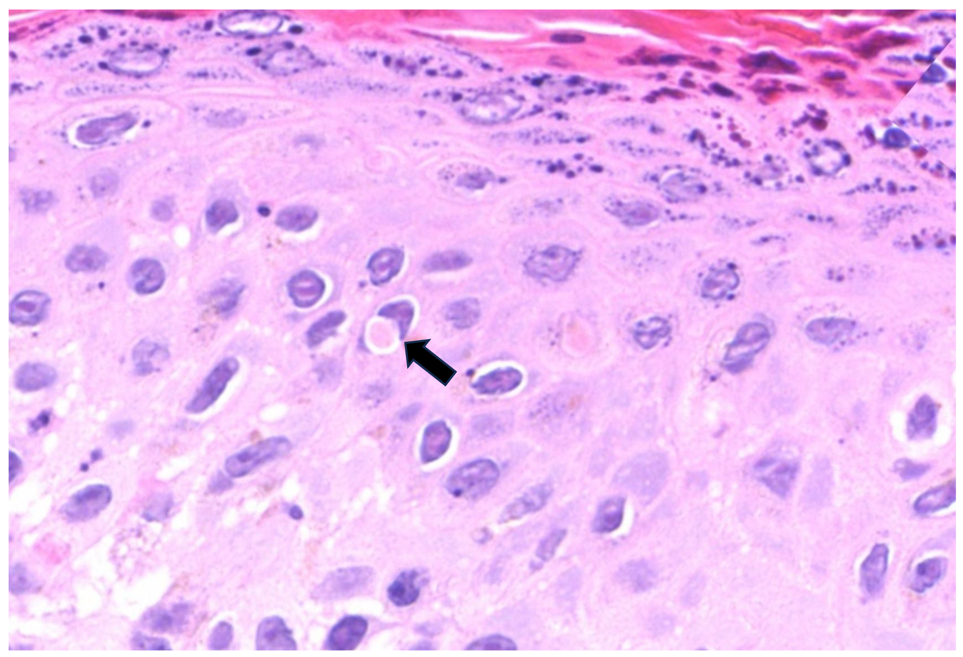
3.2. Histopathological Features
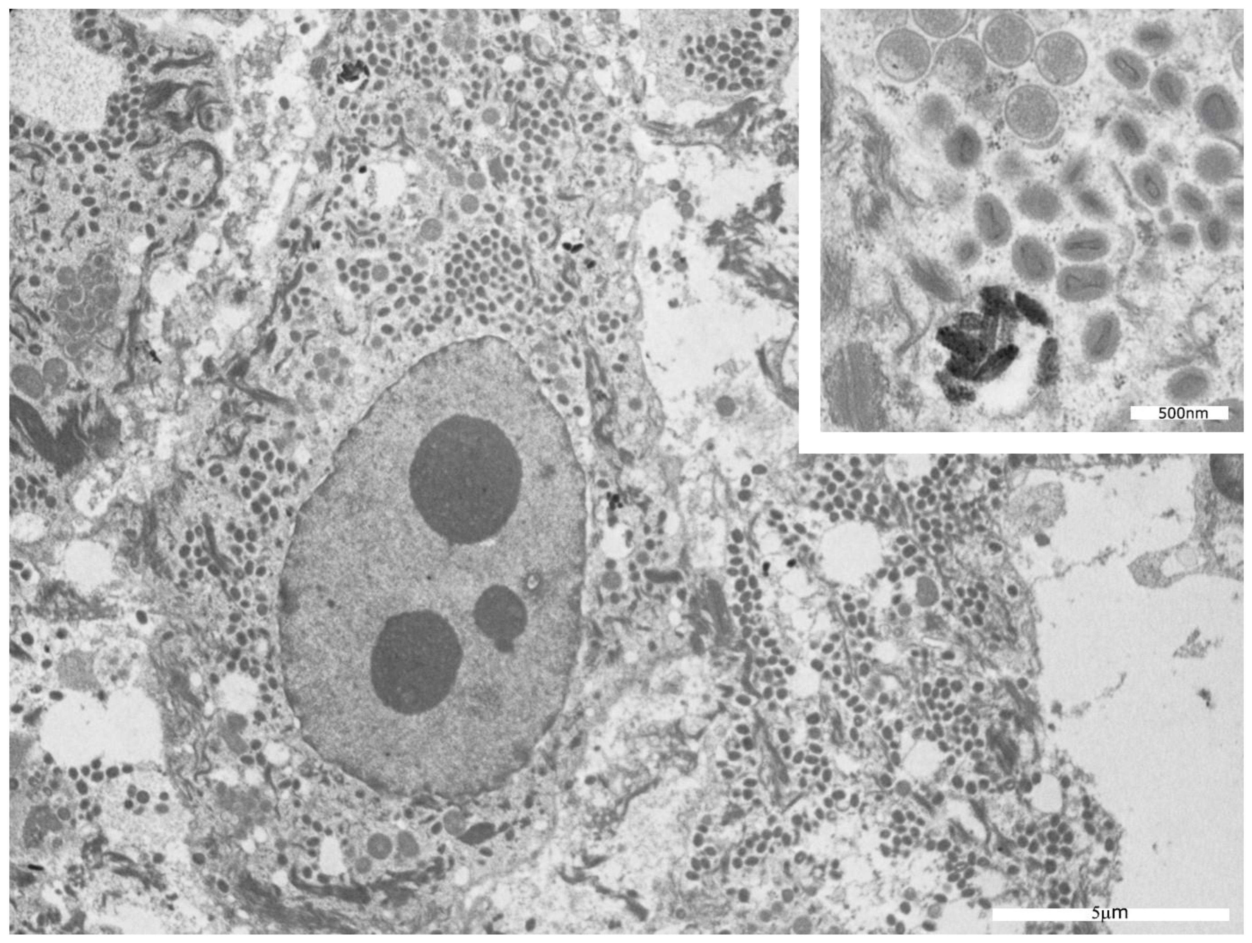
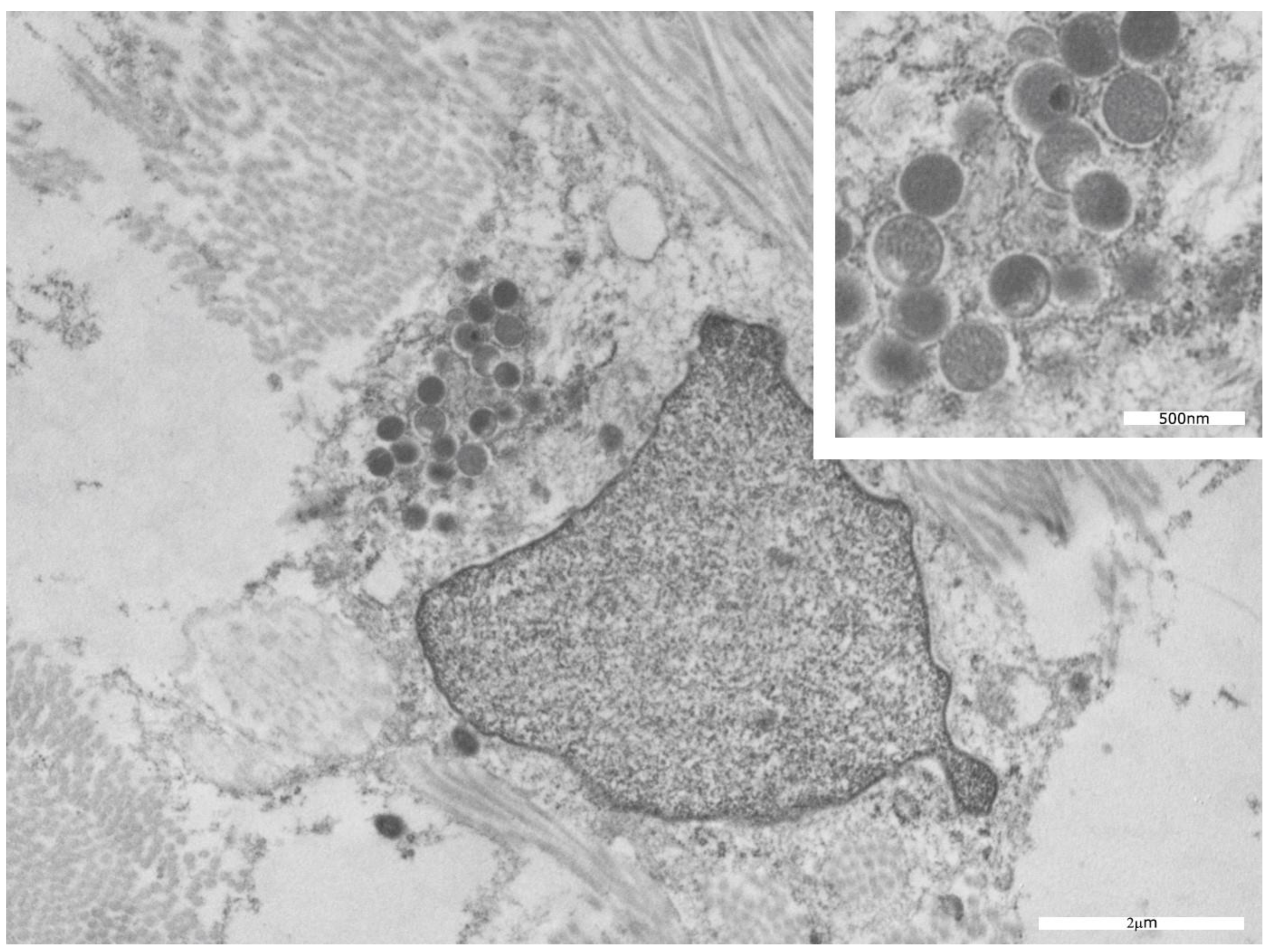
3.3. Transmission Electron Microscopy Findings
4. Discussion
5. Conclusions
Supplementary Materials
Author Contributions
Funding
Data Availability Statement
Acknowledgments
Conflicts of Interest
References
- Vaughan, A.; Aarons, E.; Astbury, J.; Brooks, T.; Chand, M.; Flegg, P.; Hardman, A.; Harper, N.; Jarvis, R.; Mawdsley, S.; et al. Human-to-human transmission of monkeypox virus, United Kingdom, October 2018. Emerg. Infect. Dis. 2020, 26, 782–785. [Google Scholar] [CrossRef] [PubMed]
- World Health Organization. The Global Eradication of Smallpox: Final Report of the Global Commission for the Certification of Smallpox Eradication; WHO: Geneva, Switzerland, 1980.
- Bryer, J.S.; Freeman, E.E.; Rosenbach, M. Monkeypox emerges on a global scale: A historical review and dermatological primer. J. Am. Acad. Dermatol. 2022, 87, 1069–1074. [Google Scholar] [CrossRef] [PubMed]
- Petersen, E.; Kantele, A.; Koopmans, M.; Asogun, D.; Yinka-Ogunleye, A.; Ihekweazu, C.; Zumla, A. Human monkeypox: Epidemiologic and clinical characteristics, diagnosis, and prevention. Infect. Dis. Clin. N. Am. 2019, 33, 1027–1043. [Google Scholar] [CrossRef]
- McCollum, A.M.; Damon, I.K. Human monkeypox. Clin. Infect. Dis. 2014, 58, 260–267. [Google Scholar] [CrossRef]
- Osadebe, L.; Hughes, C.M.; Lushima, R.S.; Kabamba, J.; Nguete, B.; Malekani, J.; Pukuta, E.; Karhemere, S.; Tamfum, J.-J.M.; Okitolonda, E.W.; et al. Enhancing case definitions for surveillance of human monkeypox in the Democratic Republic of Congo. PLoS Negl. Trop Dis. 2017, 11, e0005857. [Google Scholar] [CrossRef]
- Tarín-Vicente, E.J.; Alemany, A.; Agud-Dios, M.; Ubals, M.; Suñer, C.; Antón, A.; Arando, M.; Arroyo-Andrés, J.; Calderón-Lozano, L.; Casañ, C.; et al. Clinical presentation and virological assessment of confirmed human monkeypox virus cases in Spain: A prospective observational cohort study. Lancet 2022, 400, 661–669. [Google Scholar] [CrossRef]
- Cann, J.; Jahrling, P.; Hensley, L.; Wahl-Jensen, V. Comparative pathology of smallpox and monkeypox in man and macaques. J. Comp. Pathol. 2013, 148, 6–21. [Google Scholar] [CrossRef]
- Ortins-Pina, A.; Hegemann, B.; Saggini, A.; Deml, K.; Wallerius, K.; Hörster, S.; Kraft, S.; Weyers, W. Histopathological features of human monkeypox: Report of two cases and review of the literature. J. Cutan. Pathol. 2023; Epub ahead of print. [Google Scholar]
- Aromolo, I.F.; Maronese, C.A.; Avallone, G.; Beretta, A.; Boggio, F.L.; Murgia, G.; Marletta, D.A.; Barei, F.; Carrera, C.G.; Ramoni, S.; et al. Clinical spectrum of human monkeypox: An Italian single-centre case series. J. Eur. Acad. Dermatol. Venereol. 2023, 37, e368–e371. [Google Scholar] [CrossRef]
- Maronese, C.A.; Beretta, A.; Avallone, G.; Boggio, F.L.; Marletta, D.A.; Murgia, G.; Cusini, M.; Gori, A.; Carrera, C.G.; Di Benedetto, A.; et al. Clinical, dermoscopic and histopathological findings in localized human monkeypox: A case from northern Italy. Br. J. Dermatol. 2022, 187, 822–823. [Google Scholar] [CrossRef]
- Forni, D.; Moltrasio, C.; Sironi, M.; Mozzi, A.; Quattri, E.; Venegoni, L.; Zamprogno, M.; Citterio, A.; Clerici, M.; Marzano, A.V.; et al. Whole-genome sequencing of hMPXV1 in five Italian cases confirms the occurrence of the predominant epidemic lineage. J. Med. Virol. 2023, 95, e28493. [Google Scholar] [CrossRef]
- Fischer, A.H.; Jacobson, K.A.; Rose, J.; Zeller, R. Hematoxylin and eosin staining of tissue and cell sections. CSH Protoc 2008, 2008, pdb.prot4986. [Google Scholar] [CrossRef]
- Bayer-Garner, I.B. Monkeypox virus: Histologic, immunohistochemical and electron-microscopic findings. J. Cutan. Pathol. 2005, 32, 28–34. [Google Scholar] [CrossRef]
- Stagles, M.; Watson, A.; Boyd, J.; More, I.; McSeveney, D. The histopathology and electron microscopy of a human monkeypox lesion. Trans. R. Soc. Trop. Med. Hyg. 1985, 79, 192–202. [Google Scholar] [CrossRef]
- Rodríguez-Cuadrado, F.J.; Nájera, L.; Suárez, D.; Silvestre, G.; García-Fresnadillo, D.; Roustan, G.; Sánchez-Vázquez, L.; Jo, M.; Santonja, C.; Garrido-Ruiz, M.C.; et al. Clinical, histopathologic, immunohistochemical, and electron microscopic findings in cutaneous monkeypox: A multicenter retrospective case series in Spain. J. Am. Acad. Dermatol. 2023, 88, 856–863. [Google Scholar] [CrossRef]
- Ramoni, S.; Maronese, C.A.; Morini, N.; Avallone, G.; Quattri, E.; Carrera, C.G.; Boggio, F.L.; Marzano, A.V. Syphilis and monkeypox co-infection: Coincidence, synergy or asymptomatic carriage? Travel Med. Infect. Dis. 2022, 50, 102447. [Google Scholar] [CrossRef]
- Berna-Rico, E.; Perna, C.; Azcarraga-Llobet, C.; Garcia-Mouronte, E.; de Nicolas-Ruanes, B.; Melendez-Gispert, M.R.; Vivancos, M.J.; Martinez-Garcia, L.; Fernandez-Gonzalez, P. Monkeypox virus infection with a syphilitic-roseola-like rash and its histopathologic characterization during 2022 outbreak. J. Eur. Acad. Dermatol. Venereol. 2022, 37, e400–e402. [Google Scholar] [CrossRef] [PubMed]
- Khan, Z.M.; Cockrell, C.L. Cutaneous viral infections. In Textbook of Dermatopathology; Barnhill, R.L., Ed.; McGraw-Hill Publishers: New York, NY, USA, 1998; p. 439. [Google Scholar]
- Zaucha, G.M.; Jahrling, P.B.; Geisbert, T.W.; Swearengen, J.R.; Hensley, L. The pathology of experimental aerosolized monkeypox virus infection in cynomolgus monkeys (Macaca fascicularis). Lab. Investig. 2001, 81, 1581–1600. [Google Scholar] [CrossRef] [PubMed]
- Institute of Medicine (US) Committee on the Assessment of Future Scientific Needs for Live Variola Virus. Assessment of Future Scientific Needs for Live Variola Virus; National Academies Press: Washington, DC, USA, 1999.
- Lum, F.-M.; Torres-Ruesta, A.; Tay, M.Z.; Lin, R.T.P.; Lye, D.C.; Rénia, L.; Ng, L.F.P. Monkeypox: Disease epidemiology, host immunity and clinical interventions. Nat. Rev. Immunol. 2022, 22, 597–613. [Google Scholar] [CrossRef]
- Blank, H.; Davis, C.; Collins, C. Electron microscopy for the diagnosis of cutaneous viral infections. Br. J. Dermatol. 1970, 83, 69–80. [Google Scholar] [CrossRef] [PubMed]
- U.S. Centers for Disease Control and Prevention. Occupational Safety and Health Administration. 2022 Mpox Outbreak Global Map, as of 21 June. Available online: https://www.cdc.gov/poxvirus/monkeypox/response/2022/world-map.html (accessed on 22 June 2023).
- Golden, J.; Harryman, L.; Crofts, M.; Muir, P.; Donati, M.; Gillett, S.; Irish, C. Case of apparent mpox reinfection. Sex. Transm. Infect. 2023, 99, 283–284. [Google Scholar] [CrossRef]
- Raccagni, A.R.; Canetti, D.; Mileto, D.; Tamburini, A.M.; Candela, C.; Albarello, L.; Bracchitta, F.; Mancon, A.; Micheli, V.; Gismondo, M.R.; et al. Two individuals with potential monkeypox virus reinfection. Lancet Infect. Dis. 2023, 23, 522–524. [Google Scholar] [CrossRef] [PubMed]
- Álvarez-López, P.; Borras-Bermejo, B.; López Pérez, L.; Antón, A.; Piñana, M.; García-Pérez, J.; Descalzo, V.; Monforte, A.; Martínez-Gómez, X.; Falcó, V.; et al. Suspected case of monkeypox reinfection versus reactivation in a immunocompetent patient, Barcelona, 2022. Int. J. STD AIDS, 2023; 9564624231162426, Epub ahead of print. [Google Scholar]


| Pat ID | Sex | Sexual Behavior | Unprotected Sex | Travel History | Other STIs | Kind of Lesions | Number of Skin Lesions | Localization | Systemic Symptoms | Lymphadenopathy | Hospitalization | Outcome |
|---|---|---|---|---|---|---|---|---|---|---|---|---|
| 1 | M | MSM | Yes | No | None | Pustular lesions | >10 | Genital region; lower limb; middle finger of the right hand | Fever; headache; asthenia | No | Yes | CR |
| 2 | M | MSM | Yes | Yes (The Netherlands) | None | Pustular lesions | >5 | Penile shaft and glans | Fever; headache; myalgia; arthralgia; asthenia | No | No | CR |
| 3 | M | MSM | Yes | Yes (Spain) | None | Pustular lesions | <5 | Penile shaft and glans; pubic region; perioral region | Asthenia | Yes (laterocervical area) | No | CR |
| 4 | M | MSM | Yes | No | Late latent syphilis | Pseudo-pustular lesions | >10 | Face; penile shaft; perianal region | Hyperpyrexia; rectal pain | No | No | CR |
| 5 | M | MSM | Yes | No | None | Vesico- pustular lesions | >5 | Pubic region; penile shaft; upper limb; palm of the hand | Fever; headache; asthenia | No | No | CR |
| 6 | M | MSM | Yes | No | Syphilis | Ulcerate lesion | 1 | Pubic region | Asthenia | Yes (inguinal area) | No | CR |
| Pat ID | Site of Biopsy | Histological Stage [14] | Main Cytopathic Modification | Dermal Inflammatory Cell Infiltration |
|---|---|---|---|---|
| 1 | Left thigh; penile shaft | Pustular stage | Multinucleated keratinocytes; occasional Guarnieri bodies; extensive ballooning; ground glass nuclei; degenerative modifications in the acrosyringial epithelium | Moderate perivascular and periadnexal with neutrophils |
| 2 | Penile | Pustular stage | Ground glass nuclei; ballooning; degenerative modifications in follicular keratinocytes | Mild perivascular lymphocytic infiltration |
| 3 | Groin, left shoulder | Pustular stage | Ballooning; occasional Guarnieri bodies; degenerative modifications in follicular keratinocytes | Moderate perivascular and periadnexal with neutrophils |
| 4 | Penile shaft | Pustular stage | Guarnieri bodies; ballooning; positive immunohistochemical staining for Treponema pallidum with spirochetes in cytoplasm of keratinocytes | Moderate perivascular and periadnexal with neutrophils |
| 5 | Pubic region | Vesicular stage | Ballooning; spongiosis and achantosis; degenerative modifications in the acrosyringial epithelium | Severe perivascular, interstitial, and periadnexal with neutrophils |
| 6 | Pubic region | Not applicable | Focal follicular dyskeratosis; positive immunohistochemical staining for Treponema pallidum with perivascular and intraepithelial spirochetes | Severe periadnexal and interstitial lymphocytic infiltration with numerous plasma cells |
| Pat ID | Lesional Site | Electron Microscopy Transmission Features |
|---|---|---|
| 1 | Left thigh; penile shaft | Almost all keratinocytes contain mature and immature viral particles; virus absent in the stratum corneum and between the scales; virions identified inside the cytoplasm of mesenchymal cells. |
| 2 | Penile | Almost all keratinocytes contain mature and immature viral particles; virus absent in the stratum corneum and between the scales. |
| 3 | Groin, left shoulder | Almost all keratinocytes contain mature and immature viral particles; virus absent in the stratum corneum and between the scales; virions identified inside the cytoplasm of mesenchymal cells. |
| 4 | Penile shaft | Almost all keratinocytes contain mature and immature viral particles; virus absent in the stratum corneum and between the scales. |
| 5 | Pubic region | Some viral particles inside the cytoplasm of basal keratinocytes. |
| 6 | Pubic region | Absence of the virus. |
Disclaimer/Publisher’s Note: The statements, opinions and data contained in all publications are solely those of the individual author(s) and contributor(s) and not of MDPI and/or the editor(s). MDPI and/or the editor(s) disclaim responsibility for any injury to people or property resulting from any ideas, methods, instructions or products referred to in the content. |
© 2023 by the authors. Licensee MDPI, Basel, Switzerland. This article is an open access article distributed under the terms and conditions of the Creative Commons Attribution (CC BY) license (https://creativecommons.org/licenses/by/4.0/).
Share and Cite
Moltrasio, C.; Boggio, F.L.; Romagnuolo, M.; Cagliani, R.; Sironi, M.; Di Benedetto, A.; Marzano, A.V.; Leone, B.E.; Vergani, B. Monkeypox: A Histopathological and Transmission Electron Microscopy Study. Microorganisms 2023, 11, 1781. https://doi.org/10.3390/microorganisms11071781
Moltrasio C, Boggio FL, Romagnuolo M, Cagliani R, Sironi M, Di Benedetto A, Marzano AV, Leone BE, Vergani B. Monkeypox: A Histopathological and Transmission Electron Microscopy Study. Microorganisms. 2023; 11(7):1781. https://doi.org/10.3390/microorganisms11071781
Chicago/Turabian StyleMoltrasio, Chiara, Francesca Laura Boggio, Maurizio Romagnuolo, Rachele Cagliani, Manuela Sironi, Alessandra Di Benedetto, Angelo Valerio Marzano, Biagio Eugenio Leone, and Barbara Vergani. 2023. "Monkeypox: A Histopathological and Transmission Electron Microscopy Study" Microorganisms 11, no. 7: 1781. https://doi.org/10.3390/microorganisms11071781
APA StyleMoltrasio, C., Boggio, F. L., Romagnuolo, M., Cagliani, R., Sironi, M., Di Benedetto, A., Marzano, A. V., Leone, B. E., & Vergani, B. (2023). Monkeypox: A Histopathological and Transmission Electron Microscopy Study. Microorganisms, 11(7), 1781. https://doi.org/10.3390/microorganisms11071781


