Article

Functional Analysis of Novel alkB Genes Encoding Long-Chain n-Alkane Hydroxylases in Rhodococcus sp. Strain CH91

State Key Laboratory of Microbial Resources, Institute of Microbiology, Chinese Academy of Sciences, Beijing 100101, China
*
Author to whom correspondence should be addressed.
Microorganisms 2023, 11(6), 1537; https://doi.org/10.3390/microorganisms11061537
Submission received: 10 May 2023 / Revised: 5 June 2023 / Accepted: 7 June 2023 / Published: 9 June 2023

Abstract

:
Rhodococcus sp. strain CH91 is capable of utilizing long-chain n-alkanes as the sole carbon source. Two new genes (alkB1 and alkB2) encoding AlkB-type alkane hydroxylase were predicted by its whole-genome sequence analysis. The purpose of this study was to elucidate the functional role of alkB1 and alkB2 genes in the n-alkane degradation of strain CH91. RT-qPCR analyses revealed that the two genes were induced by n-alkanes ranging from C16 to C36 and the expression of the alkB2 gene was up-regulated much higher than that of alkB1. The knockout of the alkB1 or alkB2 gene in strain CH91 resulted in the obvious reduction of growth and degradation rates on C16-C36 n-alkanes and the alkB2 knockout mutant exhibited lower growth and degradation rate than the alkB1 knockout mutant. When gene alkB1 or alkB2 was heterologously expressed in Pseudomonas fluorescens KOB2Δ1, the two genes could restore its alkane degradation activity. These results demonstrated that both alkB1 and alkB2 genes were responsible for C16-C36 n-alkanes’ degradation of strain CH91, and alkB2 plays a more important role than alkB1. The functional characteristics of the two alkB genes in the degradation of a broad range of n-alkanes make them potential gene candidates for engineering the bacteria used for bioremediation of petroleum hydrocarbon contaminations.

1. Introduction

Long-chain n-alkanes are part of the main components of petroleum. The biodegradation of long-chain n-alkanes by bacteria has attracted considerable attention due to its potential for bioremediation of petroleum contamination and microbial-enhanced oil recovery [1,2,3,4]. Several bacteria including Acinetobacter, Pseudomonas, Dietzia, Rhodococcus, Geobacillus, etc. have been reported to be able to degrade long-chain n-alkanes [5,6]. The biodegradation of long-chain n-alkanes in these bacteria is usually initiated by the terminal oxidation of n-alkane to the corresponding alkanol, which is the key step in the n-alkane degradation pathway and catalyzed by an alkane hydroxylase. Several alkane hydroxylase systems involved in long-chain n-alkane degradation have been identified, including integral-membrane non-heme diiron monooxygenase (AlkB), cytochrome P450 monooxygenase, flavin-binding alkane hydroxylase (AlmA), and flavin-dependent alkane monooxygenase (LadA) [5,7].
The alkB-type alkane hydroxylase system of Pseudomonas putida GPo1 is the best-characterized system for alkane degradation [8,9]. It catalyzes the initial terminal oxidation of n-alkane in the form of a three-component complex, which consists of a non-heme integral-membrane alkane hydroxylase (AlkB), two rubredoxins (AlkF and AlkG), and a rubredoxin reductase (AlkT). Among the three-component complexes, rubredoxin and rubredoxin reductase are essential electron transfer components required for alkane hydroxylation by AlkB [8,10]. Similar to P. putida GPo1, Acinetobacter sp. ADP1 and 6A2 also catalyze the initial oxidation of C12–C18 n-alkanes via a three-component alkane hydroxylase system, which comprises AlkM (alkane monooxygenase), RubA (rubredoxin), and RubB (rubredoxin reductase) [11,12,13]. Whyte et al. identified two three-component alkane hydroxylase systems (AlkB1 and AlkB2) in Rhodococcus erythropolis, which are partially responsible for the initial oxidation of C12–C16 n-alkanes [14]. The alkB homologs of M. tuberculosis and P. aeruginosa were also shown to oxidize alkanes ranging from C10 to C16 [15]. The three-component alkane hydroxylase system containing alkane 1-monooxygenase (AlkB), rubredoxin (RubA), and rubredoxin reductase (RubB) is often involved in alkane hydroxylation [5,7]. Other types of alkane hydroxylation mechanisms have also been reported. Cytochrome P450 hydroxylase usually mediates the oxidation of short- and medium-chain alkanes (C5–C16). It has been reported to catalyze the terminal oxidation of n-alkane in Acinetobacter sp. EB104, Mycobacterium sp. HXN-1500, and Alcanivorax dieselolei B-5 [16]. AlmA-type alkane hydroxylase was first identified in Acinetobacter sp. DSM 17874 and oxidize C20 to >C32 n-alkanes [17]. Its homologs also have been identified in several other long-chain n-alkane-degrading strains, including Acinetobacter pittii SW-1, Acinetobacter sp. M1, Alcanivorax borkumensis SK2, and Pseudomonas aeruginosa DN1 [3,18]. LadA-type alkane monooxygenase was first reported in Geobacillus thermodenitrificans NG80-2 and could oxidize C15–C36 n-alkanes into primary alcohols. The enzyme is a two-component flavin-dependent oxygenase belonging to the bacterial luciferase family of proteins [19].
The AlkB-type alkane hydroxylases have been widely found in alkane-degrading bacteria by gene cloning, PCR detection, and genome sequencing. They are quite divergent and have different substrate ranges. The well-characterized AlkB-type alkane hydroxylases are usually reported to be involved in the metabolism of n-alkanes up to C16 [5,7]. Only a few AlkB-type alkane hydroxylases were experimentally confirmed in the degradation of n-alkanes longer than C18 [20,21]. The contributions of AlkB-type alkane hydroxylases to the long-chain alkane degradation are still far from clear. In a previous study, we isolated a novel Rhodococcus strain, CH91, with the ability to degrade long-chain n-alkanes, and two new alkB genes encoding alkane hydroxylase were annotated in the genome of this strain [22]. The enzymes encoded by the two alkB genes of strain CH91 had a lower identity with the well-characterized AlkB type alkane hydroxylases but showed significant similarity with the homologs from R. rhodochrous NCTC 10210, R. pyridinivorans DSM 44555, R. biphenylivorans TG9, and R. gordoniae NCTC13296, which belong to the R. rhodochrous subclade [22]. All of these homologs from R. rhodochrous subclade strains were annotated solely according to sequence homology, but their functions have not yet been experimentally characterized. As every AlkB enzyme is unique and showed different substrate ranges, elucidating its biochemical characterization is important for understanding the metabolic pathway of alkane degradation and realizing its biotechnological potential. In this study, we clarified the contribution of the two alkB genes to the long-chain alkane degradation of Rhodococcus sp. strain CH91 by constructing alkB gene knockout mutants and heterologously expressing the two alkB genes, together with analyzing the genetic characteristics and transcriptional expression profile of alkB genes.

2. Materials and Methods

2.1. Bacterial Strains and Cultivation

The bacterial strains and plasmids used in this study are listed in Supplementary Table S1. Rhodococcus sp. strain CH91 and its mutant derivatives were grown at 37 °C in Luria-Bertani (LB) medium or MD878 basal medium supplemented with n-alkanes or succinate as a sole carbon source [22]. P. fluorescens KOB2∆1 and its recombinants were grown at 30 °C in LB medium or E2 medium [23] supplemented with n-alkanes as sole carbon sources. E. coli DH5α was used for plasmid construction and grown at 37 °C in an LB medium. E. coli strains were transformed with plasmid DNA via calcium-dependent transformation [24]. When necessary, appropriate antibiotics were used in the medium: ampicillin (at the final concentration of 100 µg mL−1), kanamycin (50 µg mL−1), chloramphenicol (30 µg mL−1), and gentamicin (100 µg mL−1 for P. fluorescens and strain CH91, 10 µg mL−1 for E. coli).

2.2. Growth and Alkane Degradation Assays

To determine the utilization of various n-alkanes, strain CH91 and its mutant derivatives were grown in an MD878 medium supplemented with various n-alkanes as the sole carbon source. Different alkanes were first dissolved in octane and then individually added to the medium at a final concentration of 0.4% (v/v) for the liquid alkane (C16–C20) at 37 °C or 0.1% (w/v) for the solid alkane (C24–C36). The utilization of mixed n-alkanes was also tested with a final concentration of 1% (v/v) n-alkane mixture as the sole carbon source. The un-inoculated cultures were used as controls. The influence of the solvent octane on the growth of the cells was measured by using 1% (v/v) octane as the sole carbon source. All cultures were incubated on a shaker at 150 rpm at 37 °C for 10 days for C16–C20 carbon sources or 21 days for C24–C36 carbon sources or 14 days and 24 days for the n-alkane mixture. All the experiments were repeated in triplicate. The growth was examined by measuring the optical density at 600 nm and the alkane degradation was determined by analysis of the residual n-alkane by GC-MS chromatography.
The n-alkane mixture used in this study contained 5% (v/v) C16 and 1% (w/v) each of C18, C20, C24, C26, C28, C30, C32, and C36 n-alkanes dissolved in octane. Hydrocarbons such as hexane (C6), octane (C8), hexadecane (C16), octadecane (C18), eicosane (C20), tetracosane (C24), hexacosane (C26), octacosane (C28), triacontane (C30), dotriacontane (C32) and hexatriacontane (C36) were purchased from Macklin (Ghaziabad, India).

2.3. Analytical Methods

As the strain formed clumps with alkane when growing on alkane and the cells were difficultly spun down, the cultures grown on n-alkanes were firstly treated by adding n-hexane to dissociate the cells from a long-chain alkane. The entire culture was extracted twice with 1/5 volume of n-hexane following the addition of dioctyl phthalate as an internal standard. The mixtures were centrifuged at 10,000× g for 20 min at room temperature. The cell pellet was re-suspended with 1/2 volume of buffer solution and its optical density was measured at 600 nm as the growth of strain. The hexane layer was harvested and used to analyze the amount of residual alkanes using GC-MS (Agilent (Santa Clara, CA, USA) 5977A MS and 7890B GC equipped with FID detector) attached to an Agilent HP-5MS capillary column [22]. The FID detector was used for quantitative analysis. The degradation ratio of n-alkanes was calculated with the equation R (%) = (Co − Cx)/Co × 100, where R, Co, and Cx represent the n-alkane degradation ratio, the residual n-alkane concentration in the un-inoculated culture, and the concentration in the inoculated culture, respectively. All analyses were carried out with three replicates, and the values are presented as mean values ± standard deviations (SD). Data were analyzed statistically using a two-tailed T-TEST, and p-values of 0.05 or less were considered statistically significant. Calculations and graphics were performed using GraphPad Prism 8.

2.4. Reverse Transcription and Real-Time Quantitative PCR (RT-qPCR)

To investigate the induction of alkB1 and alkB2 by different n-alkanes, strain CH91 was cultured to the mid-exponential phase of growth in MD878 medium supplemented with 0.5% succinate or 0.1% (w/v) different n-alkanes as sole carbon sources. Cells were collected by centrifugation at 10,000× g for 10 min at 4 °C. Total RNA was extracted using Bacterial RNAprep Pure Kit (Tiangen, Beijing, China). Reverse transcription was performed using StarScript III All-in-one RT-PCR Kit (GenStar, Beijing, China). RT-qPCR analysis was conducted using a 2xRealStar Fast SYBR qPCR mix (GenStar, Beijing, China) on a CFX96 Real-Time System (BIO-RAD, Hercules, CA, USA). 16S rRNA gene was used as an internal control for normalization. A control reaction without reverse transcriptase was conducted to verify the absence of genomic DNA. All of the experiments were performed according to the instructions from the manufacturers of the reagents or instruments. Relative quantities were calculated using the 2−ΔΔCt method [25] with the succinate carbon source as the control. All experiments were performed in triplicate and the means and standard deviations were calculated. The primers used for RT-qPCR were designed with the Primer Premier 5.0 software package (Premier Biosoft Intl., Palo Alto, CA, USA) and are listed in Supplementary Table S2.

2.5. Construction of alkB Gene Knockout Mutants

To construct alkB gene knockout mutants, a CRISPR/Cas9-mediated triple-plasmid genome editing system was employed as previously described [26]. Briefly, wild-type strain CH91 was transformed with the plasmid pNV-Pa2-Cas9 and then made competent for the introduction of pRCTc-Pa2-Che9c60&61. The subsequent CH91 (Cas9+Che9c60&61) was transformed with 1 μg of the pBNVCm-sgRNA series and 1 μg of linear donor dsDNA. Cells were spread on LB+ 1% pyruvate plates containing 50 μg  mL−1 kanamycin, 10 μg  mL−1 tetracycline, and 25 μg  mL−1 chloramphenicol and then incubated at 28 °C for 3–5 days to obtain target strains. Knockout of the genes was confirmed by PCR amplification and sequencing of the amplified regions flanking the deletions. The genes alkB1 and alkB2 were individually knocked out by means of the procedure outlined above. The alkB-sgRNA was designed on the websites http://grna.ctegd.uga.edu/ and http://www.oligoevaluator.com/LoginServlet accessed on 15 September 2021. The primers used for gene knockout in strain CH91 are listed in Supplementary Table S2.

2.6. Heterologous Expression of alkB Genes

P. fluorescens KOB2Δ1 is an alkB1 deletion derivative of P. fluorescens CHA0 and is usually used to assess the activities of alkane hydroxylases via growth complementation by restoring alkane degradation through heterologous expression of alkB ortholog genes [27,28]. To confirm the alkane hydroxylase activity of alkB genes from Rhodococcus sp. strain CH91, both alkB genes were also heterologously expressed and evaluated in P. fluorescens KOB2Δ1. The genes alkB1 and alkB2 were individually amplified by PCR using Rhodococcus sp. CH91 chromosomal DNA as a template and cloned into pCom8 plasmid at NdeI and HindIII sites using ClonExpress® Ultra One Step Cloning Kit (Vazyme, Nanjing, China) by Gibson assembly method [29]. The primers used for PCR amplification of alkB genes were designed based on the genome sequence of strain CH91 and listed in Supplementary Table S2. The nucleotide sequences of the pCom8-alkB plasmids were confirmed to be correct by sequencing using an ABI 3730 automated DNA sequencer (Applied Biosystems, Foster City, CA, USA). Successful plasmids were transformed into P. fluorescens KOB2∆1 via electroporation as described before [30]. P. fluorescens KOB2Δ1 recombinants containing pCom8 (as negative control) and pCom8-alkB plasmids were grown in an E2 medium [23] supplemented with 1% n-alkane mixture as sole carbon sources. The cultures were incubated under shaking conditions (150 rpm) at 30 °C for 21 days. The growth was examined by measuring the optical density at 600 nm and the alkane degradation was determined by analysis of the residual n-alkane by GC-MS chromatography. The un-inoculated culture was used as a control. All the experiments were repeated in triplicate.

3. Results and Discussion

3.1. Genetic Characteristics of alkB Genes in Rhodococcus sp. CH91

Rhodococcus sp. strain CH91 is capable of utilizing n-alkanes with carbon chain lengths ranging from C16 to C36 as a sole carbon source, and no growth on C8, C12, and C40 was observed. Its complete genome was previously sequenced in our lab and two alkB-type alkane hydroxylase genes (alkB1 and alkB2) were predicted to be responsible for the first step of the n-alkane degradation pathway [22]. The open reading frame (ORF) analysis of the alkB gene loci revealed that the genetic arrangement of the two alkB gene regions had different organizations. The alkB2 gene region contained an operon-like structure that had four genes encoding an alkane hydroxylase (AlkB2), a couple of rubredoxins (RubA1 and RubA2), and a TetR transcriptional regulator (Figure 1a). Moreover, the alkB2 and rubA1 genes had 3′-end–5′-end GTGA sequence overlaps, and the rubA1 and rubA2 genes had 3′-end–5′-end ATGA sequence overlaps. The gene organization is quite similar to the alkB2 gene cluster previously demonstrated in other Rhodococcus strain genomes [14]. The alkB1 gene region contained a separate alkane hydroxylase homolog and was not flanked by rubredoxin or rubredoxin reductase genes (Figure 1a). The genes upstream alkB1 encoded a cold-shock protein and an aminotransferase. The genes downstream alkB1 encoded AbiEi family antitoxin domain-containing protein and 4Fe-4S dicluster domain-containing protein. The ORF organization of the alkB1 gene and the surrounding region was not like that of the alkB3 and alkB4 gene regions of R. erythropolis B-16531 and Q15 [14], but quite similar to that of the alkBb gene regions of R. ruber NBRC 15591 and SP2B as well as that of R. rhodochrous NCTC 10210 and NBRC 16069, R. pyridinivorans TG9 and DSM 44555, R. gordoniae NCTC13296 and R. coprophilus NCTC10994, which belong to the R. rhodochrous subclade.
Multiple sequence alignments of the AlkB1 and AlkB2 amino acid sequences were performed with other published AlkB sequences using the ClustalW algorithm in MEGA7 software (version 7.0) [31]. As shown in Figure 1b, both AlkB1 and AlkB2 proteins possessed eight histidine residues within three His boxes (Hist1, HELGHK; Hist2, EHNRGHH; and Hist3, LQRHSDHHA) and an HYG motif (NYLEHYGL), which are highly conserved in non-heme iron integral membrane alkane hydroxylases and required for catalytic activity [14,32]. In addition, both AlkB1 and AlkB2 enzymes possessed the six transmembrane helices, which are also conserved in all integral membrane alkane hydroxylase [14,33]. These structural characteristics suggest that AlkB1 and AlkB2 are membrane-bound alkane hydroxylases and might be responsible for long-chain n-alkane degradation in strain CH91.

3.2. Transcriptional Expression of n-Alkane Hydroxylase Genes in Rhodococcus sp. Strain CH91

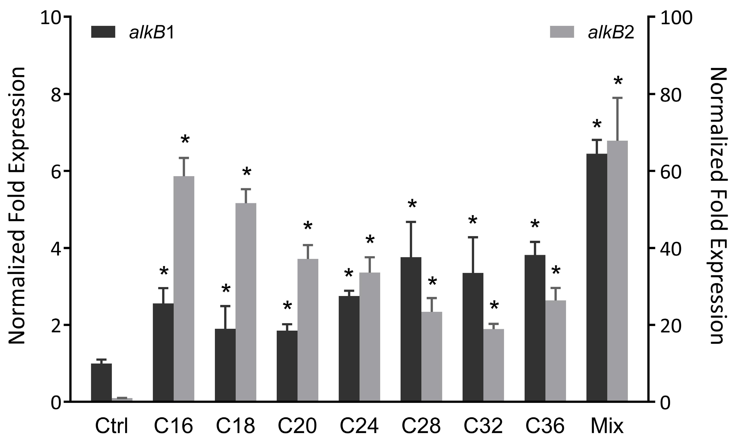
To investigate the alkB gene expression profile in strain CH91, RT-qPCR was performed to analyze the expression level of alkB1 and alkB2 genes in the presence of different n-alkanes ranging from C16 to C36. As shown in Figure 2, both the alkB genes were induced by the tested long-chain n-alkanes and showed obviously higher expression in the presence of the n-alkane mixture than in the individual n-alkane. The expression of alkB2 was remarkably induced by all of the n-alkanes tested, the largest increase (58-fold) was observed with C16, and the lower expression level was induced normally with the longer chain length of n-alkane. The expression of alkB1 was induced about 2-fold in the presence of C16–C24 and up-regulated to about 3.5-fold in the presence of C28–C36, indicating the presence of the different regulatory systems for alkB1 response to different n-alkane substrate range. Moreover, the different patterns of the transcriptional expression induced by n-alkanes between alkB1 and alkB2 suggest a different regulatory mechanism for alkB1 and alkB2. The above data supported that alkB1 and alkB2 are involved in C16–C36 n-alkane degradation of strain CH91, and alkB2 plays the predominant role in n-alkane degradation for strain CH91. The different induction manners in response to the chain length of n-alkanes were also reported in other strains. For example, Acinetobacter sp. M-1 contains two AlkB-related alkane hydroxylases, AlkMa and AlkMb, whose expressions are controlled by different regulators. The expression of alkMa is induced by n-alkanes with chain lengths more than C22, and alkMb expression is preferentially induced by C16–C22 n-alkanes [34]. Regarding other Rhodococcus members, strain TMP2 has five alkane hydroxylases. The expressions of alkB1 and alkB2 genes are induced by C16 and pristine, whereas alkB3, alkB4, and alkB5 genes were not affected by C16 or pristine [35]. Similarly, R. erythropolis PR4 possesses four alkane monooxygenases. Only alkB1 and alkB2 genes were highly upregulated by C16 and diesel oil. The other two alkB genes did not change obviously. The induction level of alkB1 was much higher than that of alkB2 [36]. Pseudomonas aeruginosa RR1, like our strain CH91, has two alkane hydroxylases. Both genes could be induced by C10–C22 n-alkanes. The induction level of alkB2 is about twice as much as that of alkB1 [37].

3.3. Utilization of n-Alkanes by Wild-Type Strain CH91 and Its Mutant Derivatives

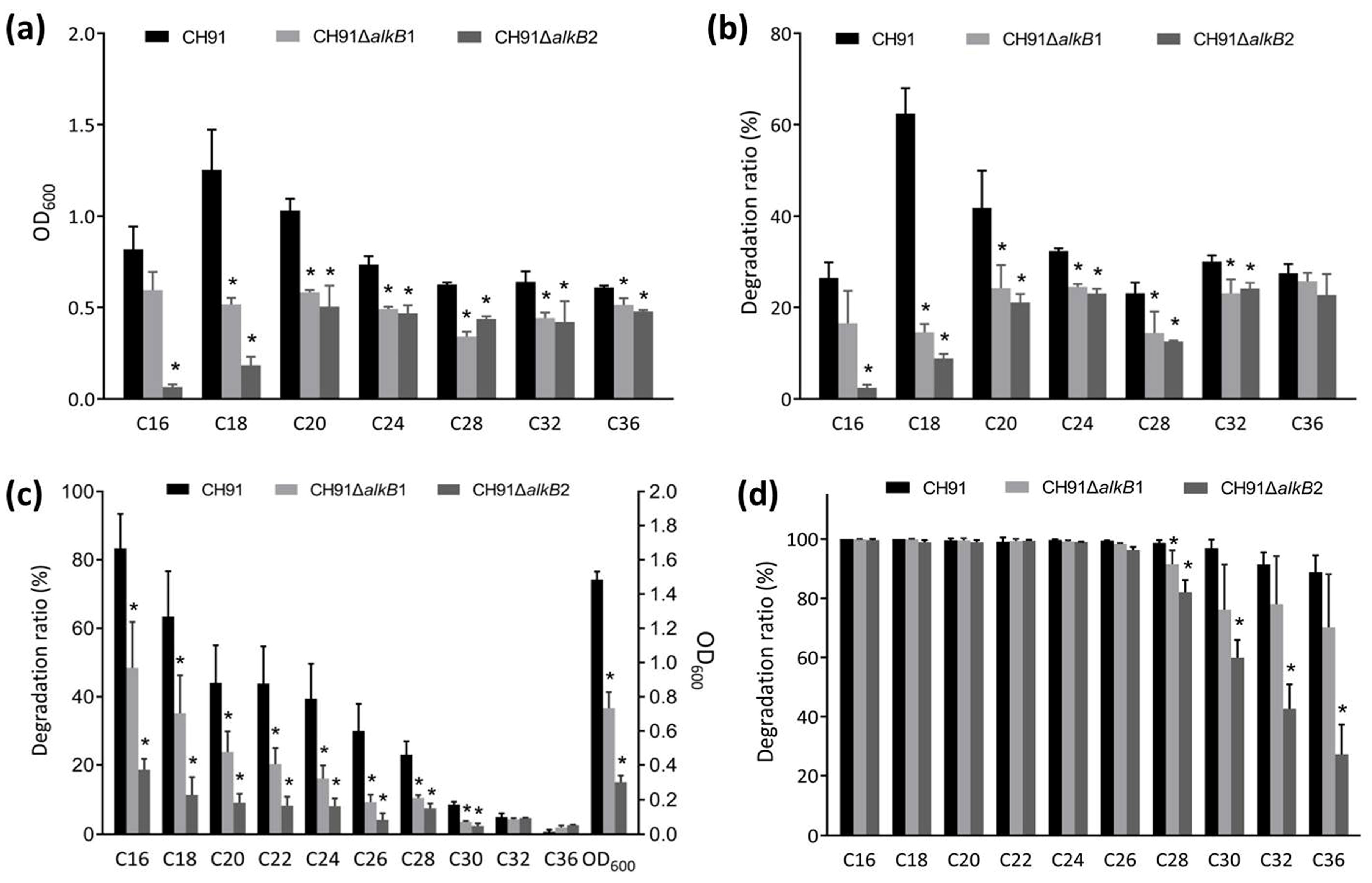
To examine the possible function of the two alkB genes in n-alkane utilization, the alkB gene knockout mutants (CH91ΔalkB1 and CH91ΔalkB2) of strain CH91 were constructed and functionally analyzed. Wild-type strain CH91 and its mutant derivatives were cultivated with different n-alkanes ranging in length from C16 to C36 as the sole carbon source. As shown in Figure 3a,b, wild-type strain CH91 had faster growth and higher degradation efficiency with C16, C18, or C20 n-alkane as the sole carbon source, while it showed some slower growth and lower degradation rate in the case of C24, C28, C32 or C36 n-alkane as sole carbon source. The best capability of growth and degradation for strain CH91 occurred on C18 as the sole carbon source and then on C20. When the alkB1 or alkB2 gene was knocked out in strain CH91, the growth and degradation efficiency were decreased to different extents in the presence of every tested n-alkanes, suggesting that alkB1 and alkB2 are responsible for C16–C36 n-alkane degradation in strain CH91. A significant decrease in the growth and degradation activity occurred in both the mutants in the case of C18 or C20 as the sole carbon source. When the mutants were grown in the presence of C16 as the sole carbon source, the mutant CH91ΔalkB2 almost completely lost the growth and degradation ability, while the mutant CH91ΔalkB1 exhibited only a slight decrease in its ability to utilize n-hexadecane. These results suggest that both alkB1 and alkB2 are essential for the utilization of C18 and C20 n-alkanes in strain CH91 and alkB2 plays a key role in the n-hexadecane utilization of strain CH91. We tried to knock out both genes alkB1 and alkB2 simultaneously in strain CH91. Unfortunately, the attempt to obtain a double mutant ΔalkB1B2 was not successful.
We also investigated the behavior of the mutants in the utilization of the long-chain n-alkane mixture including chain length from C16 to C36. As shown in Figure 3c, after 14 days of incubation, the growth of the mutants CH91ΔalkB1 and CH91ΔalkB2 decreased significantly and the mutant CH91ΔalkB2 exhibited lower growth than that of the mutant CH91ΔalkB1. GC-MS analysis of residual n-alkanes showed that the degradation ability of the two mutants was significantly decreased for C16–C30 n-alkanes and the mutant CH91ΔalkB2 had a lower degradation rate than the mutant CH91ΔalkB1. When the culturing time was prolonged to over 24 days (Figure 3d), the mutants CH91ΔalkB1 and CH91ΔalkB2 showed an obvious decrease in the degradation ability for C28–C36 n-alkanes, while C16–C26 were almost completely degraded by wild strain CH91 and its two mutants. These data also support the functional roles of alkB1 and alkB2 in the C16–C36 n-alkane utilization of strain CH91. Interestingly, the wild-type strain CH91 and the two mutants (CH91ΔalkB1 and CH91ΔalkB2) exhibited a decrement in the degradation rate of the n-alkanes in sequence from C16 to C36 in the long-chain n-alkane mixture (Figure 3c,d). The phenomenon is different from that of the strain with individual n-alkane as the sole carbon source (Figure 3b). This could be explained by the supposition of the different induction mechanisms and intensity for different n-alkane substrates inferred from the results of RT-qPCR above.
Many strains have multiple alkane hydroxylases, each one being active on certain chain-length alkanes [3,18]. For example, Alcanivorax borkumensis AP1 contains two AlkB-type alkane hydroxylases, AlkB1 and AlkB2. AlkB1 oxidizes C5–C12 n-alkanes while AlkB2 is active on C10–C16 n-alkanes [38]. Park et al. identified the presence of two genes alkB1 and alkB2 in Acinetobacter oleivorans DR1. The gene alkB1 is responsible for long-chain alkane utilization (C24–C26) and alkB2 for medium-chain alkane (C12–C16) metabolism [21]. While our mutational analysis, consistent with our RT-qPCR data, revealed that both alkB1 and alkB2 genes in strain CH91 are responsible for C16–C36 n-alkane metabolism. A similar result was found in Gordonia sp. strain SoCg. The Gordonia alkB gene is active on a wide range of long-chain n-alkanes (C16–C36) [20].

3.4. Functional Complementation of Strain CH91 alkB Genes in P. fluorescens KOB2Δ1

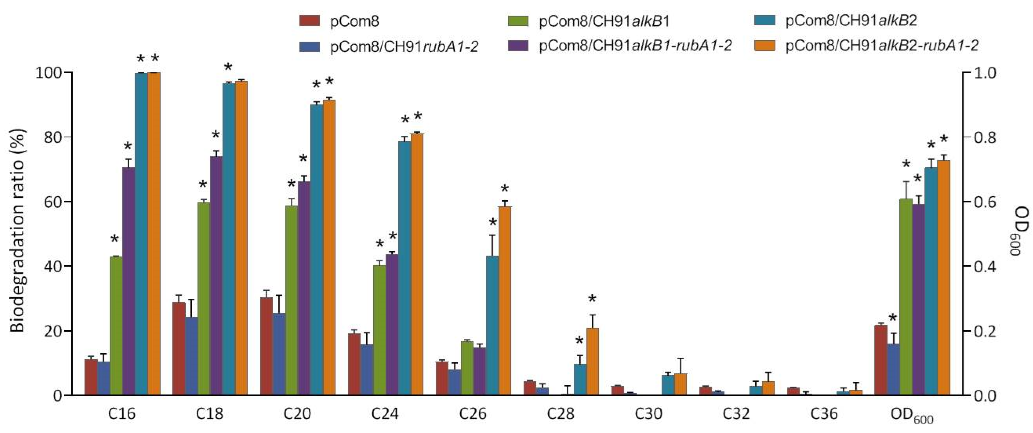
To further elucidate the functions of the two alkB genes in strain CH91, several efforts were made to complement the individual alkB1 and alkB2 CH91 mutants, however, we could not find a suitable expression vector to develop the complementing assays. Therefore, alkB1 and alkB2 genes were cloned into the vector pCom8 and expressed in P. fluorescens KOB2Δ1. The recombinants KOB2Δ1 (pCom8/CH91alkB1) and KOB2Δ1 (pCom8/CH91alkB2) showed much better growth than the control strain KOB2Δ1 (pCom8) when grown on long-chain n-alkane mixture (Figure 4). Alkane degradation analysis revealed that the recombinant KOB2Δ1 (pCom8/CH91alkB1) degraded more C16–C26 n-alkanes and the recombinants KOB2Δ1 (pCom8/CH91alkB2) showed an increased degradation on C16–C30 n-alkanes when compared to those control strains harboring vector only (Figure 4). AlkB1 and AlkB2 showed activity on the different chain length ranges of n-alkane in KOBΔ1 from that in CH91. Possible reasons may be the presence of (i) a different regulatory mechanism responsible for longer-chain alkanes between KOB2Δ1 and CH91; (ii) an unknown factor(s) in the alkane hydroxylase system of CH91; (iii) a limitation in the uptake/catabolism of C32–C36 alkanes in strain KOB2Δ1. Smits et al. reported that P. fluorescens KOB2Δ1 is no longer able to grow on C12 to C16 alkanes but could grow on C18–C28 n-alkanes [15]. However, these results could indicate that CH91AlkB1 and CH91AlkB2 can degrade long-chain n-alkanes.
When the recombinants were grown on the n-alkane mixture, the degradation rate was reduced with the increasing chain length, which is in accordance with the results of two mutants (CH91ΔalkB1 and CH91ΔalkB2) grown on the n-alkanes mixture. These data could indicate that the specific activity of CH91AlkB1 and CH91AlkB2 enzymes is reduced with increasing chain length, as it excludes the influence of different expression levels induced by different chain length n-alkane by using the n-alkanes mixture as the sole carbon source. Similar characteristics of alkane hydroxylase are also found in other studies [20,28,39]. In addition, KOB2Δ1 (pCom8/CH91alkB2) exhibited better growth and degradation ability than KOB2Δ1 (pCom8/CH91alkB1), which suggests the higher enzyme activity of CH91AlkB2 than CH91AlkB1.
The alkB2 and rubA1 genes and the rubA1 and rubA2 genes in the alkB2 cluster of strain CH91 have overlapping stop and start codons, which indicates the translational coupling of rubA1 and rubA2 to alkB2 for the production of stoichiometric amounts of the involved proteins and the important role of RubA1 and RubA2 for the function of AlkB [14]. We also tested the function of the two rubA genes by heterologous expression in KOB2Δ1. The alkB2-rubA1-2 gene coupling fragment was directly amplified by PCR and cloned into the plasmid pCom8. The alkB1-rubA1-2 gene coupling fragment was constructed by replacing alkB2 in the alkB2-rubA1-2 gene coupling fragment with the alkB1 gene using the Gibson assembly method [29]. The recombinants KOB2Δ1 (pCom8/CH91alkB1-rubA1-2) and KOB2Δ1 (pCom8/CH91alkB2-rubA1-2) showed similar growth but higher n-alkane degradability in comparison with the recombinants KOB2Δ1 (pCom8/CH91alkB1) and KOB2Δ1 (pCom8/CH91alkB2), respectively (Figure 4). The growth and n-alkane degradation was not restored by the pCom8-rubA1-2 plasmid carrying just only the two rubA genes. The results indicate that the co-expression of the coupling rubredoxins can enhance the alkane hydroxylation activities of CH91AlkB1 and CH91AlkB2 in P. fluorescens KOB2Δ1 but cannot broaden the chain length range of n-alkane degradation. This is in accordance with the previous result of Nie et al. as determined in Dietzia sp. DQ12-45-1b. They identified the rubredoxin domain as being necessary for the hydroxylation of long-chain n-alkanes with chain lengths ranging from C18 to C32 [39].

4. Conclusions

This study presented experimental evidence for elucidating the functional role of AlkB-type hydroxylase in the degradation of a broad range of n-alkanes. Combining the results of the transcriptional expression, knockout mutation, and heterologous expression of n-alkane hydroxylase genes (alkB1 and alkB2) from strain CH91, it can be concluded that both novel AlkB1 and AlkB2 hydroxylases are responsible for C16–C36 n-alkanes’ degradation in Rhodococcus sp. CH91 and AlkB2 appeared to play a more important role than AlkB1. The results of the recombinants made in Pseudomonas and the mutants in Rhodococcus grown on the n-alkane mixture indicate that the enzyme activities of CH91AlkB1 and CH91AlkB2 are reduced with increasing chain length. The different profiles of the transcriptional expression as well as the n-alkane degradation of alkB1 and alkB2 in response to different n-alkane substrates suggest the expression of alkB1 and alkB2 follows different regulatory mechanisms. The experimental results substantially contribute to understanding the metabolic pathway of the long-chain n-alkane biodegradation in Rhododcoccus sp. CH91, and also provide potential gene candidates for engineering the bacteria used for bioremediation of petroleum hydrocarbon polluted sites.

Supplementary Materials

The following supporting information can be downloaded at: https://www.mdpi.com/article/10.3390/microorganisms11061537/s1, Table S1: Bacterial strains and plasmids used in this study; Table S2: Primers used in this study.

Author Contributions

Conceptualization, Y.X., and Y.M.; methodology, W.X. and S.H.; validation, W.X.; S.H. and Y.X.; formal analysis, W.X. and S.H.; investigation, W.X. and S.H.; data curation, W.X. and Y.X.; writing—original draft preparation, W.X. and Y.X.; writing—review and editing, Y.X. and Y.M.; visualization, W.X. and Y.X.; supervision, Y.M.; project administration, Y.X. and Y.M.; funding acquisition, Y.X. All authors have read and agreed to the published version of the manuscript.

Funding

This research was funded by the National Key R&D Program of China, grant number 2018YFA0902101.

Data Availability Statement

The GenBank/EMBL/DDBJ accession numbers for the sequences reported in this paper are OM831274 for the Rhodococcus sp. strain CH91 alkB1 gene, OM831275 for the Rhodococcus sp. strain CH91 alkB2 region.

Acknowledgments

The authors appreciate Theo Smits for kindly providing P. fluorescens KOB2Δ1 and the pCom8 plasmid. We thank Huimin Yu for kindly providing the CRISPR/Cas9-mediated triple-plasmid genome editing system.

Conflicts of Interest

The authors declare no conflict of interest.

References

  1. Varjani, S.J. Microbial degradation of petroleum hydrocarbons. Bioresour. Technol. 2017, 223, 277–286. [Google Scholar] [CrossRef] [PubMed]
  2. Rodrigues, C.J.C.; de Carvalho, C.C.C.R. Phenotypic adaptations help Rhodococcus erythropolis cells during the degradation of paraffin wax. Biotechnol. J. 2019, 14, e1800598. [Google Scholar] [CrossRef] [PubMed]
  3. Kong, W.; Zhao, C.; Gao, X.; Wang, L.; Tian, Q.; Liu, Y.; Xue, S.; Han, Z.; Chen, F.; Wang, S. Characterization and transcriptome analysis of a long-chain n-alkane-degrading strain Acinetobacter pittii SW-1. Int. J. Environ. Res. Public Health 2021, 18, 6365. [Google Scholar] [CrossRef] [PubMed]
  4. Romanova, V.; Markelova, M.; Boulygina, E.; Siniagina, M.; Müller, R.; Grigoryeva, T.; Laikov, A. Significance of both alkB and P450 alkane-degrading systems in Tsukamurella tyrosinosolvens: Proteomic evidence. Appl. Microbiol. Biotechnol. 2022, 106, 3153–3171. [Google Scholar] [CrossRef]
  5. Wentzel, A.; Ellingsen, T.E.; Kotlar, H.K.; Zotchev, S.B.; Throne-Holst, M. Bacterial metabolism of long-chain n-alkanes. Appl. Microbiol. Biotechnol. 2007, 76, 1209–1221. [Google Scholar] [CrossRef]
  6. SadrAzodi, S.M.; Shavandi, M.; Amoozegar, M.A.; Mehrnia, M.R. Biodegradation of long chain alkanes in halophilic conditions by Alcanivorax sp. strain Est-02 isolated from saline soil. 3 Biotech. 2019, 9, 141. [Google Scholar] [CrossRef]
  7. Rojo, F. Degradation of alkanes by bacteria. Environ. Microbiol. 2009, 11, 2477–2490. [Google Scholar] [CrossRef]
  8. van Beilen, J.B.; Wubbolts, M.G.; Witholt, B. Genetics of alkane oxidation by Pseudomonas oleovorans. Biodegradation 1994, 5, 161–174. [Google Scholar] [CrossRef]
  9. van Beilen, J.B.; Panke, S.; Lucchini, S.; Franchini, A.G.; Röthlisberger, M.; Witholt, B. Analysis of Pseudomonas putida alkane-degradation gene clusters and flanking insertion sequences: Evolution and regulation of the alk genes. Microbiology 2001, 147 Pt 6, 1621–1630. [Google Scholar] [CrossRef] [Green Version]
  10. Williams, S.C.; Austin, R.N. An overview of the electron-transfer proteins that activate alkane monooxygenase (AlkB). Front. Microbiol. 2022, 13, 845551. [Google Scholar] [CrossRef]
  11. Geissdörfer, W.; Kok, R.G.; Ratajczak, A.; Hellingwerf, K.J.; Hillen, W. The genes rubA and rubB for alkane degradation in Acinetobacter sp. strain ADP1 are in an operon with estB, encoding an esterase, and oxyR. J. Bacteriol. 1999, 181, 4292–4298. [Google Scholar] [CrossRef] [Green Version]
  12. Ratajczak, A.; Geissdörfer, W.; Hillen, W. Alkane hydroxylase from Acinetobacter sp. strain ADP1 is encoded by alkM and belongs to a new family of bacterial integral-membrane hydrocarbon hydroxylases. Appl. Environ. Microbiol. 1998, 64, 1175–1179. [Google Scholar] [CrossRef] [Green Version]
  13. Throne-Holst, M.; Markussen, S.; Winnberg, A.; Ellingsen, T.E.; Kotlar, H.K.; Zotchev, S.B. Utilization of n-alkanes by a newly isolated strain of Acinetobacter venetianus: The role of two AlkB-type alkane hydroxylases. Appl. Microbiol. Biotechnol. 2006, 72, 353–360. [Google Scholar] [CrossRef]
  14. Whyte, L.G.; Smits, T.H.; Labbé, D.; Witholt, B.; Greer, C.W.; van Beilen, J.B. Gene cloning and characterization of multiple alkane hydroxylase systems in Rhodococcus strains Q15 and NRRL B-16531. Appl. Environ. Microbiol. 2002, 68, 5933–5942. [Google Scholar] [CrossRef] [Green Version]
  15. Smits, T.H.; Balada, S.B.; Witholt, B.; van Beilen, J.B. Functional analysis of alkane hydroxylases from gram-negative and gram-positive bacteria. J. Bacteriol. 2002, 184, 1733–1742. [Google Scholar] [CrossRef] [Green Version]
  16. van Beilen, J.B.; Funhoff, E.G. Alkane hydroxylases involved in microbial alkane degradation. Appl. Microbiol. Biotechnol. 2007, 74, 13–21. [Google Scholar] [CrossRef] [Green Version]
  17. Throne-Holst, M.; Wentzel, A.; Ellingsen, T.E.; Kotlar, H.K.; Zotchev, S.B. Identification of novel genes involved in long-chain n-alkane degradation by Acinetobacter sp. strain DSM 17874. Appl. Environ. Microbiol. 2007, 73, 3327–3332. [Google Scholar] [CrossRef] [Green Version]
  18. Li, Y.P.; Pan, J.C.; Ma, Y.L. Elucidation of multiple alkane hydroxylase systems in biodegradation of crude oil n-alkane pollution by Pseudomonas aeruginosa DN1. J. Appl. Microbiol. 2020, 128, 151–160. [Google Scholar] [CrossRef] [Green Version]
  19. Feng, L.; Wang, W.; Cheng, J.; Ren, Y.; Zhao, G.; Gao, C.; Tang, Y.; Liu, X.; Han, W.; Peng, X.; et al. Genome and proteome of long-chain alkane degrading Geobacillus thermodenitrificans NG80-2 isolated from a deep-subsurface oil reservoir. Proc. Natl. Acad. Sci. USA 2007, 104, 5602–5607. [Google Scholar] [CrossRef] [Green Version]
  20. Lo Piccolo, L.; De Pasquale, C.; Fodale, R.; Puglia, A.M.; Quatrini, P. Involvement of an alkane hydroxylase system of Gordonia sp. strain SoCg in degradation of solid n-alkanes. Appl. Environ. Microbiol. 2011, 77, 1204–1213. [Google Scholar] [CrossRef] [Green Version]
  21. Park, C.; Shin, B.; Jung, J.; Lee, Y.; Park, W. Metabolic and stress responses of Acinetobacter oleivorans DR1 during long-chain alkane degradation. Microb. Biotechnol. 2017, 10, 1809–1823. [Google Scholar] [CrossRef] [PubMed]
  22. Xiang, W.; Liang, Y.; Hong, S.; Wang, G.; You, J.; Xue, Y.; Ma, Y. Degradation of long-chain n-alkanes by a novel thermal-tolerant Rhodococcus strain. Arch. Microbiol. 2022, 204, 259. [Google Scholar] [CrossRef] [PubMed]
  23. Lageveen, R.G.; Huisman, G.W.; Preusting, H.; Ketelaar, P.; Eggink, G.; Witholt, B. Formation of polyesters by Pseudomonas oleovorans: Effect of Substrates on Formation and Composition of Poly-(R)-3-Hydroxyalkanoates and Poly-(R)-3-Hydroxyalkenoates. Appl. Environ. Microbiol. 1988, 54, 2924–2932. [Google Scholar] [CrossRef] [PubMed] [Green Version]
  24. Mandel, M.; Higa, A. Calcium-dependent bacteriophage DNA infection. J. Mol. Biol. 1970, 53, 159–162. [Google Scholar] [CrossRef]
  25. Schmittgen, T.D.; Livak, K.J. Analyzing real-time PCR data by the comparative C(T) method. Nat. Protoc. 2008, 3, 1101–1108. [Google Scholar] [CrossRef]
  26. Liang, Y.; Jiao, S.; Wang, M.; Yu, H.; Shen, Z. A CRISPR/Cas9-based genome editing system for Rhodococcus ruber TH. Metab. Eng. 2020, 57, 13–22. [Google Scholar] [CrossRef]
  27. Smits, T.H.; Seeger, M.A.; Witholt, B.; van Beilen, J.B. New alkane-responsive expression vectors for Escherichia coli and Pseudomonas. Plasmid 2001, 46, 16–24. [Google Scholar] [CrossRef]
  28. Boonmak, C.; Takahashi, Y.; Morikawa, M. Cloning and expression of three ladA-type alkane monooxygenase genes from an extremely thermophilic alkane-degrading bacterium Geobacillus thermoleovorans B23. Extremophiles 2014, 18, 515–523. [Google Scholar] [CrossRef]
  29. Casini, A.; MacDonald, J.T.; De Jonghe, J.; Christodoulou, G.; Freemont, P.S.; Baldwin, G.S.; Ellis, T. One-pot DNA construction for synthetic biology: The Modular Overlap-Directed Assembly with Linkers (MODAL) strategy. Nucleic Acids Res. 2014, 42, e7. [Google Scholar] [CrossRef] [Green Version]
  30. Højberg, O.; Schnider, U.; Winteler, H.V.; Sørensen, J.; Haas, D. Oxygen-sensing reporter strain of Pseudomonas fluorescens for monitoring the distribution of low-oxygen habitats in soil. Appl. Environ. Microbiol. 1999, 65, 4085–4093. [Google Scholar] [CrossRef] [Green Version]
  31. Kumar, S.; Stecher, G.; Tamura, K. MEGA7: Molecular Evolutionary Genetics Analysis Version 7.0 for Bigger Datasets. Mol. Biol. Evol. 2016, 33, 1870–1874. [Google Scholar] [CrossRef] [Green Version]
  32. Shanklin, J.; Whittle, E.; Fox, B.G. Eight histidine residues are catalytically essential in a membrane-associated iron enzyme, stearoyl-CoA desaturase, and are conserved in alkane hydroxylase and xylene monooxygenase. Biochemistry 1994, 33, 12787–12794. [Google Scholar] [CrossRef]
  33. van Beilen, J.B.; Penninga, D.; Witholt, B. Topology of the membrane-bound alkane hydroxylase of Pseudomonas oleovorans. J. Biol. Chem. 1992, 267, 9194–9201. [Google Scholar] [CrossRef]
  34. Tani, A.; Ishige, T.; Sakai, Y.; Kato, N. Gene structures and regulation of the alkane hydroxylase complex in Acinetobacter sp. strain M-1. J. Bacteriol. 2001, 183, 1819–1823. [Google Scholar] [CrossRef] [Green Version]
  35. Takei, D.; Washio, K.; Morikawa, M. Identification of alkane hydroxylase genes in Rhodococcus sp. strain TMP2 that degrades a branched alkane. Biotechnol. Lett. 2008, 30, 1447–1452. [Google Scholar] [CrossRef] [Green Version]
  36. Laczi, K.; Kis, Á.; Horváth, B.; Maróti, G.; Hegedüs, B.; Perei, K.; Rákhely, G. Metabolic responses of Rhodococcus erythropolis PR4 grown on diesel oil and various hydrocarbons. Appl. Microbiol. Biotechnol. 2015, 99, 9745–9759. [Google Scholar] [CrossRef] [Green Version]
  37. Marín, M.M.; Yuste, L.; Rojo, F. Differential expression of the components of the two alkane hydroxylases from Pseudomonas aeruginosa. J. Bacteriol. 2003, 185, 3232–3237. [Google Scholar] [CrossRef] [Green Version]
  38. van Beilen, J.B.; Marín, M.M.; Smits, T.H.; Röthlisberger, M.; Franchini, A.G.; Witholt, B.; Rojo, F. Characterization of two alkane hydroxylase genes from the marine hydrocarbonoclastic bacterium Alcanivorax borkumensis. Environ. Microbiol. 2004, 6, 264–273. [Google Scholar] [CrossRef]
  39. Nie, Y.; Liang, J.; Fang, H.; Tang, Y.Q.; Wu, X.L. Two novel alkane hydroxylase-rubredoxin fusion genes isolated from a Dietzia bacterium and the functions of fused rubredoxin domains in long-chain n-alkane degradation. Appl. Environ. Microbiol. 2011, 77, 7279–7288. [Google Scholar] [CrossRef] [Green Version]
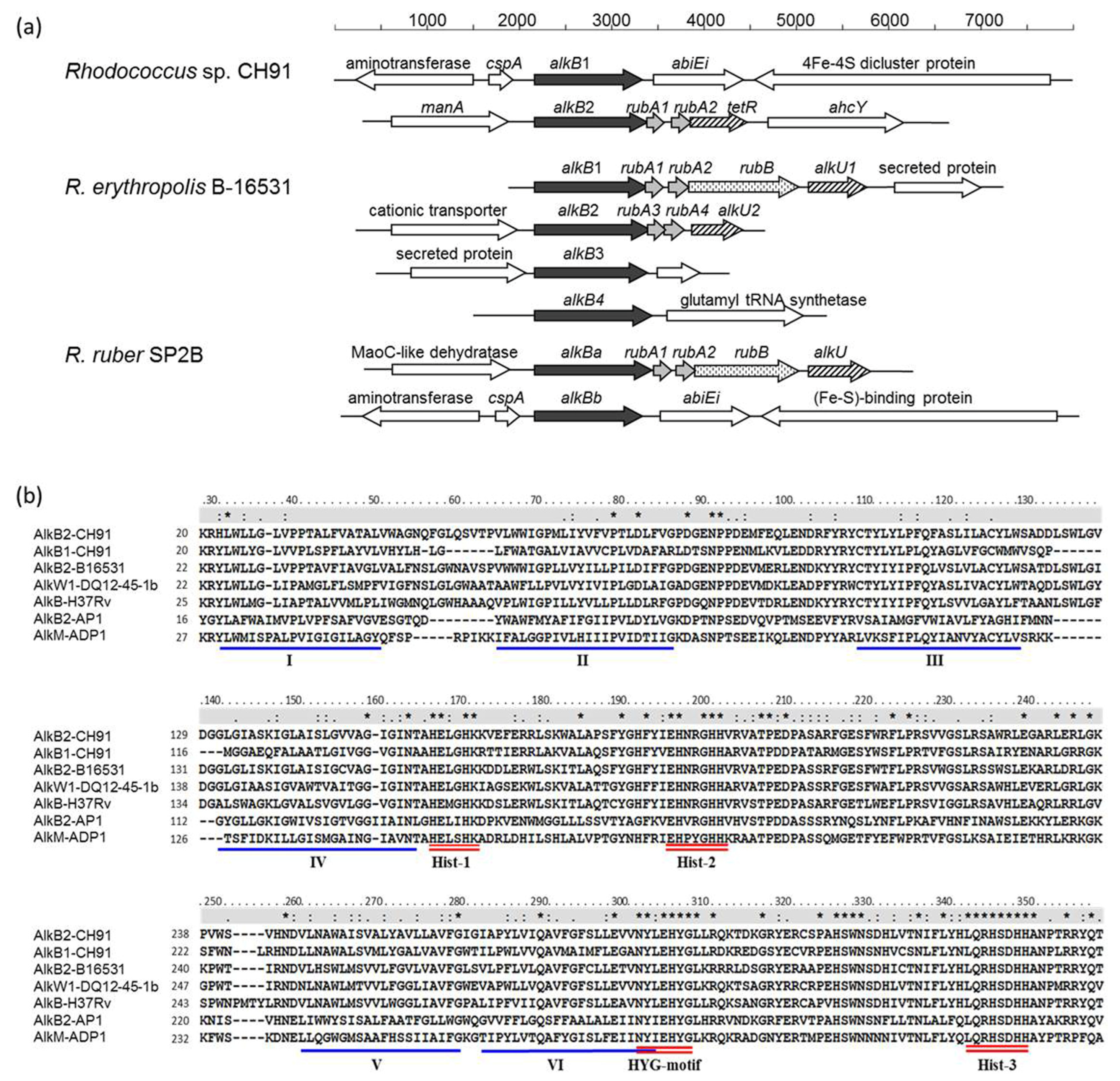
Figure 1. (a) Representative gene organizations of alkB gene regions from Rhodococcus strains. Similar shading patterns of the arrows represent similar functions: black arrow, alkB gene-encoding alkane hydroxylase; grey arrow, rubA-encoding rubredoxin; vertically striped arrow, rub-encoding rubredoxin reductase; diagonally striped arrow, tetR/alkU transcriptional regulator protein; open arrow, other genes; (b) Part of the multiple sequence alignments of Rhodococcus sp. CH91 AlkBs with some other known alkane hydroxylases. Conserved histidine boxes (Hist-1, Hist-2, and Hist-3) and the HYG motif are underlined by a red double line. The six putative transmembrane helices are underlined by a blue single line and marked by Roman numerals. The line above the alignment indicates the conserved positions: “*”, a fully conserved amino acid residue in all aligned alkane hydroxylases; “:”, a conserved strong group positions; “.”, a conserved weaker group position. AlkB2-B16531, alkane hydroxylase from R. erythropolis B-16531 (accession number CAC37038); AlkW1-DQ12-45-1b, Dietzia sp. DQ12-45-1b (AEM66514); AlkB-H37Rv, Mycobacterium tuberculosis H37Rv (NP_217769); AlkB2-AP1, Alcanivorax borkumensis AP1 (AJ577851); AlkM-ADP1, Acinetobacter sp. ADP1 (AJ002316).
Microorganisms 11 01537 g001
Figure 2. Quantitative RT-PCR analysis of the transcription levels of alkB1 and alkB2 in strain CH91 grown on n-alkanes C16 to C36 in length. 16S rRNA as an internal control for normalization. The Left Y axis represents the expression level of alkB1, and the right Y axis represents the expression level of alkB2. Mix means n-alkane mixture containing C16, C18, C20, C24, C26, C28, C30, C32 and C36. The data represent mean  ±  SD. *, p  <  0.05 compared to succinate control (n  =  3/group).
Microorganisms 11 01537 g002
Figure 3. Growth and degradation of strain CH91 wild-type and mutants on different n-alkanes. (a,b) growth and degradation, respectively, after cultivation at 37 °C in MD878 medium supplemented with 0.4% (v/v) of C16, C18, or C20 for 10 days or 0.1% (w/v) of C24, C28, C32, or C36 as the sole carbon source for 21 days; (c,d) growth and degradation after cultivation at 37 °C for 14 days and 24 days, respectively, with the n-alkane mixture (contained 0.05% C16 and 0.01% each of C18, C20, C22, C24, C26, C28, C30, C32, and C36) as the sole carbon sources. Growth was indicated by the optical density at 600 nm and n-alkane degradation was determined by GC-MS analysis of the residual n-alkane. The mean values and standard deviations are shown. *, p < 0.05 compared with wild strain CH91 (n = 3/group).
Microorganisms 11 01537 g003
Figure 4. Growth and degradation of P. fluorescens KOB2Δ1 recombinants harboring pCom8 with CH91alkB genes on n-alkanes. Cells were grown on E2 medium supplemented with the n-alkane mixture (contained 0.05% C16 and 0.01% each of C18, C20, C24, C26, C28, C30, C32 and C36) as sole carbon source at 30 °C for 21 days. Growth was indicated by the optical density at 600 nm and degradation of n-alkane was determined by GC-MS analysis of the residual n-alkane. The mean values and standard deviations are shown. *, p < 0.05 compared with pCom8 control (n = 3/group).
Microorganisms 11 01537 g004
Disclaimer/Publisher’s Note: The statements, opinions and data contained in all publications are solely those of the individual author(s) and contributor(s) and not of MDPI and/or the editor(s). MDPI and/or the editor(s) disclaim responsibility for any injury to people or property resulting from any ideas, methods, instructions or products referred to in the content.