Article

Application of Novel Short Tandem Repeat Typing for Wickerhamomyces anomalus Reveals Simultaneous Outbreaks within a Single Hospital

by
Bram Spruijtenburg
1,2,
Shivaprakash M. Rudramurthy
3,
Eelco F. J. Meijer
1,2,
Merlijn H. I. van Haren
1,
Harsimran Kaur
3,
Arunaloke Chakrabarti
3,
Jacques F. Meis
1,2,4 and
Theun de Groot
1,2,*
1
Department of Medical Microbiology and Infectious Diseases, Canisius-Wilhelmina Hospital, 6532 SZ Nijmegen, The Netherlands
2
Centre of Expertise in Mycology, Radboud University Medical Center/Canisius Wilhelmina Hospital, 6532 SZ Nijmegen, The Netherlands
3
Department of Medical Microbiology, Postgraduate Institute of Medical Education and Research, Chandigarh 160012, India
4
Department I of Internal Medicine, University of Cologne, Excellence Center for Medical Mycology, 50931 Cologne, Germany
*
Author to whom correspondence should be addressed.
Microorganisms 2023, 11(6), 1525; https://doi.org/10.3390/microorganisms11061525
Submission received: 4 May 2023 / Revised: 31 May 2023 / Accepted: 6 June 2023 / Published: 8 June 2023
(This article belongs to the Special Issue Clinical Microbiology and Antimicrobial Stewardship)

Abstract

:
Wickerhamomyces anomalus, previously known as Candida pelliculosa, occasionally causes candidemia in humans, primarily infecting neonates, and infants. The mortality rate of these invasive infections is high, and isolates with a reduced susceptibility to fluconazole have been reported. W. anomalus outbreaks are regularly reported in healthcare facilities, especially in neonatal intensive care units (NICUs). In order to rapidly genotype isolates with a high-resolution, we developed and applied a short tandem repeat (STR) typing scheme for W. anomalus. Six STR markers were selected and amplified in two multiplex PCRs, M3 and M6, respectively. In total, 90 W. anomalus isolates were typed, leading to the identification of 38 different genotypes. Four large clusters were found, unveiling simultaneous outbreak events spread across multiple units within the same hospital. STR typing results of 11 isolates were compared to whole-genome sequencing (WGS) single nucleotide polymorphism (SNP) calling, and the identified genotypic relationships were highly concordant. We performed antifungal susceptibility testing of these isolates, and a reduced susceptibility to fluconazole was found for two (2.3%) isolates. ERG11 genes of these two isolates were examined using WGS data, which revealed a novel I469L substitution in one isolate. By constructing a homology model for W. anomalus ERG11p, the substitution was found in close proximity to the fluconazole binding site. In summary, we showed multiple W. anomalus outbreak events by applying a novel STR genotyping scheme.

1. Introduction

Fungal diseases affect over a billion people, causing an estimated 1.5 million deaths every year, which appears to be increasing [1,2]. Candida species are one of the leading causative agents of invasive fungal infections, with an attributable high mortality rate primarily affecting those with underlying hematological or oncological malignancies, intensive care unit admission, recent major surgery, and/or central venous catheter placement [3,4,5]. To date, Candida albicans is the single most common cause of invasive candidiasis [6,7], although the share of non-C. albicans spp. yeast infections is increasing, and in some countries, it is surpassing C. albicans [3,7,8]. The emergence of new resistant species, including but not limited to those with acquired resistance such as (pan-)resistant C. auris, and azole-resistant C. tropicalis and C. parapsilosis, poses an additional and potentially even bigger threat [9,10,11,12]. Another emerging species is Wickerhamomyces anomalus, previously known as Candida pelliculosa, Pichia anomala and Hansenula anomala. It is a rare but increasingly reported causative agent of nosocomial fungemia outbreaks, preponderantly in low-birth-weight (LBW) infants and occasionally in immunocompromised patients [13,14,15,16,17,18]. In a multicentric study of pediatric candidemia in ICU settings in India, W. anomalus was reported as the third most common agent, especially in neonates [19].
W. anomalus is a diploid ubiquitous yeast, found in varying environments including fruits, trees, soil, waste water, marine environments and even the guts of insects [20,21]. Surprisingly, W. anomalus is highly tolerant to environmental stresses. It can develop biofilms and is adapted to a wide range of growth conditions, including temperature (3–37 °C) and pH value (2–12), and has even demonstrated unusual antimicrobial activity against other yeasts, filamentous fungi, and bacteria [22,23]. Its unique properties have made this yeast biotechnologically interesting, leading to its current use in food, environmental, industrial, and medical applications [21].
Taxonomical (mainly genus) changes in the field of mycology have recently drastically changed the naming of Candida spp., mostly due to the widespread application of molecular technologies in taxonomy, allowing for corrections in the existing system [24]. However, these novel names may not yet be globally implemented and of semantic consideration; here, W. anomalus is used instead of Candida pelliculosa, Pichia anomala, or Hansenula anomala [25]. MALDI-TOF MS using an updated valid database and molecular diagnostics like ITS sequencing or whole genome sequencing (WGS) allow accurate identification of this species, and molecular diagnostics such as ITS sequencing or whole-genome sequencing (WGS) can be used to verify species identification. Correct species identification using biochemical tests or other commercial yeast diagnostic kits can be challenging [26].
Globally, antifungal-resistant isolates are emerging, especially against azoles [5,7,27,28]. Azole resistance can either be intrinsic, for example, in Candida krusei or Candida glabrata, or can be acquired in species such as Candida tropicalis or C. parapsilosis [26,27]. Acquired fluconazole resistance in Candida species is often caused by missense mutations in ERG11, which decrease fluconazole binding affinity to ERG11 and therefore maintain membrane integrity [29,30]. Other resistance mechanisms include missense mutations in other ERG genes, or mutations in transcription factors which induce upregulation of multidrug efflux pumps [31]. The emergence of multiple W. anomalus isolates with a decreased susceptibility to fluconazole have been recently reported, although resistance mechanisms have not been described [13,32,33].
W. anomalus hospital outbreaks have been observed, predominantly in neonatal intensive care units (NICUs) or pediatric wards, often related to contaminated foods or horizontal spread from healthcare personnel due to insufficient infection prevention and control measures [13,18,34,35,36,37]. When W. anomalus is identified in multiple patients in a single healthcare facility, rapid and high-resolution genotyping is essential. With an adequate genotyping method, cross-transmission can be investigated, the source of infection can be localized, and transmission may be controlled. W. anomalus isolates have been typed with several methods, including multilocus enzyme electrophoresis (MLEE), electrophoretic karyotyping (EK), inter-repeat PCR (IR-PCR) and random amplified polymorphic DNA (RAPD) assays [18,32]. Many of these methods lack the necessary discriminatory power and are lengthy and laborious to perform. Therefore, these methods have largely been rendered futile due to recently developed PCR and sequencing-based methods. WGS has the best resolution, is reliable and has excellent reproducibility and discriminatory power; however, it is costly and has a long turn-around time, the latter being undesirable in possible outbreak settings.
An appealing alternative genotyping method with a high resolution is short tandem repeat (STR) or microsatellite genotyping, which is set up for species separately and has proved to be highly reproducible, rapid and cost-effective while being easy to execute [38,39,40,41]. STR genotyping results are straightforward, and they are suitable to be exchanged between laboratories. However, this method might not be amenable in resource limited labs due to the costs. Here, we applied a novel STR typing scheme to W. anomalus isolates, mainly retrieved from a large tertiary care center in India. After having validated the STR genotyping by WGS SNP analysis, we describe multiple large outbreak events within a single hospital.

2. Materials and Methods

2.1. Isolates

In total, 90 clinical W. anomalus isolates were used in this study (Table S1). In addition, 14 other Candida, Cryptococcus, Saccharomyces and Kodameae species were included to test the specificity of the novel typing scheme (Table S2). Isolates were stored according to standard procedures at −80 °C. All isolates were identified by matrix-assisted laser desorption ionization–time-of-flight (MALDI-TOF) mass spectrometry as previously described [42]. This study was approved by the ethics committee of the Postgraduate Institute of Medical Education and Research (PGIMER).

2.2. Culture and DNA Extraction

Isolates were taken from storage at −80 °C and grown on Sabouraud agar plates (Oxoid, Hampshire, UK) at 30 °C. For STR genotyping and WGS, a single colony of each isolate was resuspended in 400 µL MagNA Pure bacteria lysis buffer and MagNA Lyser green beads. These were mechanically lysed for 30 s at 6500 rpm using the MagNA Lyser system (all Roche Diagnostics GmbH, Mannheim, Germany). DNA was extracted and purified with the MagNA Pure 96 instrument and the MagNA Pure DNA and Viral NA Small Volume Kit (Roche Diagnostics), following the manufacturer’s instruction. For WGS, all samples were subsequently treated with RNase (Sigma-Aldrich, Burlington, MA, USA) at a final concentration of 5 µg/µL for one hour at room temperature, after which DNA was extracted and purified as described above. Purified DNA was measured with a Qubit 3.0 Fluorometer (Thermo Fisher Scientific, Waltham, MA, USA) using the double-stranded DNA (dsDNA) high sensitivity option.

2.3. Identification of STR Loci

The W. anomalus reference genome KG16 (GCA_019321765.1) was downloaded from the NCBI database and uploaded to the Tandem Repeat Finder (https://tandem.bu.edu/trf/trf.html, accessed on 4 July 2022) [43]. STRs were identified using the advanced search option (alignment parameter, 2,7,7; minimum alignment score to report repeat, 90; maximum period size, 10; maximum tandem repeat array size, 2). Repeats with insertions or deletion, exhibiting a <90% perfect match to the repeat sequence or having a copy number of <10, were excluded. From the remaining STRs, three repeats with a period size of three nucleotides and three repeats with a period size of six nucleotides were selected based on their copy number.

2.4. Whole-Genome Sequencing

Genomic libraries were prepared and sequenced with the Illumina NovaSeq 6000 platform (Illumina, San Diego, CA, USA) with 2-by-150 bp paired-end-read mode at Eurofins Genomics (Ebersberg, Germany). Read data were uploaded to the Galaxy tool, FastQC was used to assess the quality of the read data, and no trimming was performed [44]. Read data were aligned against the W. anomalus reference genome KG16 (GCA_019321765.1) using BWA-MEM [45]. PCR duplicates were removed using RmDup, local realignment was performed with BamLeftAlign, and unpaired reads were removed with BAM filter. Mapped reads with a MAPQ score < 60 were removed. WGS alignments of the six selected STRs and flanking sequences were visually inspected using JBrowse v1.16.11 to identify variants in primer binding sites [46]. All raw read data generated in this study have been submitted to the National Center for Biotechnology Information’s Sequence Read Archive (BioProject ID: PRJNA851035).

2.5. Primer Design and PCR Genotyping

Primers were designed with Primer3Plus (https://www.bioinformatics.nl/cgi-bin/primer3plus/primer3plus.cgi, accessed on 12 July 2022), using default settings, except for primer size (minimum [Min], 19; optimum [Opt], 21; maximum [Max], 24), primer melting temperature (Tm) (Min, 57.0; Opt, 59.0; Max, 62.0), Max-Poly-X (3) and GC clamp (1) [47]. Variants in STR flanking sequences were marked as excluded regions. Primers that formed no self- or cross-dimers with five or more nucleotides from the last seven nucleotides of the 3′ end of a primer according to the multiple-primer analyzer from Thermo Fisher Scientific were ordered via Eurogentec (Cologne, Germany). Multiplex PCR was performed on a thermocycler (Biometra, Westburg, Göttingen, Germany) using 1x FastStart Taq polymerase buffer without MgCl2 (Roche Diagnostics, Germany), deoxynucleotide triphosphates (dNTPs) (0.2 mM), MgCl2 (3 mM), forward (fwd) and reverse (rev) primers (0.1 to 0.5 µM), 1 U FastStart Taq polymerase (Roche Diagnostics, Germany) and isolated DNA. A thermal protocol of 10 min of denaturation at 95 °C followed by 30 cycles consisting of 30 s denaturation at 95 °C, 30 s of annealing at 60 °C and 1 min of extension at 72 °C with a final incubation step for 10 min at 72 °C was used for PCR amplification. PCR products were diluted 1:1000 in water, and 10 µL together with 0.12 µL of the Orange 600 DNA size standard (NimaGen, Nijmegen, The Netherlands) were incubated for 1 min at 95 °C and analyzed on a 3500 XL genetic analyzer (Applied Biosystems, Foster City, CA, USA).

2.6. Data Analysis

All copy numbers of the six STR markers were determined using GeneMapper 5 software (Applied Biosystems). For all markers, minus-A peaks, stutter peaks, those with an intensity of >50% below the highest peak, and bleed-through peaks were discarded. Subsequently, the size of alleles was rounded. Copy numbers for repeats were converted to a binary matrix: 1, if an isolate contained the allele, and 0, if it did not. Relatedness between isolates was analyzed using BioNumerics software v7.6.1 (Applied Maths NV, Sint-Martems-Latem, Belgium) via the unweighted pair group method with arithmetic means (UPGMA), using the multistate categorical similarity coefficient.

2.7. WGS SNP Calling

SNPs in all isolates were detected with Freebayes using the default settings except for allelic scope options (ignore indels, multiple nucleotide polymorphisms [MNPs], and complex events) [48]. Resulting SNPs in the VCF file with a read depth (DP) of <25, a quality (QUAL) of <100, an allele frequency (OA) of between 0.15 × depth (DP) and 0.45 x DP and an allele frequency of between 0.45 × DP and 0.9 × DP were removed. Previously, the WGS SNP calling pipeline was validated with a Candida auris WGS benchmark dataset [49,50]. Phylogenetic analysis was performed with VCF2PopTree, using the genetic drift algorithm, and a MEGA distance based matrix was developed [51]. The matrix was uploaded to MEGA11 v11.0.10, and a phylogenetic tree was generated using the neighbor-joining tree method [52].

2.8. Antifungal Susceptibility Testing (AFST)

The in vitro minimum inhibitory concentrations (MIC) against azoles, amphotericin B and echinocandin drugs were determined according to CLSI microbroth dilution M27 standard [53]. In short, isolates were incubated at a concentration of 1 × 10³–5 × 10³ CFU/mL in RPMI medium. MIC values were read after 24 h of incubation at 35 °C as the lowest antifungal concentration with a 50% growth reduction when compared to the growth control, except for amphotericin B, for which a 100% growth reduction was used. Epidemiological cutoff values (ECVs) were implemented according to the CLSI M57S guideline to identify non-wildtype strains [54]. Applied ECVs are as follows: for amphotericin, B 1 mg/L; for fluconazole, 8 mg/L; for itraconazole, 1 mg/L; for micafungin, 0.12 mg/L; and for voriconazole, 0.25 mg/L.

2.9. Investigation Resistance-Associated Genes

Azole resistance-associated gene sequences of ERG11 (NC_032093.1), UPC2 (JX494823.1) and ERG3 (AF069752.1) in C. albicans were identified in the W. anomalus KG16 genome using Nucleotide BLAST (https://blast.ncbi.nlm.nih.gov/Blast.cgi, accessed on 17 August 2022). Genes were visually inspected for homozygous substitutions solely present in fluconazole resistant isolates with JBrowse [46]. The coding sequence of ERG11 determined in this study of isolate 10-11-09-22 was deposited in GenBank under the accession number ON805829. The ERG11p homology model was constructed with SWISS-MODEL (https://swissmodel.expasy.org/interactive, accessed on 17 August 2022) by using the template Saccharomyces cerevisiae CYP51 (Lanosterol 14-alpha demethylase G73R mutant complexed with fluconazole (5ese.1.A) [55]. The predicted ERG11 protein model was visually inspected to ensure that all major P450 structural motifs (I-helix, FG loop, and the substrate access channel) were intact. The resulting PDB file was uploaded to PyMOL v2.0, and fluconazole and the I469 amino acid were marked [56].

3. Results

3.1. Development and Application of W. anomalus STR Genotyping

With the use of Tandem Repeat Finder, repeats in the W. anomalus reference strain KG16 were identified. Subsequently, a selection was made of three trinucleotide and three hexanucleotide repeats with the highest copy numbers (Table 1). WGS read data from five W. anomalus isolates were mapped against the KG16 genome. Flanking sequences of the selected tandem repeats were visualized to identify conserved regions, present in all five isolates. Primers were designed on these conserved regions in proximity to the repeat, screened for potential cross-dimer formation and coupled to fluorescent probes. The three trinucleotide and three hexanucleotide repeats were combined in two PCR multiplex reactions, M3 and M6, respectively, and optimal primer concentrations were determined (Table 1). Both multiplex PCRs were then applied to 90 clinical W. anomalus isolates, previously identified by MALDI-TOF (Table S1). All Indian isolates originated from the same hospital and were collected between 2018 and 2019 from neonatal and pediatric wards and ICUs. Each isolate exhibited one or two alleles per marker, which was expected because W. anomalus is a diploid species. The genotyping of all 90 W. anomalus isolates resulted in the identification of 38 different genotypes with four large clusters, consisting of between 5 and, at most, 26 isolates (Figure 1). The dates of fungemia were partially known for these closely related isolates, revealing that these outbreaks occurred in 2019 (Figure S1). In the three clusters with known information regarding involved hospital units, isolates originated from more than one hospital unit. Isolates from the Netherlands, Qatar and Italy all exhibited unique genotypes. Genotypes 1 and 2, 5 and 6, 15 and 16, and 19 and 20 were highly related to each other according to STR typing, exhibiting a zygosity difference in only one marker (Figure 1).
For specificity testing, 14 related yeast species were typed by both multiplex PCRs (Table S2). This resulted in no PCR products after amplification for any of the tested species, indicating that the STR assay is specific for W. anomalus. The stability of the STR markers was tested by subcloning five colonies of isolates 10-03-01-56 and 08-32-01-30 for ten generations. None of the copy numbers were altered, indicating high genetic stability. For reproducibility testing of the STR typing, the two previously mentioned isolates were independently genotyped, five times, in five replicate experiments. This resulted in identical results for all STR markers in all experiments, demonstrating that the genotyping method is highly reproducible.

3.2. WGS SNP Analysis

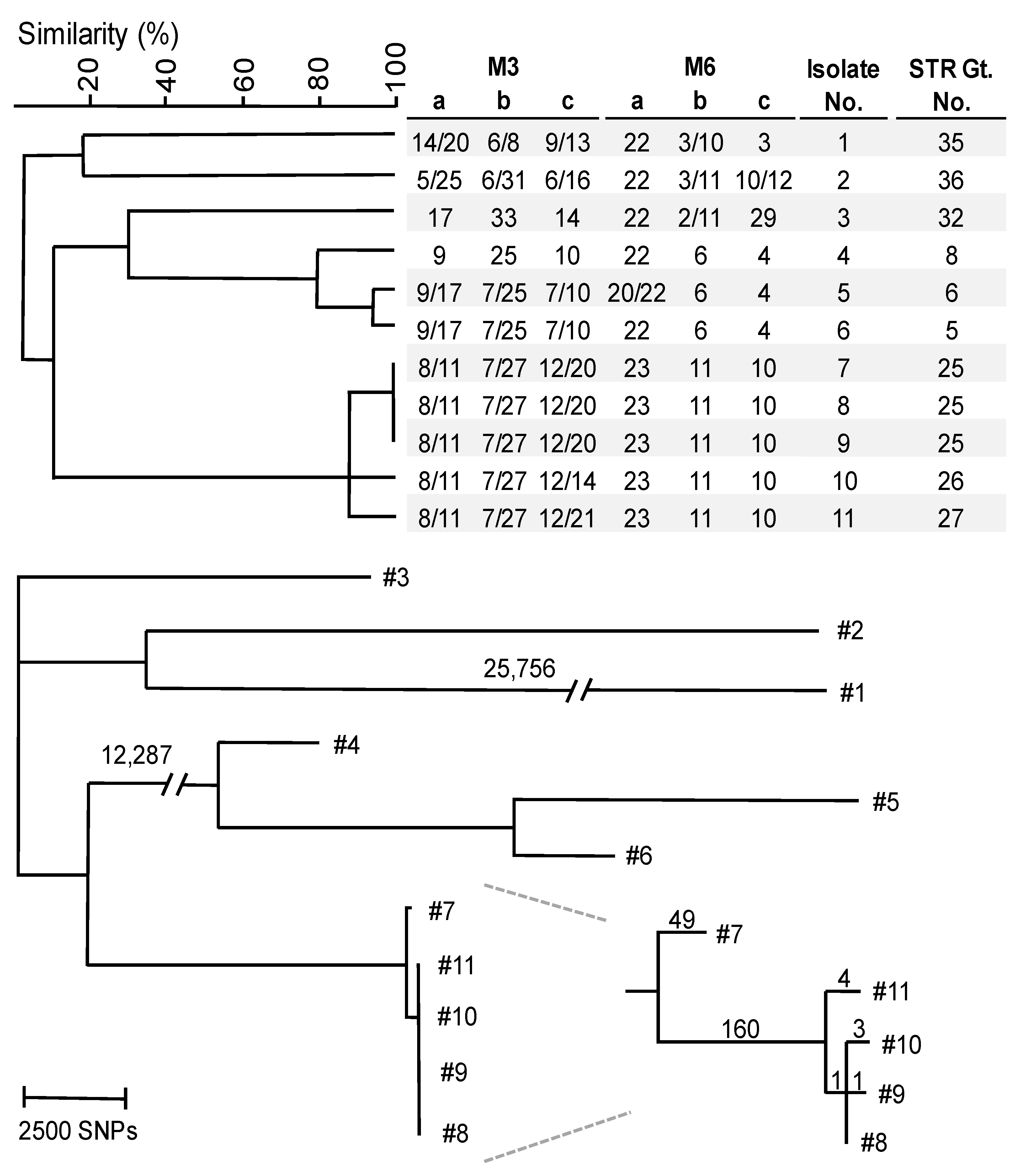
In order to validate genotypical relatedness according to STR typing, 11 isolates were submitted for WGS. Reads from each strain were mapped to the KG16 reference genome, and SNPs between isolates were called with Freebayes (Figure 2). Five isolates from India, highly related or identical according to STR typing, clustered in one group of four isolates with a difference of eight SNPs at most, which was distinguished by 210 SNPs from the remaining isolate. The examination of these 210 SNPs with JBrowse showed that 203 of these SNPs were heterozygous and located in one part of a chromosome of approximately 400 kbp. Isolates with a difference of at least 10,000 SNPs compared to other isolates all demonstrated unique STR genotypes. Copy numbers of repeats with a total length below 140 bp were all confirmed visually in silico for sequenced isolates.

3.3. AFST and Resistance-Associated Genes Investigation

AFST according to microbroth dilution was performed on 87 isolates against several azoles, amphotericin B and two echinocandins, and ECVs according to the CLSI M57S guideline were used to identify non-wildtype strains (Table S1). Only wildtype strains against micafungin were found, while some non-wildtype strains for amphotericin B, fluconazole, itraconazole and voriconazole were identified (Table 2). According to the CLSI ECVs, six isolates were non-wildtype (≥1 mg/L) to amphotericin B. Only two isolates (10-03-01-56 and 10-11-09-22) exhibited non-wildtype fluconazole MICs (8 and 16 mg/L) and originated from Qatar and the Netherlands, respectively. To search for a potential resistance mechanism, azole resistance-associated genes were located and visually inspected in the W. anomalus genome. The W. anomalus ERG11 gene was located and subsequently aligned to C. albicans ERG11 (Figure S2). Isolate 10-11-09-22 harbored an I469L substitution, while no substitutions were found in the other fluconazole non-susceptible isolate. The ERG11 I469L substitution was located in a hotspot region of C. albicans. To investigate the potential impact of this substitution on fluconazole resistance, an ERG11 homology model was constructed, based on a Saccharomyces cerevisiae CYP51 template with a 70.65% sequence identify and a MolProbity Score of 1.14 (Figure 3). The I469 position is located in close proximity to the heme-binding site, which is also used by azole derivatives. No substitutions solely present in fluconazole resistant isolates were found for UPC2 and ERG3. With nucleotide blast, efflux transcription factors TAC1 and MRR1 could not be identified based on orthologous C. albicans genes.

4. Discussion

The present study describes the development of a STR genotyping analysis for W. anomalus. With the typing scheme, six STR markers with a repeat size of three or six nucleotides were amplified by two multiplex PCRs, M3 and M6, respectively. STR genotyping of 90, mainly Indian W. anomalus isolates, resulted in the identification of 38 different genotypes and uncovered hitherto unrecognized outbreak events. WGS SNP analysis of 11 isolates demonstrated similar typing results as those obtained with STR typing, as previously also found for other clinically relevant fungi [38,39,41,57]. With AFST against common antifungals, non-wildtype strains to amphotericin B and azoles were observed for several isolates. One fluconazole non-wildtype isolate harbored an ERG11 substitution in close proximity to the azole binding site.

4.1. W. anomalus Genotyping and Loss of Heterozygosity

The development and application of this W. anomalus STR typing assay showed the phylogenetic relationship between 90 isolates. We identified 38 different genotypes, with four large Indian clusters, consisting of five or more isolates, while isolates from other countries were not closely related. To validate the genotypic relations established via STR typing, WGS SNP analysis was performed on three isolates (#7–9) with an identical STR profile and two isolates (#10 and #11) with a nearly identical STR profile. While isolates #8 to #11 differed by eight SNPs at most, they were separated by around 210 SNPs with isolate #7. The examination of these SNPs revealed that 203 SNPs were clustered in approximately 400 kbp of one chromosome and that they were heterozygous present in isolates #8 to #11. This part of the chromosome was homozygous for isolate #7. This difference in zygosity between these isolates is likely due to the loss of one allele, also known as loss of heterozygosity (LOH). LOH continuously occurs in cells, while its rate increases when cells are exposed to high levels of stress, such as exposure to antifungal drugs [58,59]. Previous research demonstrated that fluconazole can induce large LOH events involving partial or complete chromosomes [41,42,43]. As the patients with W. anomalus infections were treated with antifungals, the LOH event in the deviant isolate might have been caused by this treatment. LOH is normally repaired via the remaining template chromosome via homologous recombination [60]. The number of SNPs in the residual genome, likely accumulated via single point mutations, is only seven in comparison to the other four isolates, indicating their close relatedness. Of all isolates, genotypes 1 and 2, 5 and 6, 15 and 16, and 19 and 20 differed only in zygosity in one marker. These differences might also be due to LOH. Zygosity differences are also frequently observed in other diploid yeasts such as Candida tropicalis and Pichia kudriavzevii [40,41,60]. It is very likely that these are caused by LOH, suggesting that this phenomenon is widespread across yeast genera. Previous research on C. albicans and C. tropicalis demonstrated that LOH increases genetic diversity to rapidly adapt to changing environments by providing fitness advantages and antifungal resistance [61,62,63,64]. All aforementioned isolates were exposed to fluconazole or amphotericin B; however, we did not find a correlation between LOH and susceptibility for antifungals. Altogether, our findings suggest that the described STR scheme can distinguish non-related isolates from those causing an outbreak.

4.2. Simultaneous Outbreaks of Different W. anomalus Strains in Single Hospital

Out of the four clusters, two (genotype #5 and #7) were highly related with a single copy number difference in marker M3-a. The genotypic variations with and among the other two clusters were much larger, with differences in at least four STR markers. All these isolates originated from the same hospital, taken from patients admitted in 2018 and 2019, and spread across multiple departments. Thus, in this time frame, there were likely at least three simultaneous outbreaks of different W. anomalus strains. To date, outbreaks with W. anomalus are rarely described [18,32,33]. Thus, it is remarkable that there were three simultaneous outbreaks in this hospital, suggesting the rather exclusive presence of a certain environmental factor, enriched for W. anomalus, within or around the hospital. This is also confirmed by the other 23 genotypes of W. anomalus that were found in this hospital. As W. anomalus is ubiquitous in the environment, it might be worthwhile to search for the source of introduction within the hospital environment or a shared environmental source [22,65,66].

4.3. Antifungal Susceptibility and I469L Substitution in ERG11

AFST against common antifungals for 87 isolates identified two non-wildtype isolates against fluconazole according to CLSI ECVs [54]. It has to be noted that the ECVs are not associated with clinical outcomes. All tested isolates were wildtype against micafungin, and six isolates were non-wildtype against amphotericin B. The two isolates with the highest fluconazole MIC were examined for variations in resistance-associated genes. Two known transcription factors of efflux pumps could not be identified in the W. anomalus genome. These factors are likely highly deviant from the orthologous genes in C. albicans or might be absent in W. anomalus. The isolate with a MIC value of 16 mg/L harbored a novel I469L substitution, while the other isolate did not harbor any variations. Homology modelling of ERG11 revealed the close proximity of the I469 position to the heme binding site, to which azole derivates bind and inhibit enzyme function. Previously, multiple mutations in the same ERG11 hotspot region in C. albicans have been reported to confer fluconazole resistance [30,67]. Therefore, it is plausible that the I469L substitution alters the azole binding site, and subsequently contributes to the elevated MIC against fluconazole.
In summary, we developed and applied a high-resolution STR typing scheme for W. anomalus that is specific, reproducible and fast executable. This assay consists of two multiplex PCRs, which amplify six markers in total. The genotyping of 90 W. anomalus isolates identified 38 different genotypes, unveiling four simultaneous outbreak events spread across multiple departments within the same Indian hospital. The comparison of STR and WGS SNP genotyping correlated well, indicating the high resolution of this STR typing scheme. With the construction of an ERG11 homology model, a potential role of the I469L substitution in fluconazole resistance was suggested.

Supplementary Materials

The following supporting information can be downloaded at: https://www.mdpi.com/article/10.3390/microorganisms11061525/s1, Figure S1: Minimum-spanning tree of 90 W. anomalus isolates marked on isolates date as shown in the color key. Branch lengths indicate the similarity between isolates with thick solid lines (variation in one allele), thin solid lines (variation in two alleles), thin dashed lines (variation in three alleles) and thin dotted lines (variation in four or more alleles). Q, quarter. Figure S2: Alignment of ERG11 protein sequences of W. anomalus (Canpel_ERG11) and C. albicans (Canalb_ERG11) as generated with Omega Clustal v.1.2.4. Hotspot regions and the I469 position in W. anomalus are marked in yellow and red, respectively. Table S1: Overview of all isolates with MIC values according to broth microdilution methods. MIC values are in mg/L. Table S2: Specificity testing against 14 yeast species.

Author Contributions

Conceptualization, A.C. and J.F.M.; methodology, B.S. and M.H.I.v.H.; software, B.S.; validation, B.S., S.M.R. and M.H.I.v.H.; formal analysis, B.S., E.F.J.M. and T.d.G.; investigation, B.S., S.M.R. and H.K.; resources, S.M.R., H.K. and A.C.; data curation, B.S. and E.F.J.M.; writing—original draft preparation, B.S. and E.F.J.M.; writing—review and editing, S.M.R., M.H.I.v.H., H.K., A.C., J.F.M. and T.d.G.; visualization, B.S. and T.d.G.; supervision, J.F.M. and T.d.G.; project administration, S.M.R. and J.F.M.; funding acquisition, J.F.M. All authors have read and agreed to the published version of the manuscript.

Funding

This research received support from the Canisius Wilhelmina Hospital for the Centre of Expertise in Mycology RadboudUMC/CWZ (grant number CWZ_001421). The funder had no role in the design of the study; in the collection, analyses, or interpretation of data; in the writing of the manuscript; or in the decision to publish the results.

Data Availability Statement

All raw read data generated in this study have been submitted to the National Center for Biotechnology Information’s Sequence Read Archive (BioProject ID: PRJNA851035). The coding sequence of ERG11 determined in this study of isolate 10-11-09-22 was deposited in GenBank under the accession number ON805829.

Conflicts of Interest

The authors declare no conflict of interest.

References

  1. Rokas, A. Evolution of the human pathogenic lifestyle in fungi. Nat. Microbiol. 2022, 7, 607–619. [Google Scholar] [CrossRef] [PubMed]
  2. Bongomin, F.; Gago, S.; Oladele, R.O.; Denning, D.W. Global and Multi-National Prevalence of Fungal Diseases-Estimate Precision. J. Fungi 2017, 3, 57. [Google Scholar] [CrossRef] [Green Version]
  3. Lockhart, S.R.; Guarner, J. Emerging and reemerging fungal infections. Semin. Diagn. Pathol. 2019, 36, 177–181. [Google Scholar] [CrossRef] [PubMed]
  4. Pfaller, M.; Neofytos, D.; Diekema, D.; Azie, N.; Meier-Kriesche, H.U.; Quan, S.P.; Horn, D. Epidemiology and outcomes of candidemia in 3648 patients: Data from the Prospective Antifungal Therapy (PATH Alliance(R)) registry, 2004–2008. Diagn. Microbiol. Infect. Dis. 2012, 74, 323–331. [Google Scholar] [CrossRef]
  5. Hoenigl, M.; Egger, M.; Salmanton-Garcia, J.; Koehler, P.; Gangneux, J.P.; Arendrup, M.C.; Bicanic, T.; Arikan-Akdagli, S.; Cornely, O.A.; ECMM Study Group. Guideline adherence and survival of patients with candidaemia in Europe: Results from the ECMM Candida III multinational European observational cohort study. Lancet Infect. Dis. 2023, 9, ofac492.038. [Google Scholar] [CrossRef]
  6. Lyon, G.M.; Karatela, S.; Sunay, S.; Adiri, Y.; Candida Surveillance Study Investigators. Antifungal susceptibility testing of Candida isolates from the Candida surveillance study. J. Clin. Microbiol. 2010, 48, 1270–1275. [Google Scholar] [CrossRef] [Green Version]
  7. Pfaller, M.A.; Diekema, D.J.; Turnidge, J.D.; Castanheira, M.; Jones, R.N. Twenty Years of the SENTRY Antifungal Surveillance Program: Results for Candida Species From 1997–2016. Open Forum. Infect. Dis. 2019, 6, S79–S94. [Google Scholar] [CrossRef] [Green Version]
  8. Sharma, M.; Chakrabarti, A. Candidiasis and Other Emerging Yeasts. Curr. Fungal Infect. Rep. 2023, 17, 15–24. [Google Scholar] [CrossRef]
  9. Martini, C.; Torelli, R.; de Groot, T.; De Carolis, E.; Morandotti, G.A.; De Angelis, G.; Posteraro, B.; Meis, J.F.; Sanguinetti, M. Prevalence and Clonal Distribution of Azole-Resistant Candida parapsilosis Isolates Causing Bloodstream Infections in a Large Italian Hospital. Front. Cell. Infect. Microbiol. 2020, 10, 232. [Google Scholar] [CrossRef]
  10. Chowdhary, A.; Sharma, C.; Meis, J.F. Candida auris: A rapidly emerging cause of hospital-acquired multidrug-resistant fungal infections globally. PLoS Pathog. 2017, 13, e1006290. [Google Scholar] [CrossRef]
  11. Souza, A.C.; Fuchs, B.B.; Pinhati, H.M.S.; Siqueira, R.A.; Hagen, F.; Meis, J.F.; Mylonakis, E.; Colombo, A.L. Candida parapsilosis Resistance to Fluconazole: Molecular Mechanisms and In Vivo Impact in Infected Galleria mellonella Larvae. Antimicrob. Agents Chemother. 2015, 59, 6581–6587. [Google Scholar] [CrossRef] [Green Version]
  12. Spruijtenburg, B.; Baqueiro, C.C.S.Z.; Colombo, A.L.; Meijer, E.F.J.; de Almeida, J.N., Jr.; Berrio, I.; Fernández, N.B.; Chaves, G.M.; Meis, J.F.; de Groot, T.; et al. Short Tandem Repeat Genotyping and Antifungal Susceptibility Testing of Latin American Candida tropicalis Isolates. J. Fungi 2023, 9, 207. [Google Scholar] [CrossRef] [PubMed]
  13. Kalkanci, A.; Dizbay, M.; Turan, O.; Fidan, I.; Yalçin, B.; Hirfanoğlu, I.; Kuştimur, S.; Aktaş, F.; Sugita, T. Nosocomial transmission of Candida pelliculosa fungemia in a pediatric intensive care unit and review of the literature. Turk. J. Pediatr. 2010, 52, 42–49. [Google Scholar] [PubMed]
  14. Yang, Y.; Wu, W.; Ding, L.; Yang, L.; Su, J.; Wu, B. Two different clones of Candida pelliculosa bloodstream infection in a tertiary neonatal intensive care unit. J. Infect. Dev. Ctries. 2021, 15, 870–876. [Google Scholar] [CrossRef] [PubMed]
  15. Song, K.Y.; Park, C.; Byun, J.H.; Chun, H.S.; Choi, J.H.; Han, E.H.; Lee, S.O.; Jeong, Y.; Kim, Y.J.; Kim, S.H. Fungal arthritis with adjacent osteomyelitis caused by Candida pelliculosa: A case report. BMC Infect. Dis. 2020, 20, 438. [Google Scholar] [CrossRef] [PubMed]
  16. Mehta, V.; Mohanty, A.; Meena, S.; Rahul, J.S.; Kumar, N.U.; Chattopadhyay, D.; Bakliwal, A.; Choudhary, R.; Gupta, P. Wickerhamomyces anomalous: A Rare Cause of Fungemia Causing Febrile Neutropenia in Acute Lymphoblastic Leukemia. Case Rep. Infect. Dis. 2020, 2020, 8847853. [Google Scholar] [CrossRef] [PubMed]
  17. Chan, A.W.; Cartwright, E.J.; Reddy, S.C.; Kraft, C.S.; Wang, Y.F. Pichia anomala (Candida pelliculosa) fungemia in a patient with sickle cell disease. Mycopathologia 2013, 176, 273–277. [Google Scholar] [CrossRef] [PubMed] [Green Version]
  18. Chakrabarti, A.; Singh, K.; Narang, A.; Singhi, S.; Batra, R.; Rao, K.L.N.; Ray, P.; Gopalan, S.; Das, S.; Gupta, A.K.; et al. Outbreak of Pichia anomala infection in the pediatric service of a tertiary-care center in Northern India. J. Clin. Microbiol. 2001, 39, 1702–1706. [Google Scholar] [CrossRef] [PubMed] [Green Version]
  19. Chakrabarti, A.; Sood, P.; Rudramurthy, S.M.; Chen, S.; Jillwin, J.; Iyer, R.; Sharma, A.; Harish, B.N.; Roy, I.; Kindo, A.J.; et al. Characteristics, outcome and risk factors for mortality of paediatric patients with ICU-acquired candidemia in India: A multicentre prospective study. Mycoses 2020, 63, 1149–1163. [Google Scholar] [CrossRef] [PubMed]
  20. Passoth, V.; Olstorpe, M.; Schnurer, J. Past, present and future research directions with Pichia anomala. Antonie Van Leeuwenhoek 2011, 99, 121–125. [Google Scholar] [CrossRef]
  21. Walker, G.M. Pichia anomala: Cell physiology and biotechnology relative to other yeasts. Antonie Van Leeuwenhoek 2011, 99, 25–34. [Google Scholar] [CrossRef] [Green Version]
  22. Steyn, A.; Roets, F.; Botha, A. Yeasts Associated with Culex pipiens and Culex theileri Mosquito Larvae and the Effect of Selected Yeast Strains on the Ontogeny of Culex pipiens. Microb. Ecol. 2016, 71, 747–760. [Google Scholar] [CrossRef] [PubMed]
  23. Zhang, Z.; Cao, Y.; Li, Y.; Chen, X.; Ding, C.; Liu, Y. Risk factors and biofilm formation analyses of hospital-acquired infection of Candida pelliculosa in a neonatal intensive care unit. BMC Infect. Dis. 2021, 21, 620. [Google Scholar] [CrossRef] [PubMed]
  24. Kidd, S.E.; Abdolrasouli, A.; Hagen, F. Fungal Nomenclature: Managing Change is the Name of the Game. Open Forum. Infect. Dis. 2023, 10, ofac559. [Google Scholar] [CrossRef] [PubMed]
  25. Kurtzman, C.P. Phylogeny of the ascomycetous yeasts and the renaming of Pichia anomala to Wickerhamomyces anomalus. Antonie Van Leeuwenhoek 2011, 99, 13–23. [Google Scholar] [CrossRef]
  26. Irinyi, L.; Serena, C.; Garcia-Hermoso, D.; Arabatzis, M.; Desnos-Ollivier, M.; Duong Vu Cardinali, G.; Arthur, I.; Norman, A.C.; Giraldo, A. International Society of Human and Animal Mycology (ISHAM)-ITS reference DNA barcoding database—The quality controlled standard tool for routine identification of human and animal pathogenic fungi. Med. Mycol. 2015, 53, 313–337. [Google Scholar] [CrossRef]
  27. Perlin, D.S.; Rautemaa-Richardson, R.; Alastruey-Izquierdo, A. The global problem of antifungal resistance: Prevalence, mechanisms, and management. Lancet Infect. Dis. 2017, 17, e383–e392. [Google Scholar] [CrossRef]
  28. Arendrup, M.C. Update on antifungal resistance in Aspergillus and Candida. Clin. Microbiol. Infect. 2014, 20, 42–48. [Google Scholar] [CrossRef] [PubMed] [Green Version]
  29. Lupetti, A.; Danesi, R.; Campa, M.; Del Tacca, M.; Kelly, S. Molecular basis of resistance to azole antifungals. Trends Mol. Med. 2002, 8, 76–81. [Google Scholar] [CrossRef] [PubMed]
  30. Xiang, M.J.; Liu, J.Y.; Ni, P.H.; Wang, S.; Shi, C.; Wei, B.; Ni, Y.X.; Ge, H.L. Erg11 mutations associated with azole resistance in clinical isolates of Candida albicans. FEMS Yeast Res. 2013, 13, 386–393. [Google Scholar] [CrossRef] [Green Version]
  31. Whaley, S.G.; Berkow, E.L.; Rybak, J.M.; Nishimoto, A.T.; Barker, K.S.; Rogers, P.D. Azole Antifungal Resistance in Candida albicans and Emerging Non-albicans Candida Species. Front. Microbiol. 2016, 7, 2173. [Google Scholar] [CrossRef] [Green Version]
  32. Barchiesi, F.; Tortorano, A.M.; Flaconi Di Francesco, L.; Rigoni, A.; Giacometti, A.; Spreghini, E.; Scalise, G.; Viviani, M.A. Genotypic variation and antifungal susceptibilities of Candida pelliculosa clinical isolates. J. Med. Microbiol. 2005, 54, 279–285. [Google Scholar] [CrossRef] [Green Version]
  33. Zhang, L.; Xiao, M.; Arastehfar, A.; Ilkit, M.; Zou, J.; Deng Yuchen Xu, Y.; Liao, W.; Zhao, J.; Fang, W. Investigation of the Emerging Nosocomial Wickerhamomyces anomalus Infections at a Chinese Tertiary Teaching Hospital and a Systemic Review: Clinical Manifestations, Risk Factors, Treatment, Outcomes, and Anti-fungal Susceptibility. Front. Microbiol. 2021, 12, 744502. [Google Scholar] [CrossRef]
  34. Lin, H.C.; Lin, H.Y.; Su, B.H.; Ho, M.W.; Ho, C.M.; Lee, C.Y.; Lin, M.H.; Hsieh, H.Y.; Lin, H.C.; Li, T.C.; et al. Reporting an outbreak of Candida pelliculosa fungemia in a neonatal intensive care unit. J. Microbiol. Immunol. Infect. 2013, 46, 456–462. [Google Scholar] [CrossRef] [PubMed] [Green Version]
  35. Jung, J.; Moon, Y.S.; Yoo, J.A.; Lim, J.H.; Jeong, J.; Jun, J.B. Investigation of a nosocomial outbreak of fungemia caused by Candida pelliculosa (Pichia anomala) in a Korean tertiary care center. J. Microbiol. Immunol. Infect. 2018, 51, 794–801. [Google Scholar] [CrossRef]
  36. Yilmaz-Semerci, S.; Demirel, G.; Tastekin, A. Wickerhamomyces anomalus blood stream infection in a term newborn with pneumonia. Turk. J. Pediatr. 2017, 59, 349–351. [Google Scholar] [CrossRef] [Green Version]
  37. Suhr, M.J.; Gomes-Neto, J.C.; Banjara, N.; Florescu, D.F.; Mercer, D.F.; Iwen, P.C.; Hallen-Adams, H.E. Epidemiological investigation of Candida species causing bloodstream infection in paediatric small bowel transplant recipients. Mycoses 2017, 60, 366–374. [Google Scholar] [CrossRef]
  38. de Groot, T.; Spruijtenburg, B.; Parnell, L.A.; Chow, N.A.; Meis, J.F. Optimization and Validation of Candida auris Short Tandem Repeat Analysis. Microbiol. Spectr. 2022, 10, e0264522. [Google Scholar] [CrossRef]
  39. Spruijtenburg, B.; Bombassaro, A.; Meijer, E.F.J.; Rodrigues, A.M.; Grisolia, M.E.; Vicente, V.A.; de Queiroz-Telles, F.; Meis, J.F.; de Groot, T. Sporothrix brasiliensis genotyping reveals numerous independent zoonotic introductions in Brazil. J. Infect. 2023, 86, 610–613. [Google Scholar] [CrossRef] [PubMed]
  40. Spruijtenburg, B.; van Haren, M.H.I.; Chowdhary, A.; Meis, J.F.; de Groot, T. Development and Application of a Short Tandem Repeat Multiplex Typing Assay for Candida tropicalis. Microbiol. Spectr. 2023, 11, e0461822. [Google Scholar] [CrossRef]
  41. van Haren, M.H.I.; de Groot, T.; Spruijtenburg, B.; Jain, K.; Chowdhary, A.; Meis, J.F. Development of a Multiplex PCR Short Tandem Repeat Typing Scheme for Candida krusei. J. Clin. Microbiol. 2022, 60, e0203221. [Google Scholar] [CrossRef] [PubMed]
  42. De Carolis, E.; Vella, A.; Vaccaro, L.; Torelli, R.; Posteraro, P.; Ricciardi, W.; Sanguinetti, M.; Posteraro, B. Development and validation of an in-house database for matrix-assisted laser desorption ionization-time of flight mass spectrometry-based yeast identification using a fast protein extraction procedure. J. Clin. Microbiol. 2014, 52, 1453–1458. [Google Scholar] [CrossRef] [PubMed] [Green Version]
  43. Benson, G. Tandem repeats finder: A program to analyze DNA sequences. Nucleic Acids Res. 1999, 27, 573–580. [Google Scholar] [CrossRef] [Green Version]
  44. Afgan, E.; Baker, D.; Batut, B.; van den Beek, M.; Bouvier, D.; Cech, M.; Chilton, J.; Clements, D.; Coraor, N.; Grüning, B.A.; et al. The Galaxy platform for accessible, reproducible and collaborative biomedical analyses: 2018 update. Nucleic Acids Res. 2018, 46, W537–W544. [Google Scholar] [CrossRef] [Green Version]
  45. Li, H.; Durbin, R. Fast and accurate long-read alignment with Burrows-Wheeler transform. Bioinformatics 2010, 26, 589–595. [Google Scholar] [CrossRef] [Green Version]
  46. Skinner, M.E.; Uzilov, A.V.; Stein, L.D.; Mungall, C.J.; Holmes, I.H. JBrowse: A next-generation genome browser. Genome Res. 2009, 19, 1630–1638. [Google Scholar] [CrossRef] [Green Version]
  47. Untergasser, A.; Nijveen, H.; Rao, X.; Bisseling, T.; Geurts, R.; Leunissen, J.A.M. Primer3Plus, an enhanced web interface to Primer3. Nucleic Acids Res. 2007, 35, W71–W74. [Google Scholar] [CrossRef] [Green Version]
  48. Garrison, E.; Marth, G. Haplotype-based variant detection from short-read sequencing. arXiv 2012, arXiv:1207.3907. [Google Scholar] [CrossRef]
  49. Welsh, R.M.; Misas, E.; Forsberg, K.; Lyman, M.; Chow, N.A. Candida auris Whole-Genome Sequence Benchmark Dataset for Phylogenomic Pipelines. J. Fungi 2021, 7, 214. [Google Scholar] [CrossRef]
  50. Spruijtenburg, B.; Badali, H.; Abastabar, M.; Mirhendi, H.; Khodavaisy, S.; Sharifisooraki, J.; Armaki, M.T.; de Groot, T.; Meis, J.F. Confirmation of fifth Candida auris clade by whole genome sequencing. Emerg. Microbes. Infect. 2022, 11, 2405–2411. [Google Scholar] [CrossRef] [PubMed]
  51. Subramanian, S.; Ramasamy, U.; Chen, D. VCF2PopTree: A client-side software to construct population phylogeny from genome-wide SNPs. PeerJ 2019, 7, e8213. [Google Scholar] [CrossRef] [Green Version]
  52. Tamura, K.; Stecher, G.; Kumar, S. MEGA11: Molecular Evolutionary Genetics Analysis Version 11. Mol. Biol. Evol. 2021, 38, 3022–3027. [Google Scholar] [CrossRef] [PubMed]
  53. Clinical and Laboratory Standards Institute. Reference Method for Broth Dilution Antifungal Susceptibility Testing of Yeasts, 4th ed.; Approved Standard M27; Clinical and Laboratory Standards Institute: Wayne, PA, USA, 2017. [Google Scholar]
  54. Clinical and Laboratory Standards Institute. Epidemiological Cutoff Values for Antifungal Susceptiblity Testing, M57S, 4th ed.; Clinical and Laboratory Standards Institute: Wayne, PA, USA, 2022. [Google Scholar]
  55. Kiefer, F.; Arnold, K.; Künzli, M.; Bordoli, L.; Schwede, T. The SWISS-MODEL Repository and associated resources. Nucleic Acids Res. 2009, 37, D387–D392. [Google Scholar] [CrossRef] [Green Version]
  56. The PyMOL Molecular Graphics Systems, Version 1.2r3pre, Schrödinger LLC. Available online: https://pymol.org/2/ (accessed on 22 August 2022).
  57. Guinea, J.; Mezquita, S.; Gómez, A.; Padilla, B.; Zamora, E.; Sánchez-Luna, M.; Sánchez-Carrillo, C.; Muñoz, P.; Escribano, P. Whole genome sequencing confirms Candida albicans and Candida parapsilosis microsatellite sporadic and persistent clones causing outbreaks of candidemia in neonates. Med. Mycol. 2021, 60, myab068. [Google Scholar] [CrossRef]
  58. Forche, A.; Abbey, D.; Pisithkul, T.; Weinzierl, M.A.; Ringstrom, T.; Bruck, D.; Petersen, K.; Berman, J. Stress alters rates and types of loss of heterozygosity in Candida albicans. mBio 2011, 2, e00129-11. [Google Scholar] [CrossRef] [Green Version]
  59. Diogo, D.; Bouchier, C.; d’Enfert, C.; Bougnoux, M.E. Loss of heterozygosity in commensal isolates of the asexual diploid yeast Candida albicans. Fungal Genet Biol. 2009, 46, 159–168. [Google Scholar] [CrossRef] [PubMed]
  60. Li, X.; Heyer, W.D. Homologous recombination in DNA repair and DNA damage tolerance. Cell Res. 2008, 18, 99–113. [Google Scholar] [CrossRef] [Green Version]
  61. Coste, A.; Turner, V.; Ischer, F.; Morschhäuser, J.; Forche, A.; Selmecki, A.; Berman, J.; Bille, J.; Sanglard, D. A mutation in Tac1p, a transcription factor regulating CDR1 and CDR2, is coupled with loss of heterozygosity at chromosome 5 to mediate antifungal resistance in Candida albicans. Genetics 2006, 172, 2139–2156. [Google Scholar] [CrossRef] [Green Version]
  62. Dunkel, N.; Blass, J.; Rogers, P.D.; Morschhäuser, J. Mutations in the multi-drug resistance regulator MRR1, followed by loss of heterozygosity, are the main cause of MDR1 overexpression in fluconazole-resistant Candida albicans strains. Mol. Microbiol. 2008, 69, 827–840. [Google Scholar] [CrossRef] [PubMed] [Green Version]
  63. Niimi, K.; Monk, B.C.; Hirai, A.; Hatakenaka, K.; Umeyama, T.; Lamping, E.; Maki, K.; Tanabe, K.; Kamimura, T.; Ikeda, F.; et al. Clinically significant micafungin resistance in Candida albicans involves modification of a glucan synthase catalytic subunit GSC1 (FKS1) allele followed by loss of heterozygosity. J. Antimicrob. Chemother. 2010, 65, 842–852. [Google Scholar] [CrossRef] [PubMed] [Green Version]
  64. Chen, Y.N.; Lo, H.S.; Wu, C.C.; Ko, H.C.; CHang, T.P.; Yang, Y.L. Loss of heterozygosity of FCY2 leading to the development of flucytosine resistance in Candida tropicalis. Antimicrob. Agents Chemother. 2011, 55, 2506–2514. [Google Scholar] [CrossRef] [PubMed] [Green Version]
  65. Daniel, H.M.; Moons, M.C.; Huret, S.; Vrancken, G.; De Vuyst, L. Wickerhamomyces anomalus in the sourdough microbial ecosystem. Antonie Van Leeuwenhoek 2011, 99, 63–73. [Google Scholar] [CrossRef]
  66. Muccilli, S.; Wemhoff, S.; Restuccia, C.; Meinhardt, F. Exoglucanase-encoding genes from three Wickerhamomyces anomalus killer strains isolated from olive brine. Yeast 2013, 30, 33–43. [Google Scholar] [CrossRef] [PubMed]
  67. Marichal, P.; Koymans, L.; Willemsens, S.; Bellens, D.; Verhasselt, P.; Luyten, W.; Borgers, M.; Ramaekers, F.C.S.; Odds, F.C.; Vanden Bossche, H. Contribution of mutations in the cytochrome P450 14alpha-demethylase (Erg11p, Cyp51p) to azole resistance in Candida albicans. Microbiology 1999, 145, 2701–2713. [Google Scholar] [CrossRef] [PubMed] [Green Version]
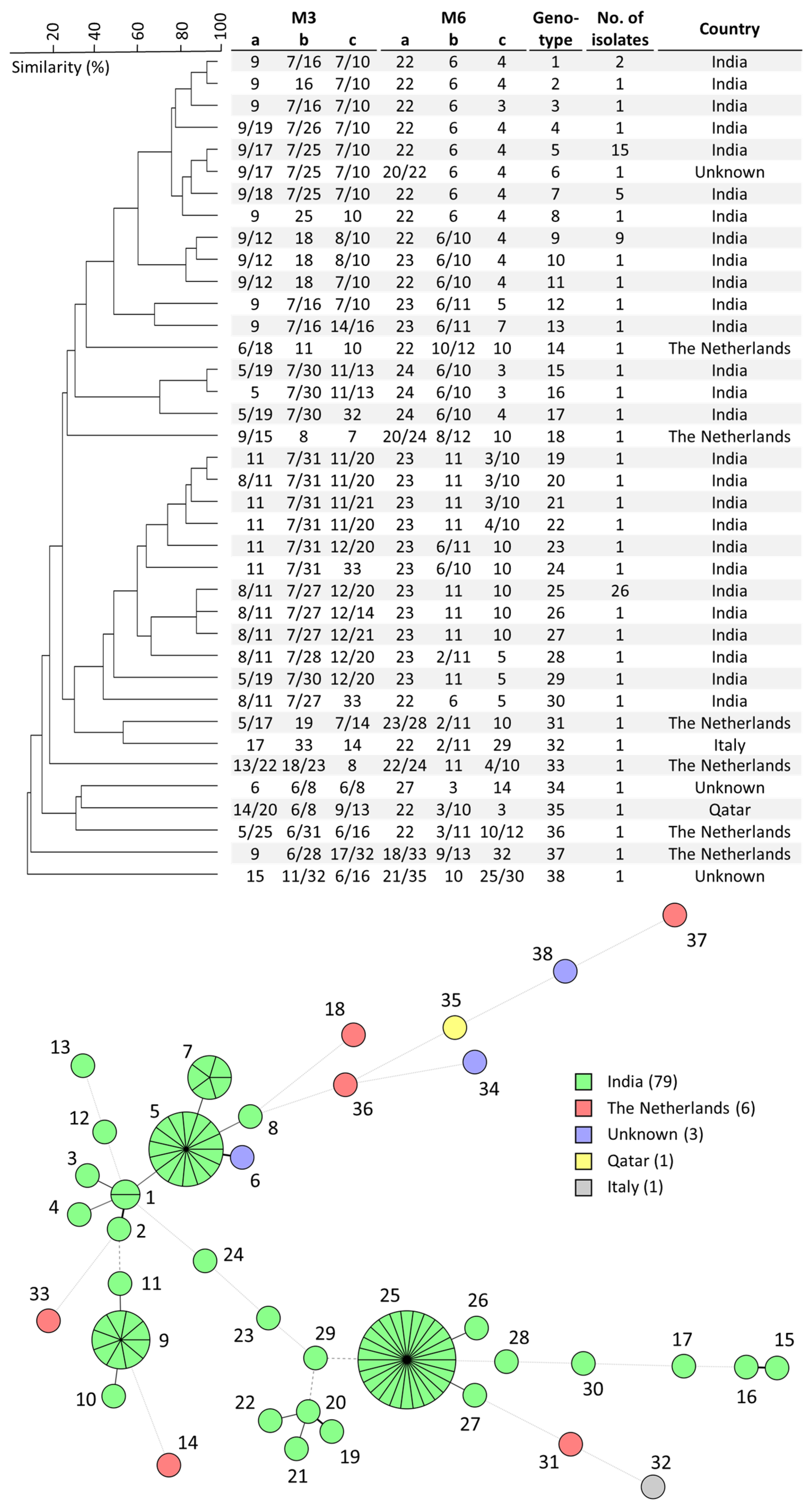
Figure 1. Cluster analysis of all W. anomalus isolates used in this study (top). Branch lengths indicate relatedness according to microsatellite alleles. The UPGMA dendrogram was generated with BioNumerics v7.5. Minimum-spanning tree of 90 W. anomalus isolates (bottom). Branch lengths indicate the similarity between isolates with thick solid lines (variation in one allele), thin solid lines (variation in two alleles), thin dashed lines (variation in three alleles) and thin dotted lines (variation in four or more alleles). Number of isolates per country is shown in the color key.
Microorganisms 11 01525 g001
Figure 2. Comparison of genetic relatedness of 11 W. anomalus isolates by STR typing (top) and WGS SNP typing (bottom). The UPGMA dendrogram was generated with BioNumerics. The numbers above the branches of the phylogenetic tree indicate the number of SNPs. The tree was generated with MEGA11 using the neighbor-joining tree method. Isolates were renamed to provide a clear overview, #1: 10-03-01-56, #2: 10-11-09-22, #3: 10-09-05-80, #4: 08-32-01-29, #5: 10-04-08-74, #6: 08-32-01-15, #7: 08-32-01-05, #8: 08-32-01-14, #9: 08-32-01-07, #10: 08-32-01-02 and #11: 08-32-01-30.
Microorganisms 11 01525 g002
Figure 3. W. anomalus homology model of ERG11 constructed with SWISS-MODEL, top view (A) and rear view (B). Amino-acids, fluconazole and the I469 position are shown in green, blue and red, respectively.
Microorganisms 11 01525 g003
Table 1. Overview of PCR primers for selected STR loci, concentration used in multiplex PCR, details of repeat characteristics and genomic sites.
PCR Panel and Primer NamePrimer Sequence (5′–3′)Conc b (µM)No. of Bases of Primer-Flanking SequenceRepeat UnitNo. of Repeats cNo. of AllelesIntragenic/
Locus Protein Coding Gene d
Forward Primer aReverse PrimerMinMax
M3
M3-aFAM-TCTTGCAAATCGTCAGACATCACCATCCTTGTTCCCTTACAAC5295TTG52516WICANDRAFT_30999
M3-bJOE-AGCTTGTAATTGTTGGGCTTGAGCTCCAGAAACTGAACCAAC5128TGT63318WICANDRAFT_34460
M3-cTAMRA-AGCTCAATTCCAAGCTGAACAATCAATCTCTGAGGGTGAAGTC5242ACA63315WICANDRAFT_91930
M6
M6-aFAM-GTTCGTTTGCAGTTTCTTTCCATCACCAACAAACTCGCTACC2123TTCACT183812WICANDRAFT_78907
M6-bJOE-TGCCTTATATGAAGGATGAAGGAGAGTCACCTCTTGGGCTATG2169GAGAGT21912WICANDRAFT_24702
M6-cTAMRA-CGGAAGGAATAAGAAGCAAAGGATGTTGGGTATTGTTGTCACTG10114AGCAAC33215WICANDRAFT_97047
a FAM, 6-carboxyfluorescein; JOE, 4′,5′-dichloro-2′,7′-dimethoxy-fluorescein; TAMRA, 6-carboxytetramethylrhodamine. b Concentrations of forward and reverse primers are identical. c Min, minimum; Max, maximum. d Reference strain is NRRL Y-366-8 (GCA_001661255.1).
Table 2. In vitro AFST metrics for W. anomalus (n = 87), according to CLSI M27-S4 standard.
AntifungalRange (mg/L)GM (mg/L)MIC50 (mg/L)MIC90 (mg/L)n Non-Wildtype (%)
AMB (n = 87)0.016–20.240.250.56 (6.9)
FLU (n = 87)0.5–161.71242 (2.3)
ITC (n = 87)0.016–10.210.250.51 (1.1)
VOR (n = 87)0.03–0.50.090.1250.1253 (3.4)
ISA (n = 49)0.031–0.1250.060.0630.063N.A.
AFG (n = 48)≤0.008–0.120.040.0310.12N.A.
MFG (n = 50)≤0.008–0.0320.010.0160.0310 (0)
AMB, amphotericin B; FLU, fluconazole; ITC, itraconazole; VOR, voriconazole; ISA, isavuconazole; AFG, anidulafungin; MFG, micafungin; GM, geometric mean; MIC, minimal inhibitory concentration; N.A., non-applicable.
Disclaimer/Publisher’s Note: The statements, opinions and data contained in all publications are solely those of the individual author(s) and contributor(s) and not of MDPI and/or the editor(s). MDPI and/or the editor(s) disclaim responsibility for any injury to people or property resulting from any ideas, methods, instructions or products referred to in the content.