Article

Effect of Essential Oils of Apiaceae, Lamiaceae, Lauraceae, Myrtaceae, and Rutaceae Family Plants on Growth, Biofilm Formation, and Quorum Sensing in Chromobacterium violaceum, Pseudomonas aeruginosa, and Enterococcus faecalis

Department of Biology, Ecology and Earth Sciences, University of Calabria, 87036 Rende, Italy
*
Author to whom correspondence should be addressed.
These authors contributed equally to this work.
Microorganisms 2023, 11(5), 1150; https://doi.org/10.3390/microorganisms11051150
Submission received: 20 March 2023 / Revised: 21 April 2023 / Accepted: 26 April 2023 / Published: 28 April 2023
(This article belongs to the Special Issue Antimicrobial Resistance: Current Status and Future Directions)

Abstract

:
The biological role played by essential oils extracted from aromatic plants is progressively being recognized. This study evaluated the potential antibacterial activity of ten essential oils against Chromobacterium violaceum, Pseudomonas aeruginosa, and Enterococcus faecalis by measuring their minimum inhibitory concentration. We found that essential oils exert different antimicrobial effects, with Origanum vulgare and Foeniculum vulgare demonstrating the most significant inhibitory effect on bacterial growth for C. violaceum and E. faecalis. The growth of P. aeruginosa was not affected by any essential oil concentration we used. Sub-inhibitory concentrations of essential oils reduced in C. violaceum and E. faecalis biofilm formation, violacein amount, and gelatinase activity, all of which are biomarkers of the Quorum Sensing process. These concentrations significantly affect the global methylation profiles of cytosines and adenines, thus leading to the hypothesis that the oils also exert their effects through epigenetic changes. Considering the results obtained, it is possible that essential oils can find a broad spectrum of applications in counteracting microbial contamination and preserving sterility of surfaces and foods, as well as inhibiting microbial growth of pathogens, alone or in combination with traditional antibiotics.

1. Introduction

The evolution and dissemination of resistance factors within bacterial populations is increasingly affirming itself. Due to the massive and uncontrolled clinical overprescription of antibiotics and their misuse by the food industry, there is an increasing need to research new compounds capable of inhibiting microbial growth. In this context, products of natural origin are showing encouraging results, with a plethora of studies demonstrating that plant extracts and essential oils (EOs) provide a huge range of complex compounds possessing antifungal, antibacterial, and antiviral properties, to the point that natural additives are spreading in the industrial field for the shelf-life extension of food products [1,2,3,4,5]. Therefore, great attention is being paid to the chemical characterization of EOs and to screening their biological activity [6,7,8]. Next to the bactericidal effects exerted by many EOs, the capacity of some EOs to interfere in Quorum Sensing (QS) processes and biofilm formation has been recently documented [9,10,11]. Several studies have reported that Origanum vulgare oil, with its main bioactive constituents, exhibited strong activity against Gram-positive and negative species such as Staphylococcus aureus, Bacillus subtilis, E. faecalis, Escherichia coli, and other pathogenic microorganisms [12,13]. More recently, Merghni et al. demonstrated its anti-biofilm, anti-swarming, and anti-QS capabilities against pathogenic bacteria such as S. aureus and Pseudomonas aeruginosa [14]. Similarly, a reduction in the biofilm formation was observed in several bacterial strains, such as E. coli, S. aureus, B. subtilis, and Candida albicans, treated with the EO extracted from peppermint [15,16,17]. Additionally, the effects of many other EOs, including those obtained from clary sage, juniper, lemon, and marjoram, and their primary components, was found to significantly inhibit the formation of bacterial and yeast biofilms and QS [18].
Although the antibacterial properties of EOs and their main components have been examined in several studies, many issues remain to be clarified about the mechanism of action by which they perform their activities. Therefore, in this study, we evaluated the potential antimicrobial activity and the inhibitory effect on biofilm formation and the QS process of ten EOs extracted from aromatic plants grown in Calabria (Southern Italy) on the Gram-negative Chromobacterium violaceum and P. aeruginosa and the Gram-positive E. faecalis. These aromatic plants are grouped into Apiaceae, Lamiaceae, Lauraceae, Myrtaceae, and Rutaceae families. Apiaceae is one of the most numerous plant families. It comprises several economically important vegetables, herbs, and spices. Umbelliferous crops such as anise, fennel, carrot, coriander, and parsley are appreciated sources of botanical flavoring agents and fragrances. Apiaceae plants produce a wide variety of specialized metabolites, including volatile phenylpropanoids, furanocoumarins, sesquiterpene coumarins, polyacetylenes, and phthalides, some of which are considered uncommon natural phytochemicals distinctive of the family [19]. Lamiaceae, or the mint family, is a widely distributed family of angiosperms that consists of 236 genera with more than 7000 species, which inhabit diverse ecosystems and have great diversity with a cosmopolitan distribution. The largest genera that belong to this family are Salvia, Scutellaria, Stachys, Ajuga, Plectranthus, Hyptis, Teucrium, Vitex, Thymus, and Nepeta. Many of them are used as spices and vegetables. The family has several species of aromatic plants that are applied in traditional medicine and in the pharmaceutical and food industries due to their biological properties. They act as a stimulant for blood circulation and digestion, as strengtheners of the central nervous system, and as expectorants, antispasmodics, antiseptics, diuretics, carminatives, and tonics [20]. Lauraceae is a large family of woody plants (except for the genus Cassytha, which is herbaceous) composed of 50 genera and 2500–3000 species. The distribution center of modern Lauraceae is in tropical to subtropical areas, including Southeast Asia, the Mediterranean, and Central and South America. The economic importance of this family lies in that many species are used in industrial sectors such as the food, timber, pharmaceutical, and perfumery industries. Lauraceae plants have been used as analgesics and applied to multiple different pathologies, such as the treatment of infectious diseases, malaria, gastrointestinal infections, female genital infections, and rheumatism [21]. Myrtaceae is one of the most important plant families, being regarded as the eighth-largest flowering plant family. It includes many genera of utmost ecological and economic importance distributed all over the world. Fruits are berry-type and have desirable pulp yield and organoleptic characteristics, as well as nutritional aspects and significant amounts of phytochemicals, such as organic acids, sugars, vitamins, polysaccharides, polyphenols, and important minerals. They can inactivate reactive oxygen species in the human body, thus promoting the risk reduction of various chronic diseases such as heart disease, stroke, cancer, atherosclerosis, diabetes, Alzheimer disease, cataracts, and pulmonary disorders [22]. Rutaceae comprises a large (about 160 genera and 1900 species), morphologically diverse, cosmopolitan family. The members are famous among phytochemists for their extraordinary array of secondary chemical compounds such as limonoids, flavonoids, coumarins, volatile oils, and alkaloids, many of which exhibit medicinal, antimicrobial, insecticidal, or herbicidal properties [23].

2. Materials and Methods

2.1. Bacterial Strains and Growth Conditions

This study was carried out on C. violaceum (ATCC 12742), P. aeruginosa (ATCC-9027), and E. faecalis OG1RF (ATCC 47077) strains. C. violaceum and P. aeruginosa were cultured in Nutrient broth containing 3 g/L of Beef extract and 5 g/L of Peptone at 30 °C. Meanwhile, E. faecalis was cultured in 36.4 g/L of Todd–Hewitt broth (THB) (Oxoid, Basingstoke, Hampshire, United Kingdom) at 37 °C under gentle agitation. The bacterial strains were kept frozen in stock cultures at −80 °C in cryovials.

2.2. Essential Oils (EOs) and Their Extraction

Ten EOs, provided from commercial producers located in Calabria (Southern Italy), were used. They were extracted from the following plants: Clinopodium nepeta L. (Kuntze, Carl Ernst Otto), Citrus bergamia (Risso, Joseph Antoine & Poiteau, Pierre Antoine), Citrus limon L. (Osbeck, Pehr), Citrus reticulata (Blanco, Francisco Manuel), Foeniculum vulgare subsp. piperitum, Laurus nobilis L., Myrtus communis L., Origanum vulgare L. subsp. viridulum (Martrin-Donos, Julien Victor) Nyman, Carl Frederik, Salvia officinalis L., and Salvia rosmarinus (Spenn, Fridolin Carl) [24].
The fruit peel of C. bergamia and C. limon, the flower, leaf, and terminal branches of C. nepeta, F. vulgare, M. communis, O. vulgare, S. officinalis, and S. rosmarinus were used, and only the leaf and terminal branches of C. reticulata and L. nobilis were used [12].
The EO from C. bergamia was mechanically extracted through an industrial cold expression process from fresh fruit. For all other species, EOs were extracted through the water-vapor-under-vacuum distillation process in a 20 L inox apparatus, starting with fresh collected raw material. The EOs were aliquoted and kept in dark glass bottles, tightly sealed at +4 °C, until use [12].

2.3. GC-MS Analysis of Essential Oils

Gas Chromatography-Mass Spectrometry (GC-MS) was performed using a gas chromatograph (Focus GC-Thermo Fisher, Milan, Italy) equipped with a Varian VF-5m (30 m × 0.25 mm × 0.25 μm) capillary column, combined with a single quadrupole mass spectrometer (DSQII-Thermo Scientific, Milan, Italy) [12]. The samples were diluted 1:1000 in ether. One microliter of the sample was injected in spitless mode at a temperature of 220 °C. The column flow rate was 1 mL min-1 using helium as carrier gas. The initial GC oven temperature was 55 °C, increased by 4 °C min−1 to 240 °C with a hold time of 3 min. The transfer line temperature was 250 °C. The MS was operated using electron impact (EI) at an ionization energy of 70 eV. The ion source temperature was set at 250 °C. The solvent delay for the mass spectrometry was set at 3 min and the EI scan mode was used for identification, covering the range of 25–350 m/z. The compound was identified by comparison with the NIST database (https://www.nist.gov/pml/atomic-spectra-database, accessed on 6 June 2021). The instrumentation performance, chromatograms, mass spectra, and initial data processing were carried out with the supplied Xcalibur software (Thermo Fisher, Milan, Italy).

2.4. Determination of Minimum Inhibitory Concentration (MIC) and Minimum Bactericidal Concentration (MBC)

Minimum inhibitory concentration (MIC) and minimum bactericidal concentration (MBC) for the ten EOs were determined using the broth dilution method. Since EOs do not dissolve in water or water-based mediums, in our study, to increase their dispersion in the aqueous medium, each EO was pre-absorbed on inulin, a fructan composed of a linear chain of connected fructose that is used in the food industry as a gelling agent and as a stabilizer for oil in water emulsions. Therefore, EOs were prepared daily by letting the oil adsorb to inulin powder to obtain a 100 µL EOs/gr inulin working solution, as previously described [12]. Approximately 109 cells of C. violaceum, P. aeruginosa, and E. faecalis from an overnight culture of the two bacterial strains were inoculated into tubes containing 3 mL of the following serial dilutions of the dissolved EOs: 0.1, 0.2, 0.3, 0.4, 0.5, 0.6, 0.7, 0.8, 0.9, 1, 2, 3, 4, 5, 6, 7, 8, 9, 10 µL of EO/mL of medium. Culture tubes were shaken at 300 rpm at 37 °C for 18 h. In all experiments, a series of controls were set up: (1) C. violaceum, P. aeruginosa, or E. faecalis cells were inoculated into tubes containing liquid growth medium in absence of EOs to verify the optimal growth conditions (positive control); (2) liquid growth medium without inoculated cells and EOs to verify no growth of microorganisms (negative control), also providing evidence of sterility and lack of contamination; (3) C. violaceum, P. aeruginosa, or E. faecalis cells were inoculated into tubes containing liquid growth medium in the presence of the different inulin dilutions (vehicle control) to evaluate the potential effect of the substance on bacterial growth; (4) liquid growth medium without inoculated cells in presence of each EO to discern the turbidity background.
Turbidity measurement was performed at 600 nm in a spectrophotometer. The MIC values were determined as the lowest concentration of EO corresponding to optical density (OD) values comparable to those of the cell-free growth medium. The MBC values were calculated by subculturing all dilutions carried out in a liquid medium on agar plates. The MBC was determined by considering the lowest concentration of EO that reduces the viability of the initial bacterial inoculum by ≥99.9%. Each experiment was carried out in quintupled, with three independent repetitions.

2.5. Biofilm Formation Evaluation

Approximately 107 cells of C. violaceum and E. faecalis from an overnight culture were inoculated into 96-well polystyrene microtiter plates containing 200 µL of growth medium in the absence and presence of the dissolved EOs at concentrations equal to and lower than those of MIC values. After 18 h of incubation, the planktonic cells were removed, and adherent cells were fixed with methanol and then stained with crystal violet solution 0.2%. After 5 min, the excess of the stain was removed by three repeated washes. Acetic acid at a concentration of 33% was added to each well and the optical density at 570 nm was determined spectrophotometrically. Each strain was examined in duplicate on each plate and the experiments were performed in quintupled. Positive and negative controls containing only the inoculated growth medium and the growth medium supplemented with EOs, respectively, were run in each experiment.

2.6. Violacein Production Evaluation

Approximately 109 cells from an overnight culture of C. violaceum were inoculated into tubes containing 3 mL of Nutrient medium in the absence and presence of the dissolved EOs at concentrations equal to and lower than those of MIC values and incubated at 30 °C for 18 h. Then, 1 mL of the above culture was collected and centrifuged at 4 °C for 20 min. The supernatant was removed, and the violacein was dissolved in DMSO at room temperature. After a 3-min centrifugation to remove the cells, violacein production was spectrophotometrically quantified by reading the absorbance at 585 nm. Each sample was examined in duplicate in each plate and the experiments were performed in quintupled.

2.7. Gelatinase Activity Evaluation

Approximately 109 cells from an overnight culture of E. faecalis were inoculated into tubes containing 3 mL of THB in the absence and presence of the dissolved EOs at concentrations equal to and lower than those of MIC values and incubated at 37 °C for 18 h. Then, 40 µL of culture supernatant was collected and incubated for 2 h with constant mixing (170 rpm) in the presence of 500 µL of azocoll substrate (10 mg/mL). After centrifugation at 20,000× g for 10 min, the protease activity of supernatant fractions was assayed spectrophotometrically by reading the absorbance at 550 nm. Each assay was performed in quintupled.

2.8. Genomic DNA Extraction

Genomic DNA was extracted from untreated C. violaceum and E. faecalis cells and cells treated with pre-inhibitory concentrations (pre-MICs) of EOs by using a DNeasy UltraClean Microbial Kit (Qiagen, Milan, Italy), according to the manufacturer’s protocol. Briefly, 3 mL of bacterial cell culture was collected and centrifuged for 10 min. The isolated pellets were resuspended in 300 µL of PowerBead Solution and transferred to PowerBead Tubes. An amount of 50 µL of Solution SL was added to each sample and, after vortexing for 10 min, the tubes were centrifuged at 10,000× g for 30 s. Supernatants were incubated at 4 °C for 5 min in the presence of 100 µL of Solution IRS and centrifugated at 10,000× g for 1 min. After the addition of 900 µL of Solution SB to the supernatants, 700 µL of the sample was loaded into MB Spin Columns and centrifuged at 10,000× g for 30 s. The centrifugation was repeated after adding 300 µL of Solution CB, and the flow-through was discarded. DNA samples were eluted by centrifugation at 10,000× g for 30 s in 50 µL of Solution EB. The DNA concentration and purity were determined spectrophotometrically by using the 260/280 nm absorbance ratio.

2.9. Quantification of Global 5-Methylcytosine Levels

Global DNA methylation levels of 5-methylcytosines (5mC) were determined by using the MethylFlash Global DNA Methylation (5mC) ELISA Easy Kit (Epigentek, Farmingdale, Nassau County, NY, USA), following the manufacturer’s instructions. Briefly, 100 ng of genomic DNA were mixed with 100 µL of Binding Solution, loaded in duplicate in a 96-well plate and then incubated at 37 °C for 60 min. After three consecutive washes of the wells with 150 μL of Wash Buffer, samples were incubated at room temperature for 50 min with 50 µL of 5-mC Detection Complex Solution, containing 5-mC antibody (1:1000), Signal Indicator (1:1000), and Enhancer Solution (1:2000). Then, the solution was removed from the wells through five consecutive washes with 150 μL of Wash Buffer, and 100 μL of Developer Solution was added to each well and incubated at room temperature for 3 min. The colorimetric reaction was stopped by the addition of 100 µL of Stop Solution and the absorbance was read at 450 nm using an ELISA plate reader. In each experiment, the percentage of 5-mC was calculated using the second-order regression equation of the standard curve obtained by diluting a positive control, provided by the kit, at different methylation percentages. The methylation values of C. violaceum and E. faecalis untreated cells were used as reference values (relative quantification) for the corresponding cells treated with the EOs.

2.10. Quantification of Global N6-Methyladenosine Levels

Global DNA methylation levels of N6-methyladenosines (m6A) were determined by using the MethylFlash m6A DNA Methylation ELISA Kit (Epigentek, Farmingdale, Nassau County, NY, USA), following the manufacturer’s instructions. Shortly, 200 ng of genomic DNA was mixed with 80 µL of Binding Solution, loaded in duplicate in a 96-well plate, and then incubated at 37 °C for 90 min. After three consecutive washes of the wells with 150 μL of Wash Buffer, samples were incubated at room temperature for 60 min with 50 µL of Capture Antibody. Then, the wells were washed thrice with 150 μL of Wash Buffer and incubated at room temperature for 3 min with 50 µL of Detection Antibody. After four consecutive washes, 50 μL of Enhancer Solution was added to each well, and the plate was incubated at room temperature for 30 min. Then, after five consecutive washes, wells were incubated at room temperature in the dark with 100 µL of Developer Solution. The colorimetric reaction was stopped by the addition of 100 µL of Stop Solution, and the absorbance was read at 450 nm using an ELISA plate reader. In each experiment, the percentage of m6A was calculated using the second-order regression equation of the standard curve obtained by diluting a positive control at different methylation percentages, as suggested by the kit. Each sample was examined in duplicate in each plate and the experiments were performed in triplicate. The methylation values of C. violaceum and E. faecalis untreated cells were used as reference values (relative quantification) for the corresponding cells treated with the EOs.

2.11. Statistical Analysis

Statistical analyses were performed using SPSS 28.0 statistical software (SPSS Inc., Chicago, IL, USA). Kruskal–Wallis one-way analysis of variance and Mann-Whitney tests were adopted. Significance level was defined as p ≤ 0.05.

3. Results

3.1. Characterization of the Essential Oils

Ten EOs were selected for the present study. Their characteristics in terms of family, chemical constituents, and their percentages and Retention Indices are presented in Table 1. Piperitone oxide, (+)-sabinene, estragole, and p-thymol represent the predominant compounds in C. nepeta, C. reticulata, F. vulgare, and O. vulgare oils, respectively. Eucalyptol is the major component of L. nobilis, M. communis, S. officinalis, and S. Rosmarinus. C. bergamia and C. limon Oils are characterized by a high content of (+)-limonene.

3.2. Effects of EOs on Cell Growth

The antibacterial activity of the EOs on the Gram-negative C. violaceum and P. aeruginosa and on the Gram-positive E. faecalis was evaluated by determining the MIC and the MBC values. Obtained results are reported in Table 2. Findings regarding P. aeruginosa are not reported as no variation in cell growth was observed at any EO concentration we used. For this reason, we then conducted the subsequent experiments only on C. violaceum and E. faecalis.
The analysis revealed that all EOs show bactericidal activity, as deduced by MIC values and confirmed by MBC. The EO from O. vulgare showed the greatest inhibitory effect on bacterial growth. The lowest MIC values, equal to 0.4 to 0.6 µL/mL in C. violaceum and E. faecalis, respectively, were identified in both bacterial strains. Under these MIC values, we also observed that the antibacterial activity of EOs from F. vulgare and Clinopodium nepeta appeared strain-specific since very low concentrations of these EOs are sufficient to inhibit the growth of C. violaceum (0.4 and 0.8 µL/mL, respectively). In comparison, higher concentrations of the two EOs are needed to inhibit that of E. faecalis (3 and 4 µL/mL, respectively). The vehicle did not show significant variations with respect to the untreated sample under any experimental conditions tested (data not shown).
Intermediate MIC values, corresponding to concentrations of 1 and 2 µL/mL, were observed following treatment with L. nobilis in C. violaceum and E. faecalis, respectively. The antibacterial activity of the remaining EOs is less effective, as evidenced by the higher MIC values, ranging from 2 to 5 µL/mL. Differences related to the bacterial strain are again observed, with E. faecalis appearing more resistant than C. violaceum to the inhibitory effects exerted by the EOs from M. communis, S. officinalis, and S. rosmarinus, and more sensitive to the EOs from C. limon and C. reticulata.

3.3. Effects of EOs on Biofilm Formation

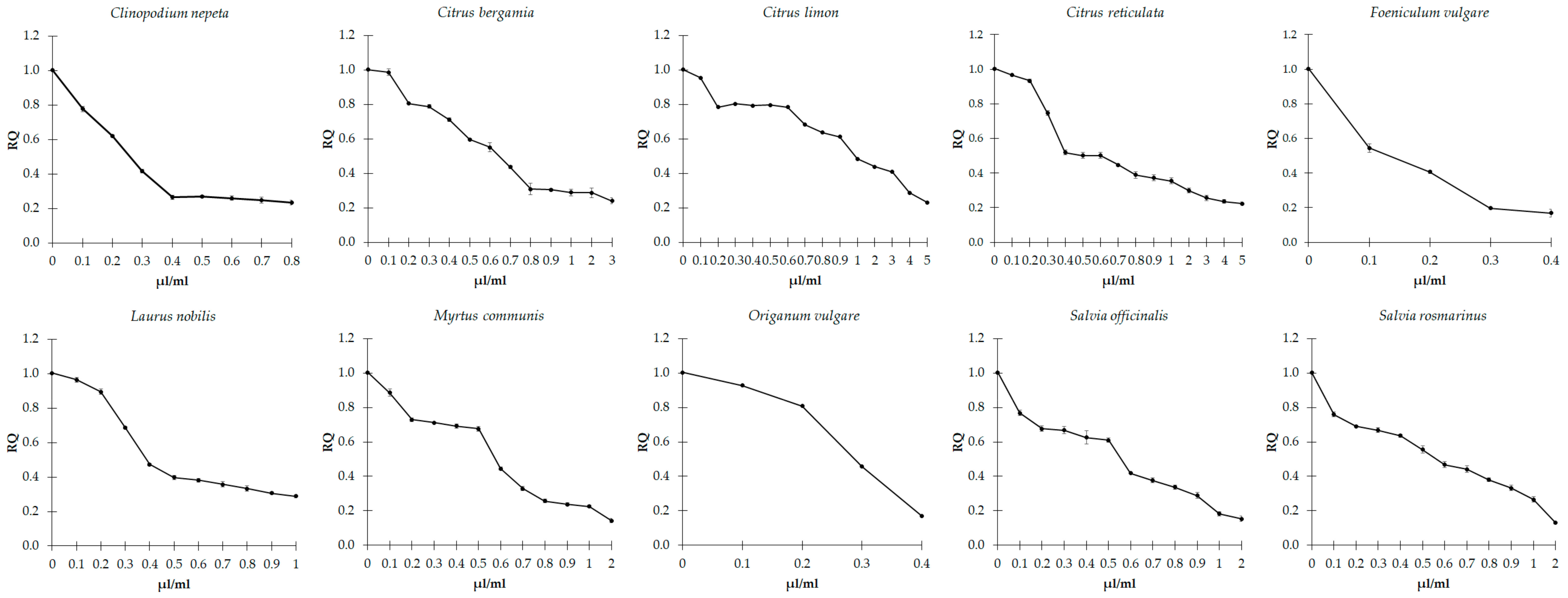
The potential effect of the ten EOs on influencing biofilm formation was evaluated by the crystal violet method in C. violaceum and E. faecalis, kept in culture in the absence and presence of each EOs at concentrations equal to and lower than those of MIC values. The action of each EO against biofilm formation is shown in Figure 1 and Figure 2.
The results showed that all EOs had considerable anti-biofilm-forming effects on both C. violaceum (Figure 1) and E. faecalis (Figure 2). Indeed, by comparing the cells before and after treatment with the EOs, we can observe that they induced a general and progressive decreased ability to form biofilms (p < 0.001), with some exceptions. Specifically, in E. faecalis, treatment with concentrations of EOs from C. limon up to 0.6 induced an increase in biofilm formation (p < 0.001), with respect to the untreated strain. In contrast, higher concentrations lead to a progressive decline (p < 0.001). Moreover, a progressive increase in biofilm formation was observed following treatment, with C. nepeta concentrations ranging from 0.3 to 0.6 µL/mL (p = 0.016), followed by a dose-dependent decrease at higher concentrations of the EO (p < 0.001).

3.4. Effects of EOs on Quorum Sensing

The ability of EOs to interfere with the QS mechanism was determined by measuring the amount of violacein in C. violaceum and the gelatinase activity in E. faecalis, both influenced by the above process. We observed that the sub-inhibitory concentrations of the ten EOs show significant concentration-dependent inhibition of violacein production in C. violaceum (p < 0.001), which depends on the QS regulation (Figure 3). Consistently, the action of sub-inhibitory concentrations of all EOs appears effective in generally reducing the gelatinase activity in E. faecalis (Figure 4), with slight differences among the different EOs.
A significant and progressive decrease in the enzymatic activity of gelatinase was observed as a function of the increasing concentration of C. nepeta, C. bergamia, and S. officinalis essential oils (p < 0.001). An approximately 80% decline in gelatinase activity was observed following the treatment with 0.1 µL/mL of F. vulgare, L. nobilis, M. communis and O. vulgare. Then the activity remained constant or further decreased under high concentrations. This activity showed a significant increase following treatment with concentrations of C. reticulata between 0.3 and 1 µL/mL (p = 0.032), and then a drastic decrease at higher concentrations (p = 0.008). Concentrations of C. limon between 0.1 and 0.4 µL/mL did not significantly influence a drastic drop in the activity of gelatinase (p = 0.008) that underwent an increase of 30% following concentrations between 0.6 and 2 µL/mL (p = 0.008). Lastly, concentrations of S. rosmarinus between 0.1 and 0.2 µL/mL significantly increased gelatinase activity by 40% (p = 0.008), which was later reduced to higher concentrations (p < 0.001).

3.5. Effects of EOs on DNA Methylation Profiles

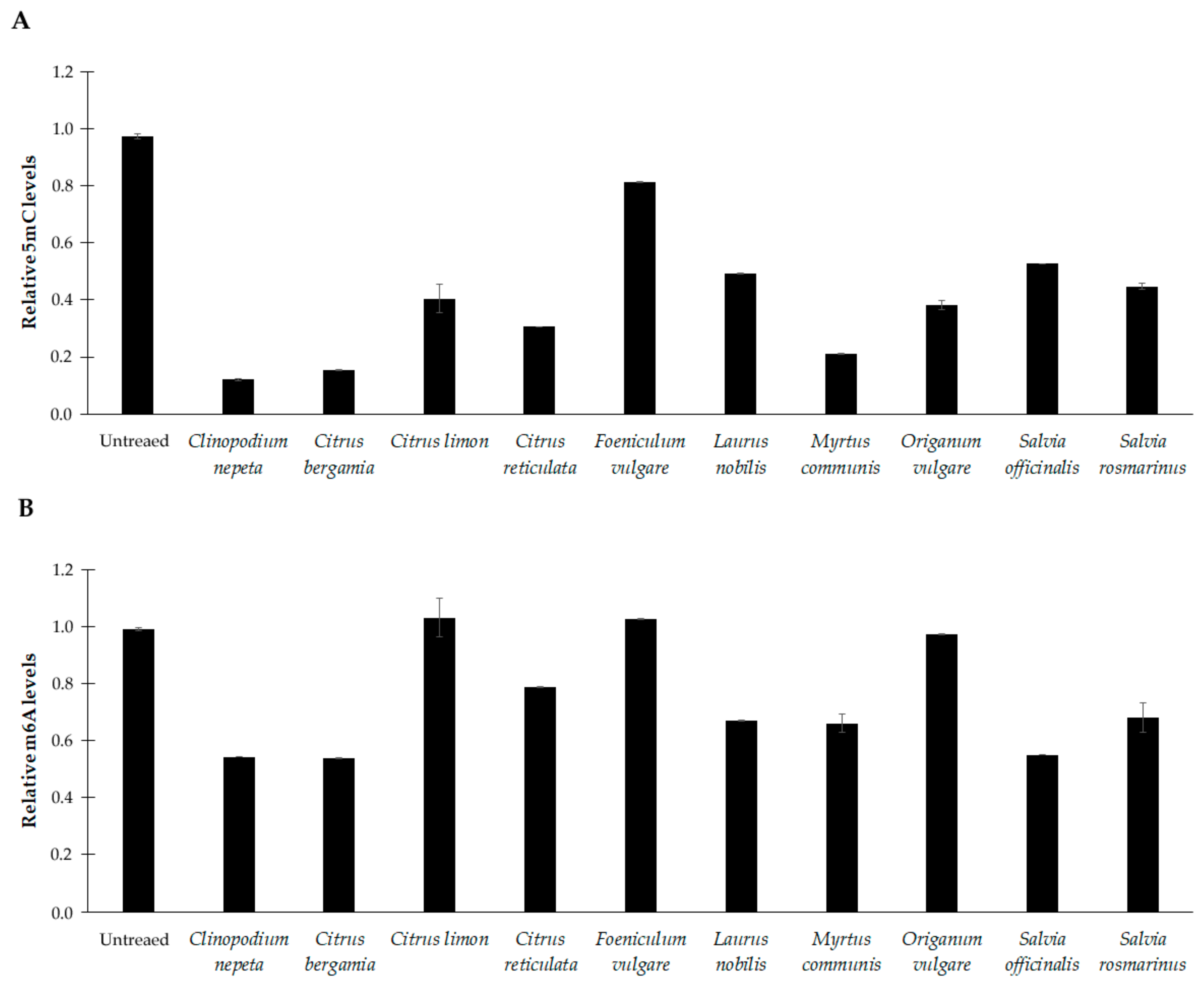
Global methylation levels of cytosine and adenine residues were evaluated in DNA samples extracted from C. violaceum and E. faecalis kept in culture in the absence and presence of each EO. Figure 4 shows the abundance of 5-methylcytosine (5mC) and N6-methyladenosine (m6A) in C. violaceum. By comparing the cells before and after treatment with the EOs at sub-inhibitory concentrations, we can observe that they induced a significant reduction in the methylation status of cytosine residues (p < 0.001) (Figure 5A). Similarly, a general down methylation of the adenine residues was detected (p < 0.05), with some exception (Figure 5B). No significant change was observed in the methylation status of adenine under the sub-inhibitory concentration of C. limon, F. vulgare or O. vulgare.
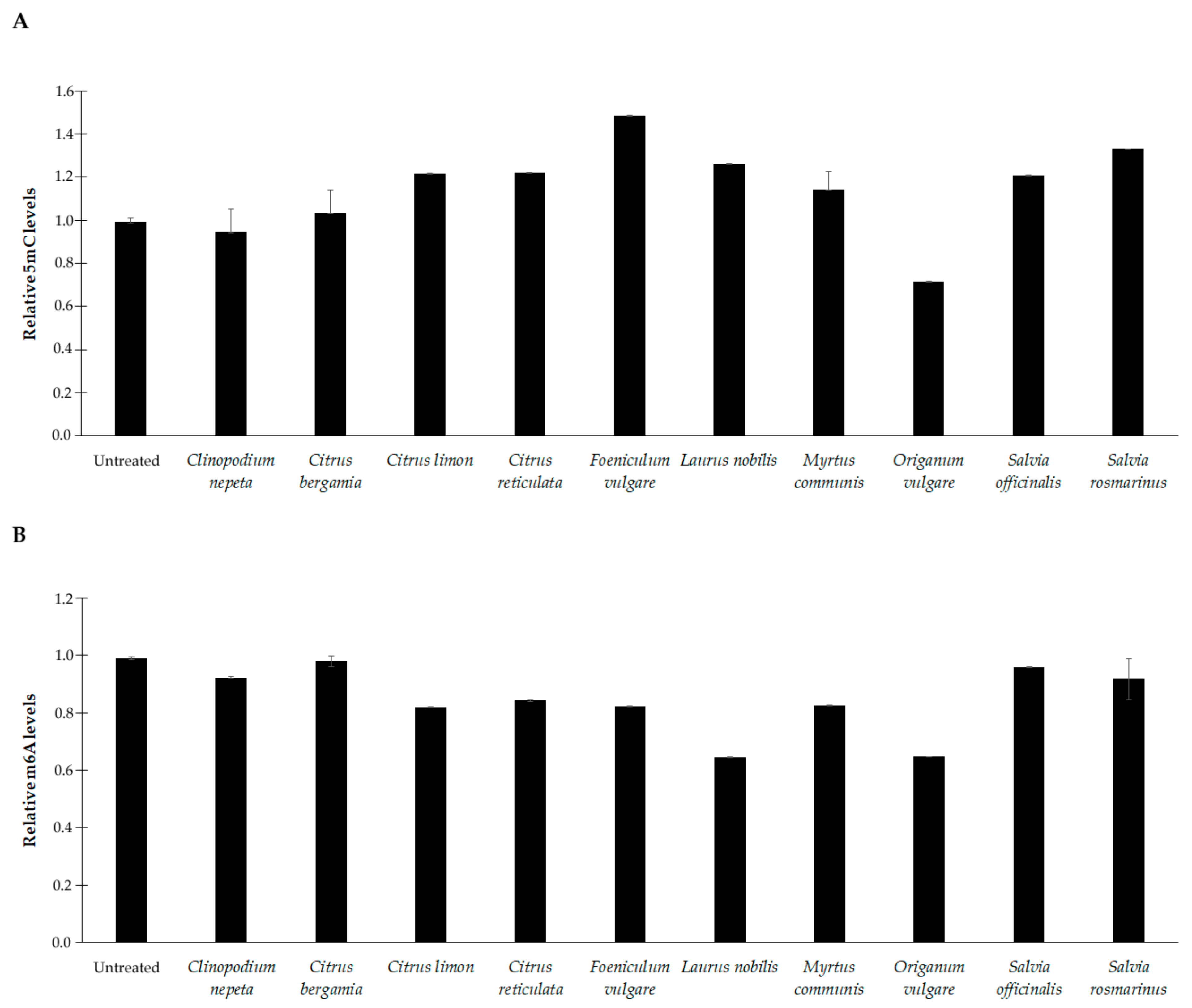
In E. faecalis, EOs treatment resulted in much more variable levels of 5mC and m6A (Figure 6). An increase in 5mC levels was observed with C. limon, C. reticulata, F. vulgare, L. nobilis, S. officinalis, and S. rosmarinus (p < 0.01). Meanwhile, a decrease was noticed for the sole O. vulgare (p < 0.001). No significant change was observed after treatment with C. nepeta, C, bergamia, or M. communis (Figure 6A).
Lastly, a general reduction in m6A was detected (p < 0.01), with some exceptions (Figure 5B). No significant change was observed with C. bergamia, S. officinalis, and S. rosmarinus (Figure 6B).

4. Discussion

The largely unregulated use of antibiotics has increased the prevalence of antibiotic-resistant bacteria. Therefore, interest in natural medicine has been growing, prompting the search for valid alternatives to non-organic synthetic antibiotic compounds. In this context, natural products, such as essential oils extracted from plants, are regarded as an important source of new molecules for drug and therapy discovery [25,26]. The historical use of EOs as antimicrobial agents has long been well-known, but only recently have a variety of experimental data demonstrated their bioactive properties in-depth. These properties can be explained through the induction of irreversible damage to the bacterial cell wall and membrane, the inhibition of metabolic pathways and protein synthesis, and interference with cell wall synthesis and DNA and RNA synthesis, according to many groups of chemical compounds which comprise them [8,27,28].
In a previous study, we reported that the essential oils extracted from C. nepeta, C. bergamia, C. limon, C. reticulata, F. vulgare, L. nobilis, M. communis, O. vulgare, S. officinalis, and S. rosmarinus aromatic plants show antibacterial activity against an E. coli strain and three lines derived by growing it at low concentrations of ampicillin, ciprofloxacin, and gentamicin [12]. Here we want to investigate whether the antibacterial properties of these oils are also effective against other strains, thus establishing that the inhibition of microbial growth mediated by the EOs is a generalized and non-strain-specific phenomenon. To this purpose, the study was conducted on Gram-negative C. violaceum and P. aeruginosa, and Gram-positive E. faecalis, commonly used for years to identify and characterize bioactive substances acting against microorganisms. C. violaceum has recently emerged as an important model of an environmental pathogen responsible for opportunistic infections that quickly lead to the host’s death. Its high pathogenicity in humans and animal models of infection involves the possession of several predicted virulence traits, including secreted chitinase and chitosanase proteins as well as proteins involved in the transport and capture of amino acids and carbohydrates, in addition to oxidative stress protection [29]. P. aeruginosa is found in soil, water, and animals, and it is an opportunistic pathogen in humans. It infects the pulmonary and urinary tracts, wounds, and burns, thus causing both severe acute and chronic infections. It is particularly critical in healthcare-associated infections and in ill and immunocompromised patients [30]. Enterococci are resilient and versatile bacteria capable of surviving in hostile environments. They have gained particular significance for human health, being the principal cause of hospital-acquired or healthcare-associated infections affecting the urinary tract, bloodstream, wounds, and the endocardium [31]. Particularly, E. faecalis, characterized by antibiotic resistance traits and biofilm formation capabilities, was also found to occur as a food contaminant and in secondary endodontic biofilm-associated infections [32].
Although E. feacalis appeared more resistant to oil stimulation, showing MIC values mostly higher than those observed in C. violaceum, the results demonstrated that all essential oils possess antibacterial activity. The EO extracted from O. vulgare was the most effective in inhibiting the growth of the two bacterial strains, with a minimum MIC of 0.4 and 0.6 µL/mL. Additionally, EOs from C. nepeta and F. vulgare had strong antibacterial activity against C. violaceum. This evidence agrees with previous studies, which reported that these EOs have strong antimicrobial activity against other bacterial and fungal species, including E. coli JM109 and its derived antibiotic-resistant cells, as well as S. aureus, Listeria monocytogenes, Salmonella typhimurium, Penicilium chrysogenum, Alternaria alternata, and Chaetomium globosum [12,33,34,35,36,37,38]. The antimicrobial effect of some monoterpenes against Gram-positive and Gram-negative bacterial strains seems to be achieved through a perturbation of the lipid fraction of bacterial plasma membranes, resulting in alterations of membrane permeability and leakage of intracellular materials [39]. No growth inhibition at the concentrations of essential oils we used was observed in P. aeruginosa. This result is substantially in line with literature data, which reported that this organism is less susceptible than other species to treatment with different essential oils, or is susceptible only to high concentrations [40,41]. In our work, we had to limit the analysis to concentrations equal or less than 10 µL/mL since the EOs do not completely dissolve in inulin at higher concentrations. In any case, once again P. aeruginosa demonstrates greater resistance to molecules with antimicrobial activity than other bacterial species.
Since biofilm formation is one of the major protective mechanisms for survival of microbes under adverse conditions, we investigated whether the antibacterial effect of the EOs was associated with a reduction in the growth ability of C. violaceum and E. faecalis in biofilm structures. Sub-inhibitory concentrations of the EOs, which were insufficient to inhibit the bacterial growth, were used for evaluating their potential antibiofilm activity. Results revealed that all EOs significantly induced a dose-dependent inhibition of the biofilm formation in both bacterial strains. Thymol and terpinene are the major components characterizing O. vulgare oil. Previous data reported that the antimicrobial effect of some monoterpenes against Gram-positive and Gram-negative bacterial strains may be due to a perturbation of the lipid fraction of bacterial plasma membranes, resulting in alterations to membrane permeability and in leakage of intracellular materials [42]. Therefore, the anti-biofilm-forming property of this oil is consistent with results showing that these two compounds may alter the permeability of plasma membranes, disrupt bacterial adhesion to the surfaces, and subsequently reduce biofilm formation by Staphylococcus epidermidis, Bacillus cereus, Pichia anomala, and Pseudomonas putida [12,19,42]. Similarly, the strong antimicrobial and antibiofilm efficacy of F. vulgare oil is consistent with the results recently observed in other bacterial strains and fungi, and suggests that it is strictly influenced by the massive presence of phenylpropanoids (estragole and anethale) and monoterpenes (α-pinene and α-phellandrene) [43,44]. It has been reported that EOs characterized by a high level of piperitenone and piperitone, such as, for example, the one obtained from C. nepeta, significantly inhibit bacterial biofilm formation and the metabolism of both Gram-positive and Gram-negative bacterial strains [45].
The decreased ability to grow in biofilm could suggest a quencher role exercised by the EOs on the QS process by inhibiting the activity of receptors and molecules involved in this pathway. Finding a significant reduction of violacein production and gelatinase activity, two widely recognized biomarkers of the QS process in C. violaceum and E. faecalis, respectively, suggests that the ten EOs analyzed interfere with the QS system and inhibit cellular communication pathways, thus preventing the assembly of the biofilm matrix.
We previously demonstrated that epigenetic modifications mediate the antibacterial properties of the EOs since they induce a profound remodeling of the methylation levels of adenine and cytosine residues in the genomes of E. coli and its derived antibiotic-resistant cells [12]. Results we observed in C. violaceum and E. faecalis corroborated this evidence, demonstrating that it is a generalized phenomenon not linked to a specific bacterial strain. In most cases, we observed down methylation at both cytosine and adenine residues after treatment with EOs, with more marked changes occurring in the C. violaceum genome. Whether these changes are correlated with the inhibition or activation of gene expression deserves further investigation.
It is plausible that a massive epigenetic remodeling at the DNA level is responsible for an equal modulation of the expression of genes involved in various processes—including cooperative behaviors, cell-to-cell communication, and virulence—that are beneficial when performed by groups of bacteria acting in synchrony. Through these mechanisms, the EOs would probably hinder the strategies adopted for the survival of the bacterial community, thus inhibiting bacterial growth.
Overall, this study reports, for the first time, that different EOs induce epigenetic changes and negatively regulate biofilm formation and the QS mechanism, providing further characterization at the molecular level of the antimicrobial properties of EOs. The antibacterial, anti-biofilm, and anti-QS efficacy of the tested EOs, especially those extracted from O. vulgare and F. vulgare, appear to be of relevance since these oils may represent effective and natural therapeutic compounds that can find applications, either alone or in combination with traditional antibiotics, in the control of bacterial infections in countless fields.

Author Contributions

Conceptualization, D.B.; Methodology P.D. and G.S.; Formal analysis, P.D. and D.B.; Data curation, D.B.; Writing-Original Draft Preparation, P.D. and D.B.; Writing-Review and editing, P.D., G.S., M.C., G.P. and D.B., Funding Acquisition, G.P. All authors have read and agreed to the published version of the manuscript.

Funding

This research was funded by the nursing homes of Sadel Spa, Sadel San Teodoro srl, Sadel CS srl, Casa di Cura Madonna dello Scoglio, AGI srl, Casa di Cura Villa del Rosario srl, Savelli Hospital srl, and Casa di Cura Villa Ermelinda, in the frame of the agreement “Attività di Ricerca e Sviluppo Sperimentale: Tecnologie avanzate per l’indagine delle relazioni tra uomo ed ambienti di vita” with the University of Calabria and by “SI.F.I.PA.CRO.DE.-Sviluppo e industrializzazione farmaci innovativi per terapia molecolare personalizzata PA.CRO.DE.” PON ARS01_00568 to G.P. granted by MIUR (Ministry of Education, University and Research) Italy. This work was supported by Progetto PON SILA (Sistema Integrato di Laboratori per l’Ambiente)—PON a3_00341A–UNICAL.

Data Availability Statement

Research data is available upon request by contacting the corresponding author of the article.

Acknowledgments

We thank Michele Crudo Farm, Vibo Valentia (Italy) for the kind gift of the essential oils.

Conflicts of Interest

The authors have no conflict of interest to declare. Michele Crudo Farm was not involved in the study design, collection, analysis, and interpretation of data, the writing of the report, or the decision to submit the article for publication. M.C., who has an equity interest in Michele Crudo Farm, contributed to the collection of aromatic plants and the extraction of essential oils.

References

  1. Bunse, M.; Daniels, R.; Gründemann, C.; Heilmann, J.; Kammerer, D.R.; Keusgen, M.; Lindequist, U.; Melzig, M.F.; Morlock, G.E.; Schulz, H.; et al. Essential Oils as Multicomponent Mixtures and Their Potential for Human Health and Well-Being. Front. Pharmacol. 2022, 13, 956541. [Google Scholar]
  2. Redondo-Blanco, S.; Fernández, J.; López-Ibáñez, S.; Miguélez, E.M.; Villar, C.J.; Lombó, F. Plant Phytochemicals in Food Preservation: Antifungal Bioactivity: A Review. J. Food Prot. 2020, 83, 163–171. [Google Scholar] [CrossRef]
  3. Tongnuanchan, P.; Benjakul, S. Essential oils: Extraction, bioactivities, and their uses for food preservation. J. Food Sci. 2014, 79, R1231–R1249. [Google Scholar] [PubMed]
  4. Bakkali, F.; Averbeck, S.; Averbeck, D.; Idaomar, M. Biological effects of essential oils-a review. Food Chem. Toxicol. 2008, 46, 446–475. [Google Scholar]
  5. Dorman, H.J.; Deans, S.G. Antimicrobial agents from plants: Antibacterial activity of plant volatile oils. J. Appl. Microbiol. 2000, 88, 308–316. [Google Scholar] [CrossRef]
  6. Rezzoug, M.; Bakchiche, B.; Gherib, A.; Roberta, A.; Flamini, G.; Kilinçarslan, Ö.; Mammadov, R.; Bardaweel, S.K. Chemical composition and bioactivity of essential oils and Ethanolic extracts of Ocimum basilicum L. and Thymus algeriensis Boiss. & Reut. from the Algerian Saharan Atlas. BMC Complement. Altern. Med. 2019, 19, 146. [Google Scholar]
  7. Bardaweel, S.K.; Bakchiche, B.; ALSalamat, H.A.; Rezzoug, M.; Gherib, A.; Flamini, G. Chemical composition, antioxidant, antimicrobial and antiproliferative activities of essential oil of Mentha spicata L. (Lamiaceae) from Algerian Saharan atlas. BMC Complement. Altern. Med. 2018, 18, 201. [Google Scholar]
  8. Dhifi, W.; Bellili, S.; Jazi, S.; Bahloul, N.; Mnif, W. Essential Oils’ Chemical Characterization and Investigation of Some Biological Activities: A Critical Review. Medicines 2016, 3, 25. [Google Scholar]
  9. Camele, I.; Elshafie, H.S.; Caputo, L.; De Feo, V. Anti-quorum Sensing and Antimicrobial Effect of Mediterranean Plant Essential Oils Against Phytopathogenic Bacteria. Front. Microbiol. 2019, 10, 2619. [Google Scholar]
  10. Al-Haidari, R.A.; Shaaban, M.I.; Ibrahim, S.R.M.; Mohamed, G.A. Anti-quorum sensing activity of some medicinal plants. Afr. J. Tradit. Complement. Altern. Med. 2016, 13, 67–71. [Google Scholar] [CrossRef]
  11. Koh, C.L.; Sam, C.K.; Yin, W.F.; Tan, L.Y.; Krishnan, T.; Chong, Y.M.; Chan, K.G. Plant-derived natural products as sources of anti-quorum sensing compounds. Sensors 2013, 13, 6217–6228. [Google Scholar]
  12. D’Aquila, P.; Paparazzo, E.; Crudo, M.; Bonacci, S.; Procopio, A.; Passarino, G.; Bellizzi, D. Antibacterial Activity and Epigenetic Remodeling of Essential Oils from Calabrian Aromatic Plants. Nutrients 2022, 14, 391. [Google Scholar] [CrossRef]
  13. Fikry, S.; Khalil, N.; Salama, O. Chemical profiling, biostatic and biocidal dynamics of Origanum vulgare L. essential oil. AMB Express 2019, 9, 41. [Google Scholar] [CrossRef]
  14. Merghni, A.; Haddaji, N.; Bouali, N.; Alabbosh, K.F.; Adnan, M.; Snoussi, M.; Noumi, E. Comparative Study of Antibacterial, Antibiofilm, Antiswarming and Antiquorum Sensing Activities of Origanum vulgare Essential Oil and Terpinene-4-ol against Pathogenic Bacteria. Life 2022, 12, 1616. [Google Scholar] [CrossRef]
  15. Benzaid, C.; Belmadani, A.; Djeribi, R.; Rouabhia, M. The Effects of Mentha × piperita Essential Oil on C. albicans Growth, Transition, Biofilm Formation, and the Expression of Secreted Aspartyl Proteinases Genes. Antibiotics 2019, 8, 10. [Google Scholar] [CrossRef]
  16. De Sousa Guedes, J.P.; de Souza, E.L. Investigation of damage to Escherichia coli, Listeria monocytogenes and Salmonella Enteritidis exposed to Mentha arvensis L. and M. piperita L. essential oils in pineapple and mango juice by flow cytometry. Food Microbiol. 2018, 76, 564–571. [Google Scholar] [CrossRef]
  17. Marwa, C.; Fikri-Benbrahim, K.; Ou-Yahia, D.; Farah, A. African peppermint (Mentha piperita) from Morocco: Chemical composition and antimicrobial properties of essential oil. J. Adv. Pharm. Technol. Res. 2017, 8, 86–90. [Google Scholar]
  18. Kerekes, E.B.; Deák, É.; Takó, M.; Tserennadmid, R.; Petkovits, T.; Vágvölgyi, C.; Krisch, J. Anti-biofilm forming and anti-quorum sensing activity of selected essential oils and their main components on food-related micro-organisms. J. Appl. Microbiol. 2013, 115, 933–942. [Google Scholar] [CrossRef]
  19. Sousa, R.M.O.F.; Cunha, A.C.; Fernandes-Ferreira, M. The potential of Apiaceae species as sources of singular phytochemicals and plant-based pesticides. Phytochemistry 2021, 187, 112714. [Google Scholar]
  20. Uritu, C.M.; Mihai, C.T.; Stanciu, G.D.; Dodi, G.; Alexa-Stratulat, T.; Luca, A.; Leon-Constantin, M.M.; Stefanescu, R.; Bild, V.; Melnic, S.; et al. Medicinal Plants of the Family Lamiaceae in Pain Therapy: A Review. Pain Res. Manag. 2018, 2018, 7801543. [Google Scholar]
  21. Damasceno, C.S.B.; Fabri Higaki, N.T.; Dias, J.F.G.; Miguel, M.D.; Miguel, O.G. Chemical Composition and Biological Activities of Essential Oils in the Family Lauraceae: A Systematic Review of the Literature. Planta Med. 2019, 85, 1054–1072. [Google Scholar] [CrossRef]
  22. Saber, F.R.; Munekata, P.E.S.; Rizwan, K.; El-Nashar, H.A.S.; Fahmy, N.M.; Aly, S.H.; El-Shazly, M.; Bouyahya, A.; Lorenzo, J.M. Family Myrtaceae: The treasure hidden in the complex/diverse composition. Crit. Rev. Food Sci. Nutr. 2023, 7, 1–19. [Google Scholar]
  23. Li, Y.; Liu, S.; Zhao, C.; Zhang, Z.; Nie, D.; Tang, W.; Li, Y. The Chemical Composition and Antibacterial and Antioxidant Activities of Five Citrus Essential Oils. Molecules 2022, 27, 7044. [Google Scholar] [CrossRef]
  24. The Royal Botanic Gardens, Kew, Harvard University Herbaria & Libraries and Australian National Herbarium. International Plant Names Index. 2023. Available online: http://www.ipni.org (accessed on 25 April 2023).
  25. Mittal, R.P.; Rana, A.; Jaitak, V. Essential Oils: An Impending Substitute of Synthetic Antimicrobial Agents to Overcome Antimicrobial Resistance. Curr. Drug Targets 2019, 20, 605–624. [Google Scholar] [PubMed]
  26. Chandra, H.; Bishnoi, P.; Yadav, A.; Patni, B.; Mishra, A.P.; Nautiyal, A.R. Antimicrobial Resistance and the Alternative Resources with Special Emphasis on Plant-Based Antimicrobials—A Review. Plants 2017, 6, 16. [Google Scholar] [CrossRef]
  27. Álvarez-Martínez, F.J.; Barrajón-Catalán, E.; Herranz-López, M.; Micol, V. Antibacterial plant compounds, extracts and essential oils: An updated review on their effects and putative mechanisms of action. Phytomedicine 2021, 90, 153626. [Google Scholar]
  28. Xu, J.G.; Liu, T.; Hu, Q.P.; Cao, X.M. Chemical Composition, Antibacterial Properties and Mechanism of Action of Essential Oil from Clove Buds against Staphylococcus aureus. Molecules 2016, 21, 1194. [Google Scholar] [CrossRef]
  29. Ciprandi, A.; da Silva, W.M.; Santos, A.V.; de Castro Pimenta, A.M.; Carepo, M.S.; Schneider, M.P.; Azevedo, V.; Silva, A. Chromobacterium violaceum: Important insights for virulence and biotechnological potential by exoproteomic studies. Curr. Microbiol. 2013, 67, 100–106. [Google Scholar] [CrossRef]
  30. Jurado-Martín, I.; Sainz-Mejías, M.; McClean, S. Pseudomonas aeruginosa: An Audacious Pathogen with an Adaptable Arsenal of Virulence Factors. Int. J. Mol. Sci. 2021, 22, 3128. [Google Scholar] [CrossRef] [PubMed]
  31. Brinkwirth, S.; Ayobami, O.; Eckmanns, T.; Markwart, R. Hospital-acquired infections caused by enterococci: A systematic review and meta-analysis, WHO European Region, 1 January 2010 to 4 February 2020. Eurosurveillance 2021, 26, 2001628. [Google Scholar]
  32. Anderson, A.C.; Jonas, D.; Huber, I.; Karygianni, L.; Wölber, J.; Hellwig, E.; Arweiler, N.; Vach, K.; Wittmer, A.; Al-Ahmad, A. Enterococcus faecalis from Food, Clinical Specimens, and Oral Sites: Prevalence of Virulence Factors in Association with Biofilm Formation. Front. Microbiol. 2016, 6, 1534. [Google Scholar] [CrossRef]
  33. Debbabi, H.; Mokni, R.E.; Chaieb, I.; Nardoni, S.; Maggi, F.; Caprioli, G.; Hammami, S. Chemical Composition, Antifungal and Insecticidal Activities of the Essential Oils from Tunisian Clinopodium nepeta subsp. nepeta and Clinopodium nepeta subsp. glandulosum. Molecules 2020, 25, 2137. [Google Scholar] [CrossRef]
  34. Arantes, S.M.; Piçarra, A.; Guerreiro, M.; Salvador, C.; Candeias, F.; Caldeira, A.T.; Martins, M.R. Toxicological and pharmacological properties of essential oils of Calamintha nepeta, Origanum virens and Thymus mastichina of alentejo (Portugal). Food Chem. Toxicol. 2019, 133, 110747. [Google Scholar] [CrossRef] [PubMed]
  35. Thielmann, J.; Muranyi, P.; Kazman, P. Screening essential oils for their antimicrobial activities against the foodborne pathogenic bacteria Escherichia coli and Staphylococcus aureus. Heliyon 2019, 5, e01860. [Google Scholar] [CrossRef] [PubMed]
  36. Puškárová, A.; Bučková, M.; Kraková, L.; Pangallo, D.; Kozics, K. The antibacterial and antifungal activity of six essential oils and their cyto/genotoxicity to human HEL 12469 cells. Sci. Rep. 2017, 7, 8211. [Google Scholar] [CrossRef]
  37. Dadalioglu, I.; Evrendilek, G.A. Chemical compositions and antibacterial effects of essential oils of Turkish oregano (Origanum minutiflorum), bay laurel (Laurus nobilis), Spanish lavender (Lavandula stoechas L.), and fennel (Foeniculum vulgare) on common foodborne pathogens. J. Agric. Food Chem. 2004, 52, 8255–8260. [Google Scholar] [CrossRef]
  38. Ruberto, G.; Baratta, M.T.; Deans, S.G.; Dorman, H.J. Antioxidant and antimicrobial activity of Foeniculum vulgare and Crithmum maritimum essential oils. Planta Med. 2000, 66, 687–693. [Google Scholar] [CrossRef]
  39. Trombetta, D.; Castelli, F.; Sarpietro, M.G.; Venuti, V.; Cristani, M.; Daniele, C.; Saija, A.; Mazzanti, G.; Bisignano, G. Mechanisms of antibacterial action of three monoterpenes. Antimicrob. Agents Chemother. 2005, 49, 2474–2478. [Google Scholar] [CrossRef]
  40. Kavanaugh, N.L.; Ribbeck, K. Selected antimicrobial essential oils eradicate Pseudomonas spp. and Staphylococcus aureus biofilms. Appl. Environ. Microbiol. 2012, 78, 4057–4061. [Google Scholar] [CrossRef]
  41. Xavier, V.; Finimundy, T.C.; Heleno, S.A.; Amaral, J.S.; Calhelha, R.C.; Vaz, J.; Pires, T.C.S.P.; Mediavilla, I.; Esteban, L.S.; Ferreira, I.C.F.R.; et al. Chemical and Bioactive Characterization of the Essential Oils Obtained from Three Mediterranean Plants. Molecules 2021, 26, 7472. [Google Scholar] [CrossRef]
  42. Nostro, A.; Roccaro, A.S.; Bisignano, G.; Marino, A.; Cannatelli, M.A.; Pizzimenti, F.C.; Cioni, P.L.; Procopio, F.; Blanco, A.R. Effects of oregano, carvacrol and thymol on Staphylococcus aureus and Staphylococcus epidermidis biofilms. J. Med. Microbiol. 2007, 56, 519–523. [Google Scholar] [CrossRef]
  43. Bomfim de Barros, D.; de Oliveira E Lima, L.; Alves da Silva, L.; Cavalcante Fonseca, M.; Ferreira, R.C.; Diniz Neto, H.; da Nóbrega Alves, D.; da Silva Rocha, W.P.; Scotti, L.; de Oliveira Lima, E.; et al. α-Pinene: Docking Study, Cytotoxicity, Mechanism of Action, and Anti-Biofilm Effect against Candida albicans. Antibiotics 2023, 12, 480. [Google Scholar] [CrossRef]
  44. Di Napoli, M.; Castagliuolo, G.; Badalamenti, N.; Maresca, V.; Basile, A.; Bruno, M.; Varcamonti, M.; Zanfardino, A. Antimicrobial, Antibiofilm, and Antioxidant Properties of Essential Oil of Foeniculum vulgare Mill. Leaves. Plants 2022, 11, 3573. [Google Scholar] [CrossRef] [PubMed]
  45. Polito, F.; Kouki, H.; Khedhri, S.; Hamrouni, L.; Mabrouk, Y.; Amri, I.; Nazzaro, F.; Fratianni, F.; De Feo, V. Chemical Composition and Phytotoxic and Antibiofilm Activity of the Essential Oils of Eucalyptus bicostata, E. gigantea, E. intertexta, E. obliqua, E. pauciflora and E. tereticornis. Plants 2022, 11, 3017. [Google Scholar] [CrossRef] [PubMed]
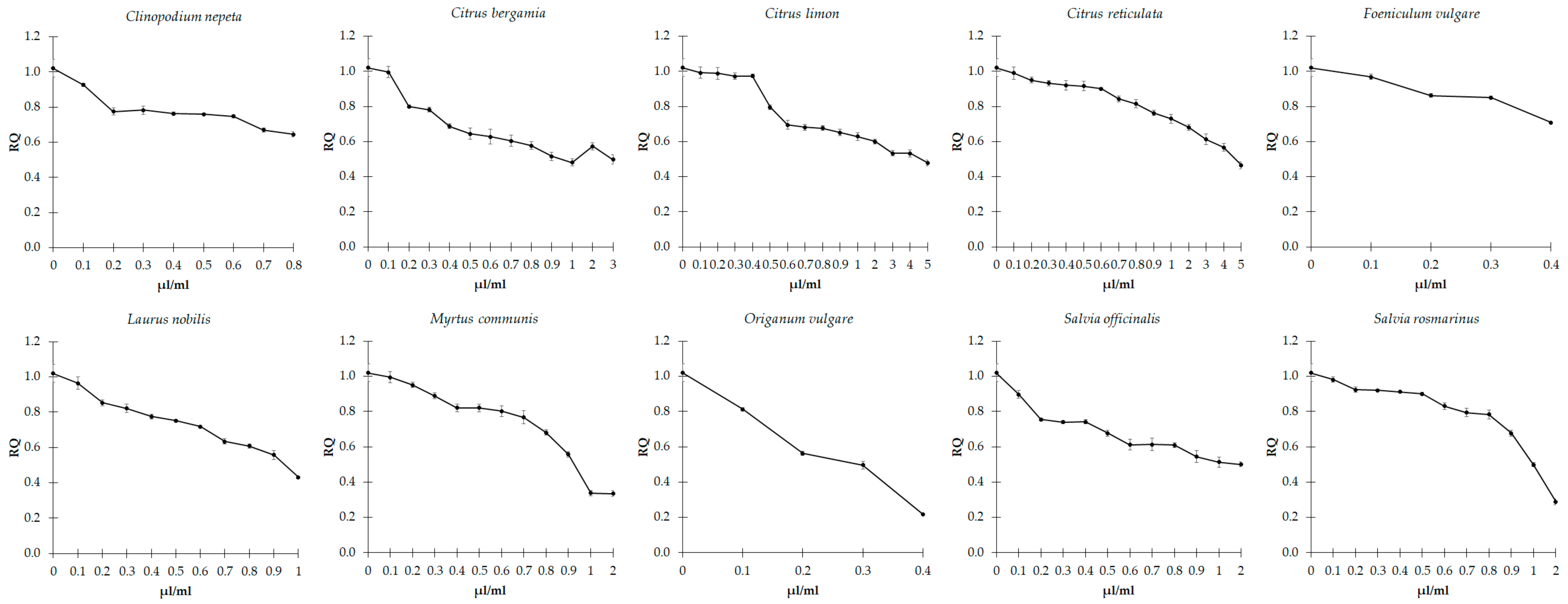
Figure 1. Effect of sub-inhibitory concentrations of essential oils on biofilm formation in C. violaceum. Values are reported as relative quantification (RQ), determined using the untreated cells as reference. Values represent the mean of five independent duplicate experiments with standard error of the mean.
Microorganisms 11 01150 g001
Figure 2. Effect of sub-inhibitory concentrations of essential oils on biofilm formation in E. faecalis. Values are reported as relative quantification (RQ), determined using the untreated cells as reference. Values represent the mean of five independent duplicate experiments with standard error of the mean.
Microorganisms 11 01150 g002
Figure 3. Effect of sub-inhibitory concentrations of essential oils on violacein production in C. violaceum. Values are reported as relative quantification (RQ), determined using the untreated cells as reference. Values represent the mean of five independent duplicate experiments with standard error of the mean.
Microorganisms 11 01150 g003
Figure 4. Effect of sub-inhibitory concentrations of essential oils on the gelatinase activity in E. faecalis. The values are reported as relative quantification (RQ), determined using the untreated cells as reference. Values represent the mean of five independent duplicate experiments with standard error of the mean.
Microorganisms 11 01150 g004
Figure 5. Methylation levels of 5-methylcytosine (5mC) (A) and N6-methyladenosine (m6A) (B) residues in DNA samples extracted from C. violaceum at basal conditions (untreated) and after treatment with sub-inhibitory concentrations of the essential oils. The values are reported as relative quantification (RQ), determined using the values of the untreated cells as reference. Values represent the mean of five independent triplicate experiments with standard error of the mean.
Microorganisms 11 01150 g005
Figure 6. Methylation levels of 5-methylcytosine (5mC) (A) and N6-methyladenosine (m6A) (B) residues in DNA samples extracted from E. faecalis at basal conditions (untreated) and after treatment with sub-inhibitory concentrations of the essential oils. The values are reported as relative quantification (RQ), determined using the values of the untreated cells as reference. Values represent the mean of five independent triplicate experiments with standard error of the mean.
Microorganisms 11 01150 g006
Table 1. Characterization of the essential oils analyzed (presence ≥ 1%).
Essential OilFamilyExtraction MethodChemical Composition%Retention
Index
Cultivation AreaTime of CollectionBatch N.
Clinopodium nepetaLamiaceaeHydro distillation from fresh collected materialpiperitone oxide34.2816.14Briatico, Province of Vibo Valentia, Calabria Region, Southern Italy
Michele Crudo Farm
July 202120210715
piperitenone oxide18.2319.72
(+)-limonene15.88.49
(+)-pulegone13.7515.6
menthone8.3212.77
isolegylacetate3.6417.92
1-terpine-4-ol1.413.56
(+)-neomenthol1.3713.25
β-pinene1.226.92
Citrus bergamiaRutaceaeMechanical extraction through industrial cold expression process from fresh fruit(+)-limonene15.8934.28Bovalino, Province of Reggio Calabria, Calabria Region, Southern Italy
Luigi Frammartino Farm
December 202120211221
lynalyl acetate10.7811.54
(+)-linalool9.386.79
α-terpinene8.4438.88
β-pinene6.875.49
α-pinene5.641.22
Citrus limon (L.)RutaceaeHydro distillation from fresh collected material(+)-limonene14.133.01Bovalino, Province of Reggio Calabria, Calabria Region, Southern Italy
Luigi Frammartino Farm
November 20212021118
α-terpinene13.591.26
β-pinene10.281.67
α-terpineol9.3611.91
α-terpinolene8.4274.41
1-Terpine-4-ol6.844.34
Citrus reticulataRutaceaeHydro distillation from fresh collected material(+)-sabinene12.61.44Briatico, Province of Vibo Valentia, Calabria Region, Southern Italy
Michele Crudo Farm
February 202120210215
(+)-linalool10.7618.27
α-phellandrene10.271.37
β-cis-ocimene9.361.14
(+)-limonene8.966.45
β-myrcene8.425.04
β-pinene7.766.54
α-pinene7.162.37
β-citronellal6.862.35
α-terpinolene6.7250.91
α-terpinene5.631.93
Foeniculum vulgare subsp. piperitumApiaceaeHydro distillation from fresh collected materialestragole17.2114.54Briatico, Province of Vibo Valentia, Calabria Region, Southern Italy
Michele Crudo Farm
September 202120210918
α-pinene14.1945.33
anethal10.4111.24
fenchone8.428.49
α-limonene7.722.51
α-phellandrene7.181.05
β-pinene6.861.65
β-myrcene5.6314.71
Laurus nobilis L.LauraceaeHydro distillation from fresh collected materialeucalyptol21.071.51Briatico, Province of Vibo Valentia, Calabria Region, Southern Italy
Michele Crudo Farm
April 202120210413
(+)-sabinene19.186.48
(+)-linalool13.61.29
terpinyl acetate10.797.38
α-pinene8.5656.61
methyleugenol6.7315.74
1-terpine-4-ol5.645.65
Myrtus communis L.MyrtaceaeHydro distillation from fresh collected materialeucalyptol20.271.88Briatico, Province of Vibo Valentia, Calabria Region, Southern Italy
Michele Crudo Farm
May 202120210521
(−)-myrtenylacetate18.4317.04
α-pinene15.923.88
(+)-limonene14.162.1
(+)-linalool10.8210.43
lynalyl acetate10.331.1
geraniol acetate9.441.09
α-terpineol9.041.58
β-ocimene8.5933.04
α-phellandrene8.4910.81
o-cymene8.351.41
terpinolene7.791.41
terpinene5.6912.33
Origanum vulgare L. subsp. viridulumLamiaceaeHydro distillation from fresh collected materialp-thymol20.484.88Briatico, Province of Vibo Valentia, Calabria Region, Southern Italy
Michele Crudo Farm
July 202120210720
terpinene16.7747.31
p-cymene16.423.52
β-caryophyllene8.6318.52
β-myrcene7.6111.78
carvacrol7.363.18
terpinolene6.553.76
α-thujene (origanene)5.151.23
α-pinene4.962.73
Salvia officinalis L.LamiaceaeHydro distillation from fresh collected materialeucalyptol22.65.54Briatico, Province of Vibo Valentia, Calabria Region, Southern Italy
Michele Crudo Farm
May 202120210504
(−)-α-thujone21.52.85
β-pinene12.459.59
(−)-camphor11.444.35
α-humulene11.0724.14
(−)-β-thujone8.5823.7
α-pinene7.232.26
(−)-β-caryophyllene6.915.1
β-myrcene6.751.13
C
camphene
6.121.88
(+)-sabinene5.673.99
Salvia rosmarinusLamiaceaeHydro distillation from fresh collected materialeucalyptol21.521.17Briatico, Province of Vibo Valentia, Calabria Region, Southern Italy
Michele Crudo Farm
April 202120210421
α-pinene13.312.28
β-pinene12.453.66
camphene8.5649.29
(−)-camphor7.211.79
isoborneol6.899.26
β -myrcene6.16.7
(−)-β-caryophyllene5.6622.84
Table 2. Minimum inhibitory concentrations (MIC) and minimum bactericidal concentrations (MBC) expressed as µL/mL of essential oils against C. violaceum and E. faecalis. The values represent the mean of five independent triplicate experiments. SEM: standard error of the mean.
Essential OilsC. violaceumE. faecalis
MICMBCMICMBC
MeanSEMMeanSEMMeanSEMMeanSEM
C. nepeta0.800.070.800.074.000.004.000.00
C. bergamia3.000.003.000.003.000.003.000.00
C. limon5.000.075.000.004.000.004.000.00
C. reticulata5.000.075.000.004.000.004.000.00
F. vulgare0.400.000.400.003.000.073.000.07
L. nobilis1.000.001.000.002.000.072.000.07
M. communis2.000.002.000.003.000.073.000.07
O. vulgare0.400.000.400.000.600.000.600.00
S. officinalis2.000.002.000.003.000.003.000.00
S. rosmarinus2.000.002.000.003.000.003.000.00
Disclaimer/Publisher’s Note: The statements, opinions and data contained in all publications are solely those of the individual author(s) and contributor(s) and not of MDPI and/or the editor(s). MDPI and/or the editor(s) disclaim responsibility for any injury to people or property resulting from any ideas, methods, instructions or products referred to in the content.