Article

Assessing the Effects of Rotifer Feed Enrichments on Turbot (Scophthalmus maximus) Larvae and Post-Larvae Gut-Associated Bacterial Communities

by
Antonio Louvado
1,
Carolina Castro
2,
Davide A. M. Silva
1,
Vanessa Oliveira
1,
Luís E. C. Conceição
3,
Daniel F. R. Cleary
1 and
Newton C. M. Gomes
1,*
1
Department of Biology and Centre for Environmental and Marine Studies (CESAM), University of Aveiro, 3810-193 Aveiro, Portugal
2
Flatlantic—Actividades Piscícolas, SA 3070-732 Praia de Mira, Portugal
3
SPAROS Lda., 8700-221 Olhao, Portugal
*
Author to whom correspondence should be addressed.
Microorganisms 2023, 11(2), 520; https://doi.org/10.3390/microorganisms11020520
Submission received: 19 January 2023 / Revised: 9 February 2023 / Accepted: 10 February 2023 / Published: 17 February 2023
(This article belongs to the Section Veterinary Microbiology)

Abstract

:
Live feed enrichments are often used in fish larvicultures as an optimized source of essential nutrients to improve larval growth and survival. In addition to this, they may also play an important role in structuring larval-associated microbial communities and may help improve their resistance to diseases. However, there is limited information available on how larval microbial communities and larviculture water are influenced by different live feed enrichments. In the present study, we investigated the effects of two commercial rotifer enrichments (ER) on turbot (Scophthalmus maximus) larval and post-larval gut-associated bacterial communities during larviculture production. We evaluated their effects on bacterial populations related to known pathogens and beneficial bacteria and their potential influence on the composition of bacterioplankton communities during larval rearing. High-throughput 16S rRNA gene sequencing was used to assess the effects of different rotifer enrichments (ER1 and ER2) on the structural diversity of bacterial communities of the whole turbot larvae 10 days after hatching (DAH), the post-larval gut 30 DAH, and the larviculture water. Our results showed that different rotifer feed enrichments were associated with significant differences in bacterial composition of turbot larvae 10 DAH, but not with the composition of larval gut communities 30 DAH or bacterioplankton communities 10 and 30 DAH. However, a more in-depth taxonomic analysis showed that there were significant differences in the abundance of Vibrionales in both 10 DAH larvae and in the 30 DAH post-larval gut fed different RE diets. Interestingly, the ER1 diet had a higher relative abundance of specific amplicon sequence variants (ASVs) related to potential Vibrio-antagonists belonging to the Roseobacter clade (e.g., Phaeobacter and Ruegeria at 10 DAH and Sulfitobacter at 30 DAH). In line with this, the diet was also associated with a lower relative abundance of Vibrio and a lower mortality. These results suggest that rotifer diets can affect colonization by Vibrio members in the guts of post-larval turbot. Overall, this study indicates that live feed enrichments can have modulatory effects on fish bacterial communities during the early stages of development, which includes the relative abundances of pathogenic and antagonist taxa in larviculture systems.

1. Introduction

Wild finfish larvae feed on a diverse array of zooplankton (e.g., copepods, cladocerans, ciliates, and rotifers [1]) in order to fulfill their nutritional requirements during early life stages [2,3], with copepods of particular importance [3,4]. Copepods are naturally rich in essential nutrients (e.g., high quality proteins, amino acids, and minerals) and polyunsaturated fatty acids (PUFAs, e.g., ω-3 and ω-5 [3,5]). PUFAs are essential for larval development, and a deficiency or imbalance in PUFAs may adversely affect fish growth and immunological development [6,7,8,9]. However, the mass cultivation of these small crustaceans is challenging, and, despite some successful small-scale ventures [10], they are not systematically used in the marine finfish larviculture sector [3,11]. As an alternative, rotifer and artemia are the most commonly used as the first feed in larviculture [5,11]. However, both artemia and rotifer are naturally poor in PUFAs. To overcome this problem, a range of commercial products, made of nutritionally balanced lipid emulsions, have been approved for the enrichment of live feed with PUFAs and are commonly used in the production of fish larvae [5,9]. An optimal nutrition of these commercial enrichments has been intensively debated in the literature for most commercial finfish species, including the turbot (Scophthalmus maximus) [12], and this has led to significant improvements in fish survival, growth, and disease resistance [13,14].
In addition to the importance of providing nutritionally complete live diets, the continuous production of marine finfish fingerlings is frequently affected by stochastic episodes of mass mortalities in the first weeks of life [2,15]. These outbreaks have typically been associated with pathogens belonging to genera Vibrio, Photobacterium, and Tenacibaculum that naturally inhabit the microbial communities of rotifers, copepods, and artemia [16,17,18]. To tackle this problem, strategies based on the exposure of live feed and fish eggs to mild doses of chemical disinfectants have been adopted as the standard procedure in the larviculture industry [19]. Although provisionally effective, these strategies may interfere with the normal development of fish larvae [20] and the quality of the live feed [21]. Furthermore, the process of disinfection affects the microbial community as a whole, creating a more unstable environment, which will favor the development of fast-growing opportunistic r-strategists and increase the probability of pathogen proliferation and fish larvae infection [22]. Despite the perception that the disinfection processes can have significantly adverse effects on the larviculture microbiome, there is a scarce amount of information on how live feed enrichments affect the initial microbe recruitment of newly hatched larvae [15,23,24,25].
The establishment of larval microbial communities is a multifactorial process, driven by both extrinsic (e.g., environment) and intrinsic processes (e.g., phylogeny and host genetics; [26,27,28]), in which diet plays a crucial role [25,29,30]. In larviculture, the rearing water and feed are the earliest sources of bacteria that colonize the skin and gastrointestinal tracts of fish [15,24,29,31]. There is, however, a lack of information on how larval bacterial communities are influenced by different commercial live feed enrichments. Therefore, in line with the need to better understand the effects of live diets on larval microbiomes, in the present study we aimed to investigate the effects of two commercial rotifer enrichments (ER1 and ER2) on turbot (Scophthalmus maximus) larval and post-larval gut-associated bacterial communities during larviculture production. We hypothesize the following: (1) Rotifer feed enrichments will have distinct effects on turbot (Scophthalmus maximus) larvae and post-larval gut-associated bacterial communities. (2) Different live feed enrichment products will have different effects on the balance of potential pathogenic and antagonistic microbes in the larviculture production system. (3) Bacterial populations of live feed enrichments will colonize turbot larvae and post-larval gut biotopes.

2. Methods

2.1. Larviculture

Water, larvae, post-larvae, and live feed samples were obtained between 17 November and the 14 December 2021 in an intensive larviculture facility for the production of turbot (S. maximus) larvae located in Portugal. The larviculture operates in a flow-through system with natural seawater collected from the adjacent Atlantic Ocean. Sampling was conducted during routine and animal husbandry practices of commercial farming operations. The sampling procedure was in line with approved methods of animal killing according to Annex IV of the European Directive Dir2010/63/EU on the protection of animals used for scientific purposes. No experimental procedure was conducted on live fish. Two products (ER1 and ER2), commercialized as enrichment diets for live feed, were used as rotifer enrichments. Both products are commercially available and have been previously approved for use for animal feeding. They are routinely used by the larviculture facility where the sampling occurred. ER1 and ER2 are dry formulas enriched in polyunsaturated fatty acids (PUFAs), vitamins, and minerals. Based on the content declarations, ER1 contains a taurine additive and has higher protein and PUFA content than ER2. ER2 has a higher vitamin content than ER1 and a probiotic additive based on viable cells of a strain of Pediococcus acidilactici. Due to the fact that this study was not designed to evaluate different commercial live feed enrichments, but instead to investigate their importance in shaping larval microbiomes, the commercial name of the products is not provided, in order to avoid misjudgment of the efficiency and quality of different products.
Water and larvae samples were obtained from independent cylindrical tanks (three for each live feed enrichment). Larviculture tanks were operated as a flow-through system with water pH 7.6–8.0, temperature 19.18 ± 0.51 °C, and salinity 34.05 ± 0.11‰. Larvae were fed rotifers, enriched by ER1 or ER2, from 3 to 15 DAH. Rotifers enriched with their respective product (ER1 or ER2) were added to rearing water of their respective tanks, together with a mature culture of Tetraselmis sp. (green water technique). From 11 to 15 DAH, the differently enriched rotifers were gradually replaced by artemia enriched with ER2. Between 16 DAH and until 30 DAH, only artemia enriched with ER2 were provided. Fish were fed manually with a three hour interval between feeds. At 30 DAH, fish were transferred to nursery tanks and they remained there until 60 DAH. During this time, any detected abnormal fish (i.e., small, deformed, diseased, or depigmented fish) were euthanized. At 60 DAH, surviving fish were counted to determine the survival rate of each tank.

2.2. Sample Collection and DNA Extraction

Composite samples of the whole turbot larvae (n = 25) and the post-larval gut (n = 5) were obtained seven (10 DAH) and twenty-seven days (30 DAH) after their transfer to the larval rearing system, respectively. One composite sample was obtained for each tank, totaling three composite samples of larvae for each combination of treatments (ER1 and ER2) and sampling points (10 and 30 DAH). Larvae were collected using a fish net (the fish net was cleaned in-between samplings with a 0.1% hydrogen peroxide solution). Collected larvae were transferred to 120 mL sterile plastic cups filled with respective rearing water. Larvae were killed using an overdose of fish anesthetic (clove oil; Sigma-Aldrich, Saint Louis, MO, USA) and then rinsed with sterile artificial seawater with salinity adjusted to 36 ppt (ASW). Dissection only occurred after proof of death (waiting for the onset of rigor mortis). Smaller larvae (10 DAH) were rinsed five times with 5 mL of ASW in a 15 mL falcon tube, while larger larvae (30 DAH) were washed on both sides for approximately 5 s with ASW, using a previously autoclaved wash bottle. Twenty-five larvae with 10 DAH, were transferred whole into bead-beating tubed from the DNA extraction kit. Five larvae with 30 DAH, were dissected and their gastro-intestinal tract was retrieved whole and pooled into bead-beating tubes from the DNA extraction kit.
Bacterioplankton samples consisted of rearing water collected from each tank at the same time that larvae samples were collected (i.e., 10 and 30 DAH). A 250 mL sterile Erlenmeyer flask was filled with rearing water and filtered through a 0.22 µm polycarbonate filter (Merck-Millipore, Rahway, NJ, USA) in an EZ-Fit™ Manifold base (Merck-Millipore, Rahway, NJ, USA) or in a Büchner filtration assembly connected to a vacuum pump. After filtration, the polycarbonate filter was cut thoroughly using sterile scissors and was transferred to a bead-beating tube. Live feeds (rotifers, algae, and artemia) were collected from respective stock cultures using a sterile 250 mL cup and transported to the laboratory. Only one sample of each stock culture of feed was collected in the experiment. Algae and rotifer samples were collected seven days after larval transfer to the rearing system, while artemia samples were collected twenty-seven days later. Algae were harvested by centrifugation at 4400× g during 30 min; supernatant was discarded and the pellet was suspended in DNA extraction buffer and transferred into bead-beating tubes from the DNA extraction kit. Rotifer and artemia samples were collected by filtering samples through a Whatman 113 paper filter (Merck-Millipore, Rahway, NJ, USA). Residues were washed three times with sterile artificial seawater, collected using a sterile spatula (300 mg), and transferred into respective bead-beating tubes from the DNA extraction kit.
All samples (larvae, bacterioplankton, and feed) were processed using FastDNATM Spin kit (MP Biomedicals, Santa Ana, CA, USA) following the manufacturer’s instructions. A blank negative control with no sample was included in the DNA extraction process each time. All samples were processed immediately; no storage was required.

2.3. High-Throughput Sequencing Data Acquisition

The hypervariable region V3/V4 of the 16S rRNA gene was amplified by PCR using the primers 314F (CCTACGGGNGGCWGCAG) and 785R (GACTACHVGGGTATCTAATCC) [32]. Library preparation and sequencing were performed using a MiSeq sequencing platform at Molecular Research LP (www.mrdnalab.com; accessed on 15 September 2022, Shallowater, TX, USA), following standard Illumina procedures (Illumina, San Diego, CA, USA). QIIME2 (version 2020.8) was used to transform the amplicon libraries to an amplicon sequence variant (ASV) abundance table [33]. Demultiplexing was performed using the “demux” algorithm in QIIME2. The dada2 algorithm from the DADA2 plugin [34] in QIIME2 was used to filter low-quality reads, merge forward and reverse reads into sequences, remove chimeras, and group sequences into ASVs. In dada2, forward and reverse sequences were truncated at 220 and 240 bp, respectively. Taxonomy was assigned to ASVs using the ‘feature-classifier’ algorithm in QIIME2 with a scikit-learn Naïve Bayes classifier based on the SILVA database of the 16S reference sequences at 99% similarity (version 138, released December 2019). The classifier was previously trained using a ‘feature-classifier’ algorithm in QIIME2 (version 2020.8) with reference sequences, trimmed and truncated at the 314F and 785R region. To simplify interpretation, a unique number was assigned to each ASV. Non-bacterial, mitochondrial, and chloroplastidial sequences were removed. Negative controls from the DNA extraction were also sequenced and ASVs that occurred in the negative controls, but which did not appear to be the result of “index hopping” [35], were removed. A list of removed ASVs is presented in Supplementary File S1.

2.4. Data Analysis and Statistics

A table containing the ASV counts per sample was imported into R and used to compare community diversity and composition and assess the relative abundance of selected higher taxa. The Shannon’s H’ diversity index was calculated using the diversity() function in the vegan package [36], rarefied richness was calculated using the rarefy() function in vegan, evenness (Pielou’s J) was calculated by dividing Shannon H’ by the logarithm of the total number of ASVs, and Fisher’s alpha was calculated using the fisher.alpha() function in vegan. Since diversity parameter data were not normally distributed, we tested for significant variations for each factor separately: diet, biotope (water and larvae), and sampling time; using the Kruskal–Wallis rank sum test with the kruskal.test() function in the stats package. Relative abundances of selected bacterial higher taxa were tested for significant variation between diets, biotopes (water and larvae), and sampling times using the anova() function in R with the F test applied to a general-linearized model using the glm() function in R [37]. Since a number of these variables included an excess of zero counts in the samples, we set the family argument to ‘tweedie’ using the tweedie() function in the statmod package in R, with var.power = 1.5 and link.power = 0 (a compound Poisson–gamma distribution). To perform pairwise comparisons among treatments, we used the emmeans() function from the emmeans package with the p-value adjustment set to the false discovery rate (i.e., p.adjust = ”fdr”). Biplot ordinations of ASV composition were produced for the full dataset and separately for each sampling point (10 and 30 DAH). First, a phyloseq object was generated using the phyloseq() function from the ‘phyloseq’ package [38]. The ordinate() function in ‘phyloseq’ was subsequently used with the phyloseq object as input, the method argument set to ‘PCoA’, and the distance argument set to ‘bray’. A biplot was then produced using the plot_ordination() function in ‘phyloseq’ with the type argument set to ‘biplot’. Significant differences among factors (diet (ER1 vs. ER2) and sampling point (10 vs. 30 DAH) and biotope (water vs. larvae)) and their interactions were determined using the adonis() function in vegan for a permutational multivariate analysis of variance (PERMANOVA). The BLAST search tool (http://www.ncbi.nlm.nih.gov/; accessed on 15 September 2022) was used to compare representative sequences of the 50 most abundant ASVs to sequences in the NCBI 16S ribosomal RNA (Bacteria and Archaea type strains) database using standard parameter settings [39,40]. Sequences that exhibited the highest levels of similarity were considered to be closely related organisms.

3. Results and Discussion

After quality control and the removal of non-bacterial sequences, the dataset consisted of 2486757 sequences binned into 2336 ASVs. Overall, our results showed similar α-diversity patterns for water and fish (larvae 10 DAH and post-larval gut 30 DAH) bacterial communities in tanks with different rotifer diets (ER1 and ER2) (Figure 1; Kruskal Wallis–Wallis: p > 0.05). This is in line with other studies, which have shown that live feed manipulation did not affect rarefied richness of the gut-associated bacterial communities of 34 DAH gilthead seabream post-larvae (Sparus aurata; [15]). Bacterioplankton communities, however, were richer (Kruskal–Wallis: H(1) = 17.38; p < 0.001) than the fish samples, but there were no significant differences in evenness or Shannon’s H’ diversity. Previous studies also showed that bacterioplankton communities tended to be richer than the host-associated bacterial communities [41,42]. There were differences in the diversity and evenness of bacterioplankton communities from different sampling events (10 vs. 30 DAH), but these differences were not significant following the p-value correction using the FDR method (Kruskal–Wallis: FDR-P > 0.05).
A PCO ordination highlighting the differences in ASV composition is presented in Figure 2. Biotope (water vs. larvae) and sampling time (10 vs. 30 DAH) were better predictors of the bacterial community structure than the rotifer’s diet (Figure 2). The rotifer’s diet, however, did have a significant independent and interaction effect on the ASV composition (PERMANOVA: p < 0.05, Table 1). Separate PCO ordinations for 10 and 30 DAH samples are presented in Supplementary Figure S1 in order to better evaluate the interactions between the sampling time and rotifer diets. Our results showed that the differences between different diets (ER1 and ER2) were more prevalent for the 10 DAH whole larvae samples as opposed to the post-larval gut and water samples, showing a clear separation of the effects of the different diets along the second axis. In line with this, the results of the PERMANOVA showed that the rotifer’s diet was a significant predictor of ASV composition at 10 DAH (PERMANOVA: F1,11 = 5.21; p = 0.002), but not at 30 DAH (PERMANOVA: F1,11 = 1.324; p = 0.202). Previous studies have reported similar results in cod larvae that suggested that the environmental setting (e.g., water and biofilm) is more influential than live feed during initial microbiome recruitment in larval guts [29]. Some speculate that the maladaptation of feed’s microbes to the gut environment impedes a successful gut colonization [24].
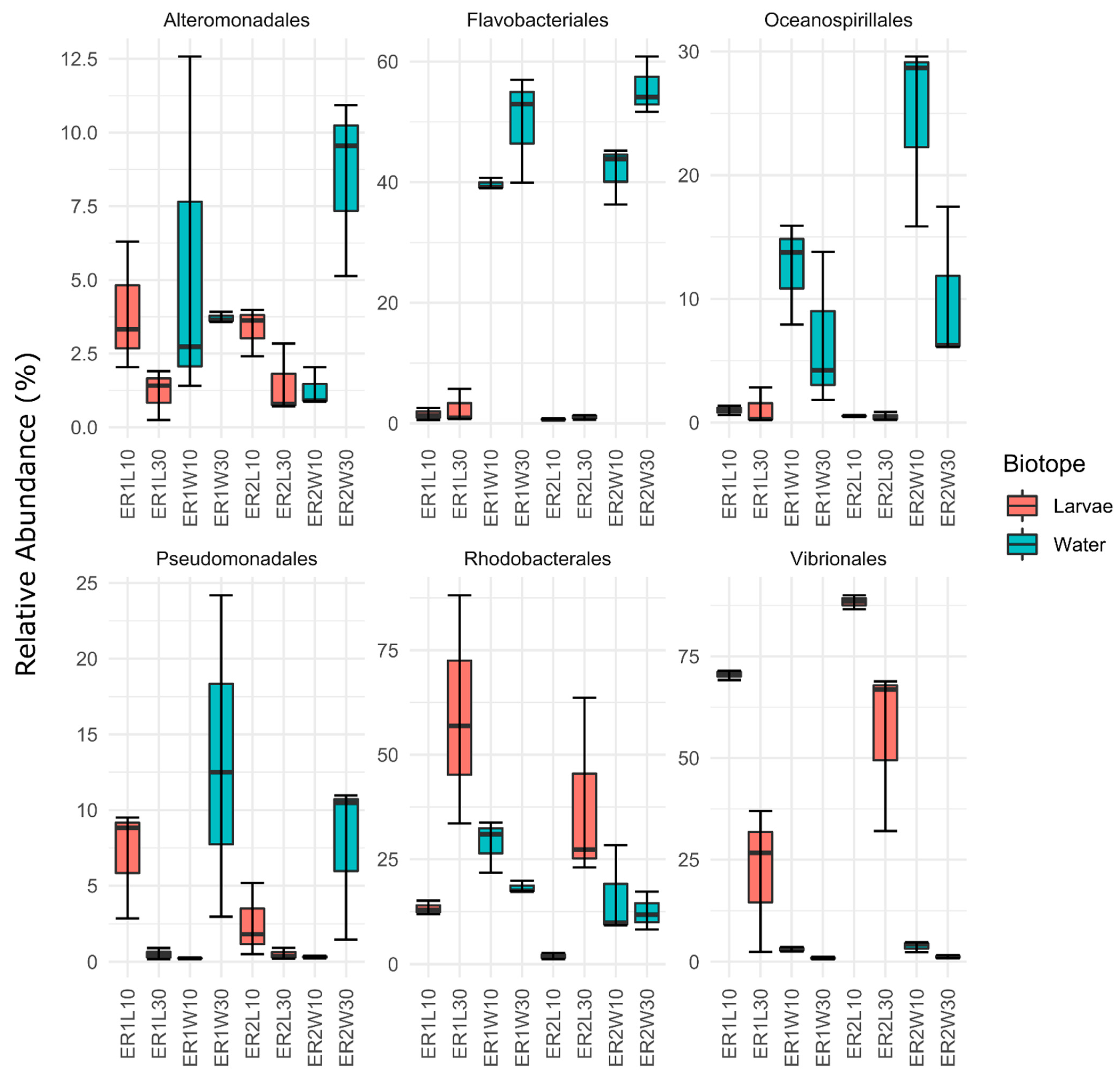
The taxonomic composition of the dataset is presented in Figure 3. Overall, our results showed that, in contrast to the fish larvae 10 DAH, the taxonomic abundances of the bacterial communities inhabiting the post-larval gut 30 DAH were highly variable among the replicates. Larval microbiomes changed rapidly during development, a process which may result in varying degrees of resistance (open niches and/or lack of antagonistic traits) to the invasion by fast-growing opportunistic microbes [43]. Our results also showed that the bacterial communities of the larvae 10 DAH and the post-larval gut 30 DAH were dominated by members of the order Vibrionales. The predominance of Vibrionales during larval development is in line with other studies on turbot larvae [44,45]. Although not all Vibrionales strains cause infection in fish, some members of this order (e.g., Photobacterium damselae, V. parahaemolyticus, V. anguillarum, and V. harveyi) are responsible for mass mortalities in aquaculture systems [46,47]. Invertebrates are recognized as natural reservoirs of Vibrio and, in enclosed aquaculture facilities, their administration as live feed is considered one of the main causes of Vibrio infections in marine larvicultures [16,31,48]. This is why many studies have investigated the potential of probiotics, prebiotics, postbiotics, and other microbiome-modulating strategies to control Vibrio populations in live feed [15,16,44,49,50]. Here, we also observed that the order Rhodobacterales showed a stronger dominance in the bacterial communities of the post-larval gut 30 DAH than in the whole larvae 10 DAH. This enrichment was, furthermore, more pronounced for fish larvae fed rotifers enriched with ER1 than with ER2. Overall, the majority of sequences assigned to the Rhodobacterales order were assigned to members of the Roseobacter clade (Supplementary Table S1). The Roseobacter clade is a paraphyletic group belonging to the Rhodobacterales order, with several members known to possess antagonistic activity against Vibrio spp. and other aquaculture pathogens, e.g., Phaeobacter inhibens, P. gallaeciensis, P. piscinae, Ruegeria sp., and Sulfitobacter sp. [51,52,53].
In this study, the bacterioplankton was dominated by the orders Flavobacteriales and Rhodobacterales at both sampling points (10 and 30 DAH), with a high abundance of order Oceanospirillales at 10 DAH and Pseudomonadales at 30 DAH (Figure 3). All orders except Pseudomonadales have been previously found to be abundant in the rearing water of RAS for adult turbot [54]. The most abundant ASVs of order Flavobacteriales were most similar (>98%) to sequences in the NCBI database identified as belonging to genera Polaribacter, Winogradskyella, or Meridianimaribacter (Supplementary Table S1). All these genera are commonly found in marine settings, including aquaculture systems [54,55,56,57]. The most abundant ASVs assigned to the Oceanospirillales order were assigned to family Nitrincolaceae and had very low similarity (<93%) with type strains of the NCBI database. Members of the Nitrincolaceae family are heterotrophic bacteria usually found as core members of bacterioplankton communities and involved in nitrogen and carbon cycling following phytoplankton blooms [58,59]. The enrichment of Pseudomonadales in the rearing water 30 DAH was mainly due to the enrichment of two ASVs (ASVs 10 and 13; Supplementary Table S1) assigned to Paraperlucidibaca baekdonensis (Supplementary Table S1). This specie was first isolated from marine waters [60], but was later found to be a core member of the bacterioplankton of marine aquacultures. However, its potential functional roles in aquaculture systems remain unknown [61,62].
A more in-depth pairwise analysis preformed on the six most abundant orders in the dataset showed that larvae 10 DAH had a significant and higher abundance of Vibrionales than all the other groups analyzed (Supplementary File S3, EMMEANS: p < 0.05; Figure 4). Our results also showed that the abundance of this order was significantly higher in fish fed rotifers enriched with ER2 (88.384 ± 1.728% in larvae 10 DAH and 55. 919 ± 20.682% in the post-larval gut 30 DAH) than with ER1 (70.52 ± 1.187% in larvae 10 DAH and 22.033 ± 17.74% in the post-larval gut 30 DAH). Among the 50 most abundant ASVs, all those assigned to order Vibrionales were also assigned to the Vibrio genus (Supplementary Table S1). Interestingly, a Venn diagram analysis showed that about 92 ASVs detected in rotifers (combined ER1 and ER2) were also detected in the bacterial communities of the post-larval gut and/or in the bacterioplankton 30 DAH, algae, and artemia (Supplementary Figure S2). We also observed that 51 of these ASVs were either present in fish guts’ bacterial communities alone or in both the fish guts’ bacterial communities and the bacterioplankton. This suggests that these ASVs may have a more specific association with the turbot post-larval gut. The relative abundance analysis showed that the majority of these ASVs belonged to the rare biosphere and only sixteen ASVs had relative abundances exceeding 0.1% (Supplementary File S4). Ten of these sixteen ASVs were classified to genus Vibrio (ASV 51, 52, 53, 56, 57, 61, 62, 65, 69, and 76) and the remaining to genera Litoribacillus (ASV 59), Peptoniphilus (ASV 68), Paraperlucidibaca (ASV 10), Conchiformibius (ASV 75), and Thalassolituus (ASV 81). In general, members of Litoribacillus, Paraperlucidibaca, and Thalassolituus are often associated with marine bacterioplankton [60,63,64], while Conchiformibius and Peptoniphilus were previously found to be associated with oral cavities [65] and the gut of animal hosts [66,67], respectively. ASV-51 (Vibrio sp.) was the only ASV to have a relatively high abundance in both diets (ER1 and ER2). In general, ASVs related to Vibrio were less abundant in the bacterial communities of the post-larval gut fed rotifers enriched with ER1. Our results suggest that rotifer diets contribute to colonization by Vibrio members in the gut of turbot post-larvae, and the degree of colonization depends on the type of enrichment used.
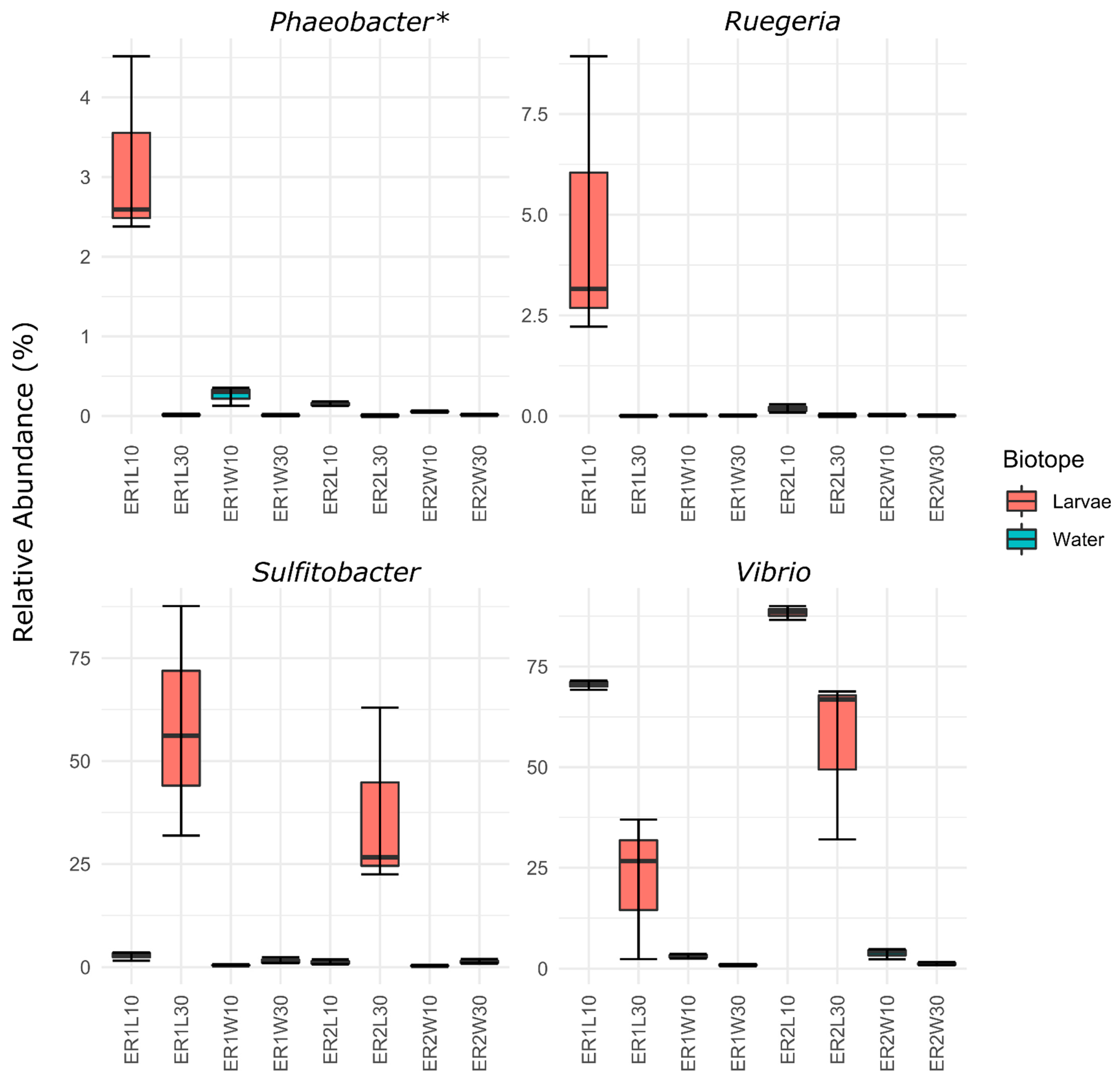
The Vibrio genus is known to comprise both non-pathogenic and pathogenic strains, with the latter being a major concern for the aquaculture sector [46,47]. Interestingly, in line with the reduction in the relative abundance of Vibrio in ER1 samples, we also observed that this diet increased the relative abundance of ASVs assigned to the genera Ruegeria (ASV 47) and Nautella (ASV 49) at 10 DAH and Sulfitobacter at 30 DAH (ASV-2; Figure 5 and Supplementary Figure S3). Nonetheless, sequence similarity analysis using the NCBI type strain database showed that ASV 49 had the highest similarity (100%) with a strain previously identified as Nautella italica, but later reclassified as Phaeobacter italicus (Supplementary Table S1; [68]). Ruegeria, Phaeobacter, and Sulfitobacter are bacterial members of the Roseobacter clade [69] and are known to comprise several bacterial strains with antagonistic activity against bacterial pathogens, including Vibrio species [52,70]. Interestingly, the lower Vibrio abundance and higher abundance of potential antagonistic bacteria (Ruegeria, Phaeobacter and Sulfitobacter) detected in ER1 tanks was associated with a significantly higher fish survival rate (8.0 ± 0.9%) in comparison to ER2 (4.34 ± 0.89%). The survival rates reported here at 60 DAH are similar to previous studies of turbot larviculture [14,71]. It has been recognized that the survival of the turbot larvae is often lower than that of other marine fish [72], but in the past decades, larval survival has been a major undertaking for hatchery management, with significant advances obtained through improvements in the nutritional formulation of live feed enrichment [13,14].

4. Conclusions

This study showed that different rotifer enrichments were associated with significant differences in the bacterial community structure of turbot larvae 10 DAH but not with post-larval gut communities 30 DAH. However, a more in-depth analysis showed that the ER1 diet was associated with a lower abundance of Vibrio in whole larvae and post-larval gut-associated communities, and a higher abundance of the ASVs related to potential Vibrio antagonists (i.e., members of the Roseobacter clade). Overall, our findings indicate that live feed enrichment formulas can have modulatory effects on fish communities during their early stages of development with the potential to affect the balance of pathogenic and antagonist microbes in larviculture. In addition to the above, different enrichments potentially contribute to Vibrio colonization in the guts of post-larval turbot; the degree of colonization may also vary depending on the enrichment used. Our findings highlight the importance of analyzing the effects of differentially enriched rotifer feeds on the structural composition of microbial communities in fish larviculture. Given the uniqueness of microbial communities at different larviculture systems and the well-known effects of the host microbiome on animal health and growth [73], aquaculture producers should consider the strategy applied in the present study to evaluate and optimize larval fish diets.

Supplementary Materials

The following supporting information can be downloaded at: https://www.mdpi.com/article/10.3390/microorganisms11020520/s1. Supplementary File S1, list of deleted sequences; Supplementary File S2 which contains Supplementary Figure S1, first two axis of PCO of samples of the host and water associated bacterial communities at 10 DAH and 30 DAH ), Supplementary Figure S2, Venn diagram of whole larvae at 10 DAH (Larvae), post-larvae gut at 30 DAH (Post-larvae gut), feed samples (Artemia, Rotifer and Algae) and water samples (Water) at 10 and 30 DAH, irrespective of diet, Supplementary Figure S3, heatmap analysis of the 50 most abundant ASV, and Supplementary Table S1, detailed description of the 50 most abundant ASVs; Supplementary File S3, emmeans results of pairwise comparisons of relative abundances of the six most abundant bacterial orders; Supplementary File S4, table of relative abundance per sample of all ASVs only found in rotifers and 30 DAH samples (post-larval gut and bacterioplankton communities) in the Venn diagram in Figure 5.

Author Contributions

A.L.: methodology, investigation, formal analysis, data curation, and writing—original draft; C.C.: methodology, investigation, formal analysis, and writing—review and editing; D.A.M.S.: investigation; V.O.: investigation; L.E.C.C.: resources, funding acquisition; D.F.R.C.: data curation, formal analysis, and writing—review and editing, visualization; N.C.M.G.: conceptualization, resources, and writing—review and editing, supervision, and funding acquisition. All authors have read and agreed to the published version of the manuscript.

Funding

This research was funded by Portugal and the European Union through FEDER, COMPETE2020 and CRESC Algarve2020, in the framework of Portugal2020 through project MAXIMUS.PT grant number [POCI-01-0247-FEDER-069769] and co-financed by the Portuguese Foundation for Science and Technology/Ministry of Science and Higher Education (FCT/MCTES) through project UIDB/04326/2020 to CCMAR. We acknowledge financial support to CESAM by FCT/MCTES (UIDP/50017/2020+UIDB/50017/2020+ LA/P/0094/2020), through national funds. VO was funded by national funds, through FCT, in the scope of the framework contract foreseen in the numbers 4, 5 and 6 of the article 23, of the Decree-Law 57/2016, of August 29, changed by Law 57/2017, of July 19. DAMS was funded by the FCT (2020.05774.BD), co-financed by the European Social Fund within the framework of Programa Operacional Centro 2020.

Data Availability Statement

Sequences used in this study were uploaded to the NCBI ShortRead Archive (BioProject PRJNA911963; Biosamples SAMN32217043—SAMN32217070).

Conflicts of Interest

The authors declare no conflict of interest.

References

  1. Manickam, N.; Bhavan, P.S.; Santhanam, P.; Muralisankar, T. Influence of Wild Mixed Zooplankton on Growth and Muscle Biochemical Composition of the Freshwater Prawn Macrobrachium rosenbergii Post Larvae. Aquaculture 2020, 522, 735110. [Google Scholar] [CrossRef]
  2. Vadstein, O.; Bergh, Ø.; Gatesoupe, F.; Galindo-Villegas, J.; Mulero, V.; Picchietti, S.; Scapigliati, G.; Makridis, P.; Olsen, Y.; Dierckens, K. Microbiology and Immunology of Fish Larvae. Rev. Aquac. 2013, 5, S1–S25. [Google Scholar] [CrossRef] [Green Version]
  3. Lee, C.-S.; O’Bryen, P.J.; Marcus, N.H. Copepods in Aquaculture; John Wiley & Sons: Hoboken, NJ, USA, 2008; ISBN 0470276304. [Google Scholar]
  4. Zeng, C.; Shao, L.; Ricketts, A.; Moorhead, J. The Importance of Copepods as Live Feed for Larval Rearing of the Green Mandarin Fish Synchiropus splendidus. Aquaculture 2018, 491, 65–71. [Google Scholar] [CrossRef]
  5. Haché, R.; Plante, S. The Relationship between Enrichment, Fatty Acid Profiles and Bacterial Load in Cultured Rotifers (Brachionus plicatilis L-Strain) and Artemia (Artemia salina strain Franciscana). Aquaculture 2011, 311, 201–208. [Google Scholar] [CrossRef]
  6. Koven, W.M.; Tandler, A.; Kissil, G.W.; Sklan, D.; Friezlander, O.; Harel, M. The Effect of Dietary (N− 3) Polyunsaturated Fatty Acids on Growth, Survival and Swim Bladder Development in Sparus aurata Larvae. Aquaculture 1990, 91, 131–141. [Google Scholar] [CrossRef]
  7. Bell, J.G.; Castell, J.D.; Tocher, D.R.; MacDonald, F.M.; Sargent, J.R. Effects of Different Dietary Arachidonic Acid: Docosahexaenoic Acid Ratios on Phospholipid Fatty Acid Compositions and Prostaglandin Production in Juvenile Turbot (Scophthalmus maximus). Fish Physiol. Biochem. 1995, 14, 139–151. [Google Scholar] [CrossRef]
  8. Sargent, J.; Bell, G.; McEvoy, L.; Tocher, D.; Estevez, A. Recent Developments in the Essential Fatty Acid Nutrition of Fish. Aquaculture 1999, 177, 191–199. [Google Scholar] [CrossRef]
  9. Chee, W.-L.; Turchini, G.M.; Teoh, C.-Y.; Ng, W.-K. Dietary Arachidonic Acid and the Impact on Growth Performance, Health and Tissues Fatty Acids in Malabar Red Snapper (Lutjanus malabaricus) Fingerlings. Aquaculture 2020, 519, 734757. [Google Scholar] [CrossRef]
  10. Jepsen, P.M.; van Someren Gréve, H.; Jørgensen, K.N.; Kjær, K.G.W.; Hansen, B.W. Evaluation of High-Density Tank Cultivation of the Live-Feed Cyclopoid Copepod Apocyclops royi (Lindberg 1940). Aquaculture 2021, 533, 736125. [Google Scholar] [CrossRef]
  11. Drillet, G.; Frouël, S.; Sichlau, M.H.; Jepsen, P.M.; Højgaard, J.K.; Joarder, A.K.; Hansen, B.W. Status and Recommendations on Marine Copepod Cultivation for Use as Live Feed. Aquaculture 2011, 315, 155–166. [Google Scholar] [CrossRef]
  12. Estevez, A.; McEvoy, L.A.; Bell, J.G.; Sargent, J.R. Growth, Survival, Lipid Composition and Pigmentation of Turbot (Scophthalmus maximus) Larvae Fed Live-Prey Enriched in Arachidonic and Eicosapentaenoic Acids. Aquaculture 1999, 180, 321–343. [Google Scholar] [CrossRef]
  13. Conceição, L.E.C.; Yúfera, M.; Makridis, P.; Morais, S.; Dinis, M.T. Live Feeds for Early Stages of Fish Rearing. Aquac. Res. 2010, 41, 613–640. [Google Scholar] [CrossRef]
  14. Robin, J.H. Use of Borage Oil in Rotifer Production and Artemia Enrichment: Effect on Growth and Survival of Turbot (Scophthalmus maximus) Larvae. Aquaculture 1998, 161, 323–331. [Google Scholar] [CrossRef]
  15. Sanches-Fernandes, G.M.M.; Califano, G.; Castanho, S.; Soares, F.; Ribeiro, L.; Pousão-Ferreira, P.; Mata, L.; Costa, R. Effects of Live Feed Manipulation with Algal-Derived Antimicrobial Metabolites on Fish Larvae Microbiome Assembly: A Molecular-Based Assessment. Aquac. Res. 2022, 53, 1062–1083. [Google Scholar] [CrossRef]
  16. D’Alvise, P.W.; Lillebø, S.; Prol-Garcia, M.J.; Wergeland, H.I.; Nielsen, K.F.; Bergh, Ø.; Gram, L. Phaeobacter gallaeciensis Reduces Vibrio anguillarum in Cultures of Microalgae and Rotifers, and Prevents Vibriosis in Cod Larvae. PLoS ONE 2012, 7, e43996. [Google Scholar] [CrossRef] [PubMed] [Green Version]
  17. Verner-Jeffreys, D.W.; Shields, R.J.; Birkbeck, T.H. Bacterial Influences on Atlantic Halibut Hippoglossus hippoglossus Yolk Sac Larval Survival and Start Feed Response. Dis. Aquat. Organ. 2003, 56, 105–113. [Google Scholar] [CrossRef]
  18. Lagadec, E.; Småge, S.B.; Trösse, C.; Nylund, A. Phylogenetic Analyses of Norwegian Tenacibaculum Strains Confirm High Bacterial Diversity and Suggest Circulation of Ubiquitous Virulent Strains. PLoS ONE 2021, 16, e0259215. [Google Scholar] [CrossRef]
  19. Shields, R.J. Larviculture of Marine Finfish in Europe. Aquaculture 2001, 200, 55–88. [Google Scholar] [CrossRef]
  20. Hansen, T.K.; Falk-Petersen, I.B. Effects of Egg Disinfection and Incubation Temperature on Early Life Stages of Spotted Wolffish. Aquac. Int. 2001, 9, 333–344. [Google Scholar] [CrossRef]
  21. Pati, A.C.; Belmonte, G. Disinfection Efficacy on Cyst Viability of Artemia franciscana (Crustacea), Hexarthra fennica (Rotifera) and Fabrea salina (Ciliophora). Mar. Biol. 2003, 142, 895–904. [Google Scholar] [CrossRef]
  22. Attramadal, K.J.K.; Salvesen, I.; Xue, R.; Øie, G.; Størseth, T.R.; Vadstein, O.; Olsen, Y. Recirculation as a Possible Microbial Control Strategy in the Production of Marine Larvae. Aquac. Eng. 2012, 46, 27–39. [Google Scholar] [CrossRef]
  23. Bakke, I.; Coward, E.; Andersen, T.; Vadstein, O. Selection in the Host Structures the Microbiota Associated with Developing Cod Larvae (Gadus morhua). Environ. Microbiol. 2015, 17, 3914–3924. [Google Scholar] [CrossRef] [PubMed]
  24. Minich, J.J.; Nowak, B.; Elizur, A.; Knight, R.; Fielder, S.; Allen, E.E. Impacts of the Marine Hatchery Built Environment, Water and Feed on Mucosal Microbiome Colonization across Ontogeny in Yellowtail Kingfish, Seriola lalandi. Front. Mar. Sci. 2021, 8, 676731. [Google Scholar] [CrossRef] [PubMed]
  25. Wilkes Walburn, J.; Wemheuer, B.; Thomas, T.; Copeland, E.; O’Connor, W.; Booth, M.; Fielder, S.; Egan, S. Diet and Diet-Associated Bacteria Shape Early Microbiome Development in Yellowtail Kingfish (Seriola lalandi). Microb. Biotechnol. 2019, 12, 275–288. [Google Scholar] [CrossRef] [Green Version]
  26. Rawls, J.F.; Mahowald, M.A.; Ley, R.E.; Gordon, J.I. Reciprocal Gut Microbiota Transplants from Zebrafish and Mice to Germ-Free Recipients Reveal Host Habitat Selection. Cell 2006, 127, 423–433. [Google Scholar] [CrossRef] [Green Version]
  27. Sullam, K.E.; Essinger, S.D.; Lozupone, C.A.; O’CONNOR, M.P.; Rosen, G.L.; Knight, R.O.B.; Kilham, S.S.; Russell, J.A. Environmental and Ecological Factors That Shape the Gut Bacterial Communities of Fish: A Meta-analysis. Mol. Ecol. 2012, 21, 3363–3378. [Google Scholar] [CrossRef] [Green Version]
  28. Turnbaugh, P.J.; Bäckhed, F.; Fulton, L.; Gordon, J.I. Diet-Induced Obesity Is Linked to Marked but Reversible Alterations in the Mouse Distal Gut Microbiome. Cell Host Microbe 2008, 3, 213–223. [Google Scholar] [CrossRef] [Green Version]
  29. Bakke, I.; Skjermo, J.; Vo, T.A.; Vadstein, O. Live Feed Is Not a Major Determinant of the Microbiota Associated with Cod Larvae (Gadus morhua). Environ. Microbiol. Rep. 2013, 5, 537–548. [Google Scholar] [CrossRef]
  30. Nayak, S.K. Role of Gastrointestinal Microbiota in Fish. Aquac. Res. 2010, 41, 1553–1573. [Google Scholar] [CrossRef]
  31. Olafsen, J.A. Interactions between Fish Larvae and Bacteria in Marine Aquaculture. Aquaculture 2001, 200, 223–247. [Google Scholar] [CrossRef]
  32. Klindworth, A.; Pruesse, E.; Schweer, T.; Peplies, J.; Quast, C.; Horn, M.; Glöckner, F.O. Evaluation of General 16S Ribosomal RNA Gene PCR Primers for Classical and Next-Generation Sequencing-Based Diversity Studies. Nucleic Acids Res. 2013, 41, e1. [Google Scholar] [CrossRef] [PubMed]
  33. Bolyen, E.; Rideout, J.R.; Dillon, M.R.; Bokulich, N.A.; Abnet, C.C.; Al-Ghalith, G.A.; Alexander, H.; Alm, E.J. Reproducible, Interactive, Scalable and Extensible Microbiome Data Science Using QIIME 2. Nat. Biotechnol. 2019, 37, 852–857. [Google Scholar] [CrossRef] [PubMed]
  34. Callahan, B.J.; McMurdie, P.J.; Rosen, M.J.; Han, A.W.; Johnson, A.J.A.; Holmes, S.P. DADA2: High-Resolution Sample Inference from Illumina Amplicon Data. Nat. Methods 2016, 13, 581–583. [Google Scholar] [CrossRef] [PubMed] [Green Version]
  35. Hornung, B.V.H.; Zwittink, R.D.; Kuijper, E.J. Issues and Current Standards of Controls in Microbiome Research. FEMS Microbiol. Ecol. 2019, 95, fiz045. [Google Scholar] [CrossRef] [PubMed] [Green Version]
  36. Oksanen, J.; Blanchet, F.G.; Friendly, M.; Kindt, R.; Legendre, P.; McGlinn, D.; Minchin, P.R.; O’Hara, R.B.; Simpson, G.L.; Solymos, P. Vegan: Community Ecology Package. R Package Version 2.5-6 ed. 2019. R package. 2020.
  37. R Core Team. A Language and Environment for Statistical Computing. Found. Stat. Comput. 2020. [Google Scholar]
  38. McMurdie, P.J.; Holmes, S. Phyloseq: An R Package for Reproducible Interactive Analysis and Graphics of Microbiome Census Data. PLoS ONE 2013, 8, e61217. [Google Scholar] [CrossRef] [PubMed] [Green Version]
  39. Madden, T. The BLAST Sequence Analysis Tool; NCBI Handbook Bethesda (MD); National Center for Biotechnology Information (US): Bethesda, MD, USA, 2003. [Google Scholar]
  40. Zhang, Z.; Schwartz, S.; Wagner, L.; Miller, W. A Greedy Algorithm for Aligning DNA Sequences. J. Comput. Biol. 2000, 7, 203–214. [Google Scholar] [CrossRef]
  41. Quero, G.M.; Piredda, R.; Basili, M.; Maricchiolo, G.; Mirto, S.; Manini, E.; Seyfarth, A.M.; Candela, M.; Luna, G.M. Host-Associated and Environmental Microbiomes in an Open-Sea Mediterranean Gilthead Sea Bream Fish Farm. Microb. Ecol. 2022, 1–12. [Google Scholar] [CrossRef]
  42. Cleary, D.F.R.; Swierts, T.; Coelho, F.J.R.C.; Polónia, A.R.M.; Huang, Y.M.; Ferreira, M.R.S.; Putchakarn, S.; Carvalheiro, L.; van der Ent, E.; Ueng, J.-P. The Sponge Microbiome within the Greater Coral Reef Microbial Metacommunity. Nat. Commun. 2019, 10, 1–12. [Google Scholar] [CrossRef] [Green Version]
  43. De Schryver, P.; Vadstein, O. Ecological Theory as a Foundation to Control Pathogenic Invasion in Aquaculture. ISME J. 2014, 8, 2360–2368. [Google Scholar] [CrossRef] [Green Version]
  44. Jiang, Y.; Wang, Y.; Zhang, Z.; Liao, M.; Li, B.; Rong, X.; Chen, G. Responses of Microbial Community Structure in Turbot (Scophthalmus maximus) Larval Intestine to the Regulation of Probiotic Introduced through Live Feed. PLoS ONE 2019, 14, e0216590. [Google Scholar] [CrossRef] [PubMed] [Green Version]
  45. Dittmann, K.K.; Rasmussen, B.B.; Melchiorsen, J.; Sonnenschein, E.C.; Gram, L.; Bentzon-Tilia, M. Changes in the Microbiome of Mariculture Feed Organisms after Treatment with a Potentially Probiotic Strain of Phaeobacter inhibens. Appl. Environ. Microbiol. 2020, 86, e00499-20. [Google Scholar] [CrossRef] [PubMed]
  46. Liu, L.; Ge, M.; Zheng, X.; Tao, Z.; Zhou, S.; Wang, G. Investigation of Vibrio alginolyticus, V. Harveyi, and V. Parahaemolyticus in Large Yellow Croaker, Pseudosciaena crocea (Richardson) Reared in Xiangshan Bay, China. Aquac. Rep. 2016, 3, 220–224. [Google Scholar] [CrossRef] [Green Version]
  47. Ina-Salwany, M.Y.; Al-saari, N.; Mohamad, A.; Mursidi, F.-A.; Mohd-Aris, A.; Amal, M.N.A.; Kasai, H.; Mino, S.; Sawabe, T.; Zamri-Saad, M. Vibriosis in Fish: A Review on Disease Development and Prevention. J. Aquat. Anim. Health 2019, 31, 3–22. [Google Scholar] [CrossRef] [PubMed]
  48. Reid, H.I.; Treasurer, J.W.; Adam, B.; Birkbeck, T.H. Analysis of Bacterial Populations in the Gut of Developing Cod Larvae and Identification of Vibrio logei, Vibrio anguillarum and Vibrio splendidus as Pathogens of Cod Larvae. Aquaculture 2009, 288, 36–43. [Google Scholar] [CrossRef]
  49. Vargas, O.; Gutiérrez, M.S.; Caruffo, M.; Valderrama, B.; Medina, D.A.; García, K.; Reyes-Jara, A.; Toro, M.; Feijóo, C.G.; Navarrete, P. Probiotic Yeasts and Vibrio anguillarum Infection Modify the Microbiome of Zebrafish Larvae. Front. Microbiol. 2021, 12, 1639. [Google Scholar] [CrossRef]
  50. Castanho, S.; Califano, G.; Soares, F.; Costa, R.; Mata, L.; Pousão-Ferreira, P.; Ribeiro, L. The Effect of Live Feeds Bathed with the Red Seaweed Asparagopsis armata on the Survival, Growth and Physiology Status of Sparus aurata Larvae. Fish Physiol. Biochem. 2017, 43, 1043–1054. [Google Scholar] [CrossRef]
  51. Shen, H.; Song, T.; Lu, J.; Qiu, Q.; Chen, J.; Xiong, J. Shrimp AHPND Causing Vibrio anguillarum Infection: Quantitative Diagnosis and Identifying Antagonistic Bacteria. Mar. Biotechnol. 2021, 23, 964–975. [Google Scholar] [CrossRef]
  52. Porsby, C.H.; Nielsen, K.F.; Gram, L. Phaeobacter and Ruegeria Species of the Roseobacter Clade Colonize Separate Niches in a Danish Turbot (Scophthalmus maximus)-Rearing Farm and Antagonize Vibrio anguillarum under Different Growth Conditions. Appl. Environ. Microbiol. 2008, 74, 7356–7364. [Google Scholar] [CrossRef] [Green Version]
  53. Bibi, F.; Yasir, M.; Al-Sofyani, A.; Naseer, M.I.; Azhar, E.I. Antimicrobial Activity of Bacteria from Marine Sponge Suberea mollis and Bioactive Metabolites of Vibrio sp. EA348. Saudi J. Biol. Sci. 2020, 27, 1139–1147. [Google Scholar] [CrossRef]
  54. Martins, P.; Coelho, F.J.R.C.; Cleary, D.F.R.; Pires, A.C.C.; Marques, B.; Rodrigues, A.M.; Quintino, V.; Gomes, N.C.M. Seasonal Patterns of Bacterioplankton Composition in a Semi-Intensive European Seabass (Dicentrarchus labrax) Aquaculture System. Aquaculture 2018, 490, 240–250. [Google Scholar] [CrossRef]
  55. Rosado, D.; Perez-Losada, M.; Severino, R.; Cable, J.; Xavier, R. Characterization of the Skin and Gill Microbiomes of the Farmed Seabass (Dicentrarchus labrax) and Seabream (Sparus aurata). Aquaculture 2019, 500, 57–64. [Google Scholar] [CrossRef]
  56. Nedashkovskaya, O.I.; Vancanneyt, M.; Kim, S.B.; Zhukova, N. V Winogradskyella echinorum sp. nov., a Marine Bacterium of the Family Flavobacteriaceae Isolated from the Sea Urchin Strongylocentrotus intermedius. Int. J. Syst. Evol. Microbiol. 2009, 59, 1465–1468. [Google Scholar] [CrossRef] [PubMed]
  57. Wang, B.; Sun, F.; Du, Y.; Liu, X.; Li, G.; Lai, Q.; Luo, J.; Shao, Z. Meridianimaribacter flavus gen. nov., sp. nov., a Member of the Family Flavobacteriaceae Isolated from Marine Sediment of the South China Sea. Int. J. Syst. Evol. Microbiol. 2010, 60, 121–127. [Google Scholar] [CrossRef] [Green Version]
  58. Liu, Y.; Blain, S.; Crispi, O.; Rembauville, M.; Obernosterer, I. Seasonal Dynamics of Prokaryotes and Their Associations with Diatoms in the Southern Ocean as Revealed by an Autonomous Sampler. Environ. Microbiol. 2020, 22, 3968–3984. [Google Scholar] [CrossRef]
  59. Lian, J.; Zheng, X.; Zhuo, X.; Chen, Y.-L.; He, C.; Zheng, Q.; Lin, T.-H.; Sun, J.; Guo, W.; Shi, Q.; et al. Microbial Transformation of Distinct Exogenous Substrates into Analogous Composition of Recalcitrant Dissolved Organic Matter. Environ. Microbiol. 2021, 23, 2389–2403. [Google Scholar] [CrossRef]
  60. Oh, K.-H.; Lee, S.-Y.; Lee, M.-H.; Oh, T.-K.; Yoon, J.-H. Paraperlucidibaca baekdonensis gen. nov., sp. nov., Isolated from Seawater. Int. J. Syst. Evol. Microbiol. 2011, 61, 1382–1385. [Google Scholar] [CrossRef] [Green Version]
  61. Levican, A.; Fisher, J.C.; McLellan, S.L.; Avendaño-Herrera, R. Microbial Communities Associated with Farmed Genypterus chilensis: Detection in Water Prior to Bacterial Outbreaks Using Culturing and High-Throughput Sequencing. Animals 2020, 10, 1055. [Google Scholar] [CrossRef]
  62. Schoina, E.; Doulgeraki, A.I.; Miliou, H.; Nychas, G.-J.E. Dynamics of Water and Biofilm Bacterial Community Composition in a Mediterranean Recirculation Aquaculture System. Aquac. J. 2022, 2, 164–179. [Google Scholar] [CrossRef]
  63. Zhao, R.; Ji, S.; Wang, Y.; Yu, T.; Li, Z.; Li, B.; Shi, X.; Zhang, X.-H. Litoribacillus peritrichatus gen. nov., sp. nov., Isolated from Coastal Sediment of an Amphioxus Breeding Zone in Qingdao, China. Antonie Van Leeuwenhoek 2013, 103, 357–366. [Google Scholar] [CrossRef]
  64. Wang, G.; Jia, Q.; Li, T.; Dai, S.; Wu, H.; He, H.; Fan, J.; Xiang, W.; Li, X. Bacterioplanes sanyensis Gen. Nov., sp. nov., a PHB-Accumulating Bacterium Isolated from a Pool of Spirulina platensis Cultivation. Arch. Microbiol. 2014, 196, 739–744. [Google Scholar] [CrossRef] [PubMed]
  65. Hedlund, B.P.; Staley, J.T. Phylogeny of the Genus Simonsiella and Other Members of the Neisseriaceae. Int. J. Syst. Evol. Microbiol. 2002, 52, 1377–1382. [Google Scholar] [PubMed] [Green Version]
  66. Aujoulat, F.; Mazuet, C.; Criscuolo, A.; Popoff, M.R.; Enault, C.; Diancourt, L.; Jumas-Bilak, E.; Lavigne, J.-P.; Marchandin, H. Peptoniphilus nemausensis sp. Nov. A New Gram-Positive Anaerobic Coccus Isolated from Human Clinical Samples, an Emendated Description of the Genus Peptoniphilus and an Evaluation of the Taxonomic Status of Peptoniphilus Species with Not Validly Published. Syst. Appl. Microbiol. 2021, 44, 126235. [Google Scholar] [CrossRef] [PubMed]
  67. Murdoch, D.A.; Collins, M.D.; Willems, A.; Hardie, J.M.; Young, K.A.; Magee, J.T. Description of Three New Species of the Genus Peptostreptococcus from Human Clinical Specimens: Peptostreptococcus harei sp. Nov., Peptostreptococcus ivorii sp. nov., and Peptostreptococcus octavius sp. nov. Int. J. Syst. Evol. Microbiol. 1997, 47, 781–787. [Google Scholar] [CrossRef] [Green Version]
  68. Wirth, J.S.; Whitman, W.B. Phylogenomic Analyses of a Clade within the Roseobacter Group Suggest Taxonomic Reassignments of Species of the Genera Aestuariivita, Citreicella, Loktanella, Nautella, Pelagibaca, Ruegeria, Thalassobius, Thiobacimonas and Tropicibacter, and the Proposal. Int. J. Syst. Evol. Microbiol. 2018, 68, 2393–2411. [Google Scholar] [CrossRef] [PubMed]
  69. Buchan, A.; González, J.M.; Moran, M.A. Overview of the Marine Roseobacter Lineage. Appl. Environ. Microbiol. 2005, 71, 5665–5677. [Google Scholar] [CrossRef] [PubMed] [Green Version]
  70. Sonnenschein, E.C.; Jimenez, G.; Castex, M.; Gram, L. The Roseobacter-Group Bacterium Phaeobacter as a Safe Probiotic Solution for Aquaculture. Appl. Environ. Microbiol. 2020, 87, e02581-20. [Google Scholar] [CrossRef]
  71. Curé, K.; Gajardo, G.; Coutteau, P. The Effect of DHA/EPA Ratio in Live Feed on the Fatty Acid Composition, Survival, Growth and Pigmentation of Turbot larvae Scophthalmus maximus L. In Improvement of the Commercial Production of Marine Aquaculture Species: Proceedings of a Workshop on Fish and Mollusc Larviculture; Impresora Creces: Santiago, Chile, 1996; pp. 108–118. [Google Scholar]
  72. Aydın, İ.; Öztürk, R.Ç.; Küçük, E.; Polat, H.; Altınok, İ. Temperature Appeared to Be an Effective Factor on Growth Performance of Diploid and Triploid Turbot, Scophthalmus maximus, during the Early Life Stage. Aquaculture 2022, 546, 737316. [Google Scholar] [CrossRef]
  73. Heintz, C.; Mair, W. You Are What You Host: Microbiome Modulation of the Aging Process. Cell 2014, 156, 408–411. [Google Scholar] [CrossRef] [Green Version]
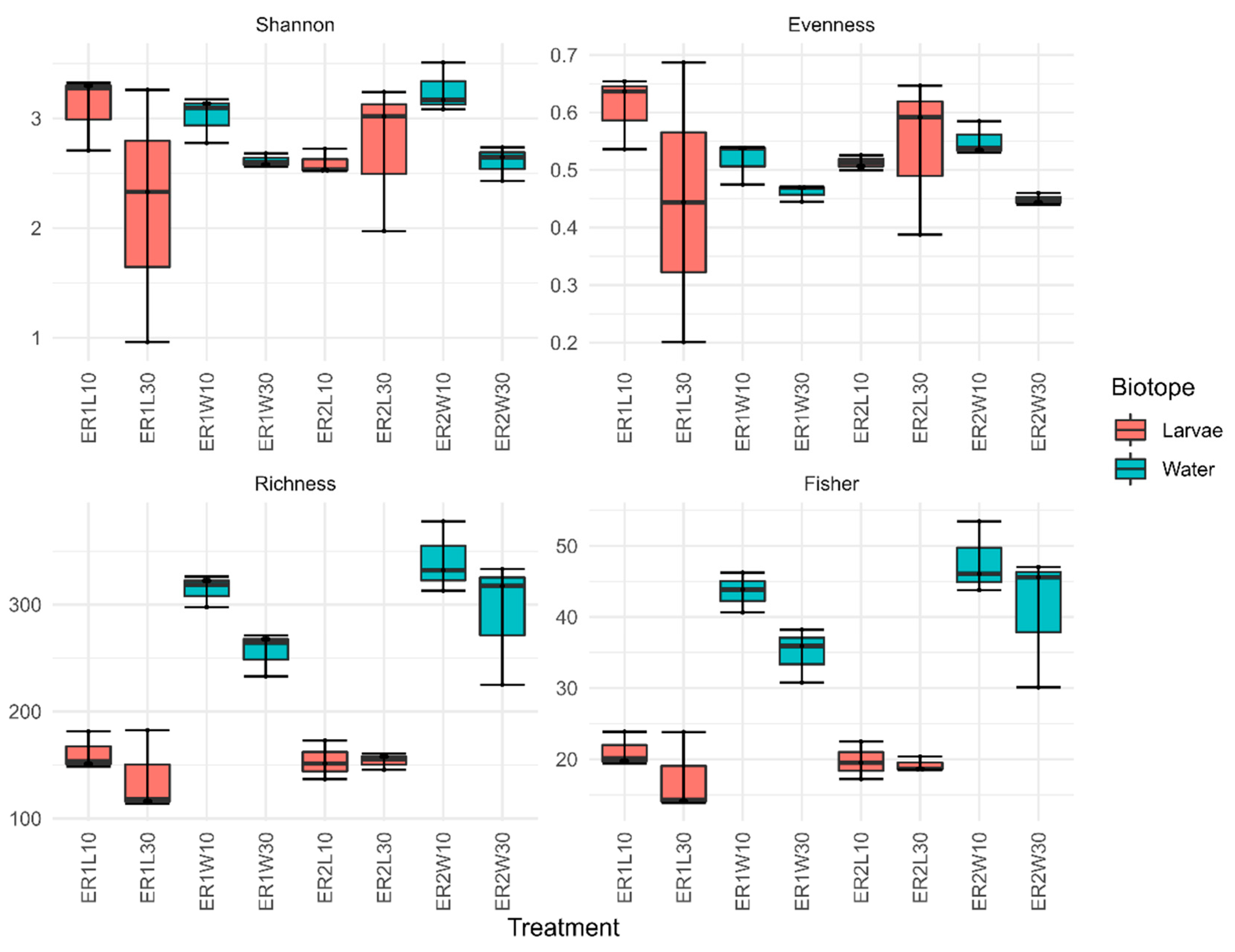
Figure 1. Diversity indices (Peilou’s J (evenness), richness, Shannon’s H’ (Shannon), and Fisher’s α (Fisher)) of bacterial communities of larvae (10 DAH), post-larval gut (30 DAH) and respective water samples, in tanks fed rotifer enriched with different commercial products (ER1 and ER2). ER1L10 and ER2L10 (whole larvae 10 DAH); ER1L30 and ER2L30 (post-larval gut 30 DAH); ER1W10 and ER2W10 (rearing water 10 DAH); and ER1W30 and ER2W30 (rearing water 30 DAH).
Microorganisms 11 00520 g001
Figure 2. First two axes of a principal coordinates analysis (PCO) of ASV composition. Colored symbols are samples. Gray symbols are weighted averages scores for ASVs (size is proportional to abundance). ER1 and ER2 corresponds to the different rotifer diets. ART (Artemia 30 DAH); GW (Algae 10 DAH); Rot_ER1 and Rot_ER2 (Rotifers 10 DAH); ER1L10 and ER2L10 (whole larvae 10 DAH); ER1L30 and ER2L30 (post-larval gut 30 DAH); ER1W10 and ER2W10 (rearing water 10 DAH); and ER1W30 and ER2W30 (rearing water 30 DAH).
Microorganisms 11 00520 g002
Figure 3. Taxonomic composition at order level for feed, larval, and water samples in tanks fed rotifers enriched with different commercial products (ER1 and ER2) at 10 and 30 DAH. ART (artemia); GW (green water (i.e., mature culture of Tetraselmis sp.)); Rot_ER1 and Rot_ER2 (rotifer); ER1L10 and ER2L10 (whole larvae 10 DAH); ER1L30 and ER2L30 (post-larval gut 30 DAH); ER1W10 and ER2W10 (rearing water 10DAH); and ER1W30 and ER2W30 (rearing water 30 DAH). Small letters indicate tank-replicates. Data are present as relative abundances of the ten most abundant orders for individual tank-replicates.
Microorganisms 11 00520 g003
Figure 4. Relative abundances of the six most abundant bacterial orders for larval and water samples in rotifer-enriched feeds with different commercial products (ER1 and ER2). ER1L10 and ER2L10 (whole larvae 10 DAH); ER1L30 and ER2L30 (post-larval gut 30 DAH); ER1W10 and ER2W10 (rearing water 10 DAH); and ER1W30 and ER2W30 (rearing water 30 DAH).
Microorganisms 11 00520 g004
Figure 5. Relative abundance of ASVs assigned to the genera Phaeobacter, Sulfitobacter, Ruegeria, and Vibrio in rotifer-enriched feeds with different commercial products (ER1 and ER2). ER1L10 and ER2L10 (whole larvae 10 DAH); ER1L30 and ER2L30 (post-larval gut 30 DAH); ER1W10 and ER2W10 (rearing water 10 DAH); and ER1W30 and ER2W30 (rearing water 30 DAH). * Genus Phaeobacter includes all ASVs classified as Phaeobacter by the naïve Bayes classifier based using the SILVA database of the 16S reference sequences at 99% similarity (version 138, released December 2019) and all ASVs classified as Nautella but with high similarity (>99%) to type strains of the genus Phaeobacter in sequence similarity search of the NCBI 16S database using the BLAST algorithm.
Microorganisms 11 00520 g005
Table 1. PERMANOVA results of variation in ASV composition among groups and their interactions using the adonis() function in vegan applied to the Bray–Curtis dissimilarity matrix of a log-transformed ASV table and with permutations set to 999. Factors tested include rotifer’s diet (Diet; ER1 and ER2), Biotope (water and larvae), and fish age (Age; 10 and 30 DAH). p-values (P) were adjusted by false-discovery rate (FDR-P). Significant values (Sig.) are presented as **—FDR-P < 0.01, *—FDR-P < 0.05, and ns—not significant.
FactorF1,23R2PFDR-PSig.
Diet2.49990.028440.0370.432*
Biotope25.78430.293310.0010.002**
Age19.00440.216190.0010.002**
Diet:Biotope2.00050.022760.0640.064ns
Diet:Age3.09730.035230.0100.015*
Biotope:age16.58380.188650.0010.002**
Diet:Biotope:Age2.93680.033410.0110.015*
Disclaimer/Publisher’s Note: The statements, opinions and data contained in all publications are solely those of the individual author(s) and contributor(s) and not of MDPI and/or the editor(s). MDPI and/or the editor(s) disclaim responsibility for any injury to people or property resulting from any ideas, methods, instructions or products referred to in the content.