Article

Discovery, Identification, and Insecticidal Activity of an Aspergillus flavus Strain Isolated from a Saline–Alkali Soil Sample

1
College of Life Sciences, Northwest A&F University, Xianyang 712100, China
2
College of Plant Protection, Northwest A&F University, Xianyang 712100, China
3
Institute of Pesticides, Northwest A&F University, Xianyang 712100, China
*
Author to whom correspondence should be addressed.
Microorganisms 2023, 11(11), 2788; https://doi.org/10.3390/microorganisms11112788
Submission received: 11 October 2023 / Revised: 4 November 2023 / Accepted: 14 November 2023 / Published: 16 November 2023
(This article belongs to the Section Plant Microbe Interactions)

Abstract

:
Aphids are one of the most destructive pests in agricultural production. In addition, aphids are able to easily develop resistance to chemical insecticides due to their rapid reproduction and short generation periods. To explore an effective and environmentally friendly aphid control strategy, we isolated and examined a fungus with aphid-parasitizing activity. The strain (YJNfs21.11) was identified as Aspergillus flavus by ITS, 28S, and BenA gene sequence analysis. Scanning electron microscopy and transmission electron microscopy revealed that the infection hyphae of ‘YJNfs21.11’ colonized and penetrated the aphid epidermal layer and subsequently colonized the body cavity. Field experiments showed that ‘YJNfs21.11’ and its fermentation products exerted considerable control on aphids, with a corrected efficacy of 96.87%. The lipase, protease, and chitinase secreted by fungi help aphid cuticle degradation, thus assisting spores in completing the infection process. Additionally, changes were observed in the mobility and physical signs of aphids, with death occurring within 60 h of infection. Our results demonstrate that A. flavus ‘YJNfs21.11’ exhibits considerable control on Aphis gossypii Glover and Hyalopterus arundimis Fabricius, making it a suitable biological control agent.

Graphical Abstract

1. Introduction

Aphids are highly diverse, with 10 families and 4400 species discovered to date [1]. Unfortunately, aphids are also one of the most destructive crop pests, reducing both quality and yield in diverse plants, including wheat [2], melons [3,4], peaches [5,6], and cotton [7], among others. Currently, chemical pesticide application is considered the most efficacious method of control. However, chemical pesticides alter the molecular physiology of target insects, resulting in altered enzyme activity, genetic mutations, and pesticide resistance [8]. In addition, chemical pesticides have been linked to both environmental and food safety risks.
To address these issues, the importance of entomopathogenic fungi as alternative pest control agents is increasing. For example, certain fungi have been found to effectively control aphids [9]. These include Beauveria bassiana [10,11,12], the Metarhizium [13,14] and Verticillium [15,16] genera, as well as fungi belonging to the Entomophthorales order [17]. These mycopesticides mainly use propagules such as conidia, blastospores, or hyphae. These propagules have the advantages of directly killing the target pest as well as secondary infection via horizontal transmission of spores from cadavers with mycosis. Several of these fungi have been used to develop wettable powder-based biopesticides for use against foliar sap-sucking pests [18].
Based on initial reports, the cosmopolitan saprophytic fungus Aspergillus flavus may be a promising aphid biocontrol agent [19,20]. Laboratory bioassays indicate that A. flavus is pathogenic to aphids at doses of 1.23 × 10³ spores/mL (LC50) and 1.34 × 10⁷ spores/mL (LC90) [21]. In another study involving cabbage and wheat, 33% of cabbage aphids and 37% of wheat aphids were found to be susceptible to fungal biocontrol, with A. flavus among the most effective pathogens [22]. However, the mechanism by which A. flavus exerts control over aphids, and the active substances involved, remains largely unknown.
In this study, we isolated A. flavus from a sample of Chinese saline–alkali soil. The insecticidal activity of the strain was verified with lab-based and field-based bioassays. Subsequently, microscopic techniques were used to analyze histopathological changes in infected aphids and the influence of enzyme activity on biocontrol efficacy was explored. The results presented here will be valuable for the realization of sustainable aphid control strategies.

2. Experimental Procedures

2.1. Isolation and Identification of the Strain

In October 2021, we isolated a strain in saline–alkali soil sample from Ningxia Autonomous Region, China. The soil sample was first cleared of debris and filtered using a 100-mesh screen, then washed and rinsed three times with sterile water [23]. Subsequently, the soil was diluted 10×, 102×, 103×, 104×, 105×, 106×, 107×, and 108× with sterile water and inoculated (100 µL) onto potato dextrose agar (PDA) using the spread plate technique [24,25]. Each gradient was repeated three times and was cultured for 3–5 d in a constant temperature (28 °C) incubator. Purification was repeated until a single colony was obtained [26].
The strain was identified based on ITS, LSU (28S), and BenA gene sequence (Supplementary Materials) analysis. Briefly, genomic DNA was isolated with a DNA extraction kit (Solarbio Science & Technology Co., Ltd., Beijing, China) and used as the PCR amplification template. The forward primer and reverse primer were synthesized by Beijing Qingke Biotechnology (Beijing, China). The amplified products were subjected to Sanger two-way sequencing. The ITS gene was amplified using the ITS1 primer (5′-TCCGTAGGTGAACCTGCGG-3′) and ITS4 primer (5′-TCCTCCGCTTATTGATATGC-3′); the LSU (28S) gene was amplified using the LR0R (5′-GTACCCGCTGAACTTAAGC-3′) and LR12 (5′-GACTTAGAGGCGTTCAG) primers; and the BenA gene was amplified using the Bt2a (5′-GGTAACCAAATCGGTGCTGCTTTC-3′) and Bt2b (5′-ACCCTCAGTGTAGTGACCCTTGGC-3′) primers. A BLAST search (NCBI) was conducted and the strain was identified as the species sharing the highest homology (>97%). Colony morphology was evaluated using a stereo microscope (NIKON, Tokyo, Japan) and a tungsten filament scanning electron microscope (JSM-6360LV, Nippon Electronics, Tokyo, Japan).

2.2. Extraction and Separation of Active Substances and Preparation of Reagents

2.2.1. Preparation of Spore Suspension

To prepare the spore suspension, incubation plates were washed with sterile water on a sterile operating table. The spores were collected in a conical flask with glass beads, shaken thoroughly, and scattered. A single spore suspension was obtained via filtration with a cotton-stuffed syringe. The concentration was adjusted to 1 × 10⁷ spores/mL using a hemocytometer. Finally, 0.1% tween-20 was added and the suspension was refrigerated (4 °C) for subsequent analyses [27,28].

2.2.2. Preparation of Fermentation Solution

The strain was fermented under aerobic conditions. The fermentation medium (35 g soluble starch, 15 g sucrose, 12.5 g yeast extract, 7.5 g soybean cake powder, 1.0 g KH2PO4, 1.1 g anhydrous MgSO4, 1.0 g NaCl) was prepared in 1 L of distilled water and distributed into ten 250 mL conical flasks. The flasks were sealed with eight layers of gauze and then autoclaved for 20 min. The spore suspension was injected into the fermentation medium at a concentration of 5%. Following inoculation, the flasks were again sealed with eight layers of gauze and cultured for 3 d in a shaker (165 r/min) at 32 °C. Next, the fermentation solution was homogenized (JJ-2, Hangzhou, China) to prevent mycelium from clogging the nozzle. Finally, 0.1% tween-20 was added. We found that sealing with gauze was more beneficial to the growth of A. flavus. This may be due to the breathability of the gauze. In order to prevent bacterial contamination, we used gauze to seal the conical bottles and then coated the gauze in kraft paper to maintain sterility and prevent water vapor pollution. In addition, after inoculation we used ultraviolet radiation to sterilize the replacement gauze.

2.2.3. Preparation of Mycelium Reagents

The fermented broth was filtered with eight layers of gauze and eluted three times with water. Next, the mycelium was weighed, homogenized, and diluted 100×. Finally, 0.1% tween-20 was added and the suspension was refrigerated (4 °C) for subsequent analyses.

2.2.4. Preparation of Methanol Extract Reagent

After the fermentation solution was filtered with eight layers of gauze, the mycelium was weighed. Next, the mycelium was mixed with methanol at a ratio of 1:10 and ultrasonicated for 30 min. This process was repeated twice, followed by dilution with 0.1% tween-20 to ensure that the organic solvent content was ≤1%. The resulting methanol extract reagent was refrigerated (4 °C) for subsequent analyses.

2.2.5. Preparation of Fermentation Filtrate

After filtration with 8 layers of gauze, the fermentation solution was centrifuged (12,000 r/min) for 5 min and the supernatant was filtered using a 0.22 µm microporous filtration membrane. Finally, 0.1% tween-20 was added to the filtered supernatant and the suspension was refrigerated (4 °C) for subsequent analyses.

2.3. Evaluation of Biocontrol Efficacy

Here, we used naturally occurring A. gossypii and H. arundimis in order to better test the aphicidal activity of the strain. Naturally occurring aphid populations are more resilient and resistant to biotic and abiotic stressors than lab-reared aphids. In addition, due to exposure to environmental toxins and pesticides, naturally occurring aphids are much more resistant to biotic and abiotic stressors, and reproduce more quickly, than lab-reared aphids.
In the lab-based bioassay [29,30,31], we tested the efficacy of this strain against H. arundimis, which thrive on peach trees and are often difficult to control because of their thick waxy layers. Screening experiments were performed to measure the efficacy of the strain on aphid nymphs. All treatments were carried out in 90 mm Petri dishes. Peach leaves exhibiting vigorous growth and hosting large numbers of aphids were excised, and the petioles were wrapped in cotton moistened with water. The experimental leaves were immersed in both the original and diluted (2×, 10×, and 50×) fermentation broths for approximately 3 s [32], with 0.1% tween-20 used as a negative control, followed by blotting with absorbent paper to remove excess liquid. The aphids were counted and then transferred to sterile Petri dishes containing a 9 cm diameter section of filter paper. Three parallel experiments were carried out for each group. Dishes were placed in a culture chamber with a temperature of 25 (±1) °C and a relative humidity of ≥85% [33]. Aphid mortality rates were recorded at 16 h, 24 h, and 48 h after treatment. Subsequently, the population reduction rate and corrected efficacy were calculated according to the equations below.
In the field-based bioassay, we tested the aphicidal efficacy of this strain against A. gossypii, which are a major pest of fruit and vegetable crops such as melons. Both the original and diluted (2×, 10×, and 50×) fermentation broths were sprayed evenly on the leaves of vigorously growing muskmelon, with 0.1% tween-20 used as a negative control. Five to eight muskmelon plants were used for each treatment. Three parallel experiments were carried out for each group. The test area (1 m2) was randomly arranged in blocks, and no other chemical agents were applied to the field during the test [34]. The number of aphids was recorded before application, and the number of dead aphids was recorded on the 1st, 3rd, and 5th day after application. Subsequently, the population reduction rate and corrected efficacy were calculated according to the equations below. Corrected efficacy refers to a statistical method for correcting calculations of control efficacy over a specific treatment area to account for changes in insect body condition. The day of the experiment was sunny with a suitable temperature. The experiment was carried out in the key open dryland test field of Northwest A&F University, China. The experimental field contained loamy soil of medium fertility.
P o p u l a t i o n   r e d u c t i o n   r a t e   ( % ) = i n i t i a l   a p h i d   c o u n t a p h i d   c o u n t   a f t e r   t r e a t m e n t i n i t i a l   a p h i d   c o u n t × 100 %
C o r r e c t e d   e f f i c a c y   ( % ) = ( 1 i n i t i a l   c o n t r o l   a p h i d   c o u n t × e x p e r i m e n t a l   a p h i d   c o u n t   a f t e r   t r e a t m e n t i n i t i a l   e x p e r i m e n t a l   a p h i d   c o u n t × c o n t r o l   a p h i d   c o u n t   a f t e r   t r e a t m e n t ) × 100 %

2.4. Evaluation of Histopathologic Changes in Infected Aphids

Subsequent to this strain exposure, the feeding ability, activity [35], and symptoms of infected and control A. gossypii were observed continuously with a stereomicroscope (LEICA M165 FC, LEICA, Wetzlar, Germany). At 24 h, 36 h, 48 h, and 60 h post infection, A. gossypii were fixed with 2.5% glutaraldehyde at 4 °C. Then, the levels of this strain adhesion, germination, and invasion were observed using scanning electron microscopy. In addition, at 24 h, 36 h, 48 h, and 60 h post infection, the susceptible A. gossypii were fixed for 3 h with paraformaldehyde at 4 °C. The fixed specimens were infiltrated and embedded in Epon 812, then sliced (80 µm thickness) using an ultrathin slicer (EMUC7, Leica Instrument Co. LTD, Vienna, Austria), fixed on a copper mesh, stained with uranium acetate and lead citrate, and observed using transmission electron microscopy (TECNAI G2 SPIRIT BIO, FEI Corporation, Hillsboro, OR, USA).

2.5. Evaluation of the Effect of the Strain Infection on Aphid Body Wall Extracellular Enzyme Degradation

2.5.1. Formulation of Culture Medium from Aphid Cuticles

H. arundimis were carefully removed from peach tree branches with paintbrushes and placed in beakers, and then subsequently rinsed repeatedly with tap water to remove impurities. The internal organs of the aphids were removed using a dissecting microscope (SZ51, Beijing, China), and the wax shells and body walls of the aphids were dried in an oven at 65 °C. Next, the dried body walls were ground into powder and sieved (40 mesh). In total, 5 g of aphid body wall powder was added to the base medium (1.5 g KH2PO4, 0.6 g MgSO4, 0.5 g KCl, 1 mg FeSO4·7H2O, 1 mg ZnSO4·7H2O, 1 L distilled water). A total of 40 mL of the mixture was allocated to each 150 mL conical vial and autoclaved (120 °C for 30 min). This process ensured that the liquid medium contained aphid body walls and wax shells as the only carbon sources.

2.5.2. Extraction of Crude Enzyme Solution

Each conical vial was inoculated with 10 mL of spore suspension and placed in a thermostatic shaking table (28 °C, 150 r/min) for 8 d. Three 0.5 mL samples were collected each day and centrifuged (10,000 r/min for 20 min, 4 °C). The supernatant was collected as the crude enzyme solution and stored in an ultralow temperature refrigerator at −80 °C for subsequent analyses.

2.5.3. Determination of Lipase Activity

Briefly, 3 mg of p-nitrophenyl palmitate (p-NPP) was dissolved in 1 mL of isopropyl alcohol, and the mixture was subsequently dissolved in 9 mL of Tris-HCl (0.05 mol/L, pH 8.0) containing 20.7 mg sodium deoxycholate and 10 mg gum arabic to prepare the matrix solution. During the reaction, 200 μL of the substrate solution was preheated in a 37 °C water bath for 10 min, and then 20 μL of enzyme solution was added and stirred uniformly. Next, 300 μL of trichloroacetic acid was added after water bath heating (37 °C, 20 min) and stirred well, then left to stand at room temperature for 5 min to terminate the reaction. Finally, 320 μL of NaOH (0.05 mol/L) was added. For the control group, trichloroacetic acid was added first, followed by the enzyme solution. OD values (410 nm) were measured using a microplate reader (Bio Tek Synergy H1, Winooski, VT, USA). The enzyme activity was calculated according to the p-nitrophenol standard curve. A lipase activity unit was defined as the amount of enzyme catalyzing the decomposition of fat to produce 1 μg of p-nitroaniline per minute.

2.5.4. Determination of Protease Activity

A 1% (w/v) casein solution was prepared with Tris-HCl buffer (0.05 mol/L, pH 8.5). In total, 1 mL of this solution was mixed with 1 mL of the prepared enzyme solution in a centrifuge tube and heated in a water bath (37 °C for 30 min). The reaction was terminated with 3 mL trichloroacetic acid (0.4 mol/L). For the control group, 1 mL of enzyme solution and 3 mL of trichloroacetic acid (0.4 mol/L) were successively added to the centrifuge tube, and then 1 mL of casein solution was added after mixing. Next, 1 mL of the filtrate was added to 5 mL of 0.55 mol/mL Na2CO3 and mixed well. Finally, 1 mL of Folin reagent was added and the solution was mixed well and reacted for 30 min. OD values (680 nm) were measured using a microplate reader. The enzyme activity was calculated according to the tyrosine standard curve. A protease activity unit was defined as the amount of enzyme catalyzing the decomposition of protein to produce 1 μg of tyrosine per minute.

2.5.5. Determination of Chitinase Activity

Both 0.1 mL of enzyme solution and 0.1 mL of colloidal chitin were mixed in a centrifuge tube, water-bathed (37 °C for 4 h), and then centrifuged (8000 r/min for 5 min) to terminate the enzymatic hydrolysis reaction. A total of 50 μL of the supernatant was mixed with 20 μL of potassium tetraborate solution, shaken thoroughly, reacted in a boiling water bath for 5 min, and then quickly cooled to room temperature with tap water. Next, 300 μL of 10% dimethylaminobenzaldehyde reagent was added and the mixture was water-bathed (37 °C for 20 min) and subsequently cooled to room temperature with tap water. OD values (585 nm) were measured using a microplate reader. The enzyme activity was calculated according to the N-acetylglucosamine standard curve. A chitinase activity unit was defined as the amount of enzyme catalyzing the decomposition of chitin to produce 1 μg of N-acetylglucosamine per minute.

2.6. Statistical Analyses

In order to eliminate certain analytical errors, exact values were utilized for statistical analyses. SPSS 26.0 was used to perform single-factor analysis of variance, and the results were expressed as the mean ± standard deviation. Duncan’s method was used for multiple comparisons. Statistical significance was determined at the p < 0.05 level.

3. Results

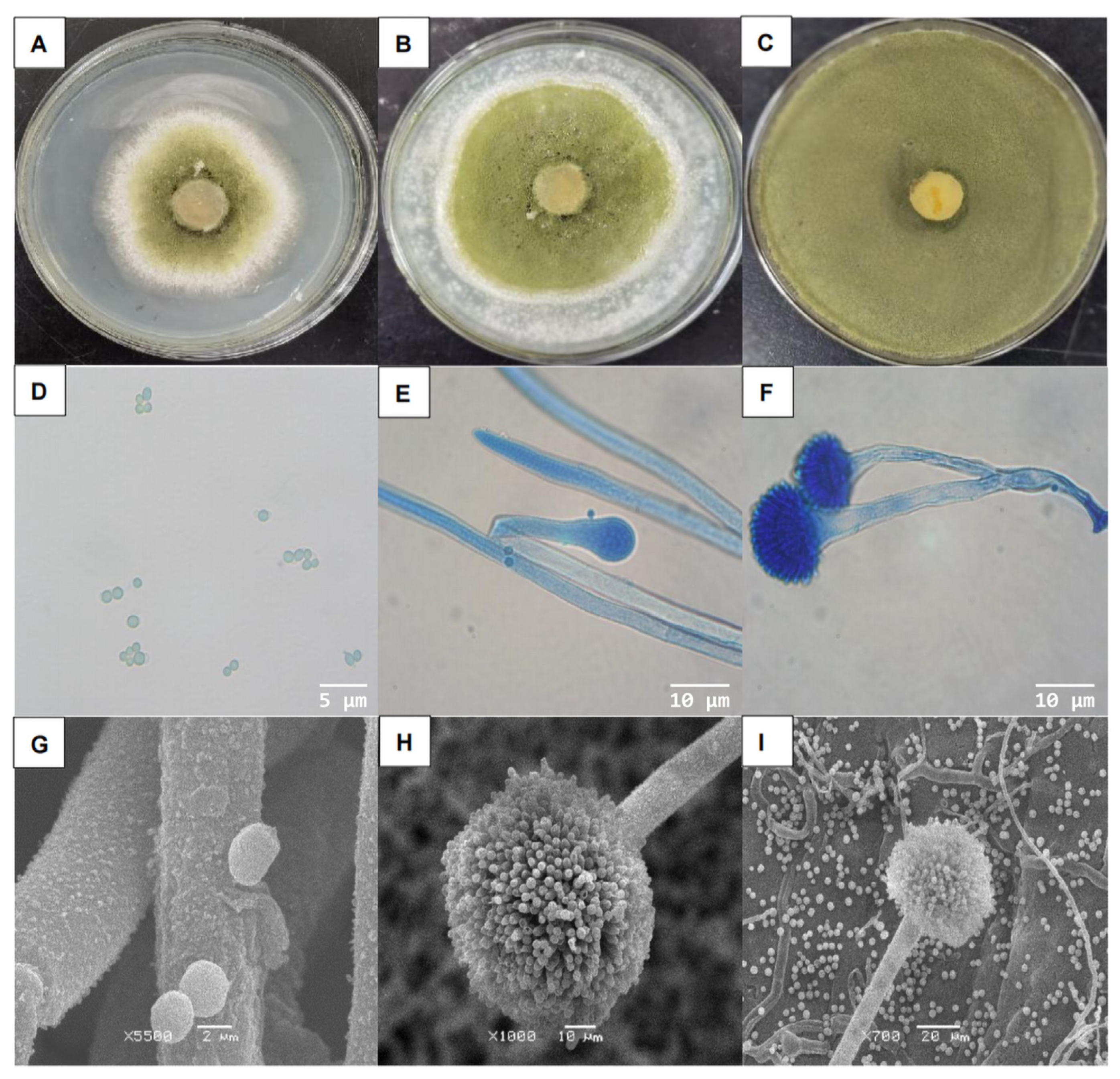
3.1. Isolation and Identification of the Strain

After 24 h of culture on PDA, the strain formed loose, flat, regular-edged colonies 2–3 cm in diameter (Figure 1A). The colonies were initially white with short, fluffy hyphae growing outward (Figure 1B). The center of the colonies later turned green, with a large number of green spores growing (Figure 1C,D). The nutritional hyphae exhibited septa, and a portion of the aerial hyphae formed a long, rough conidium, giving rise to a nearly spherical apical sac (Figure 1E). The surface gave rise to several small peduncles bearing clusters of coarse-surfaced spherical conidia (Figure 1F). Additionally, rough-surfaced spherical conidia were generated on the surface of the small peduncle (Figure 1G–I).
Through an ITS gene sequence analysis and search in the BLAST database (NCBI), the strain was identified as Aspergillus flavus (GenBank: OP071264), and the homology reached 99.33%. Then, we further analyzed the gene sequences of LSU (28S) and BenA, and compared them with the sequences reported in the BLAST database (NCBI). We speculated that this might be a new strain, and we temporarily named it ‘YJNfs11’ for the convenience of subsequent studies. A sample specimen was deposited in the China General Microbiological Culture Collection Center (CGMCC) with the deposit number 40260. It will be used as a new portal for further research, such as whole-genome sequencing analysis. Phylogenetic tree based on concatenated ITS, LSU (28S), and BenA of strain ‘YJNfs21.11’ was shown in Figure 2.

3.2. Biocontrol Efficacy of A. flavus

For laboratory experiments, the corrected control efficacy of the 100× diluted spore suspension reached 85.69% after 48 h (Table 1). These results suggest that the best infective agents were spores. The biocontrol efficacy of each solution, before and after application, is shown in Figure 3a.
Field experiments revealed that A. flavus ‘YJNfs21.11’ and its fermentation products exerted considerable control on aphids, with a corrected control efficacy of 96.87% after 5 days of treatment with the undiluted fermentation liquid (Table 2). The biocontrol efficacy of each solution, before and after application, is shown in Figure 3b.

3.3. Histopathological Changes of Infected Aphids

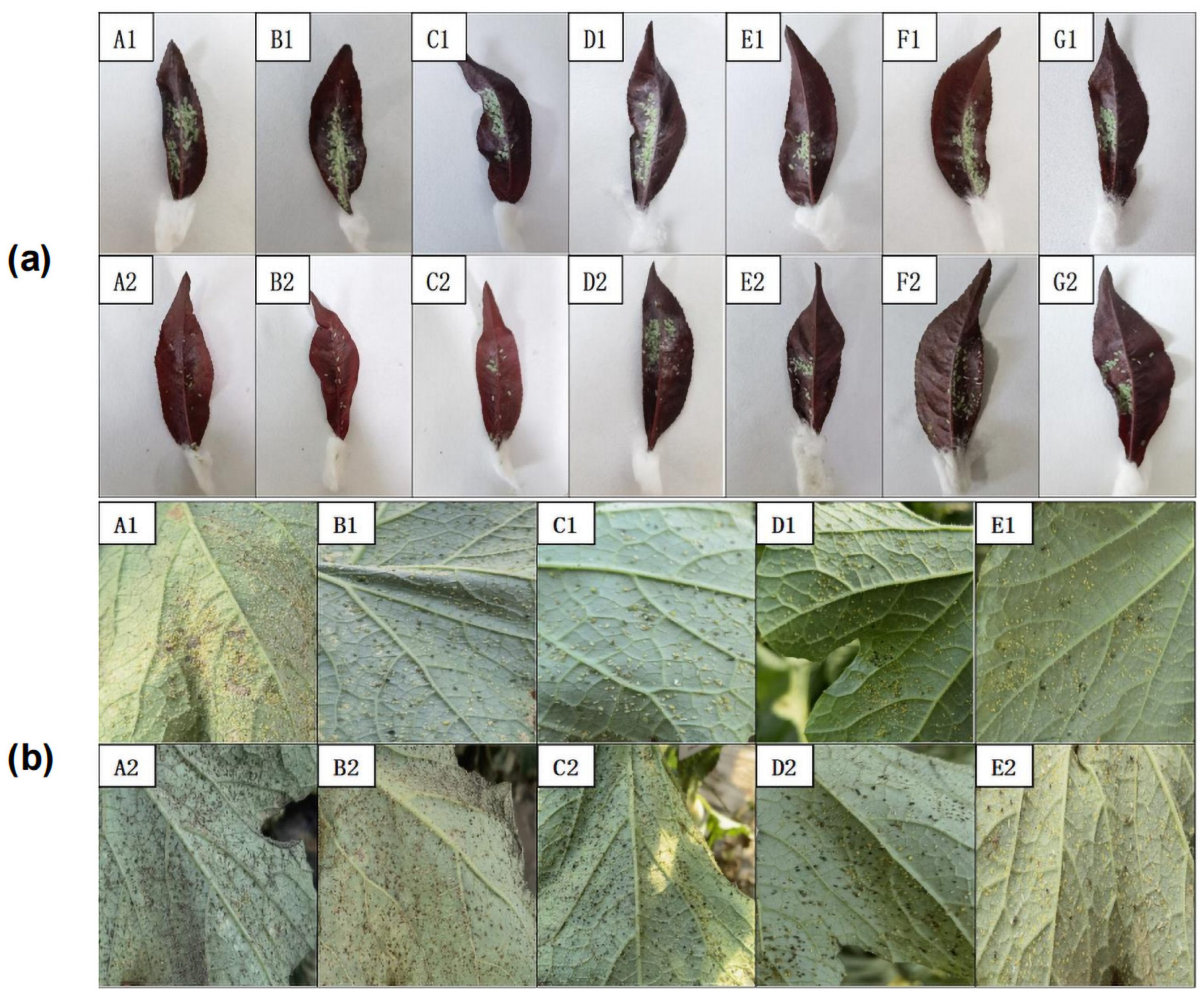
3.3.1. External Symptoms of Aphids Infected with A. flavus ‘YJNfs21.11’

Observation under a stereomicroscope revealed no significant changes in the mobility or physical signs of aphids between 24 h and 36 h after infection (Figure 4A,B). However, aphid mobility decreased considerably after 48 h of infection, with aphids exhibiting a darkened body color and black intersegmental folds (Figure 4C). After 60 h of infection, white hyphae were observed on the heads, abdomens, and tails of aphids (Figure 4D), indicating that they had died [36]. Furthermore, their bodies had shrunk and stiffened. Aphids infected via spore transmission can form new sources of infection that continue to invade other aphids [37].

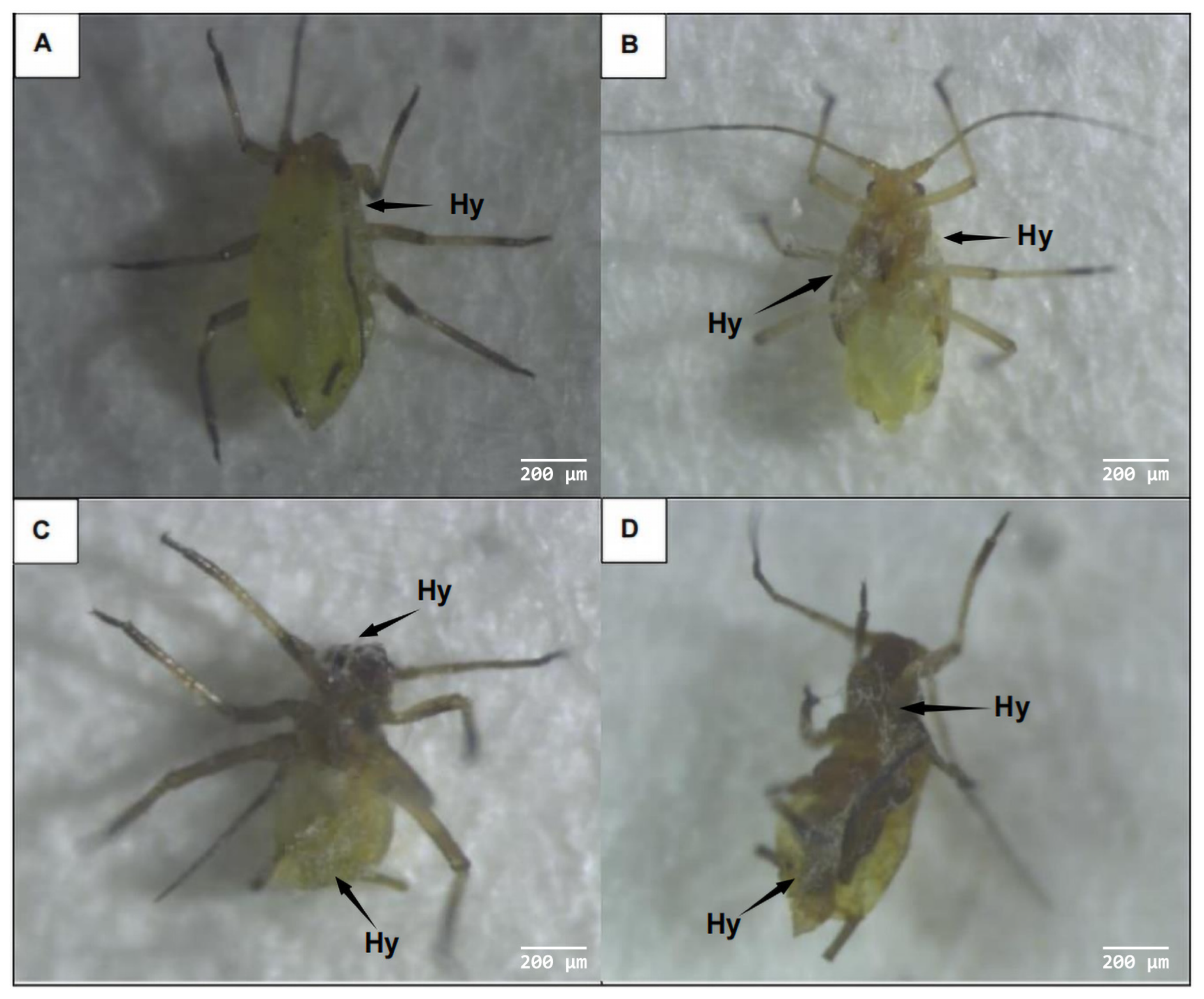
3.3.2. Adhesion and Germination of A. flavus ‘YJNfs21.11’ on the Surface of Aphids

Scanning electron microscopy revealed that A. flavus ‘YJNfs21.11’ primarily invaded aphid bodies by penetrating the epidermis. After 24 h of infection, the conidia were observed to be attached to the head vertices, antennae, and bases of the mouthparts. The conidia further attached to the spinous projections and intersegmental folds on the thorax and abdomen (Figure 5A,B). After 48 h of infection, the conidia gradually germinated to form germ tubes specialized in appressoria and penetration pegs at the invasion site (Figure 5C,D). The conidia penetrated the epidermis using mechanical force, causing cracks, blackening the aphid body surface, and deforming the epidermis (Figure 5E). During the invasion process, the epidermal tissue was destroyed and cracks and cavities appeared, likely because the fungus secreted enzymes on the surface of the insect (Figure 5F). Many spores penetrated the body wall through these cracks, thereby completing the invasion process (Figure 5G). Through examination of magnified photographs, we observed that the mycelium had many conidia attached to it (Supplementary Materials). We also examined photographs of aphid bristles and, through comparison, we identified the structure as mycelium rather than bristles. The mucilage produced by these spores promoted spore adhesion, germination, and aphid invasion (Figure 5H). Some of the hyphae grew and extended around the setae (Figure 5E). Fungal growth in the peripheral space of the anterior epidermis is generally lateral and can rupture the epidermis, thereby facilitating invasion [38]. At the invasion site, the body surface appeared concave, and the body wall appeared darker in color. In summary, fungal invasion destroyed the epidermal layer and resulted in cracks forming on the surface of the aphid body, allowing A. flavus ‘YJNfs21.11’ to enter (Figure 5I).

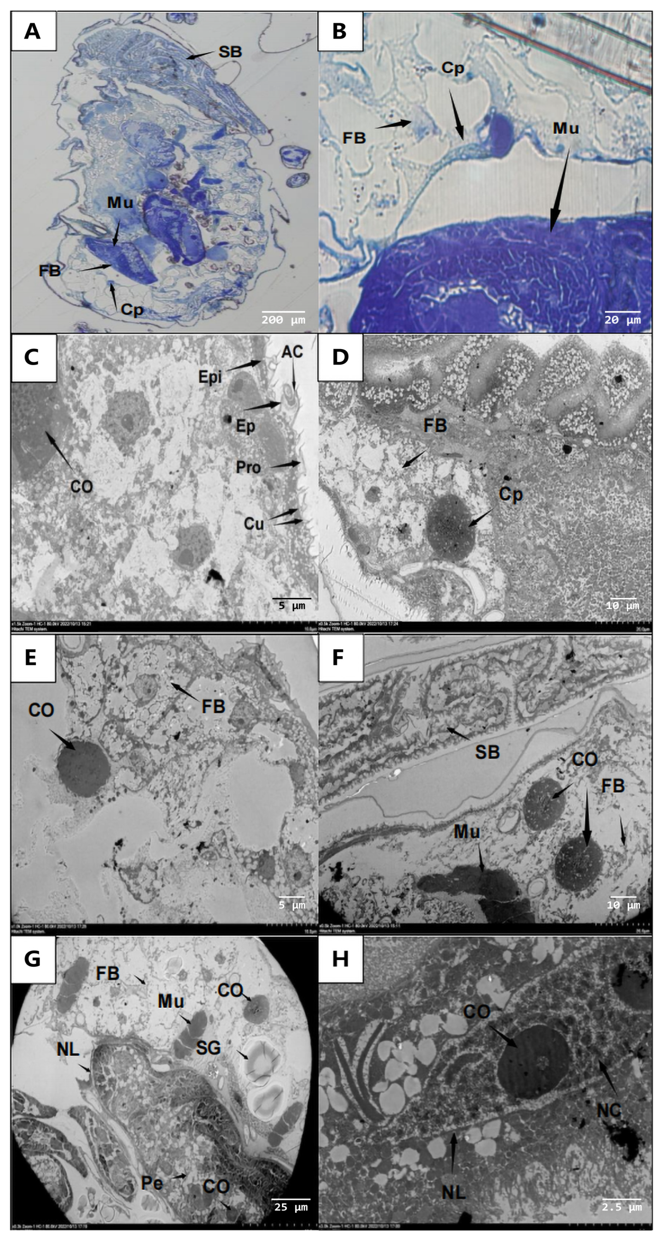
3.3.3. Invasion of the Aphid Internal Organs by A. flavus ‘YJNfs21.11’

Tissue sections and transmission electron microscopy revealed that the overall aphid body structure was intact after 24 h of infection (Figure 6A,B). From the outside to the inside, the aphid body wall consists of the upper epidermis, pro-epidermis, and dermis (Figure 6C). Muscles and fat are distributed in the hemocoel between the digestive tract and body wall. The muscles are attached to the inner surface of the body wall and the surface of the digestive tract in a strip or block shape. In addition, body fat is linearly distributed in the hemocoel (Figure 6D). After 48 h of infection, the spores had invaded the aphid body, resulting in the loosening of fat structures, the shredding of muscle tissues, and the infection of nerve ganglia (Figure 6E,F). In addition, the nerve cell bodies appeared slightly blackened and some had dissolved (Figure 6G,H). In the late stages of infection, fat was consumed by the spores and the tissue became unstructured.

3.4. Effect of Strain YJNfs21.11 on Extracellular Enzyme Activity during Aphid Body Wall Degradation

During the degradation of the H. arundimis body wall, the activities of lipase, protease, and chitinase tended to first increase and then decrease over time (Figure 7). Both lipase activity (2.212 U/mL) and chitinase activity (0.032 U/mL) were highest on the 3rd day. Protease activity peaked last, and its activity (0.906 U/mL) was highest on the 4th day. During days 3–6, the body wall cracked, the aphid body stiffened and atrophied, and many tissues and organs collapsed, resulting in a decline in lipase activity. In terms of absorbance, the content of the lipase-specific cleaved substrate was significantly higher than that of chitinase or protease. Taken together, these results indicate that lipase played an important role in the degradation of the aphid body wall.

3.5. Relationship between Extracellular Enzyme Activity and A. flavus Pathogenesis

Linear regression was performed between enzymatic activity and corrected mortality rates on days 1, 3, and 5 of infection (Table 3). The regression curve of corrected efficacy and lipase activity was y = 36.254x − 1.7864 and the correlation coefficient (R2) was 0.7191. The regression curve of corrected efficacy and protease activity was y = 98.036x + 12.999 and R2 = 0.8707. The regression curve of corrected efficacy and chitinase activity was y = 2433x − 0.92 and R2 = 0.6681. In conclusion, the activities of lipase, protease, and chitinase were positively correlated with aphid mortality. These results suggest that during the late stage of infection, cracks appearing in the aphid body wall were the result of enzymatic breakdown. In this study, the effects of extracellular enzymes on aphid body wall degradation and their virulence were investigated, which provides a theoretical basis for the application of A. flavus for the biological control of aphids.

4. Discussion

Certain fungi penetrate the host body through a combination of mechanical pressure and enzymatic degradation [39,40]. After germination, spores form germ tubes and appressoria under suitable conditions [41,42]. Each appressorium produces only one penetration peg which exerts a selective effect on nutrients before invading the body, resulting in physical pressure and forcing the penetration peg to invade the nutrient-rich aphid body [43,44].
Using transmission electron microscopy, we observed considerable degradation of the aphid epidermis around the infection site [45]. In fact, conidial germ tubes are known to secrete substances that degrade extracellular enzymes, decompose or soften the insect body wall [46], and aid in the invasion of the insect body [47]. These enzymes, such as esterase, protease, and chitinase, can also provide nutrients required for fungal growth and reproduction [48,49]. During the late stages of infection, a portion of the upper aphid epidermis disappeared, indicating that A. flavus spores may secrete extracellular lipases, proteases, and chitinases during invasion. This was later confirmed when lipases activity was found to reach the maximum on the 3rd day.
To date, the majority of research on fungal biocontrol agents has focused on simple insecticidal assays [50,51]. These simple experiments do not address the process of fungal infection nor the mechanism by which fungi exert control on their insect hosts, and generally do not study the enzymatic digestion process in detail. In this study, the mechanism by which A. flavus ‘YJNfs21.11’ infects aphids was studied in depth, including the enzymatic dynamics associated with the infection process. Because spores were identified as the infective agent, our study focused on spores. However, we cannot rule out that either the mycelia or other bioactive metabolites may be involved in the process, and suggest further study.
Aspergillus flavus is distributed all over the world, especially in warm and moist fields [52]. It can produce a variety of secondary metabolites [53], among which aflatoxins have been most studied because of their strong carcinogenicity [54], which includes AFB1, AFB2, AFG1, and AFG2 [55]. A. flavus strains AF13 and K54A are notorious for producing aflatoxins [56]. However, not all A. flavus produce aflatoxin [57,58,59,60]; for example, NRRL 21882 [61] and SU-16 [62,63] are non-toxin-producing strains and have been used for biological control of plant diseases and brewing of yellow rice wine, respectively [64]. In this study, the fermentation products of A. flavus ‘YJNfs21.11’ were detected with LC-MS (AB SCIEX, Singapore), and no aflatoxins were detected (Supplementary Materials). Meanwhile, we also did not detect the presence of aflatoxin synthesis-related genes using PCR (Supplementary Materials). A. flavus ‘YJNfs21.11’ was isolated and extracted from soil, and its metabolites were natural products; these natural products can be degraded and reused by microorganisms. In field and laboratory bioassays, the study was carried out between biological half-lives [65]. Therefore, A. flavus ‘YJNfs21.11’ is safe for the environment and people. In addition, strategies for the degradation of other aflatoxin-producing strains of aflatoxin are now available. One major determinant of aflatoxin production in A. flavus is temperature, and oxidative stress may also indeed be a pre-requisite for aflatoxin production [66,67]. The most promising strategy currently being used to reduce preharvest contamination of crops with aflatoxin is to introduce non-aflatoxin (biocontrol) A. flavus into the crop environment [68,69]. In conclusion, A. flavus is worth exploring as a promising biocontrol agent. We believe that this work will provide new ideas and directions for related research.

5. Conclusions

A. flavus ‘YJNfs21.11’ effectively infects aphids through conidial spores. The infection process results in the destruction of the epidermal tissue and internal organs, and the eventual death and digestion of the aphid. The activities of lipase, protease, and chitinase are positively correlated with aphid mortality, and they complete the infection process by decomposing aphid epidermal auxiliary spores. These findings highlight that biocontrol agents can successfully replace chemical pesticides for aphid control [70,71,72] and A. flavus is worth exploring as a promising biocontrol agent [73,74].

Supplementary Materials

The following supporting information can be downloaded at: https://www.mdpi.com/article/10.3390/microorganisms11112788/s1, Figure S1. LC-MS chromatogram. Figure S2. Aflatoxin synthesis gene was detected using PCR. Figure S3. Attachment and germination of A. flavus ‘YJNfs21.11’ on aphid body surface. Table S1. Sequences of the nucleotide primers used in PCR study. Refs. [73,74] can be found in Supplementary Materials.

Author Contributions

Conceptualization, Y.S., X.L., S.F. and M.R.; methodology, Y.S., Z.Q, W.W. and K.Z.; software, Y.S. and X.L.; validation, Z.Q. and W.W.; formal analysis, X.L., J.X. and H.X.; investigation, Y.S., S.F., J.X., K.Z. and M.R.; resources, Z.Q., W.W., H.X. and B.Q.; data curation, Y.S., X.L. and K.Z.; writing—original draft preparation, Y.S.; writing—review and editing, Y.S. and X.L.; visualization, W.W.; supervision, B.Q.; project administration, B.Q.; funding acquisition, B.Q. All authors have read and agreed to the published version of the manuscript.

Funding

This work was supported by the Science and Technology Department of Shaanxi Province, China (Grant 2021ZDLNY05-05).

Data Availability Statement

All experimental data are included in this paper and the Supplementary Materials.

Acknowledgments

We thank the Teaching and Research Core Facility at the College of Life Science, Life Sciences Research Core Services, Northwest A&F University for their technical support and thank Elsevier for their linguistic assistance during the preparation of this manuscript.

Conflicts of Interest

The authors declare no conflict of interest.

References

  1. Smith, C.M.; Chuang, W.-P. Plant Resistance to Aphid Feeding: Behavioral, Physiological, Genetic and Molecular Cues Regulate Aphid Host Selection and Feeding: Plant Resistance to Aphid Feeding. Pest. Manag. Sci. 2014, 70, 528–540. [Google Scholar] [CrossRef]
  2. Elliott, N.C.; Giles, K.L.; Baum, K.A.; Elzay, S.D. Quantitative Assessment of Aphid Parasitoids and Predators in Central Oklahoma Wheat Fields during Five Growing Seasons. Southwest. Entomol. 2021, 46, 833–842. [Google Scholar] [CrossRef]
  3. Ismail, H.M.; Freed, S.; Naeem, A.; Malik, S.; Ali, N. The Effect of Entomopathogenic Fungi on Enzymatic Activity in Chlorpyrifos-Resistant Mosquitoes, Culex Quinquefasciatus (Diptera: Culicidae). J. Med. Entomol. 2020, 57, 204–213. [Google Scholar] [CrossRef] [PubMed]
  4. Liang, D.; Hu, Q.; Xu, Q.; Qi, X.; Zhou, F.; Chen, X. Genetic Inheritance Analysis of Melon Aphid (Aphis gossypii Glover) Resistance in Cucumber (Cucumis sativus L.). Euphytica 2015, 205, 361–367. [Google Scholar] [CrossRef]
  5. Jordan, M.; Sauge, M.; Vercambre, G. Chemical and Growth Traits of the Peach Tree May Induce Higher Infestation Rates of the Green Peach Aphid, Myzus persicae (Sulzer). Pest. Manag. Sci. 2020, 76, 797–806. [Google Scholar] [CrossRef]
  6. Davis, J.A.; Radcliffe, E.B.; Ragsdale, D.W. Effects of High and Fluctuating Temperatures on Myzus persica (Hemiptera: Aphididae). Environ. Entomol. 2006, 35, 1461–1468. [Google Scholar] [CrossRef]
  7. Wang, L.; Zhang, S.; Luo, J.-Y.; Wang, C.-Y.; Lv, L.-M.; Zhu, X.-Z.; Li, C.-H.; Cui, J.-J. Identification of Aphis gossypii Glover (Hemiptera: Aphididae) Biotypes from Different Host Plants in North China. PLoS ONE 2016, 11, e0146345. [Google Scholar] [CrossRef]
  8. Kaleem Ullah, R.M.; Gao, F.; Sikandar, A.; Wu, H. Insights into the Effects of Insecticides on Aphids (Hemiptera: Aphididae): Resistance Mechanisms and Molecular Basis. IJMS 2023, 24, 6750. [Google Scholar] [CrossRef]
  9. Zaki, F.N. Efficiency of the Entomopathogenic Fungus, Beauveria bassiana (Bals), against Aphis crassivora Koch and Bemesia tabaci, Gennandius. J. Appl. Entomol. 1998, 122, 397–399. [Google Scholar] [CrossRef]
  10. Mantzoukas, S.; Daskalaki, E.; Kitsiou, F.; Papantzikos, V.; Servis, D.; Bitivanos, S.; Patakioutas, G.; Eliopoulos, P.A. Dual Action of Beauveria bassiana (Hypocreales; Cordycipitaceae) Endophytic Stains as Biocontrol Agents against Sucking Pests and Plant Growth Biostimulants on Melon and Strawberry Field Plants. Microorganisms 2022, 10, 2306. [Google Scholar] [CrossRef]
  11. Homayoonzadeh, M.; Esmaeily, M.; Talebi, K.; Allahyari, H.; Reitz, S.; Michaud, J.P. Inoculation of Cucumber Plants with Beauveria Bassiana Enhances Resistance to Aphis gossypii (Hemiptera: Aphididae) and Increases Aphid Susceptibility to Pirimicarb. Eur. J. Entomol. 2022, 119, 1–11. [Google Scholar] [CrossRef]
  12. Poprawski, T.J.; Parker, P.E.; Tsai, J.H. Laboratory and Field Evaluation of Hyphomycete Insect Pathogenic Fungi for Control of Brown Citrus Aphid (Homoptera: Aphididae). Environ. Entomol. 1999, 28, 315–321. [Google Scholar] [CrossRef]
  13. Reingold, V.; Kottakota, C.; Birnbaum, N.; Goldenberg, M.; Lebedev, G.; Ghanim, M.; Ment, D. Intraspecies Variation of Metarhizium brunneum against the Green Peach Aphid, Myzus persicae, Provides Insight into the Complexity of Disease Progression. Pest Manag. Sci. 2021, 77, 2557–2567. [Google Scholar] [CrossRef]
  14. Qubbaj, T.; Samara, R. Efficacy of Three Entomopathogenic Fungi Beauveria bassiana, Metarhizium anisopliae and Lecanicillium lecanii Isolates against Black Bean Aphid, Aphis fabae (Scop.) (Hemiptera: Aphididae) on Faba Bean (Vicia faba L.). LR 2022, 45, 1572–1579. [Google Scholar] [CrossRef]
  15. Zare, R.; Gams, W. A Revision of Verticillium Section Prostrata. IV. The Genera Lecanicillium and Simplicillium Gen. Nov. Nova Hedwig. 2000, 71, 1–50. [Google Scholar] [CrossRef]
  16. Ritika; Joshi, N.; Sharma, N. Chitinase Enzyme Activity and Pathogenicity of Lecanicillium lecanii against Mustard Aphid Lipaphis erysimi (Kalt.). IJE 2021, 84, 833–836. [Google Scholar] [CrossRef]
  17. Manfrino, R.G.; Gutierrez, A.C.; Rueda Páramo, M.E.; Salto, C.E.; López Lastra, C.C. Prevalence of Entomophthoralean Fungi (Entomophthoromycota) of Aphids in Relation to Developmental Stages: Prevalence of Entomophthoralean Fungi on Aphids. Pest. Manag. Sci. 2016, 72, 1566–1571. [Google Scholar] [CrossRef]
  18. Feng, M.G.; Poprawski, T.J.; Khachatourians, G.G. Production, Formulation and Application of the Entomopathogenic Fungus Beauveria Bassiana for Insect Control: Current Status. Biocontrol Sci. Technol. 1994, 4, 3–34. [Google Scholar] [CrossRef]
  19. Mamo, F.T.; Shang, B.; Selvaraj, J.N.; Wang, Y.; Liu, Y. Study on Distribution and Characterization of Aspergillus flavus Strains to Screen Potential Biocontrol Agents from Different Geographical Locations of China. In Proceedings of the 2016 Academic Annual meeting of Chinese Bacteriology Society, Fuzhou, China, 19–21 August 2016; p. 1. [Google Scholar]
  20. Naz, I.; Khan, R.A.A.; Masood, T.; Baig, A.; Siddique, I.; Haq, S. Biological Control of Root Knot Nematode, Meloidogyne Incognita, in Vitro, Greenhouse and Field in Cucumber. Biol. Control 2021, 152, 104429. [Google Scholar] [CrossRef]
  21. Seye, F.; Bawin, T.; Boukraa, S.; Zimmer, J.-Y.; Ndiaye, M.; Delvigne, F.; Francis, F. Effect of Entomopathogenic Aspergillus Strains against the Pea Aphid, Acyrthosiphon pisum (Hemiptera: Aphididae). Appl. Entomol. Zool. 2014, 49, 453–458. [Google Scholar] [CrossRef]
  22. Chen, B.; Li, Z.Y.; Feng, M.G. Occurrence of Entomopathogenic Fungi in Migratory Alate Aphids in Yunnan Province of China. BioControl 2008, 53, 317–326. [Google Scholar] [CrossRef]
  23. Masi, C.; Tebiso, A.; Selva Kumar, K.V. Isolation and Characterization of Potential Multiple Extracellular Enzyme-Producing Bacteria from Waste Dumping Area in Addis Ababa. Heliyon 2023, 9, e12645. [Google Scholar] [CrossRef]
  24. Khusnul; Nafisa, G.; Hidana, R.; Virgianti, D.P. Influence of the Growth of Candida Albicans on Several Alternative Medium. In Proceedings of the 2nd Bakti Tunas Husada-Health Science International Conference (BTH-HSIC 2019), Tasikmalaya, Indonesia, 5–6 October 2019. [Google Scholar]
  25. Besas, J.R.; Dizon, E.I. Biochemical Changes of Salt-Fermented Tuna Viscera(Dayok) and Its Effect on Histamine Content during Fermentation. Int. Conf. Food Secur. Nutr. 2014, 67, 101–106. [Google Scholar]
  26. De Aldana, B.R.V.; Arellano, J.B.; Cuesta, M.J.; Mellado-Ortega, E.; González, V.; Zabalgogeazcoa, I. Screening Fungal Endophytes from a Wild Grass for Growth Promotion in Tritordeum, an Agricultural Cereal. Plant Sci. 2021, 303, 110762. [Google Scholar] [CrossRef]
  27. Sun, H.F.; Yang, H.Y.; Mu, X.J.; Wang, H.; Li, N.; Yan, Y.; Wei, M.Y. First Report of Alternaria alternata Causing Leaf Blight on Actaea dahurica in China. Plant Dis. 2022, 106, 3207. [Google Scholar] [CrossRef]
  28. Li, Y.; Shi, M.; Mo, Z.; Zhai, M.; Shang, C.; Xuan, J.; Hu, L. First Report of Leaf Spot on Pecan (Carya illinoinensis) Caused by Stemphylium eturmiunum in China. Plant Dis. 2023, 107, 2544. [Google Scholar] [CrossRef] [PubMed]
  29. Padaliya, P.J.; Desai, H.R.; Bhanderi, G.R.; Patel, R.D. Toxicity of Selected Insecticides against Cotton Mealybug (Phenacoccus Solenopsis Tinsley) in Laboratory Bioassays. IJEP 2022, 9, 164–169. [Google Scholar] [CrossRef]
  30. Kichu, N.; Imtinaro, L.; Neog, P.; Ozukum, L. Bioassay on Toxicity of Plant Extracts against Aphis craccivora Koch (Hemiptera: Aphididae) in French Bean (Phaseolus vulgaris L.). IJECC 2022, 12, 1216–1221. [Google Scholar] [CrossRef]
  31. Wei, X.; Langston, D.B.; Mehl, H.L. Comparison of Current Peanut Fungicides against Athelia rolfsii through a Laboratory Bioassay of Detached Plant Tissues. Plant Dis. 2022, 106, 2046–2052. [Google Scholar] [CrossRef]
  32. Wei, Q.; Yu, H.-Y.; Niu, C.-D.; Yao, R.; Wu, S.-F.; Chen, Z.; Gao, C.-F. Comparison of Insecticide Susceptibilities of Empoasca vitis (Hemiptera: Cicadellidae) from Three Main Tea-Growing Regions in China. J. Econ. Entomol. 2015, 108, 1251–1259. [Google Scholar] [CrossRef]
  33. Golizadeh, A.; Kamali, K.; Fathipour, Y.; Abbasipour, H. Life Table and Temperature-Dependent Development of Diadegma anurum (Hymenoptera: Ichneumonidae) on Its Host Plutella xylostella (Lepidoptera: Plutellidae). Environ. Entomol. 2008, 37, 38–44. [Google Scholar] [CrossRef] [PubMed]
  34. Li, B.; Yuan, B.; Duan, J.; Qin, Y.; Shen, H.; Ren, J.; Francis, F.; Chen, M.; Li, G. Identification of Fcl-29 as an Effective Antifungal Natural Product against Fusarium graminearum and Combinatorial Engineering Strategy for Improving Its Yield. J. Agric. Food Chem. 2023, 71, 5554–5564. [Google Scholar] [CrossRef] [PubMed]
  35. Gao, Y.; Xie, Y.P.; Xiong, Q.; Liu, W.M.; Xue, J.L. Ultrastructural Exploration on the Histopathological Change in Phenacoccus fraxinus Infected with Lecanicillium lecanii. PLoS ONE 2015, 10, e0117428. [Google Scholar] [CrossRef] [PubMed]
  36. Martínez, L.C.; Plata-Rueda, A.; Ramírez, A.; Serrão, J.E. Susceptibility of Demotispa neivai (Coleoptera: Chrysomelidae) to Beauveria bassiana and Metarhizium anisopliae Entomopathogenic Fungal Isolates. Pest Manag. Sci. 2022, 78, 126–133. [Google Scholar] [CrossRef] [PubMed]
  37. Khun, K.K.; Ash, G.J.; Stevens, M.M.; Huwer, R.K.; Wilson, B.A.L. Transmission of Metarhizium anisopliae and Beauveria bassiana to Adults of Kuschelorhynchus macadamiae (Coleoptera: Curculionidae) from Infected Adults and Conidiated Cadavers. Sci. Rep. 2021, 11, 2188. [Google Scholar] [CrossRef]
  38. Brey, P.T.; Latge, J.P.; Prevost, M.C. Integumental Penetration of the Pea Aphid, Acyrthosiphon pisum, by Conidiobolus obscurus (Entomophthoracease). J. Invertebr. Pathol. 1986, 48, 34–41. [Google Scholar] [CrossRef]
  39. Mahboub, H.H.; Shaheen, A.A. Mycological and Histopathological Identification of Potential Fish Pathogens in Nile Tilapia. Aquaculture 2021, 530, 735849. [Google Scholar] [CrossRef]
  40. Chen, Y.; Wang, T.; Zhang, X.; Zhang, F.; Linhardt, R.J. Structural and Immunological Studies on the Polysaccharide from Spores of a Medicinal Entomogenous Fungus Paecilomyces cicadae. Carbohydr. Polym. 2021, 254, 117462. [Google Scholar] [CrossRef]
  41. Steenberg, T.; Ravn, H.P. Effect of Beauveria bassiana against Overwintering Pea Leaf Weevil, Sitona lineatus. Bull. OILB/SROP 1996, 19, 183–185. [Google Scholar]
  42. Sanad, S.M.; Gamaal, M.A.A.; Hemmaid, D.K. Histopathological Changes in the Liver of the Nile Fish Oreochromis niloticus Fed on the Blue-Green Algae Microcystis aeruginosa under Laboratory Conditions. In Proceedings of the International Conference on Biological, Civil and Environmental Engineering (BCEE-2015), Bali, Indonesia, 3–4 February 2015; pp. 75–90. [Google Scholar]
  43. Khoja, S.; Eltayef, K.M.; Baxter, I.; Myrta, A.; Bull, J.C.; Butt, T. Volatiles of the Entomopathogenic Fungus, Metarhizium Brunneum, Attract and Kill Plant Parasitic Nematodes. Biol. Control 2021, 152, 104472. [Google Scholar] [CrossRef]
  44. Waqar, I.; Muhammad, A.; Asad, S.; Hassan, N.; Saddeeq, A.Y.; Muhammad, Q.; Muhammad, T.; Ali, N.; Shahid, N.M.; Ali, K.K.; et al. Insect-Fungal-Interactions: A Detailed Review on Entomopathogenic Fungi Pathogenicity to Combat Insect Pests. Microb. Pathog. 2021, 159, 105122. [Google Scholar]
  45. Chenyin, L.; Tianyan, G.; Rui, M.; Wei, Y.; Yuyang, H.; Jingang, G.; Guogang, Z. Biochemical Characterization of a GH19 Chitinase from Streptomyces Alfalfae and Its Applications in Crystalline Chitin Conversion and Biocontrol. Int. J. Biol. Macromol. 2021, 167, 193–201. [Google Scholar]
  46. Lai, Y.; Chen, H.; Wei, G.; Wang, G.; Li, F.; Wang, S. In vivo gene expression profiling of the entomopathogenic fungus Beauveria bassiana elucidates its infection stratagems in Anopheles mosquito. Sci. China Life Sci. 2017, 60, 839–851. [Google Scholar] [CrossRef] [PubMed]
  47. Keyhani, N.O. Lipid Biology in Fungal Stress and Virulence: Entomopathogenic Fungi. Fungal Biol. 2018, 122, 420–429. [Google Scholar] [CrossRef] [PubMed]
  48. Motomura, M.; Toyomasu, T.; Mizuno, K.; Shinozawa, T. Purification and Characterization of an Aflatoxin Degradation Enzyme from Pleurotus ostreatus. Microbiol. Res. 2003, 158, 237–242. [Google Scholar] [CrossRef]
  49. Fernandes, E.G.; Valério, H.M.; Feltrin, T.; Sand, S.T.V.D. Variability in the Production of Extracellular Enzymes by Entomopathogenic Fungi Grown on Different Substrates. Braz. J. Microbiol. 2012, 43, 827–833. [Google Scholar] [CrossRef]
  50. Ajinath, D.; Sangeeta, P. Biological Control of Fusarium Wilt and Growth Promotion in Pigeon Pea (Cajanus cajan) by Antagonistic Rhizobacteria, Displaying Multiple Modes of Pathogen Inhibition. Rhizosphere 2021, 17, 100278. [Google Scholar]
  51. Sehrawat, A.; Sindhu, S.S.; Glick, B.R. Hydrogen Cyanide Production by Soil Bacteria: Biological Control of Pests and Promotion of Plant Growth in Sustainable Agriculture. Pedosphere 2022, 32, 15–38. [Google Scholar] [CrossRef]
  52. Bai, Y.; Wang, S.; Zhong, H.; Yang, Q.; Zhang, F.; Zhuang, Z.; Yuan, J.; Nie, X.; Wang, S. Integrative analyses reveals the transcriptome-proteome correlation in biological pathways and secondary metabolism clusters in A. flavus in response to temperature. Sci. Rep. 2015, 2, 14582. [Google Scholar] [CrossRef]
  53. Reverberi, M.; Ricelli, A.; Zjalic, S.; Fabbri, A.A.; Fanelli, C. Natural functions of mycotoxins and control of their biosynthesis in fungi. Appl. Microbiol. Biotechnol. 2010, 87, 899–911. [Google Scholar] [CrossRef]
  54. Klich, M.A. Aspergillus flavus: The major producer of aflatoxin. Mol. Plant Pathol. 2007, 8, 713–722. [Google Scholar] [CrossRef]
  55. John, D.; Groopman, D.J.A.T.K. Aflatoxin and hepatitis B virus biomarkers: A paradigm for complex environmental exposures and cancer risk. Cancer Biomark. 2005, 1, 5–14. [Google Scholar]
  56. Kjærbølling, I.; Vesth, T.; Frisvad, J.C.; Nybo, J.L.; Theobald, S.; Kildgaard, S.; Petersen, T.I.; Kuo, A.; Sato, A.; Lyhne, E.K.; et al. A comparative genomics study of 23 Aspergillus species from section Flavi. Nat. Commun. 2020, 11, 1106. [Google Scholar] [CrossRef]
  57. Chen, Z.; Lin, H.; Wang, F.; Adade, S.Y.S.S.; Peng, T.; Chen, Q. Discrimination of toxigenic and non-toxigenic Aspergillus flavus in wheat based on nanocomposite colorimetric sensor array. Food Chem. 2023, 430, 137048. [Google Scholar] [CrossRef] [PubMed]
  58. Nsabiyumva, G.; Mutegi, C.K.; Wagacha, J.M.; Mohamed, A.B.; Njeru, N.K.; Ndayihanzamaso, P.; Niyuhire, M.C.; Atehnkeng, J.; Njukwe, E.; Callicott, K.A.; et al. Aflatoxin contamination of maize and groundnut in Burundi: Distribution of contamination, identification of causal agents and potential biocontrol genotypes of Aspergillus flavus. Front. Microbiol. 2023, 14, 1106543. [Google Scholar] [CrossRef]
  59. Kim, G.; Lee, S.-E. Antifungal and Antiaflatoxigenic Properties of Naphthoquinones toward Aspergillus Flavus and Their Mode of Inhibitory Action on Aflatoxin Biosynthesis. Food Control 2021, 119, 107506. [Google Scholar] [CrossRef]
  60. Hojnik, N.; Modic, M.; Walsh, J.L.; Žigon, D.; Javornik, U.; Plavec, J.; Žegura, B.; Filipič, M.; Cvelbar, U. Unravelling the Pathways of Air Plasma Induced Aflatoxin B 1 Degradation and Detoxification. J. Hazard. Mater. 2021, 403, 123593. [Google Scholar] [CrossRef] [PubMed]
  61. Tianyu, Q.; Haiming, W.; Yang, Y.; Jian, Y.; Jian, J.; Jiadi, S.; Shuang, Z.; Xiulan, S. Exploration of Biodegradation Mechanism by AFB1-Degrading Strain Aspergillus Niger FS10 and Its Metabolic Feedback. Food Control 2021, 121, 107609. [Google Scholar]
  62. Reis, T.A.; Oliveira, T.D.D.; Zorzete, P.; Faria, P.; Corrêa, B. A non-toxigenic Aspergillus flavus strain prevents the spreading of Fusarium verticillioides and fumonisins in maize. Toxicon, 2020; 181, prepublish. [Google Scholar] [CrossRef]
  63. Ehrlich, K.C. Non-aflatoxigenic Aspergillus flavus to prevent aflatoxin contamination in crops: Advantages and limitations. Front. Microbiol. 2014, 5, 50. [Google Scholar] [CrossRef]
  64. Sun, H.; Liu, S.; Zhang, J.; Zhang, S.; Mao, J.; Xu, Y.; Zhou, J.; Mao, J. Safety evaluation and comparative genomics analysis of the industrial strain Aspergillus flavus SU-16 used for huangjiu brewing. Int. J. Food Microbiol. 2022, 380, 109859. [Google Scholar] [CrossRef]
  65. Tran-Dinh, N.; Pitt, J.I.; Markwell, P.J. Selection of non-toxigenic strains of Aspergillus flavus for biocontrol of aflatoxins in maize in Thailand. Biocontrol Sci. Technol. 2014, 24, 652–661. [Google Scholar] [CrossRef]
  66. Maxwell, L.A.; Callicott, K.A.; Bandyopadhyay, R.; Mehl, H.L.; Orbach, M.J.; Cotty, P.J. Degradation of Aflatoxins B1 by Atoxigenic Aspergillus flavus Biocontrol Agents. Plant Dis. 2021, 105, 2343–2350. [Google Scholar] [CrossRef] [PubMed]
  67. Lattab, N.; Kalai, S.; Bensoussan, M.; Dantigny, P. Effect of storage conditions (relative humidity, duration, and temperature) on the germination time of Aspergillus carbonarius and Penicillium chrysogenum. Int. J. Food Microbiol. 2012, 160, 80–84. [Google Scholar] [CrossRef] [PubMed]
  68. Smith, C.A.; Robertson, D.; Yates, B.; Nielsen, D.M.; Brown, D.; Dean, R.A.; Payne, G.A. The effect of temperature on Natural Antisense Transcript (NAT) expression in Aspergillus flavus. Curr. Genet 2008, 54, 241–269. [Google Scholar] [CrossRef]
  69. Wang, J.; Ogata, M.; Hirai, H.; Kawagishi, H. Detoxification of Aflatoxin B1 by Manganese Peroxidase from the White-rot Fungus Phanerochaete sordida YK-624. FEMS Microbiol. Lett. 2010, 314, 164–169. [Google Scholar] [CrossRef]
  70. Eshelli, M.; Harvey, L.; Edrada-Ebel, R.; McNeil, B. Metabolomics of the Bio-Degradation Process of Aflatoxin B1 by Actinomycetes at an Initial PH of 6.0. Toxins 2015, 7, 439–456. [Google Scholar] [CrossRef]
  71. Yin, J.; Bai, R.; Yuan, L.; Huang, J. Application of Ceriporia Lacerata HG2011 as Biocontrol Agent against Multiple Phytopathogenic Fungi and Oomycetes. Pestic. Biochem. Physiol. 2023, 190, 105316. [Google Scholar] [CrossRef]
  72. Mitra, D.; De Los Santos-Villalobos, S.; Parra-Cota, F.I.; Montelongo, A.M.G.; Blanco, E.L.; Lira, V.L.; Olatunbosun, A.N.; Khoshru, B.; Mondal, R.; Chidambaranathan, P.; et al. Rice (Oryza sativa L.) Plant Protection Using Dual Biological Control and Plant Growth-Promoting Agents: Current Scenarios and Future Prospects. Pedosphere 2023, 33, 268–286. [Google Scholar] [CrossRef]
  73. Modupeade, C.A.; Lubanza, N.; Olusegun, O.A.; Mulunda, M. A polyphasic method for the identification of aflatoxigenic Aspergilla from cashew nuts. World J. Microbiol. Biotechnol. 2019, 35, 15. [Google Scholar]
  74. Kumari, P.; Gogoi, R.; Srinivasa, N.; Shekhar, M. Characterization of aflatoxigenic Aspergillus flavus associated with aflatoxin B1 (AFB1) production in maize kernel in India. Indian Phytopathol. 2020, 74, 103–112. [Google Scholar] [CrossRef]
Figure 1. Colony morphology and microscopic examination of A. flavus ‘YJNfs21.11’. Note: (A) ‘YJNfs21.11’ colony morphology after 2 d of growth; (B) ‘YJNfs21.11’ colony morphology after 4 d of growth; (C) ‘YJNfs21.11’ colony morphology after 7 d of growth; (D) conidia of strain ‘YJNfs21.11’ (×1000); (E) cephalic stalk of strain ‘YJNfs21.11’ (×400); (F) sporophore of strain ‘YJNfs21.11’ (×400); (G) conidiophores of strain ‘YJNfs21.1’ under scanning electron microscope (×5500); (H) sporophore of strain ‘YJNfs21.1’ under scanning electron microscope (×1000); (I) conidial and mycelial morphology of strain ‘YJNfs21.11’ under scanning electron microscope (×700).
Microorganisms 11 02788 g001
Figure 2. Phylogenetic tree based on concatenated ITS, LSU (28S), and BenA of strain ‘YJNfs21.11’. Note: (AF) represent different branches of the phylogenetic tree.
Microorganisms 11 02788 g002
Figure 3. Comparison of the efficacy of each reagent against H. arundimis and A. gossypii before and after application. Note: (a): A1: efficacy of 100× diluted spore suspension 1 h after application; A2: efficacy of 100× diluted spore suspension 48 h after application; B1: efficacy of 200× diluted spore suspension 1 h after application; B2: efficacy of 200× diluted spore suspension 48 h after application; C1: efficacy of 300× diluted spore suspension 1 h after application; C2: efficacy of 300× diluted spore suspension 48 h after application; D1: efficacy of 100× diluted mycelium reagent 1 h after application; D2: efficacy of 100× diluted mycelium reagent 48 h after application; E1: efficacy of methanol extract reagent 1 h after application; E2: efficacy of methanol extract reagent 48 h after application; F1: efficacy of fermentation filtrate 1 h after application; F2: efficacy of fermentation filtrate 48 h after application; G1: efficacy of 0.1% tween20 1 h after application; G2: efficacy of 0.1% tween20 48 h after application. (b): A1: efficacy of stock solution 1 d after application; A2: efficacy of stock solution 5 d after application; B1: efficacy of 2× diluted stock solution 1 d after application; B2: efficacy of 2× diluted stock solution 5 d after application; C1: efficacy of 10× diluted stock solution 1 d after application; C2: efficacy of 10× diluted stock solution 5 d after application; D1: efficacy of 50× diluted stock solution 1 d after application; D2: efficacy of 50× diluted stock solution 5 d after application; E1: efficacy of 0.1% tween20 1 d after application; E2: efficacy of 0.1% tween20 5 d after application.
Microorganisms 11 02788 g003
Figure 4. External symptoms of aphids infected with A. flavus ‘YJNfs21.11’. Note: (A) aphids infected with strain ‘YJNfs21.11’ for 24 h exhibited no change in body color; (B) aphids infected with strain ‘YJNfs21.11’ for 36 h, with mycelium attached to the aphid’s abdomen resulting in a darker color; (C) aphids infected with strain ‘YJNfs21.11’ for 48 h exhibited significantly decreased mobility and stiffened limbs; (D) aphids infected with strain ‘YJNfs21.11’ for 60 h, with mycelium attached to the aphid’s head, abdomen, and tail, resulting in shrinkage.
Microorganisms 11 02788 g004
Figure 5. Attachment and germination of A. flavus ‘YJNfs21.11’ on aphid body surface. Note: (A) conidia (Co) attach to the aphid near acanthoid processes (AC); (B) growth and extension of germ tubes to form mycelium (Hy); (C) mycelium (Hy) attaches to the surface of the aphid near scaly hairs (SH); (D) portion of mycelium (Hy) attached to aphid legs; (E) mycelium (Hy) searches for invasion sites near setae (Se); (F) the mycelial tip (Hy) at the abdominal internode fold is specialized into an invasive nail (Peg); (G) mycelium (Hy) invades the abdominal aphid surface; (H) mycelium (Hy) and sporophore (Cp) invade aphid tail; (I) cracks and voids appear in the aphid body wall and mycelium was observed inside the aphid body.
Microorganisms 11 02788 g005
Figure 6. Invasion of aphid fatty layer, muscle, and sheath cell layer by A. flavus ‘YJNfs21.11’. Note: (A) relatively complete aphid body structure as observed under ultrathin section; (B) sporophores (Cp) were observed in the fatty layer; (C) from outside to inside, the aphid epidermis consists of spinous process (AC), upper epidermis (Ep), proto-epidermis (Pro), epidermis (Cu), and dermis (Epi); (D) sporophore (Cp) observed in aphid body; (E) conidia (Co) destroy the fat structure (FB) in vivo; (F) muscle (Mu) is loosely divided and small intestine (SB) is relatively intact; (G) conidia (Co) enter the sheath cell layer (Pe) through the nerve perinema (NL); (H) the nerve cell body (NC) is dissolved and blackened, although the structure of the nerve perimembrane (NL) is relatively intact.
Microorganisms 11 02788 g006
Figure 7. Enzymatic activity over time.
Microorganisms 11 02788 g007
Table 1. Mortality and corrected efficacy of A. flavus ‘YJNfs21.11’ against H. arundimis.
Process16 h24 h48 h
NumberMortality (%)Corrected
Efficacy (%)
Mortality (%)Corrected
Efficacy (%)
Mortality (%)Corrected
Efficacy (%)
spore suspension diluted 100×(48.04 ± 6.13) b(47.385 ± 6.21) b(62.93 ± 3.87) ab(54.62 ± 4.74) ab(91.89 ± 3.07) a(85.69 ± 5.41) a
spore suspension diluted 200×(36.78 ± 6.97) bc(35.99 ± 7.05) bc(47.30 ± 1.54) bc(35.49 ± 1.88) bc(84.56 ± 2.19) abc(72.76 ± 3.86) abc
spore suspension diluted 300×(23.11 ± 5.37) cd(22.15 ± 5.43) cd(34.03 ± 4.77) cd(19.24 ± 5.83) cd(78.21 ± 0.40) bc(61.57 ± 0.71) bc
mycelium reagent diluted 100×(31.85 ± 16.27) bc(31.00 ± 16.48) bc(44.21 ± 4.16) bc(41.39 ± 20.42) bc(76.55 ± 3.94) c(58.63 ± 6.96) c
methanol extract(3.98 ± 9.34) de(2.78 ± 9.45) de(8.25 ± 17.78) e(−13.80 ± 21.16) e(45.66 ± 15.95) d(4.13 ± 28.13) d
fermentation filtrate(71.07 ± 9.90) a(70.71 ± 10.03) a(82.64 ± 10.12) a(79.59 ± 13.13) a(90.62 ± 3.47) ab(83.46 ± 6.12) ab
negative control(0.24 ± 18.09) e(−1.01 ± 18.32) e(17.37 ± 17.45) de(−1.16 ± 21.36) de(36.89 ± 4.35) d(−11.34 ± 7.67) d
FF = 14.384F = 14.384F = 17.635F = 14.029F = 32.053F = 32.054
p < 0.001p < 0.001p < 0.001p < 0.001p < 0.001p < 0.001
Note: All data are mean ± standard deviation. Different lowercase letters within the same column indicate significant differences between treatments (p < 0.05).
Table 2. Mortality and corrected efficacy of A. flavus ‘YJNfs21.11’ against A. gossypii.
Process1 d3 d5 d
NumberMortality (%)Corrected
Efficacy (%)
Mortality (%)Corrected
Efficacy (%)
Mortality (%)Corrected
Efficacy (%)
stock solution(73.63 ± 6.03) a(75.19 ± 5.68) a(80.61 ± 1.89) a(80.58 ± 1.89) a(98.32 ± 1.01) a(96.87 ± 1.88) a
stock solution diluted 2×(68.69 ± 4.15) a(70.54 ± 3.90) a(77.61 ± 2.24) ab(77.58 ± 2.24) ab(92.51 ± 3.53) ab(90.30 ± 2.33) ab
stock solution diluted 10×(60.64 ± 6.18) a(62.97 ± 5.82) a(78.30 ± 0.93) ab(78.27 ± 0.93) ab(90.70 ± 0.65) b(82.72 ± 1.20) bc
stock solution diluted 50×(30.40 ± 9.47) b(34.22 ± 9.31) b(72.17 ± 4.78) b(72.13 ± 4.79) b(89.50 ± 5.57) b(75.43 ± 1.68) c
negative control(−5.05 ± 14.46) c(1.17 ± 13.60) c(9.23 ± 7.40) c(9.10 ± 7.41) c(49.42 ± 4.27) c(6.02 ± 7.93) d
FF = 41.702F = 40.932F = 160.826F = 160.828F = 91.950F = 266.98
p < 0.001p < 0.001p < 0.001p < 0.001p < 0.001p < 0.001
Note: All data are mean ± standard deviation. Different lowercase letters within the same column indicate significant differences between treatments (p < 0.05).
Table 3. Mortality and corrected efficacy of spore suspension against H. arundimis.
Process
Number
1 d3 d5 d
Mortality (%)Corrected
Efficacy (%)
Mortality (%)Corrected
Efficacy (%)
Mortality (%)Corrected
Efficacy (%)
Spore suspension(30.49 ± 2.17)(26.29 ± 2.30)(67.76 ± 4.44)(64.90 ± 4.84)(92.47 ± 1.75)(91.26 ± 2.03)
Blank control(6.81 ± 5.02)-(9.52 ± 4.14)-(16.24 ± 5.51)-
Disclaimer/Publisher’s Note: The statements, opinions and data contained in all publications are solely those of the individual author(s) and contributor(s) and not of MDPI and/or the editor(s). MDPI and/or the editor(s) disclaim responsibility for any injury to people or property resulting from any ideas, methods, instructions or products referred to in the content.

