Article

Rhodoalgimonas zhirmunskyi gen. nov., sp. nov., a Marine Alphaproteobacterium Isolated from the Pacific Red Alga Ahnfeltia tobuchiensis: Phenotypic Characterization and Pan-Genome Analysis

by
Olga Nedashkovskaya
1,*,
Nadezhda Otstavnykh
1,
Larissa Balabanova
1,
Evgenia Bystritskaya
1,
Song-Gun Kim
2,
Natalia Zhukova
3,
Liudmila Tekutyeva
4,5 and
Marina Isaeva
1,*
1
G.B. Elyakov Pacific Institute of Bioorganic Chemistry, Far Eastern Branch, Russian Academy of Sciences, Prospect 100 Let Vladivostoku, 159, Vladivostok 690022, Russia
2
Korean Collection for Type Cultures, Biological Resource Center, Korea Research Institute of Bioscience and Biotechnology, 181 Ipsin-gil, Jeongeup-si 56212, Republic of Korea
3
A.V. Zhirmunsky National Scientific Center of Marine Biology, Far Eastern Branch, Russian Academy of Sciences, Palchevskogo Street 17, Vladivostok 690041, Russia
4
Innovative Technology Center, Far Eastern Federal University, 8 Suhanova St., Vladivostok 690950, Russia
5
ARNIKA, Territory of PDA Nadezhdinskaya, Centralnaya St. 42, Volno-Nadezhdinskoye, Vladivostok 692481, Russia
*
Authors to whom correspondence should be addressed.
Microorganisms 2023, 11(10), 2463; https://doi.org/10.3390/microorganisms11102463
Submission received: 17 August 2023 / Revised: 25 September 2023 / Accepted: 26 September 2023 / Published: 30 September 2023
(This article belongs to the Special Issue 10th Anniversary of Microorganisms: Past, Present and Future)

Abstract

:
A novel Gram-staining negative, strictly aerobic, rod-shaped, and non-motile bacterium, designated strain 10Alg 79T, was isolated from the red alga Ahnfeltia tobuchiensis. A phylogenetic analysis based on 16S rRNA gene sequences placed the novel strain within the family Roseobacteraceae, class Alphaproteobacteria, phylum Pseudomonadota, where the nearest neighbor was Shimia sediminis ZQ172T (97.33% of identity). However, a phylogenomic study clearly showed that strain 10Alg 79T forms a distinct evolutionary lineage at the genus level within the family Roseobacteraceae combining with strains Aquicoccus porphyridii L1 8-17T, Marimonas arenosa KCTC 52189T, and Lentibacter algarum DSM 24677T. The ANI, AAI, and dDDH values between them were 75.63–78.15%, 67.41–73.08%, and 18.8–19.8%, respectively. The genome comprises 3,754,741 bp with a DNA GC content of 62.1 mol%. The prevalent fatty acids of strain 10Alg 79T were C18:1 ω7c and C16:0. The polar lipid profile consisted of phosphatidylethanolamine, phosphatidylglycerol, phosphatidylcholine, an unidentified aminolipid, an unidentified phospholipid and an unidentified lipid. A pan-genome analysis showed that the unique part of the 10Alg 79T genome consists of 13 genus-specific clusters and 413 singletons. The annotated singletons were more often related to transport protein systems, transcriptional regulators, and enzymes. A functional annotation of the draft genome sequence revealed that this bacterium could be a source of a new phosphorylase, which may be used for phosphoglycoside synthesis. A combination of the genotypic and phenotypic data showed that the bacterial isolate represents a novel species and a novel genus, for which the name Rhodoalgimonas zhirmunskyi gen. nov., sp. nov. is proposed. The type strain is 10Alg 79T (=KCTC 72611T = KMM 6723T).

1. Introduction

Red algae are widely distributed in marine environments and tend to be colonized by rich and diverse communities of bacteria, some of which may be cultured under laboratory conditions [1,2,3,4]. Many novel bacterial species associated with different red algae have been described in recent years. The strains belonging to a new genus and species Algibacillus agarilyticus (the family Alteromonadaceae, class Gammaproteobacteria), and two new species Marinomonas algarum (the family Oceanospirillaceae, class Gammaproteobacteria) and Maribacter algarum (the family Flavobacteriaceae, class Flavobacteriia) were isolated from the red alga Gelidium amansii collected from coastal waters in Wenhai (Shandong province, China) [5,6,7]. The novel members of the genera Marinomonas and Maribacter, Marinomonas agarivorans and Maribacter algicola, were also recovered from the surfaces of the red algae Gracillaria bladgettii and Porphyridium marinum, respectively [8,9]. The representatives of three new species of the family Flavobacteriaceae, Muricauda (formerly Flagellimonas) algicola, Psychroserpens luteolus, and Tenacibaculum aquimarinum, were described as epiphytic bacteria of the red algae Asparagopsis taxiformis, Gelidium sp., and Chondrus sp. collected from Korean and Chinese coastal waters [10,11,12]. The new species Thalassotalea algicola (family Colwelliaceae, class Gammaproteobacteria) was isolated from a red alga Porphyra sp. collected from the Chinese coast in Weihai [13]. The new genus and species Hwanghaeella grinnelliae (the family Rhodospirillaceae, class Alphaproteobacteria) was isolated from a red marine alga Grinnellia sp. in the Yellow Sea of the Republic of Korea [14]. Other representatives of the class Alphaproteobacteria associated with the Pacific red alga Polysiphonia sp. were placed as new species of the family Roseobacteraceae (formerly Rhodobacteraceae), Roseibium (formerly Labrenzia) polysiphoniae [15]. The new species Aquimarina algiphila (the family Flavobacteriaceae, class Flavobacteriia), and the new genus and species Algicella marina (the family Paracoccaceae, class Alphaproteobacteria) were recovered from a common inhabitant of the Sea of Japan, Tichocarpus crinitus [16,17]. Three new species of the family Flavobacteriaceae, Aureibaculum algae, Flavobacterium ahnfeltiae, and Polaribacter staleyi, were isolated from the red alga Ahnfeltia tobuchiensis collected from coastal waters of the Okhotsk Sea [17,18,19].
The aim of this report is to provide a genomic characterization and taxonomic description of another associate of the red alga Ahnfeltia tobuchiensis, designated as strain 10Alg 79T (KMM 6723T). This isolate is proposed as a new member of the family Roseobacteraceae (formerly Rhodobacteraceae) based on the study of its phenotypic, chemotaxonomic, and genomic characteristics.

2. Materials and Methods

2.1. Bacterial Isolation and Maintenance

Strain 10Alg 79T was isolated from the red alga Ahnfeltia tobuchiensis collected near Island Paramushir, Kuril Isls, Okhotsk Sea, Russia, using a standard dilution plating method. The sample of algal fronds (5 g) was homogenized in 10 mL sterile seawater in a glass homogenizer, and 0.1 mL of homogenate was spread onto Marine Agar 2216 (MA, Difco) plates. The novel isolates were obtained from a single colony after incubation of the plate at 28 °C for 7 days. After primary isolation and purification, the strains were cultivated at 28 °C on the same medium and stored at −80 °C in marine broth (Difco, Sparks, MD, USA) supplemented with 20% (v/v) glycerol. The type strains Aquicoccus porphyridii JCM 31543T and Marimonas arenosa KCTC 52189T obtained from the Japan Collection of Microorganisms (JCM) and the Korean Collection for Type Cultures (KCTC), respectively, were used as reference strains in the parallel tests for this study. The data for Lentibacter algarum ZXM100T neighboring to the new isolate on the genomic tree were also included in all tables as a reference strain.

2.2. Phenotypic and Chemotaxonomic Characterization

The physiological, morphological, and biochemical characteristics of strain 10Alg 79T were determined following standard methods. The novel isolate was also examined using the API 20E, API 20NE, API ID 32GN, API 50CH, and API ZYM galleries (bioMérieux, Marcy l’Etoile, France) according to the manufacturer’s instructions. All galleries were incubated at 28 °C. Cell morphology was examined using transmission electron microscopy using cells grown for 48 h on MA at 28 °C. Gram-staining was performed as recommended by Gerhardt et al. [20]. Oxidative or fermentative utilization of glucose was determined on Hugh and Leifson’s medium modified for marine bacteria [21]. Catalase activity was tested with the addition of 3% (v/v) H2O2 solution to a bacterial colony and observation for the appearance of gas. Oxidase activity was determined using N,N,N,N-tetramethyl-p-phenylenediamine. Degradation of agar, starch, casein, gelatin, chitin, DNA, and urea; growth at the pH 5–11 range (using increments of 1 pH unit); production of acid from carbohydrates, hydrolysis of Tweens 20, 40, and 80, nitrate reduction; and production of hydrogen sulfide were tested according to Smibert and Krieg [22]. The temperature range for growth was 4–42 °C for 1 °C intervals and was assessed on MA. Tolerance to NaCl was checked in a medium containing 5 g Bacto Peptone (Difco), 2 g Bacto yeast extract (Difco), 1 g glucose, 0.02 g KH2PO4, and 0.05 g MgSO4·7H2O per liter of distilled water with 0, 0.5, 1.0, 1.5, 2.0, 2.5, 3, 4, 5, 6, 8, and 10% (w/v) of NaCl. Susceptibility to antibiotics was examined using the routine disc diffusion plate method. Discs were impregnated with the following antibiotics: ampicillin (10 μg), benzylpenicillin (10 U), carbenicillin (100 μg), cefalexin (30 μg), cefazolin (30 μg), chloramphenicol (30 μg), erythromycin (15 μg), doxycycline (10 μg), gentamicin (10 μg), kanamycin (30 μg), lincomycin (15 μg), nalidixic acid (30 μg), neomycin (30 μg), ofloxacin (5 μg), olean domycin (15 μg), oxacillin (10 μg), polymyxin B (300 U), rifampicin (5 μg), streptomycin (30 μg), tetracycline (5 μg), and vancomycin (30 μg).
Fatty acid methyl esters and polar lipids of strain 10Alg 79T and its closest phylogenetic relatives, Aquicoccus porphyridii JCM 31543T and Marimonas arenosa KCTC 52189T, were extracted and analyzed as described previously [23] using cells grown on MA for 48 h at 28 °C. Isoprenoid quinones were extracted with chloroform/methanol (2:1, v/v) and purified with TLC using a mixture of n-hexane and diethyl ether (85:15, v/v) as the solvent. Isoprenoid quinone composition was characterized with HPLC (Shimadzu LC-10A, Shimadzu, Kyoto, Japan) using a reversed phase type Supelcosil LC-18 column (15 cm × 4.6 mm) and acetonitrile/2-propanol (65:35, v/v) as a mobile phase at a flow rate of 0.5 mL min−1, as described previously [24]. The column was kept at 40 °C. Quinones were detected by monitoring at 275 nm.

2.3. 16S rRNA and RpoC Sequences and Phylogenetic Analysis

Genomic DNA extracted from strain 10Alg 79T with the NucleoSpin Tissue kit (Macherey-Nagel, Düren, Germany) was used to amplify 16S rRNA genes with 27F (5′-AGAGTTTGATCMTGGCTCAG-3′) and 1492R (5′-TACGGTTACCTTGTTACGACTT-3′) primers [25] and sequences using an ABI Prism 3130xL DNA analyzer (Applied Biosystems, Hitachi, Japan).
The analysis of 16S rRNA gene sequences was performed on the EzBioCloud server [26], and the phylogenetic analysis was conducted using MEGA X software, version 10.2.1 [27], with the neighbor-joining (NJ), maximum-likelihood (ML), and maximum-parsimony (MP) methods. Genetic distances were calculated according to the Kimura two-parameter model [28], and bootstrap values were obtained from 500–1000 alternative trees.
The maximum-likelihood phylogeny of RpoC sequences translated from genome sequences was calculated using the IQ-TREE web server, version 1.6.12 [29], with 100 non-parametric bootstrap replicates and the LG+I+G4 substitution model determined using ModelFinder [30].

2.4. Whole-Genome Sequencing and Genome-Based Phylogenetic Analysis

The genomic DNA of strain 10Alg 79T extracted as described above in Section 2.4 was verified using DNA gel electrophoresis and quantified in a Qubit 4.0 Fluorometer (Thermo Fisher Scientific, Waltham, MA, USA). The DNA sequencing library was prepared using Nextera DNA Flex kits (Illumina, San Diego, CA, USA) and was sequenced using paired-end runs on an Illumina MiSeq platform with a 150 bp read length. The row reads were trimmed using Trimmomatic, version 0.39 [31], and their quality was assessed using FastQC, version 0.11.8 (https://www.bioinformatics.babraham.ac.uk/projects/fastqc/, accessed on 21 August 2021). Filtered reads were assembled into contigs with SPAdes, version 3.15.3 [32]. The quality of assembly was analyzed using QUAST, version 5.0.2 [33]. Genome completeness and contamination were estimated using CheckM, version 1.1.3, based on the taxonomic-specific workflow (family Rhodobacteriaceae) [34]. The genome was annotated using the NCBI Prokaryotic Genome Annotation Pipeline (PGAP), Rapid Annotation using Subsystem Technology (RAST), and the Pathosystems Resource Integration Center (PATRIC) [35,36,37]. Comparisons of the average nucleotide identity (ANI), average amino acid identity (AAI), and in silico DNA-DNA hybridization (dDDH) values of strain 10Alg 79T and its closest neighbors were performed with the online server ANI/AAI-Matrix [38] and TYGS platform [39], respectively.
The phylogenomic analysis was performed using PhyloPhlAn software, version 3.0.1, based on a set of 400 conserved bacterial protein sequences [40]. A genome-wide analysis of orthologous clusters and singleton genes was carried out using OrthoVenn2 and OrthoVenn3 (https://orthovenn3.bioinfotoolkits.net/home, accessed on 15 August 2023) [41,42].
To identify carbohydrate-active enzymes (CAZymes), the dbCAN2 meta server, version 11, was used with default settings (http://cys.bios.niu.edu/dbCAN2, accessed on 1 December 2022) [43]. Predictions using two of the three algorithms integrated within the server (DIAMOND, HMMER, and dbCAN-sub) were considered sufficient for CAZy family assignments. The relative abundances of CAZymess were visualized with heat maps using pheatmap, version 1.0.12, in RStudio, version 2022.02.0+443, with R, version 4.1.3. Annotation of secondary metabolite biosynthetic gene clusters was conducted using antiSMASH server, version 7.0.0beta1-67b538a9 (https://antismash.secondarymetabolites.org/#!/start, accessed on 12 December 2022) [44]. The genomic regions containing NRPS-like fragment and Type I PKS (Polyketide synthase) biosynthetic gene clusters were extracted from GBK files of the genomes using Geneious Pro software, version 4.8 [45]. Generated GBK files were modified by adding custom color feature qualifiers, according to antiSMASH conventional coloring. Pairwise comparisons of each locus between four genomes were carried out using BLASTn (BLAST version 2.11.0+) run in EasyFig (version 2.2.5) [46]. Synteny plots were visualized using Easyfig with a minimum of 100 bp BLAST hits. Fonts and sizes in all figures were edited manually in Adobe Photoshop CC 2018 for better visualization.
The draft genome of M. arenosa KCTC 52189T was obtained in this study as described above for the draft genome of 10Alg 79T and used in the comparative genome analysis.

3. Results and Discussion

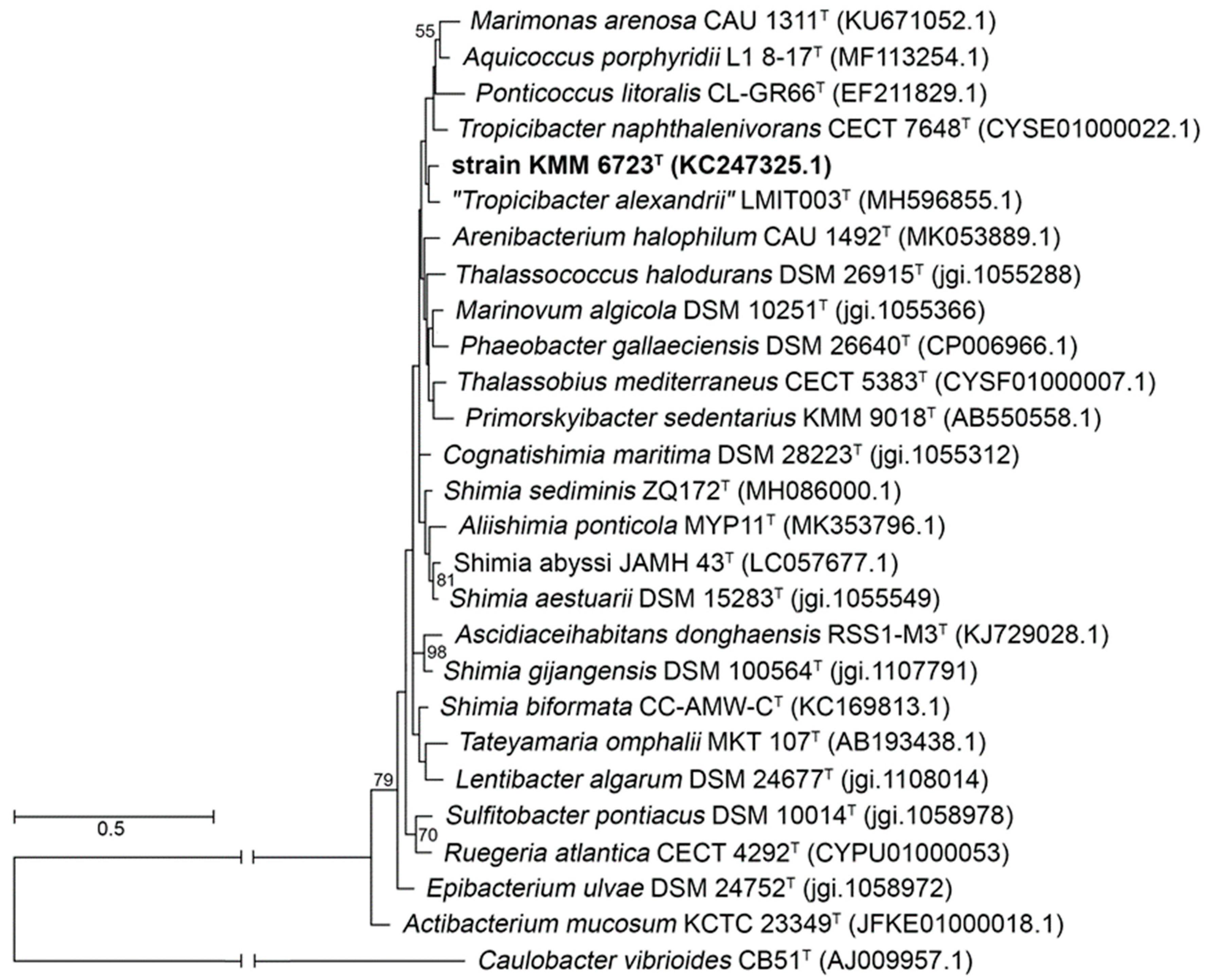
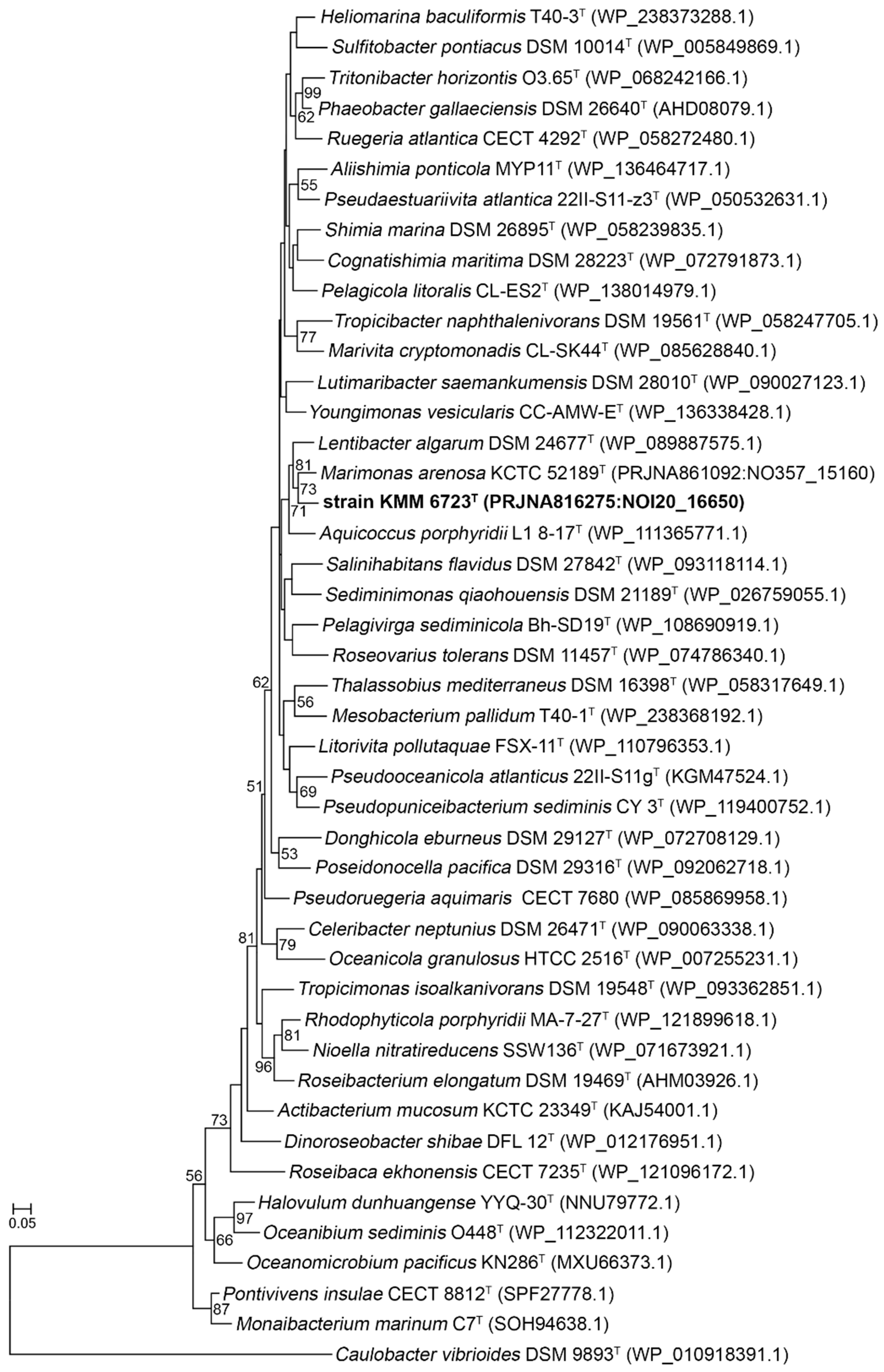
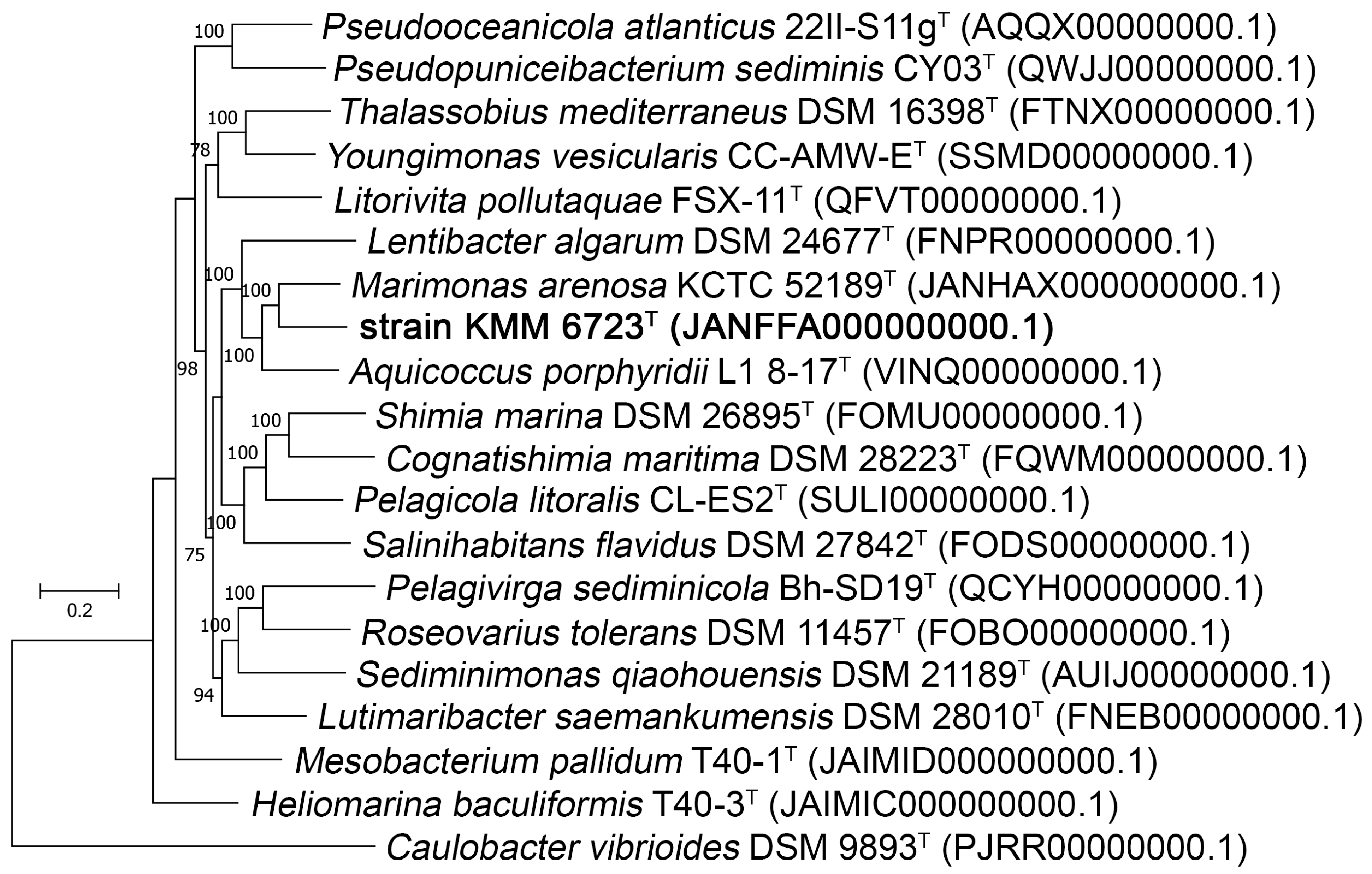
3.1. Phylogenetic and Phylogenomic Analyses

At first, the 1470 bp length 16S rRNA gene sequence of 10Alg 79T was amplified and used to perform 16S rRNA comparisons on the EzBioCloud server [26]. From this analysis, 10Alg 79T showed the highest similarity of 97.40% with ‘Tropicibacter alexandrii’ LMIT003T, followed by Shimia sediminis ZQ172T (97.33%), Shimia aestuarii DSM 15283T (96.75%), and Aliishimia ponticola MYP11T (96.75%). However, the 16S rRNA phylogenetic trees reconstructed with ML (Figure 1); MP, and NJ algorithms demonstrated very low-support branches and topological discordance.
Therefore, the position of strain 10Alg 79T was further determined using rpoC gene sequences extracted from type strain genomes of genera affiliated with the family Roseobacteraceae (formerly Rhodobacteraceae). RpoC protein sequences have been shown to be suitable for the Roseobacter group phylogeny [17,47]. An ML phylogenetic tree inferred on 43 RpoC protein sequences showed that 10Alg 79T forms a poorly supported clade together with M. arenosa KCTC 52189T [48], A. porphyridii L1 8-17T [49], and L. algarum DSM 24677T [50] (Figure 2).
Further, genomes of 18 type species of RpoC-related genera were selected for genomic tree building using PhyloPhlAn 3.0 software [40] using the concatenated sequence alignment of 400 conservative proteins. The resulting tree showed that 10Alg 79T forms a distinct genus-level lineage (Figure 3). Despite the higher identity of 16S rRNA sequences with those of ‘T. alexandrii’ LMIT003T (97.40%) and S. sediminis ZQ172T (97.33%), 10Alg 79T comprises the same clade with M. arenosa KCTC 52189T (96.03%), A. porphyridii L1 8-17T (96.17%), and L. algarum DSM 24677T (95.95%) on the genomic tree.
The ANI/AAI values between the genomes of strains 10Alg 79T and strains of three closely related genera M. arenosa KCTC 52189T, A. porphyridii L1 8-17T, and L. algarum DSM 24677T were 78.15%/73.08%, 77.85%/71.35%, and 75.63%/67.41%, respectively. In addition, the 10Alg 79T genome shared low values of dDDH (formula d4) with M. arenosa KCTC 52189T (19.4%), A. porphyridii L1 8-17T (19.8%), and L. algarum DSM 24677T (18.8%).
These values were significantly below the boundaries for the demarcation of bacterial species [51,52]. The AAI values between 10Alg 79T and the representatives were 67.41–73.08%, which did not exceed the declared genus boundaries of 80% [53]. Thus, the phylogenomic analysis based on the genome relatedness indexes clearly supported 10Alg 79T as a novel type species of a new genus in the family Rosebacteraceae (formerly Rhodobacteraceae).
Remarkably, a search for 16S rRNA sequences homologous to 10Alg 79T in NCBI, Greengenes, EzBioCloud, and Silva databases revealed only two 16S rRNAs with 99% identity (JQ215453.1 and JQ212029.1). Both originated from uncultured bacterium clones isolated from the dolphin Tursiops truncates in 2011 and 2013. This means that 10Alg 79T represents a new species of the rare genus of the family Roseobacteraceae.

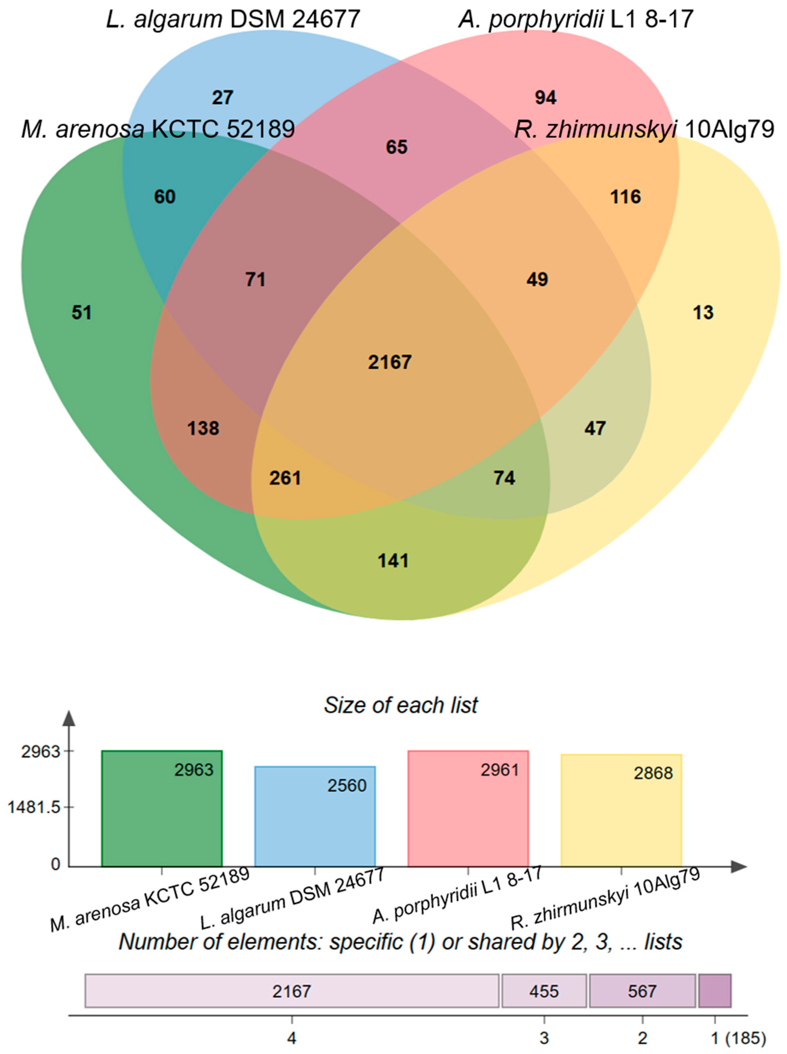
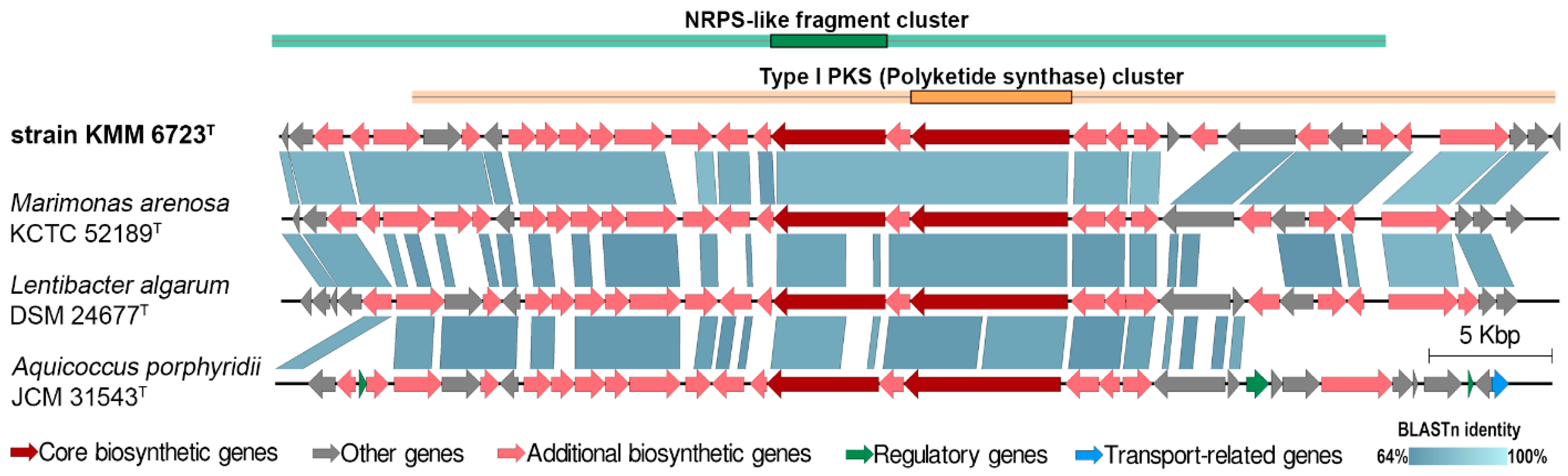
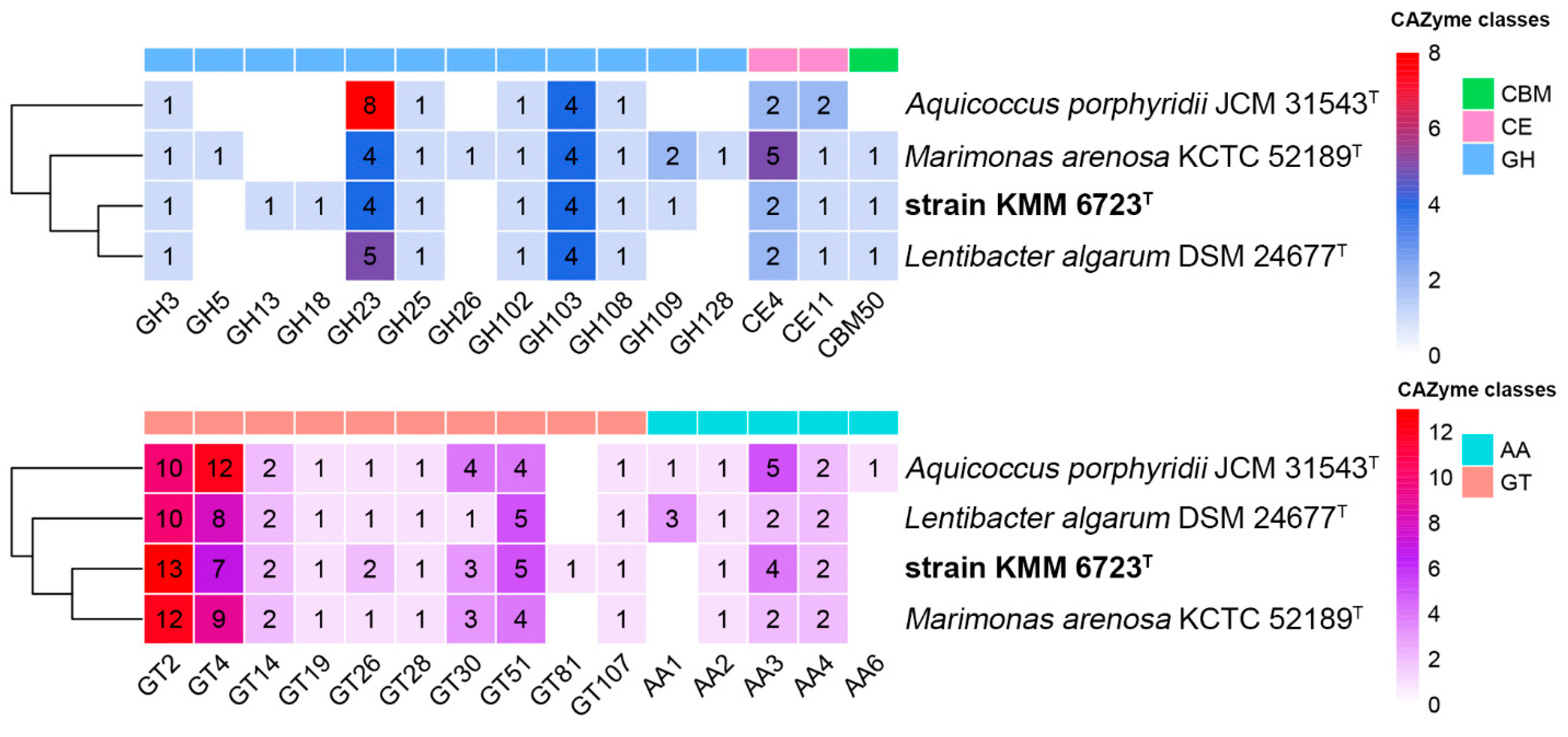
3.2. Genomic Characteristics and Analysis of Genus-Related Features

The draft genome of strain 10Alg 79T was de novo assembled into 73 contigs, with an N50 value of 702,161 bp and an L50 value of two. The genome size was estimated at 3,754,741 bp in length with an overall GC content of 62.1%. The genome-extracted 16S rRNA gene sequence was 100% identical to the PCR-amplified one. The genome contains 3739 coding sequences, 41 tRNAs, and 4 rRNA genes (one each of 16S and 23S and two genes of 5S). A comparison between genome features of 10Alg 79T and the type strains of closely related genera is presented in Table 1. These characteristics satisfy the proposed minimal standards for bacterial taxonomy [54].
To clarify genus-related features, a pan-genome analysis of 10Alg 79T and representatives of three closely related genera (M. arenosa KCTC 52189T, A. porphyridii L1 8-17T, and L. algarum DSM 24677T) was performed using orthologous clustering with the OrthoVenn2 and OrthoVenn3 servers [41,42]. The analysis revealed 3374 orthologous gene clusters (gene families); 1602 single-copy clusters consisted of only single-copy orthologs presented in all lineages of the clade. The core and accessory (without unique genes) genomes consisted of 2167 and 1022 orthologous clusters, respectively (Figure 4). Most of the 10Alg 79T clusters were shared with M. arenosa KCTC 52189T (141) and A. porphyridii L1 8-17T (116) (Figure 4). The results are in good agreement with the taxonomic position of 10Alg 79T on the genomic tree (Figure 3); M. arenosa KCTC 52189T and A. porphyridii L1 8-17T were more related to 10Alg 79T than L. algarum DSM 24677T. A total of 185 were identified as genus-specific clusters (Figure 4).
To predict metabolism of representatives of the nearest genera, genomes were first screened for the presence of genes of central and peripheral carbon metabolism due to the ability to grow on various carbon resources (Table 2). Based on the KEGG annotation, the genomes possess almost all genes of the glycolysis/gluconeogenesis pathways except for a key gene encoding 6-phosphofructokinase (EC 2.7.1.11) in the Embden–Meyerhof pathway. However, a modified pathway might incorporate the pentose phosphate pathway (PPP) due to genes encoding transaldolase (EC 2.2.1.2) and transketolase (EC 2.2.1.1). Moreover, genes found for the Entner–Doudoroff pathway (EDP), excluding L. algarum DSM 24677T, might encode the metabolism of glucose-6-phosphate into glyceraldehyde-3-phosphate and pyruvate. All the genomes were devoid of pgd encoding 6-phosphogluconate dehydrogenase (EC 1.1.1.44) in the oxidative phase of PPP. Semi-phosphorylative and non-phosphorilative EDP were presented by a partial set of genes. This means that the species are not able to utilize glucose through these canonical pathways and produce NADPH using PPP. All the genomes encode a complete tricarboxylic acid (TCA) cycle. Interestingly, M. arenosa KCTC 52189T also encodes a complete Calvin–Benson–Bassham cycle, in which a key enzyme RuBisCO (EC 4.1.1.39), is encoded by rbcLS and accompanied by molecular chaperon genes cbbQ and ccbO to enhance CO2 fixation activity [55].
All four genomes shared genes and operons involved in phosphate metabolism, such as pstSCAB (high-affinity phosphate-specific transporter, TC 3.A.1.7.1), phnCDE2E1 (phosphonate ABC transporter, TC 3.A.1.9.1), phnGHIJKLNM (carbon–phosphorus lyase enzyme complex), ugpBAEC (glycerol-3-phosphate transporter), ugpQ (cytosolic glycerophosphoryl diester phosphodiesterase, EC 3.1.4.46), ppx (exopolyphosphatase, EC 3.6.1.11), ppa (pyrophosphatase, EC 3.6.1.1), and ppk (polyphosphate kinase, EC 2.7.4.1).
All four genomes possessed a set of orthologous genes involved in ammonia assimilation, such as gdhA (NAD-specific glutamate dehydrogenase, 1.4.1.2; NADP-glutamate dehydrogenase, 1.4.1.4), glnA (glutamine synthetase, EC 6.3.1.2), gltBD (glutamate synthase, EC 1.4.1.13), gltB (ferredoxin-dependent glutamate synthase, EC 1.4.7.1), and nadE (ammonium-dependent NAD synthetase, EC 6.3.1.5). However, no genomes possessed the complete set of genes for denitrification; both the A. porphyridii L1 8-17T and M. arenosa KCTC 52189T genomes encode enzymes for nitrite, nitric and nitrous oxide reduction, while the 10Alg 79T genome encodes only for nitrite, and nitric oxide reduction, whereas the L. algarum DSM 24677T genome had none. Except for L. algarum DSM 24677T, most had ncd2 (nitronate monooxygenase, EC 1.13.12.16), which is responsible for the oxidation of nitroalkane into nitrite.
In addition, to compare the carbon utilization potential, genes encoding CAZymes in 10Alg 79T, A. porphyridii L1 8-17T, M. arenosa KCTC 52189T, and L. algarum DSM 24677T were predicted using the dbCAN2 server [43] (Figure 5). The 10Alg 79T genome code 60 CAZymes, including 15 glycoside hydrolases (GHs), 36 glycosyltransferases (GTs), three carbohydrate esterases (CEs), seven auxiliary activities (AAs), and one carbohydrate-binding module (CBM).
Genome mining of 10Alg 79T for secondary metabolite biosynthetic gene clusters (BGS) revealed five biotechnologically significant BGSs for the putative synthesis of non-ribosomal peptide synthetase (NRPS)-like and polyketide synthetase (PKS/NRPS hybrid), ribosomally synthesized and post-translationally modified peptides (RiPP-like, with methanobactin-like DUF692 domain), two homoserine lactones (hserlactone), and one important osmoprotectant ectoine. The most of these BGSs were also characteristic for the closely related genera Marimonas, Lentibacter, and Aquicoccus. For example, 10Alg 79T sheared up to 90% of genes encoding the PKS/NRPS hybrid system with 50–88% of gene identities (Figure 6), as estimated by [44].
Out of 16,163 genes identified in the clade pan-genome, 2312 genes were assigned as singletons without any orthologues among the compared genomes. Among them 413 singletons were found in the 10Alg 79T genome, followed by L. algarum DSM 24677T (410), A. porphyridii L1 8-17T (713), and M. arenosa KCTC 52189T (775). An analysis of a unique part of each genome showed that most were annotated as hypothetical proteins (more than 60%). The annotated singletons were more often related to transport protein systems, transcriptional regulators, and enzymes.
To illustrate the novelty of the 10Alg 79T genome, a detailed analysis of its singleton genome was carried out. In contrast to A. porphyridii L1 8-17T, M. arenosa KCTC 52189T, and L. algarum DSM 24677T, the 10Alg 79T genome demonstrates the presence of additional genes encoding periplasmic glycerophosphodiester phosphodiesterase (GlpQ, EC 3.1.4.46), and sn-glycerol-3-phosphate (g3p) transport system permease (UgpA, TC 3.A.1.1.3) participating in transport and hydrolysis of organic phosphorus [56]. In addition, this genome contained six singleton genes and one orthologous gene encoding alkaline phosphatases (EC 3.1.3.1) indicating the role of mineralization in the transported organic phosphorus. For comparison, the M. arenosa KCTC 52189T genome represents five orthologous genes and one singleton gene for alkaline phosphatases, followed by L. algarum DSM 24677T with only two orthologous genes and A. porphyridii L1 8-17T with only one orthologous gene. Therefore, attention should be paid to nitrogen metabolisms due to the number of singleton genes belonging to these subsystems. Remarkably, the 10Alg 79T genome contains two urease operons for urea catabolism; the first ureDABCEFG was shared with those of the other three genomes, and the second ureEFACGD was composed of singleton genes homologous to one of the numerous urease operons of Salipiger spp. (about 80% amino acid identity). This second ure operon was accompanied by an additional urea transport operon utrABCDE, which is like that in Salipiger spp, indicating past lateral gene transfer events. Additionally, 10Alg79 harbors singletons encoding N-methylhydantoinase A and B (EC 3.5.2.14), involved in the metabolism of nitrogen-containing compounds.
Importantly, 10Alg 79T, in addition to dddP, has additional DMSP cleavage gene dddL, indicating its role in ocean DMSP metabolism. In addition, it can be speculated that singletons encoding CAZymes GH13_18 (EC 2.4.1.7), GH18 (EC 3.2.1.-), and GT81 (EC 2.4.1.266) provide genus-related features of 10Alg 79T. It should be noted that GH13_18 can be a 2-O-glucosylglycerate phosphorylase (GGaP; EC 2.4.1.352) based on the characteristic motifs of GGaP in a loop A (PYELN) and an acid/base loop (TETN) [57]. This novel phosphorylase catalyzes the reversible phosphorolysis of glucosylglycerate and has great potential for the synthesis of valuable phosphoglycosides [56].

3.3. Morphological, Physiological, and Biochemical Characteristics

Strain 10Alg 79T has many properties in common with its closest relatives. They were found to be strictly aerobic, heterotrophic, and non-motile rods that can produce alkaline phosphatase, esterase (C4), esterase lipase (C8), leucine arylamidase, acid phosphatase, naphthol-AS-BI-phosphohydrolase, and catalase and oxidase but not lipase (C14), cystine arylamidase, trypsin, α-chymotrypsin, α-galactosidase, β-galactosidase, β-glucuronidase, α-glucosidase, β-glucosidase, N-acetyl-β-glucosaminidase, α-mannosidase, α-fucosidase, agarase, or caseinase. The novel isolate was distinguished from all neighbors by the minimal temperature for growth and amylase production. The sets of phenotypic traits including the ability to grow without NaCl or seawater, to reduce nitrate to nitrite, to produce acetoin and hydrogen sulfide, to hydrolyze aesculun, gelatin, tyrosine, and Tweens 40 and 80, taken together with the ability to utilize various carbohydrates, can help to discriminate strain 10Alg 79T from each of its relatives (Table 2).

3.4. Chemotaxonomy

The fatty acid composition of strain 10Alg 79T and reference strains A. porphyridii JCM 31543T and M. arenosa KCTC 52189T are listed in Table 3. The predominant fatty acids of strain 10Alg 79T were C18:1 ω7c (82.1%) and C16:0 (8.2%) (Table 3). The fatty acid composition of the strain studied was similar to that of the reference strains, although differences were found in the proportions of some fatty acids (Table 3). The polar lipid profile of strain 10Alg 79T contained phosphatidylethanolamine, phosphatidylglycerol, phosphatidylcholine, an unidentified phospholipid, an unidentified aminolipid, and an unidentified lipid and was consistent with that of the reference species (Supplementary Figure S1; Table 3). However, the presence of an unidentified phospholipid distinguished the bacterial isolate from M. arenosa KCTC 52189T and L. algarum ZXM100T. Moreover, strain 10Alg 79T can be differentiated from M. arenosa KCTC 52189T by the absence of an unknown glycolipid (Table 3). The predominant respiratory quinone was Q-10.

4. Conclusions

Description of Rhodoalgimonas gen. nov.
Rhodoalgimonas (Rho.do.al.gi.mo’nas. Gr. adj. rhodos red (rose); L. fem. n. alga seaweed, L. fem. n. monas a monad, a unit; N.L. fem. n. Rhodoalgimonas, a red (rose) monad isolated from seaweed).
Cells are Gram-stain-negative, strictly aerobic, rod-shaped, non-spore-forming, and non-motile. Catalase- and oxidase-positive. The dominant fatty acids (>5%) are summed feature 8, comprising C18:1 ω7c and C16:0. The polar lipids are phosphatidylethanolamine, phosphatidylglycerol, phosphatidylcholine, unidentified aminolipid, unidentified phospholipid and unidentified lipid. The major respiratory quinone is Q-10. Phylogenetically, the genus belongs to the family Rhodobacteraceae, the class Alphaproteobacteria, and the phylum Pseudomonadota. The type species is Rhodoalgimonas zhirmunskyi
Description of Rhodoalgimonas zhirmunskyi sp. nov.
Rhodoalgimonas zhirmunskyi (zhir.mun’skyi. N.L. gen. masc. n. zhirmunskyi named in honor of the academician A.V. Zhirmunskyi for his great contribution to the development of marine biology in the Russian Far East).
Cells are heterotrophic, strictly aerobic, non-motile, Gram-stain-negative rods, and 0.3–0.7 μm wide and 0.8–2.1 μm long. On marine agar, colonies are circular, shiny, slightly convex, with entire edges, 1–2 mm in diameter, and beige-colored. Growth occurs at 4–40 °C (optimum is 30–32 °C) and pH 6.0–9.0 (optimum is 7.0–8.0) and with 0–5% NaCl (optimum is 1.5–3% NaCl). Seawater or artificial seawater is substantial for growth. Catalase and oxidase activities are present. Arginine dihydrolase, lysine decarboxylase, ornithine decarboxylase, and tryptophan deaminase activities are absent. Aesculin, starch, and Tweens 20, 40, and 80 are hydrolyzed but agar, casein, gelatin, chitin, CM-cellulose, DNA, L-tyrosine, and urea are not. Acid is not produced from L-arabinose, D-cellobiose, D-fructose, D-galactose, D-glucose, D-lactose, maltose, mannose, melibiose, raffinose, L-rhamnose, ribose, sucrose, trehalose, D-xylose, N-acetylglucosamine, mannitol, glycerol, sorbitol, or citrate. Growth is observed on L-arabinose, D-cellebiose, D-fructose, D-galactose, D-glucose, D-lactose, maltose, D-mannose, D-melibiose, D-raffinose, L-rhamnose, D-trehalose, D-xylose, N-acetylglucosamine, L-alanine, L-asparagine, L-histidine, L-methionine, L-proline, L-triptophane, L-treonine, L-tyrosin, and L-valine. In the API 20NE gallery, positive for aesculin hydrolysis and assimilation of D-mannose, D-mannitol, maltose, gluconate, adipate, malate, and phenylacetate tests. In the API 20E and API 50 CH kits, no results were positive. In the API ID 32GN kit, maltose, D-mannitol, salicin, D-melibiose, propionic acid, valeric acid, and 3-hydroxybutiric acid were assimilated. According to the API ZYM tests, alkaline phosphatase, esterase (C4), esterase lipase (C8), leucine arylamidase, acid phosphatase, and naphtol-AS-BI-phosphohydrolase activities are present but lipase (C14), cystine arylamidase, trypsin, α-chymotrypsin, α-galactosidase, β-galactosidase, β-glucuronidase, α-glucosidase, β-glucosidase, N-acetyl-β-glucosaminidase, α-mannosidase, and α-fucosidase activities are absent. Valine arylamidase activity was weakly positive. Nitrate is not reduced. Hydrogen sulfide, indole, and acetoin are not produced. The prevalent fatty acids are summed feature 8, comprising C18:1 ω7c and C16:0. The polar lipid profile consists of phosphatidylethanolamine, phosphatidylglycerol, phosphatidylcholine, an unidentified aminolipid, an unidentified phospholipid, and an unidentified lipid. The main ubiquinone is Q-10. The genomic DNA GC content of the type strain is 62.1 mol%.

Supplementary Materials

The following supporting information can be downloaded at: https://www.mdpi.com/article/10.3390/microorganisms11102463/s1, Figure S1: Two-dimensional TLC of the total polar lipids of strain 10Alg 79T. First dimension, chloroform/methanol/water (65:25:4, v/v/v); second dimension, chloroform/methanol/acetic acid/water (80:12:15:4, v/v/v/v). PE—phosphatidylethanolamine, PG—phosphatidylglycerol, PC—phosphatidylcholine, AL—an unidentified aminolipid, PL—an unidentified phospholipid, and L—an unidentified lipid.

Author Contributions

Investigation, O.N., N.O., L.B., E.B., N.Z. and M.I.; methodology, O.N., N.O., L.B., S.-G.K., E.B. and N.Z.; project administration, M.I.; resources, S.-G.K., L.T. and M.I.; software, N.O. and M.I.; writing—original draft, O.N., N.O., L.B. and M.I.; writing—review and editing, O.N. and M.I. All authors have read and agreed to the published version of this manuscript.

Funding

This research was funded by a grant from the Ministry of Science and Higher Education, Russian Federation 15.BRK.21.0004 (contract no. 075-15-2021-1052).

Data Availability Statement

The type strain of the species is strain 10Alg 79T isolated from the red alga Ahnfeltia tobuchiensis collected near Island Paramushir, Kuril Isles, the Sea of Okhotsk, Pacific Ocean, Russia. The DDBJ/ENA/GenBank accession numbers for the 16S rRNA gene and the whole-genome shotgun sequences are KC247325 and JANFFA000000000, respectively. Strain 10Alg 79T was deposited in the Collection of Marine Microorganisms (WFCC acronym is KMM) under the number KMM 6723T, and in the Korean Collection for Type Cultures (KCTC) under the number KACC 72611T.

Conflicts of Interest

The authors declare no conflict of interest.

References

  1. Barbato, M.; Vacchini, V.; Engelen, A.H.; Patania, G.; Mapelli, F.; Borin, S.; Crotti, E. What lies on macroalgal surface: Diversity of polysaccharide degraders in culturable epiphytic bacteria. AMB Express 2022, 12, 98. [Google Scholar] [CrossRef] [PubMed]
  2. Kanagasabhapathy, M.; Sasak, H. Phylogenetic identification of epibiotic bacteria possessing antimicrobial activities isolated from red algal species of Japan. World J. Microbiol. Biotechnol. 2008, 24, 2315–2321. [Google Scholar] [CrossRef]
  3. Miranda, L.N.; Hutchison, K. Diversity and abundance of the bacterial community of the red macroalga Porphyra umbilicalis: Did bacterial farmers produce macroalgae? PLoS ONE 2013, 8, e58269. [Google Scholar] [CrossRef] [PubMed]
  4. Tan, T.T.; Song, S.L. The effect of grazing on the microbiome of two commercially important agarophytes, Gracilaria firma and G. salicornia (Gracilariaceae, Rhodophyta). J. Appl. Phycol. 2020, 32, 2549–2559. [Google Scholar] [CrossRef]
  5. Meng, X.; Chang, Y.Q. Algibacillus agarilyticus gen. nov., sp. nov., isolated from the surface of the red algae Gelidium amansii. Int. J. Syst. Evol. Microbiol. 2021, 71, 4558. [Google Scholar] [CrossRef]
  6. Xue, J.H.; Zhang, B.N. Comparative genomic analysis of the genus Marinomonas and taxonomic study of Marinomonas algarum sp. nov., isolated from red algae Gelidium amansii. Arch. Microbiol. 2022, 204, 586. [Google Scholar] [CrossRef]
  7. Zhang, J.Y.; Xia, Y. Maribacter algarum sp. nov., a new member of the family Flavobacteriaceae isolated from the red alga Gelidium amansii. Int. J. Syst. Evol. Microbiol. 2020, 70, 3679–3685. [Google Scholar] [CrossRef]
  8. Khan, S.A.; Jeong, S.E. Maribacter algicola sp. nov., isolated from a marine red alga, Porphyridium marinum, and transfer of Maripseudobacter aurantiacus Chen et al. 2017 to the genus Maribacter as Maribacter aurantiacus comb. nov. Int. J. Syst. Evol. Microbiol. 2020, 70, 797–804. [Google Scholar] [CrossRef]
  9. Yu, W.N.; Du, Z.Z. Marinomonas agarivorans sp. nov., an agar-degrading marine bacterium isolated from red algae. Int. J. Syst. Evol. Microbiol. 2020, 70, 100–104. [Google Scholar] [CrossRef]
  10. Kim, J.; Kim, K.H. Flagellimonas algicola sp. nov., isolated from a marine red alga, Asparagopsis taxiformis. Curr. Microbiol. 2020, 77, 294–299. [Google Scholar] [CrossRef]
  11. Ping, X.Y.; Wang, K. Psychroserpens luteolus sp. nov., isolated from Gelidium, reclassification of Ichthyenterobacterium magnum as Psychroserpens magnus comb. nov., Flavihalobacter algicola as Psychroserpens algicola comb. nov., Arcticiflavibacter luteus as Psychroserpens luteus comb. nov. Arch. Microbiol. 2022, 204, 279. [Google Scholar]
  12. Kristyanto, S.; Kim, K.R. Tenacibaculum aquimarinum sp. nov., isolated from a marine alga and sea-water. Int. J. Syst. Evol. Microbiol. 2022, 72, 5477. [Google Scholar]
  13. Lian, F.B.; Jiang, S. Thalassotalea algicola sp. nov., an alginate-utilizing bacterium isolated from a red al-ga. Antonie Leeuwenhoek 2021, 114, 835–844. [Google Scholar] [CrossRef] [PubMed]
  14. Kim, J.; Jeong, S.E. Hwanghaeella grinnelliae gen. nov., sp. nov., isolated from a marine red alga. Int. J. Syst. Evol. Microbiol. 2019, 69, 3544–3550. [Google Scholar] [CrossRef] [PubMed]
  15. Romanenko, L.A.; Kurilenko, V.V. Characterization of Labrenzia polysiphoniae sp. nov. isolated from red alga Polysiphonia sp. Arch. Microbiol. 2019, 201, 705–712. [Google Scholar] [CrossRef]
  16. Nedashkovskaya, O.I.; Kim, S.G. Aquimarina algiphila sp. nov., a chitin degrading bacterium isolated from the red alga Tichocarpus crinitus. Int. J. Syst. Evol. Microbiol. 2018, 68, 892–898. [Google Scholar] [CrossRef]
  17. Nedashkovskaya, O.I.; Kukhlevskiy, A.D. Aureibaculum algae sp. nov. isolated from the Pacific red alga Ahnfeltia tobuchiensis. Arch. Microbiol. 2022, 204, 153. [Google Scholar] [CrossRef]
  18. Nedashkovskaya, O.I.; Balabanova, L.A.; Zhukova, N.V.; Kim, S.J.; Bakunina, I.Y.; Rhee, S.K. Flavobacterium ahnfeltiae sp. nov., a new marine polysaccharide-degrading bacterium isolated from a Pacific red alga. Arch. Microbiol. 2014, 196, 745–752. [Google Scholar] [CrossRef]
  19. Nedashkovskaya, O.I.; Kim, S.G. Polaribacter staleyi sp. nov., a polysaccharide-degrading marine bacterium isolated from the red alga Ahnfeltia tobuchiensis. Int. J. Syst. Evol. Microbiol. 2018, 68, 623–629. [Google Scholar] [CrossRef]
  20. Gerhardt, P.; Murray, R.G.E.; Wood, W.A.; Krieg, N.R. Methods for General and Molecular Bacteriology; American Society for Microbiology: Washington, DC, USA, 1994; p. 224. [Google Scholar]
  21. Lemos, M.L.; Toranzo, A.E. Modified medium for oxidation-fermentation test in the identification of marine bacteria. Appl. Environ. Microbiol. 1985, 40, 1541–1543. [Google Scholar] [CrossRef]
  22. Smibert, R.M.; Krieg, N.R. Phenotypic characterization. In Methods for General and Molecular Bacteriology; Gerhardt, P., Murray, R.G.E., Wood, W.A., Krieg, N.R., Eds.; American Society for Microbiology: Washington, DC, USA, 1994; pp. 607–654. [Google Scholar]
  23. Nedashkovskaya, O.I.; Kim, S.B. Mesonia mobilis sp. nov., isolated from seawater, and emended description of the genus Mesonia. Int. J. Syst. Evol. Microbiol. 2006, 56, 2433–2436. [Google Scholar] [CrossRef] [PubMed]
  24. Komagata, K.; Suzuki, K.I. Lipid and cell wall analysis in bacterial systematics. Methods Microbiol. 1987, 19, 161–207. [Google Scholar]
  25. Lane, D.J. 16S/23S rRNA sequencing. In Nucleic Acid Techniques in Bacterial Systematics; Stackebrandt, E., Goodfellow, M., Eds.; John Wiley & Sons: New York, NY, USA, 1991; pp. 115–147. [Google Scholar]
  26. Yoon, S.H.; Ha, S.M.; Kwon, S.; Lim, J.; Kim, Y.; Seo, H.; Chun, J. Introducing EzBioCloud: A taxonomi-cally united database of 16S rRNA gene sequences and whole-genome assemblies. Int. J. Syst. Evol. Microbiol. 2017, 67, 1613–1617. [Google Scholar] [CrossRef] [PubMed]
  27. Kumar, S.; Stecher, G.; Li, M.; Knyaz, C.; Tamura, K. MEGA X: Molecular evolutionary genetics analysis across computing platforms. Mol. Biol. Evol. 2018, 35, 1547–1549. [Google Scholar] [CrossRef]
  28. Kimura, M. A simple method for estimating evolutionary rate of base substitutions through comparative studies of nucleotide sequences. Mol. Biol. Evol. 1980, 16, 111–120. [Google Scholar] [CrossRef]
  29. Nguyen, L.T.; Schmidt, H.A.; von Haeseler, A.; Minh, B.Q. IQ-TREE: A fast and effective stochastic algorithm for estimating maximum-likelihood phylogenies. Mol. Biol. Evol. 2015, 32, 268–274. [Google Scholar] [CrossRef]
  30. Kalyaanamoorthy, S.; Minh, B.Q.; Wong, T.K.F.; von Haeseler, A.; Jermiin, L.S. Modelfinder: Fast model selection for accurate phylogenetic estimates. Nat. Methods 2017, 14, 587–589. [Google Scholar] [CrossRef]
  31. Bolger, A.M.; Lohse, M.; Usadel, B. Trimmomatic: A flexible trimmer for Illumina sequence data. Bioinformatics 2014, 30, 2114–2120. [Google Scholar] [CrossRef]
  32. Bankevich, A.; Nurk, S.; Antipov, D.; Gurevich, A.A.; Dvorkin, M.; Kulikov, A.S.; Lesin, V.M.; Nikolenko, S.I.; Pham, S.; Prjibelski, A.D.; et al. SPAdes: A new genome assembly algorithm and its applications to single-cell sequencing. J. Comput. Biol. 2012, 19, 455–477. [Google Scholar] [CrossRef]
  33. Gurevich, A.; Saveliev, V.; Vyahhi, N.; Tesler, G. QUAST: Quality assessment tool for genome assemblies. Bioinformatics 2013, 29, 1072–1075. [Google Scholar] [CrossRef]
  34. Parks, D.H.; Imelfort, M.; Skennerton, C.T.; Hugenholtz, P.; Tyson, G.W. CheckM: Assessing the quality of microbial genomes recovered from isolates, single cells, and metagenomes. Genome Res. 2015, 25, 1043–1055. [Google Scholar] [CrossRef] [PubMed]
  35. Tatusova, T.; DiCuccio, M.; Badretdin, A.; Chetvernin, V.; Nawrocki, E.P.; Zaslavsky, L.; Lomsadze, A.; Pruitt, K.D.; Borodovsky, M.; Ostell, J. NCBI prokaryotic genome annotation pipeline. Nucleic Acids Res. 2016, 44, 6614–6624. [Google Scholar] [CrossRef] [PubMed]
  36. Aziz, R.K.; Bartels, D.; Best, A.A.; DeJongh, M.; Disz, T.; Edwards, R.A.; Formsma, K.; Gerdes, S.; Glass, E.M.; Kubal, M.; et al. The RAST Server: Rapid annotations using subsystems technology. BMC Genom. 2008, 9, 75. [Google Scholar] [CrossRef] [PubMed]
  37. Wattam, A.R.; Abraham, D.; Dalay, O.; Disz, T.L.; Driscoll, T.; Gabbard, J.L.; Gillespie, J.J.; Gough, R.; Hix, D.; Kenyon, R.; et al. PATRIC, the bacterial bioinformatics database and analysis resource. Nucleic Acids Res. 2014, 42, D581–D591. [Google Scholar] [CrossRef] [PubMed]
  38. Rodriguez-R, L.M.; Konstantinidis, K.T. The enveomics collection: A toolbox for specialized analyses of microbial genomes and metagenomes. PeerJ Prepr. 2016, 4, e1900v1. [Google Scholar]
  39. Meier-Kolthoff, J.P.; Göker, M. TYGS is an automated high-throughput platform for state-of-the-art genome-based taxonomy. Nat. Commun. 2019, 10, 2182. [Google Scholar] [CrossRef]
  40. Asnicar, F.; Thomas, A.M.; Beghini, F.; Mengoni, C.; Manara, S.; Manghi, P.; Zhu, Q.; Bolzan, M.; Cumbo, F.; May, U.; et al. Precise phylogenetic analysis of microbial isolates and genomes from meta-genomes using PhyloPhlAn 3.0. Nat. Commun. 2020, 11, 2500. [Google Scholar] [CrossRef]
  41. Xu, L.; Dong, Z.; Fang, L.; Luo, Y.; Wei, Z.; Guo, H.; Zhang, G.; Gu, Y.Q.; Coleman-Derr, D.; Xia, Q.; et al. OrthoVenn2: A web server for whole-genome comparison and annotation of orthologous clusters across multiple species. Nucleic Acids Res. 2019, 47, W52–W58. [Google Scholar] [CrossRef]
  42. Sun, J.; Lu, F.; Luo, Y.; Bie, L.; Xu, L.; Wang, Y. OrthoVenn3: An integrated platform for exploring and visualizing orthologous data across genomes. Nucleic Acids Res. 2013, 1, 13–14. [Google Scholar] [CrossRef]
  43. Zhang, H.; Yohe, T.; Huang, L.; Entwistle, S.; Wu, P.; Yang, Z.; Busk, P.K.; Xu, Y.; Yin, Y. dbCAN2: A meta server for automated carbohydrate-active enzyme annotation. Nucleic Acids Res. 2018, 46, W95–W101. [Google Scholar] [CrossRef]
  44. Blin, K.; Shaw, S.; Kloosterman, A.M.; Charlop-Powers, Z.; van Wezel, G.P.; Medema, M.H.; Weber, T. AntiSMASH 6.0: Improving Cluster Detection and Comparison Capabilities. Nucleic Acids Res. 2021, 49, W29–W35. [Google Scholar] [CrossRef]
  45. Kearse, M.; Moir, R.; Wilson, A.; Stones-Havas, S.; Cheung, M.; Sturrock, S.; Buxton, S.; Cooper, A.; Markowitz, S.; Duran, C. Geneious Basic: An integrated and extendable desktop software platform for the organization and analysis of sequence data. Bioinformatics 2012, 28, 1647–1649. [Google Scholar] [CrossRef] [PubMed]
  46. Sullivan, M.J.; Petty, N.K.; Beatson, S.A. Easyfig: A genome comparison visualizer. Bioinformatics 2011, 27, 1009–1010. [Google Scholar] [CrossRef] [PubMed]
  47. Wirth, J.S.; Whitman, W.B. Phylogenomic analyses of a clade within the roseobacter group suggest taxonomic reassignments of species of the genera Aestuariivita, Citreicella, Loktanella, Nautella, Pelagibaca, Ruegeria, Thalassobius, Thiobacimonas and Tropicibacter, and the proposal of six novel genera. Int. J. Syst. Evol. Microbiol. 2018, 68, 2393–2411. [Google Scholar]
  48. Thongphrom, C.; Kim, J.H. Marimonas arenosa gen. nov., sp. nov., isolated from sea sand. Int. J. Syst. Evol. Microbiol. 2017, 67, 121–126. [Google Scholar] [CrossRef]
  49. Feng, T.; Kim, K.H. Aquicoccus porphyridii gen. nov., sp. nov., isolated from a small marine red alga, Porphyridium marinum. Int. J. Syst. Evol. Microbiol. 2018, 68, 283–288. [Google Scholar] [CrossRef]
  50. Li, Z.; Qu, Z. Lentibacter algarum gen. nov., sp. nov., isolated from coastal water during a massive green algae bloom. Int. J. Syst. Evol. Microbiol. 2012, 62, 1042–1047. [Google Scholar] [CrossRef]
  51. Richter, M.; Rosselló-Móra, R. Shifting the genomic gold standard for the prokaryotic species definition. Proc. Natl. Acad. Sci. USA 2009, 106, 19126–19131. [Google Scholar] [CrossRef] [PubMed]
  52. Colston, S.M.; Fullmer, M.S.; Beka, L.; Lamy, B.; Gogarten, J.P.; Graf, J. Bioinformatic genome comparisons for taxonomic and phylogenetic assignments using Aeromonas as a test case. mBio 2014, 5, e02136. [Google Scholar] [CrossRef] [PubMed]
  53. Luo, C.; Rodriguez-R, L.M.; Konstantinidis, K.T. MyTaxa: An advanced taxonomic classifier for genomic and metagenomic sequences. Nucleic Acids Res. 2014, 42, e73. [Google Scholar] [CrossRef]
  54. Chun, J.; Oren, A.; Ventosa, A.; Christensen, H.; Arahal, D.R.; da Costa, M.S.; Rooney, A.P.; Yi, H.; Xu, X.W.; De Meyer, S.; et al. Proposed minimal standards for the use of genome data for the taxonomy of prokaryotes. Int. J. Syst. Evol. Microbiol. 2018, 68, 461–466. [Google Scholar] [CrossRef] [PubMed]
  55. Tsai, Y.C.; Lapina, M.C.; Bhushan, S.; Mueller-Cajar, O. Identification and characterization of multiple rubisco activases in chemoautotrophic bacteria. Nat. Commun. 2015, 6, 8883. [Google Scholar] [CrossRef] [PubMed]
  56. Franceus, J.; Pinel, D.; Desmet, T. Glucosylglycerate phosphorylase, an enzyme with novel specificity involved in compatible solute vetabolism. Appl. Environ. Microbiol. 2017, 83, e01434-17. [Google Scholar] [CrossRef]
  57. Brzoska, P.; Boos, W. Characteristics of a ugp-encoded and phoB-dependent glycerophosphoryl diester phosphodiesterase which is physically dependent on the ugp transport system of Escherichia coli. J. Bacteriol. 1988, 170, 4125–4135. [Google Scholar] [CrossRef] [PubMed]
Figure 1. ML 16S rRNA tree showing phylogenetic relationships between the novel strain 10Alg 79T (KMM 6723T) and members of the family Rosebacteraceae (formerly Rhodobacteriaceae). Bootstrap values shown as percentage are based on 500 replicates. Bars are 0.5 substitutions per nucleotide. Strain Caulobacter vibrioides CB51T/DSM 9893T was used as an outgroup. GenBank/EMBL/DDB accession numbers are given in parentheses.
Microorganisms 11 02463 g001
Figure 2. ML RpoC tree showing phylogenetic relationships between the novel strain 10Alg 79T (KMM 6723T) and 43 members of the family Rosebacteraceae (formerly Rhodobacteriaceae). Bootstrap values are based on 500 replicates and shown as percentages greater than 50. Bars are 0.5 substitutions per amino acid position. Strain Caulobacter vibrioides DSM 9893T was used as an outgroup. GeBank/EMBL/DDB accession numbers are given in parentheses.
Microorganisms 11 02463 g002
Figure 3. ML genomic tree showing phylogenetic relationships between the strain 10Alg 79T (KMM 6723T) and members of the family Rosebacteraceae (formerly Rhodobacteriaceae). Bootstrap values shown as percentages greater than 50 are based on 100 replicates. Bar is 0.2 substitutions per amino acid position. Strain Caulobacter vibrioides DSM 9893T was used as an outgroup. GenBank/EMBL/DDB accession numbers are given in parentheses.
Microorganisms 11 02463 g003
Figure 4. Venn diagram showing the distribution of shared orthologous clusters among genomes of 10Alg 79T, A. porphyridii L1 8-17T, M. arenosa KCTC 52189T, and L. algarum DSM 24677T. The orthologous clusters are comprised of proteins predicted using RAST [36]. The number of orthologous clusters for each genome is shown in vertical bar chart.
Microorganisms 11 02463 g004
Figure 5. The distribution of CAZymes across genomes of KMM 6723T (=10Alg 79T) and close relatives. The heat maps show the number of genes assigned to individual CAZyme families. Rows are clustered using Euclidean distances. GH, glycoside hydrolases; GT, glycosyltransferases, CE, carbohydrate esterase; AA, auxiliary activity; CBM, carbohydrate-binding module.
Microorganisms 11 02463 g005
Figure 6. Comparative analysis of synteny between NRPS-like fragment and Type I PKS BGCs in the genomes of strain 10Alg 79T (KMM 6723T) and close relatives. Genes are colored based on their annotation as indicated at the bottom. The locus regions are NOI20_03420 to NOI20_03580 in the KMM 6723T genome, NO357_05410 to NO357_05560 in the M. arenosa KCTC 52189T genome, BLV93_RS04080 to BLV93_RS03920 in the L. algarum DSM 24677T genome, and FLO80_RS04395 to FLO80_RS04560 in the A. porphyridii JCM 31543T genome.
Microorganisms 11 02463 g006
Table 1. Genomic features of 10Alg 79T and three type strains of closely related genera.
Feature1234
Assembly levelContigContigContigContig
Genome size (bp)3,754,7414,513,8374,380,8393,293,487
Number of contigs732563821
GC content (mol%)62.163.26355.5
N50 (bp)702,161179,538656,426890,063
L50 (bp)2732
Coverage19x100x90x311.0x
Total genes3609454242993318
Protein coding genes3535437342053262
rRNAs(5S/16S/23S)2/1/12/1/11/1/162/2/2
tRNA41424444
checkM completeness (%)98.2410099.4299.69
checkM contamination (%)0.042.60.340.31
WGS project JANFFA01VINQ01JANHAX01FNPR01
Annotated genome assemblyASM3084892v1ASM836910v1ASM3084894v1IMG-taxon 2693429861
Submitted GenBank assemblyGCA_030848925.1GCA_008369105.1GCA_030848945.1GCA_900107355.1
Number of Subsystems (RAST)300324315285
Strains: 1, 10Alg 79T; 2, Aquicoccus porphyridii L1 8-17T; 3, Marimonas arenosa KCTC 52189T; 4, Lentibacter algarum DSM 24677T.
Table 2. Phenotypic characteristics differentiating strain 10Alg 79T and related genera of the family Roseobacteraceae.
Characteristics1234
Source of isolationMarine red algaMarine red algaSea sandSeawater
Colony colorBeigeWhitishBeigeWhitish
MorphologyRodCoccusRodRod
Temperature range for growth (°C) *:4–4020–4020–3722–28
Salinity range for growth (% NaCl) *:0–50–70–63–9
Nitrate reduction---+
Acetoin production (3-hydroxybutanone or acetyl methyl carbinol)---+
H2S production-+--
Degradation of:
AesculinW--+
Gelatin --+-
StarchW---
Tweens 40 and 80+--+
Tyrosine-+-ND
Acid production from:
D-fructose, D-glucose, maltose, sucrose, D-xylose---+
Utilization of:
L-arabinose, maltose++-+
D-cellobiose, D-fructose,+--+
D-lactose, D-melibiose+--ND
D-glucose, D-mannose+--+
D-mannitol+-++
N-acetylglucosamine+---
Adipate+-+-
Citrate ---+
Gluconate+-++
Malate+++-
Phenylacetate++--
Susceptibility to:
Ampicillin, carbenicillin+-++
Gentamicin+-+-
Bensylpenicillin +--+
Kanamycin+++-
Oxacillin+---
Tetracycline ++--
Vancomycin++-+
Major polar lipids **PC, PG, PE,
AL, PL, L
PC, PG, PE,
AL, PL, L
PC, PG, PE,
AL, GL
PC, PG, PE,
AL, L
Strains: 1, 10Alg 79T (this study); 2, Aquicoccus porphyridii JCM 31543T (this study); 3, Marimonas arenosa KCTC 52189T (this study); 4, Lentibacter algarum ZXM100T (data from [50]). All strains were positive for the following tests: respiratory type of metabolism; alkaline phosphatase, esterase (C4), esterase lipase (C8), leucine arylamidase, acid phosphatase, naphthol-AS-BI-phosphohydrolase, catalase and oxidase activities; susceptibility to erythromycin, and resistance to polymixin. All strains were negative for the following tests: motility; indole production; hydrolysis of agar, casein; acid production from D-lactose, D-melibiose, L-rhamnose, and ribose; lipase (C14), cystine arylamidase, trypsin, α-chymotrypsin, α-galactosidase, β-galactosidase, β-glucuronidase, α-glucosidase, β-glucosidase, N-acetyl-β-glucosaminidase, α-mannosidase and α-fucosidase activities. +, Positive; -, negative; W, a weak reaction; ND, data are not detected. *, Data taken from [48,49] **, PC, phosphatidylcholine; PG, phosphatidylglycerol; PE, phosphatidylethanolamine; AL, unidentified aminolipid; PL, unidentified phospholipid; L, unidentified lipid; and GL, unidentified glycolipid.
Table 3. Fatty acid profiles of strain 10Alg 79T and the type strains of the related genera of the family Roseobacteraceae.
Fatty Acid1234
Straight fatty acid
C15:01.3tr--
C16:08.27.74.512.0
C17:01.61.5-2.0
C18:0Tr2.33.920.0
Unsaturated fatty acid
C16:1 ω7c1.3trTr1.0
C18:1 ω7c82.181.880.660.0
11-methyl C18:1 ω7cTr4.34.12.0
Hydroxy fatty acid
C10:0 3-OH---2.0
C12:0 3-OH4.51.04.0-
C12:1 3-OH-1.2--
Strains: 1, 10Alg 79T; 2, Aquicoccus porphyridii JCM 31543T; 3, Marimonas arenosa KCTC 52189T; 4, Lentibacter algarum ZXM100T (data from [48]). Data are from the present study. Values are percentages of the total fatty acids. tr, trace amount (<1%).
Disclaimer/Publisher’s Note: The statements, opinions and data contained in all publications are solely those of the individual author(s) and contributor(s) and not of MDPI and/or the editor(s). MDPI and/or the editor(s) disclaim responsibility for any injury to people or property resulting from any ideas, methods, instructions or products referred to in the content.

