Article

Domiciliation of Trichoderma asperellum Suppresses Globiosporangium ultimum and Promotes Pea Growth, Ultrastructure, and Metabolic Features

by
Zeiad Moussa
1,*,
Yasmene F. Alanazi
2,
Aiah Mustafa Khateb
3,
Noha M. Eldadamony
4,*,
Marwa M. Ismail
5,
WesamEldin I. A. Saber
1,* and
Doaa Bahaa Eldin Darwish
6
1
Microbial Activity Unit, Microbiology Department, Soils, Water and Environment Research Institute, Agricultural Research Center, Giza 12619, Egypt
2
Department of Biochemistry, Faculty of Science, University of Tabuk, Tabuk 71421, Saudi Arabia
3
Medical Laboratory Technology Department, College of Applied Medical Sciences, Taibah University, Madinah 42353, Saudi Arabia
4
Seed Pathology Research Department, Plant Pathology Research Institute, Agricultural Research Center, Giza 12619, Egypt
5
Central Lab of Biotechnology, Plant Pathology Research Institute, Agricultural Research Center, Giza 12619, Egypt
6
Botany Department, Faculty of Science, Mansoura University, Mansoura 35516, Egypt
*
Authors to whom correspondence should be addressed.
Microorganisms 2023, 11(1), 198; https://doi.org/10.3390/microorganisms11010198
Submission received: 8 December 2022 / Revised: 29 December 2022 / Accepted: 9 January 2023 / Published: 12 January 2023
(This article belongs to the Section Plant Microbe Interactions)

Abstract

:
The beneficial microorganisms represent a new and hopeful solution for a sustainable environment and development. In this investigation, Trichoderma asperellum ZNW, isolated from seeds, was domiciliated within the pea plant for improving growth, disease management, and enhancement of productivity. Globisporangium ultimum NZW was isolated from deformed pea seeds, representing the first record of the pathogen caused by pea damping-off. Both fungi were molecularly identified. T. asperellum ZNW produced several lytic enzymes and bioactive metabolites as detected by GC-MC. The SEM illustrated the mycoparasitic behavior of T. asperellum ZNW on G. ultimum NZW mycelia. In the pot experiment, T. asperellum domiciliated the root and grew as an endophytic fungus, leading to root vessel lignification. Under soil infection, T. asperellum reduced damping-off, by enhancing peroxidase, polyphenol, total phenols, and photosynthetic pigments content. The vegetative growth, yield, and soil dehydrogenase activity were improved, with an enhancement in the numerical diversity of the microbial rhizosphere. This work may enable more understanding of the plant-fungal interaction, yet, working on domiciliation is recommended as a new approach to plant protection and growth promotion under various ecological setups.

1. Introduction

The current era shows great awareness of environmental problems. Microbial diversity can help in sustaining a clean and balanced ecosystem. Plant-associated microorganisms, especially endophytes, represent a hopeful tool for ecological maintenance and sustainable development. To do so, beneficial microorganisms have several mechanisms, e.g., acting as biofertilizers, bio-stimulants, and increasing resistance against abiotic and biotic stresses. Economically, applying plant-associated microorganisms is expected to drop production budgets [1].
Besides the ecological role, plant growth-promoting fungi (PGPF) is useful fungi that have dual uses; they stimulate the growth of plants and protect plants from pathogens. The group of PGPF contains diverse species of fungi, e.g., Trichoderma spp., non-pathogenic species of Aspergillus, and Penicillium, however, more investigations to elucidate the biological role of PGPF are encouraged [2].
Trichoderma spp. contains diverse fungi that are present in different environments, mostly in soils. Trichoderma spp. can be linked with plants such as epiphytes, endophytes, and/or in the rhizosphere region, acting as promotors of plant growth and/or bioagents against various plant diseases. Trichoderma species secrete diverse volatile and nonvolatile compounds that display antibiotic activity [3]. Additionally, Trichoderma spp. produces many compounds that improve the plant’s defense, growth, and resistance to numerous biotic and abiotic stresses like heavy metals, salinity drought, cold, etc. Several biocontrol modes were reported by Trichoderma spp., i.e., (i) the production of hydrolytic enzymes, antibiotics, and many bioactive metabolites, (ii) competition with the pathogen for nutrients, and space, (iii) mycoparasitism, and/or (iv) induction of defense-related genes and systemic resistance in plants [4].
The bioactive metabolites, especially lytic enzymes squirted by Trichoderma spp., hinder the growth of plants’ pathogens. For example, proteases, glucanases, and chitinases have been stated to hydrolyze the cell wall of the pathogen. Also, extracellular proteins produced by Trichoderma spp. have a vital task in the inhibition of phytopathogens, as well as the enhancement of plants’ immune response [5]. These modes of action may indirectly or directly suppress the phytopathogens’ growth and pathogenicity.
Furthermore, as bio-stimulants, Trichoderma spp. improves plant health and productivity through the production of peptides, hormones, volatiles, and nonvolatile metabolites that enhance the growth, and rooting system [6]. Trichoderma spp. induces the plant to produce defense-related enzymes, such as polyphenol oxidase and peroxidase, which improve defense response against fungal, viral, and bacterial plant pathogens [7,8,9]. Ecologically, the presence of Trichoderma spp. in soil improves soil microbial communities leading to improvement of plant growth and yield [10].
Pea (Pisum sativum L.) is grown in numerous regions in the world and is consumed by animals and humans. It is known as a low-cost, readily available source of protein, minerals, vitamins, and complex carbohydrates [11]. The world cultivated area of pea reached 2.5 million ha, yielding 19.8 million tons [12]. Pea is exposed to the attack by several fungal phytopathogens, causing significant decreases in quality and yield. For instance, the damping-off disease, caused by different abiotic or biotic stresses, leads to an obvious decline in seed germination and seedling emergence, or development. Plant pathogenic fungi have been stated as the most vital biotic stress. Fusarium spp., Pythium spp., Phytophthora spp., and Rhizoctonia spp. are the greatest commonly damping-off pathogens [13]. Thus, this disease is one of the most significant yield constraints in fields and nurseries.
To the authors’ information, the existing pioneer work is the first state of domiciliation of a new Trichoderma sp., isolated from pea seeds, into pea plant tissue as a new habitat for the bioagent fungus. The growth and health-promoting action, as well as the biocontrol of pea damping-off disease, were investigated. Evidence of the domiciliation process of the endophytic fungus was followed up by ultrastructural and biochemical studies. Additional investigation on the changes in the rhizosphere microbial biodiversity was carried out.

2. Materials and Methods

2.1. Isolation Trial

Two types of samples of pea seeds (Master B) were collected. The first were deformed seeds obtained during a routine work of seed investigation. This sample was used for the isolation of the causal pathogenic fungus. The second was healthy-looking seeds and was used for the isolation of the bioagent. The seed-borne fungi were isolated by the standard blotter method. About 100 seeds for each seed type were separately surface sterilized by sodium hypochlorite (5 min), then washed away with sterilized water (10 min), and dried with filter paper under aseptic conditions. Ten seeds of each type were plated before being incubated at 25 ± 2 °C with daily follow-up until the appearance of fungi, which were isolated, purified, and kept on PDA slants until testing [14]. The common phytopathogenic fungus that repeatedly appeared on the deformed seeds was isolated and further subjected to the pathogenicity test on pea seeds.

2.2. Pathogenicity Test

To confirm the severity of the pathogenic fungus, the pathogenicity test was performed. The inoculum of the pathogenic fungus was prepared by growing a 5 mm fungal disk (obtained from the edge of a 5-day-old colony) on PDA plates. After incubation, the spores’ suspension was prepared by scrapping the fungal spores into 50 mL of sterilized distilled water. For the pathogenicity test, surface-sterilized (using 1% sodium hypochlorite) healthy seeds of pea were immersed in each spore suspension of the tested fungus, containing Arabic gum solution (2%, for 15 min), being used as an adhesive agent, the seeds were left for drying at room temperature. The control treatment was prepared by immersing healthy seeds in distilled water. Under greenhouse conditions, 10 seeds per pot were planted. The most aggressive fungal isolate was selected for additional study.

2.3. Antagonism Test

The dual culturing method was used to evaluate the efficiency of the selected Trichoderma spp. isolate against the damping-off causal fungus [15]. A 5-mm mycelial disc of the tested bioagent was paired against a mycelial disc of the fungal pathogen at the opposite side of a 9-cm diameter PDA plate. Both the discs of the antagonist and the pathogen were inoculated at 1 cm apart from the plate edge, then incubated at 25 °C. The radial growth of the control and the pathogen were measured. The inhibition in radial growth of the pathogen was calculated.
To estimate the antifungal activity of the volatile metabolites of Trichoderma spp., two bottoms of Petri dishes with PDA were separately inoculated with either a disc of Trichoderma spp. or a disc of the tested fungal phytopathogen. The two bottoms were adjusted and connected by tape. The control treatment was prepared without the pathogen only [16].
The mycoparasitic behavior was inspected using scanning electron microscopy (SEM). After the first contact between Trichoderma spp. and phytopathogen in a dual culture, the mycelial interaction zone (0.5 cm) was cut from the interface region. The sample was vapor-fixed for 20 h with osmium tetroxide (2%, w/v), dried, and held in a desiccator [16,17], then mounted on copper, before being coated with gold-palladium (sputter coater, JFC-1100). The examination was done at different magnifications by SEM (TEM-2100, JEOL, Tokyo, Japan) connected to an accelerating voltage unit.

2.4. Morphological, Microscopic, and Molecular Identification

The isolated fungi were subjected to cultural, light microscopic, and scanning electron microscopy identifications [18,19], followed by molecular identification and evolutionary relationship.
For molecular identification, the internal transcribed spacer (ITS) analysis was performed using a polymerase chain reaction (PCR). The reaction of the PCR amplification was achieved in 50 µL composed of 1.5 mM MgCl2, 2.5 mM dNTPs, 1X reaction buffer, 1U Taq DNA polymerase (Promega, Madison, WI, USA), and 30 picomole of each primer (ITS4 R; 5′-TCCTCCGCTTATTGATATGC-3′ and ITS1 F; 5′-TCCGTAGGTGAACCTGCGG-3′) with a product size of 600 bp, and 30 ng DNA genome.
The thermo-cycling PCR technique was applied for the amplification process (Perkin-Elmer/GeneAmp® PCR System 9700, PE Applied Biosystems, Waltham, MA, USA) through fulfilling forty cycles after a cycle of initial denaturation at 94 °C for five min. Each cycle is composed of the following steps; thirty-sec denaturation (94 °C), thirty-sec annealing (45 °C), and one min elongation (72 °C). The final cycle of the primer extension segment continued for seven min (72 °C). The PCR outcomes were resolved by agarose gel electrophoresis (1.5%) and ethidium bromide (0.5 µg/mL) at 95 volts in 1X TBE buffer. A molecular size standard was used (100 bp DNA ladder). UV light was utilized for visualizing PCR products and a BIO-RAD-2000 Gel Documentation System was used for photographing these products.
The amplified and purified PCR products were performed by mixing the PCR mixture into a microfuge tube (1.5 mL) with three volumes of binding buffer, then transferring it to the EZ-10 spin column, left at room temperature for two min, and centrifuged. Next, it was mixed with 750 µL of wash solution and centrifuged (10,000 rpm, 2 min); the procedure was repeated to remove any residuals, followed by transferring it to a microfuge tube (1.5 mL) with elution buffer (50 µL).
The analysis of ITS sequencing of the PCR product was achieved (Automatic sequencer ABI PRISM 3730XL Analyzer, Thermo Fisher Scientific, Cincinnati, OH, USA) by utilizing sequencing kits (Big Dye TM Terminator Cycle). Using the Rbcl Forward primer, single-pass sequencing was accomplished on every template. The product was purified and exposed to electrophoresis (ABI 3730xl sequencer, Microgen Company, Moscow, Russia). The alignment was performed (Align Sequences Nucleotide BLASTn (http://www.ncbi.nlm.nih.gov/BLAST, accessed on 31 May 2021). The relationships of taxa were compared based on the Neighbor-Joining method [20]. The replicate trees’ percentage (1000 replicates) was compared [21]. The distance of the evolution was calculated for each sequence pair, using the pairwise deletion option [22]. The ambiguous positions were eliminated. MEGA11 was used for evolutionary analyses [23].

2.5. Biochemical Features of Trichoderma sp.

2.5.1. Assay of Lytic Enzymes

The lytic capacity of Trichoderma spp. was evaluated. The inoculum was prepared by growing the isolate on Czapek Dox agar plates. Two 0.5 mm disks of 7 days-old culture were used to inject the fermentation medium. The culturing technique was done using dried crushed pea plant (1.0 g) as a solid-state fermentation (SSF) medium after being humidified with 5 mL tap water in 250 mL Erlenmeyer flasks. The flasks were sterilized, inoculated (15 min, 121 °C), and incubated (30 °C, for 7 days). Distilled water (10 mL) was added to the grown culture and shaken (30 min, 150 rpm), then centrifuged (5000 rpm, 20 min). The resultant filtrate was assessed for lytic enzymes.
Xylanase enzyme was assessed in the fungal filtrate by using xylan as substrate [24], while for cellulase, microcrystalline cellulose was used [25]. Polygalacturonase (pectinase) was also assessed in a mixture of pectin and filtrate [26]. The reducing units, due to the enzymatic actions of the three enzymes, were determined at A575 nm [27]. The enzyme unit (U) was the amount that liberates one µmol/g/min of xylose (xylanase), glucose (cellulase), or D-galacturonic acid (pectinase) under the assay.
Proteinase enzyme was measured in casein as a substrate. The free amino acids were determined at A280 [28]. One protease U was expressed as the quantity that resulted in the liberation of one µg equivalent of tyrosine/g/min.
Chitin azure was used to measure chitinase at A575 nm. The U of chitinase was expressed as the amount that increases the absorbance by 0.01 [29].

2.5.2. Gas Chromatography-Mass Spectrometry Analysis (GC-MS)

For derivatizations of metabolites, Trichoderma spp. was grown in potato dextrose broth after incubation (150 rpm and 25 °C for 7 days; then, the grown fungal culture was centrifuged at 10,000 rpm for 20 min). The filtrate was resuspended (50 µL of BSTFA incubated in a Dry Block Heater) at 70 °C for 30 min. The GC-MS (Agilent Technologies, Santa Clara, CA, USA) was supported with a mass spectrometer detector (5977A) and a gas chromatograph (7890B). The GC was supported with columns (HP-5MS, 30 m × 0.25 mm diameter and 0.25 μm film width). The carrier gas (hydrogen) was added at 1.0 mL/min, with initial thermo-programming (50 °C for 1 min), which rose 10 °C/min before holding (20 min) at 300 °C. The injector and detector were held at 250 °C. The mass spectra were obtained by electron ionization at 70 eV; by a spectral range of 30–700 m/z and a solvent delay of 9 min. The mass temperature was 230 °C and Quad 150 °C. The different ingredients were compared with the spectrum patterns (Wiley and NIST Mass Spectral Library data).

2.6. Greenhouse Experiment

2.6.1. Pea Seeds and Fungal Inocula

The master B cultivar of pea seeds was obtained from the Research Institute of Horticulture, Agricultural Research Center, Giza, Egypt.
The inoculum of the damping-off pathogen (Globisporangium ultimum NZW) was on plates of PDA after incubation for five days (at 23 °C), then mycelial plugs were transferred to sterilizing medium of sorghum: water: coarse sand: (2:2:1 v/v) and incubated (23 °C for 10 days).
The inoculum of the bioagent (Trichoderma asperellum ZNW) was prepared in potato dextrose broth under static conditions (7 days at 25 °C incubation temperature).

2.6.2. Setting-Up the Pot Experiment

The experiment was performed to study the proficiency of Trichoderma asperellum (the bioagent) to enhance the health and the growth of pea and to antagonize the damping-off phytopathogen, Globisporangium ultimum. The experiment was carried out from 17 December 2020 to 17 February 2021, at TagElezz Agricultural Research Station, Dakahlia, Egypt (Lat. 030° 57′ 25″ N, Long. 031°35′ 54″ E).
Natural soil (sand: clay, 1:2 v/v) was used for filling pots (8 kg/pot). Half the number of the pots was singly inoculated with G. ultimum NZW (0.4%, w/w), irrigated, and left for 7 days to confirm the spreading of the fungus into the soil. While the other half of the pots were non-infested by the pathogen. At the time of planting, the pea seeds were soaked in the filtrate of T. asperellum ZNW for one hour, while others were soaked in fungicide. Chemical fungicide (Unidron, SC 56%) was used at the recommended dose that recommended by the Egyptian Ministry of Agriculture (250 cm/100 L).
Accordingly, the applied treatments were; (1) fungicide, (2) control (without any treatments), (3) T. asperellum ZNW (T), (4) Fungicide + G. ultimum NZW (5), G. ultimum NZW, and (6) T. asperellum ZNW + G. ultimum NZW.

2.6.3. Disease Assessment, Vegetative Growth, and Yield

After 15 days of planting, the rotted pea percentage (un-emerged peas) and post-infected seedlings were recorded. The plant survival after 30 days from planting was estimated.
The vegetative growth parameters were measured for each treatment. Three plants were neatly harvested after 5 weeks from planting and rinsed with tap water to eliminate the soil’s particles. Shoot, root, and plant lengths (cm), the fresh and dry weight of root (g), the fresh and dry weight of plant (g), and leaf area (cm2) were measured.
Three months after planting, the yield parameters were determined in terms of the number of pods, the fresh and dry weight of pods, and the 100 seeds’ weight.

2.6.4. Root Anatomy

Changes in the anatomy in pea root due to the phytopathogen and bioagent treatments were studied in the root cross sections, after gold-coating, and examined by SEM (TEM-2100, JEOL, Tokyo, Japan) connected to an accelerating voltage of 30 kV [30].

2.6.5. Physiology Features

The biochemical features of pea plants under different treatments were measured after 45 days of planting. Total phenolic content was estimated spectrophotometrically at 650 nm [31]. The activities of peroxidase and polyphenol oxidase were measured spectrophotometrically at 470 and 495 nm, respectively. The enzyme unit was expressed as U min/g plant fresh weight [32].

2.6.6. Chlorophylls and Carotenoids Content

Chlorophylls (a, b, and total), and carotenoids were determined. A 0.05 g sample of fresh leaves taken from the first plant leaflet was soaked overnight in 10 mL of methanol with a trace of sodium bicarbonate (4 °C) to hinder chlorophyllase enzyme activity [33]. The chlorophyll contents were then assessed spectrophotometrically at 452.5, 650, and 665 nm, then chlorophylls and carotenoids were estimated [34].

2.7. Biological Features of Soil

2.7.1. Soil Dehydrogenase Activity

After 60 days of planting, dehydrogenase activity was determined in the rhizosphere soil [35]. One gram of sieved soil was mixed with 1 mL of 3% aqueous solution (w/v) 2,3,5-triphenyl tetrazolium chloride (TTC) and stirred well with a glass rod. After the incubation (96 h and 27 °C), 10 mL of ethanol was added to each test tube, then vortexed (30 s), and incubated for 1 h to permit the settling of the suspended soil. Five ml of the resultant filtrate was transferred to a test tube to measure dehydrogenases that lead to the conversion of TTC to triphenyl-formazan (TPF). The formazan’s absorbance was recorded spectrophotometrically at A485 nm. The unit of dehydrogenase activity is expressed as µg TPFg−1 dry soil min−1.

2.7.2. Soil Microflora

After 60 and 90 (the end of the experiment) days, the rhizosphere soil was gathered, and the plants’ debris was removed. Bacterial and fungal counts were determined [36]. Ten grams of soil were shaken (120 rpm/min for 30 min) with sterile water (90 mL), then serial dilution was prepared. Plates of nutrient agar medium were used for counting the total bacteria after incubation (3 days at 28 °C). The PDA medium was used for the estimation of total fungal count after incubation (25 °C for 7 days). The number of colonies was determined and the log value of CFU g−1 dry soil was calculated.

2.8. Statistical Analysis

At least all trials were performed in triplicates. The greenhouse trial was arranged in a completely randomized block. The ANOVA was performed, followed by means comparison (Tukey’s HSD) at a probability (p ≤ 0.05). The CoStat software package (version 6.450, CoHort Software, Monterey, CA, USA) was used.

3. Results

3.1. Fungal Isolates

Trichoderma asperellum ZNW and the pathogen (G. ultimum NZW) were isolated from the healthy and deformed pea seeds, respectively. The two fungi were grown on the PDA plate. Trichoderma asperellum had white mycelia with a cottony texture and green spores. The mycelia of the most aggressive damping-off phytopathogenic fungus had a white color.

3.2. Antagonism Test

The antagonism between the bioagent and pathogen was tested directly on the agar plate. Trichoderma asperellum ZNW caused a reduction of the growth fungal pathogen; the reduction increased with the incubation time, recording 43.01 ± 3.02, 72.17 ± 2.05, and 82.25 ± 1.10% after 24, 72, and 120 h, respectively. Trichoderma asperellum ZNW could overgrow G. ultimum NZW, the causative agent of the damping-off of the pea (Figure 1).
Another antagonistic activity test based on the volatile metabolites of Trichoderma spp. ZNW was performed. There is no observed inhibition activity of the volatile compounds of T. asperellum against the fungal pathogen where there was no difference between the fungal pathogen’s growth on the control and the dual culture plates.

3.3. Morphology and Antagonism Investigation by SEM

The morphology of the two fungi was further examined by SEM. The conidia of T. asperellum are oval or slightly globose, measuring 3.75 um in length and 2.5 um in width. The branched conidiophores measure 1.5 um in width. Every five conidia are arranged together to resemble a flower (Figure 2A).
The diameter of the hyphae of the pathogenic fungus has about 2.5 um diameter with terminal hyphal swellings (Figure 2B). Based on cultural, light microscopy, and SEM investigations, both fungi were identified as Trichoderma asperellum ZNW, and Globisporangium ultimum NZW.
The images of mycoparasitism of T. asperellum ZNW on the pathogenic fungus; G. ultimum NZW under SEM showed the coiling of T. asperellum ZNW hyphae around G. ultimum NZW (host) and caused the breakdown and collapse of the host hyphae (Figure 3) at different stages of mycoparasite. As shown, T. asperellum ZNW overgrows G. ultimum NZW (Figure 3A), then T. asperellum ZNW hyphae coiled around G. ultimum NZW hyphae (Figure 3B,C), leading to the breakdown and collapse of G. ultimum NZW hyphae due to the pressure of T. asperellum ZNW hyphae (Figure 3D,E).

3.4. Molecular Identification

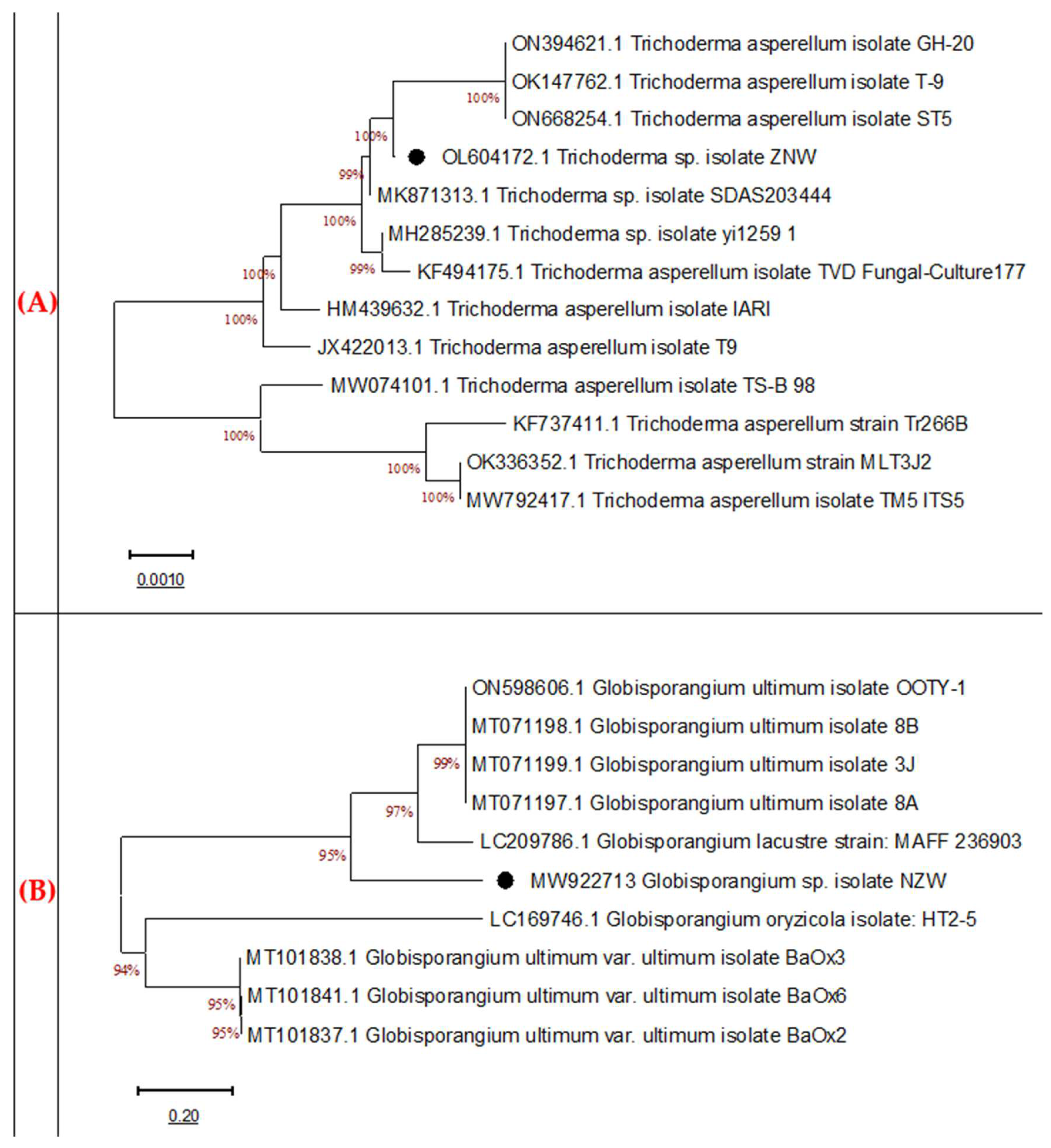
The fungal isolates were identified molecularly, after PCR amplification, through ITS sequencing, which codes for the 18S rRNA gene. The gel electrophoresis showed that the amplified ITS fragment (600 bp) of both fungi was similar to the target sequence of the related fungal. The phylogeny was nonstructured (Figure 4A,B). The nucleotide sequences and the BLAST assessment show that the bioagent strain (ZNW) exhibited a high similarity (99%) with the Trichoderma asperellum on the GenBank (Figure 5A). The fungus was identified as Trichoderma asperellum, and the GenBank accession number is OL604172. The pathogenic fungal isolate NZW showed similarity with more than 95% with the closely related Globisporangium ultimum on the GenBank (Figure 4B). The strain NZW was identified and classified as Globisporangium ultimum NZW, with accession number MW922713.

3.5. Profile of Lytic Features

The prototype of the hydrolytic enzymes of the bioagent fungus was assayed (Figure 5) as a possible biocontrol method. The fungus produced all tested enzymes, including xylanase, cellulase, pectinase, protease, and chitinase. Trichoderma asperellum ZNW in general demonstrated a high amount of all tested enzymes at various levels.

3.6. GC-MS Assessment

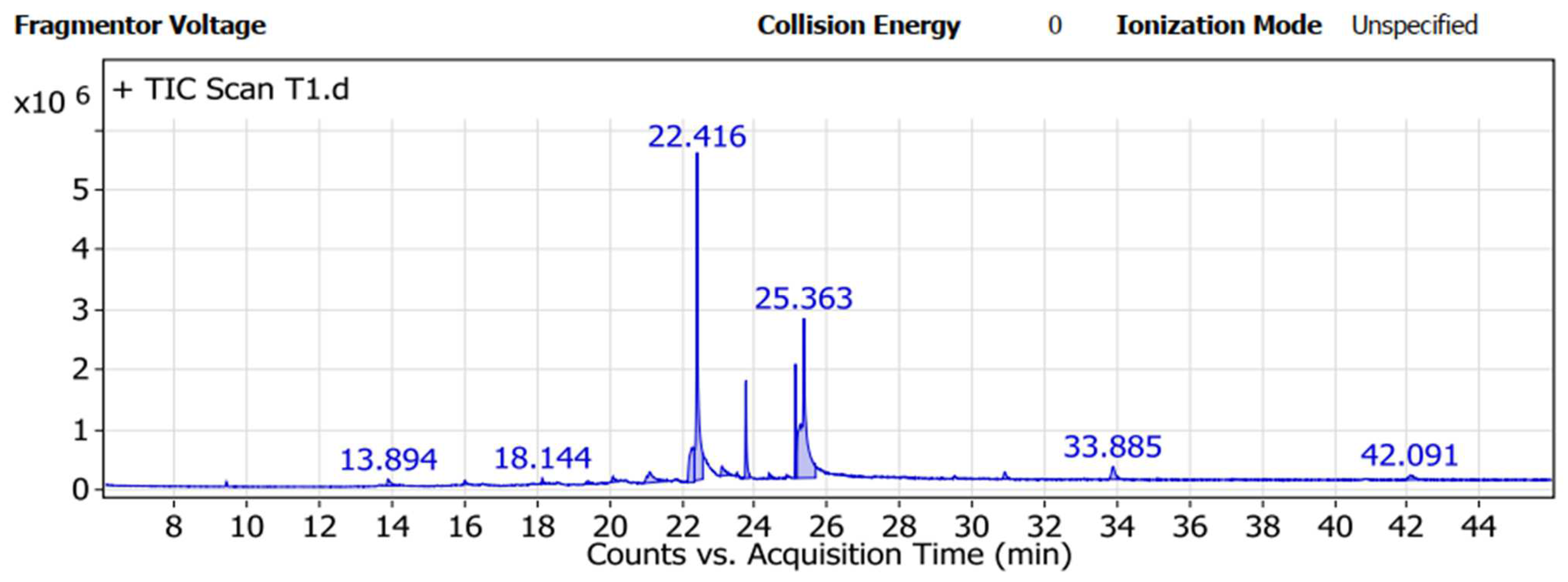
The T. asperellum ZNW metabolite was investigated. Results of the GC-MS analysis (Table 1 and Figure 6) indicated that the main components of T. asperellum ZNW filtrate were 9-octadecenoic acid (Z) (peak area-36.32%), Oleamide, TMS derivative (peak area-29.89%),1,2-15,16-diepoxyhexadecane (peak area-8.13%), 1-Monopalmitin, 2TMS derivative (peak area-5.74%), and 9-hexadecenoic acid (palmitic acid) (peak area-3.96%). Whereas, other components were present in low percentages; acetic acid, (1,2,3,4,5,6,7,8-octahydro-3,8,8-trimethylnaphth-2-yl), methyl ester (peak area-2.5%), 9,12-octadecadienoyl chloride (1.81%), 1,4-Bis(trimethylsilyl)benzene (peak area-1.36%), and butylated hydroxytoluene (peak area-1.16%). On the other hand, there are many components present in traces (peak area less than 1%).

3.7. Greenhouse Experiment

3.7.1. Disease Development

The bioagent was further evaluated under greenhouse conditions as a possible biocontrol for damping-off disease. Infection by G. ultimum NZW (infested soil only) recorded the highest values of rotted seeds and infected seedlings, being 46.67% and 18.33%, respectively, and the lowest number of plants that survived, 35% (Table 2). On the other hand, there were no significant differences among the other treatments, where priming pea seeds by fungicide or T. asperellum ZNW increased the percentage of survival and decreased the percentage of rotted seeds and infected seedlings in the presence or absence of G. ultimum NZW. Generally, the highest survival percentages were recorded in both fungicide (80%) and T. asperellum ZNW (80%) treatments, followed by T. asperellum + G. ultimum (78.33%). Whereas the survival percentage of the control and fungicide + G. ultimum treatments was 70% each. Finally, T. asperellum ZNW alone led to an increase in plant survival by 10 compared to non-infected control, and by 45% under the soil infestation with G. ultimum, in addition to the decrease in the rotted seeds and infected seedlings.

3.7.2. Vegetative Growth Parameters

All treatments by T. asperellum ZNW or fungicide had higher vegetative growth parameters than those non-treated ones. Trichoderma asperellum ZNW positively improved plant growth (root, shoot, and plant lengths, as well as the fresh and dry weight of root, the fresh and dry weight of the plant, and leaf area) (Table 3). Moreover, under infection, T. asperellum ZNW improved all previous vegetative characteristics as compared with pathogen-only treatment. The highest recorded value of root length was 10 cm, recorded by T. asperellum ZNW + G. ultimum NZW treatment, while the lowest root length (5.33 cm) was observed with the treatment of infested soil by G. ultimum NZW. Also, the plants treated with fungicide improved (significantly or insignificantly) the root and plant weight (fresh and dry) and leaf area more than the untreated ones.

3.7.3. Ultrastructural Changes of Pea Root

The cross-section magnified by SEM showed normal control plants (Figure 7A), and also showed the ability of T. asperellum hyphae to domiciliate and grow as an endophytic fungus in pea roots (Figure 7B). Moreover, T. asperellum ZNW led to thickening in the cell wall of vessels (xylem). The cross-section of pea plants that were planted in the infested soil by G. ultimum NZW showed an accumulation of G. ultimum NZW spores in plant vessels, leading to damping-off (Figure 7C). Moreover, the cross-section of pea root treated by T. asperellum ZNW and planted in soil infested by G. ultimum NZW showed the growth of T. asperellum ZNW and thickening in the cell wall of vessels (the lower part of the photo) that prevents G. ultimum NZW spores (that accumulate in the upper part of the photo) to invade the region of root in which T. asperellum ZNW present (Figure 7D).

3.7.4. Defense Response

The total phenols, peroxidase, and polyphenol oxidase of different treatments, that were planted in non-infested and infested soil with G. ultimum NZW, are illustrated in Table 4. Generally, the plants treated with T. asperellum ZNW and fungicide showed enhancement in the accumulation of total phenols and the obvious activity of both defense-related enzymes. Under infection, T. asperellum ZNW also significantly enhanced the total phenol content and the activity of defense-related enzymes as compared with G. ultimum NZW treatment. The highest peroxidase activity was observed with the treatment of T. asperellum ZNW + G. ultimum NZW (14 U). Also, no significant variation was observed in the value of peroxidase activity of the two treatments, the fungicide, and the control. G. ultimum NZW treatment has the lowest value of peroxidase activity (4.7 U), and polyphenol oxidase activity (4 U). Other treatments had a significant increase in polyphenol oxidase activity where there was no significant variation between T. asperellum ZNW + G. ultimum NZW, T. asperellum ZNW, fungicide, and the control, being 11.3, 11.3, 10, and 8 U, respectively. Moreover, plants treated with G. ultimum NZW had the lowest value of total phenols (43 mg), whereas those treated with T. asperellum ZNW + G. ultimum NZW had the highest value of total phenols (71.0 mg).

3.7.5. Photosynthetic Pigmentation

Chlorophylls (a, b, and total), and carotenoids were determined as a sign of metabolic changes (Table 5). Under infested soil, G. ultimum NZW showed a variable significant number of photosynthetic pigments in pea leaves when compared with other treatments. Under infection stress, T. asperellum ZNW showed a significant variation in total chlorophyll compared to the other treatments. The treatment of T. asperellum ZNW + G. ultimum NZW had the highest significant values of chlorophyll a (2.71 mg g−1 fresh weight), chlorophyll b (0.987 mg g−1 fresh weight), and total chlorophylls (3.694 mg g−1 fresh weight). Whereas no significant alterations were noticed among values of carotenoids of different treatments.

3.7.6. Yield Parameters

Finally, the recorded yield parameter of pea plants treated by T. asperellum ZNW or fungicide were significantly increased than the corresponding values of untreated plants (Table 6). The treatment by T. asperellum ZNW planted in infested soil (T. asperellum ZNW + G. ultimum NZW) had the highest values of yield parameters (70.67 pods, 81.07 g fresh weight of pods, 14.28 g pods dry weight, and 8.63 g weight of 100 seeds). On the contrary, G. ultimum NZW had the lowest values of yield parameters (35 pod of 28.65 and 5.05 g of fresh and dry weights, respectively, and 6.23 g of 100 seeds).

3.8. Biological Activity of the Rhizosphere Soil

3.8.1. Dehydrogenase Activity

The treatment of fungicide and T. asperellum + G. ultimum led to enhancements of dehydrogenase activity in the rhizosphere soil (0.283 and 0.404 µg TPF g−1 dry soil min−1, respectively) when compared with the other treatments (Table 7). The treatment of infested soil only had the lowest recorded value (0.211 µg TPF g−1 dry soil min−1).

3.8.2. Population of Soil Microflora

The total bacterial and fungal counts in the rhizosphere soil after 60 days of planting were more than the corresponding values after 90 days of planting (Table 8). After 60 days of planting, the highest recorded total bacterial count was observed with the infested soil by G. ultimum NZW (log 6.99), while the value of the total bacterial count of the treatments of T. asperellum ZNW and fungicide was lower than the corresponding value in the infested or non-infested soil.
After 90 days of planting, the highest total bacterial count was observed with the control treatment (log 6.29), whereas the lowest value was reported with infested soil with G. ultimum NZW (log 5.64). However, the total fungal count in T. asperellum and fungicide treatments in the non-infested soil was lower than that of the control treatment.
The highest count of soil fungal count, after 60 days, was recorded with the treatment by T. asperellum ZNW (log 4.99) while the lowest value was reported with the treatment T. asperellum ZNW + G. ultimum NZW (log 4.46). The fungal count of plants treated with T. asperellum ZNW or fungicide was higher than the corresponding value of the control and T. asperellum + G. ultimum treatments. After 90 days, the control and fungicide + G. ultimum treatments, followed by T. asperellum ZNW, recorded the highest soil fungal counts.

4. Discussion

Owing to their biodiversity in various ecosystems, beneficial fungi must be considered when evolving plant protection strategies. Unfortunately, public awareness, mostly, neglected fungal biodiversity and its critical role in this respect. Therefore, there is a great need for investigating the diverse fungal communities for new fungal strains, having new roles, instead of the already available ones.
This study was carried out to draw attention to the possibility of domiciliating a new beneficial microbiome into the plant tissue. In this connection, a pioneer isolate of T. asperellum ZNW, an endophytic fungus isolated from pea seeds, was domiciliated into the pea plant, representing a case study for the settlement of the bioagent fungus into plant tissue.
Trichoderma spp. is a diverse group of wonderful bioagents widely used in sustainable agriculture owing to their ability to enhance plant growth and productivity, and further alleviate abiotic and biotic stresses [37]. Trichoderma spp. are used in various remedies (foliar application, seed treatment, and/or soil remedy) for the management of diseases caused by bacteria, fungi, and nematodes [38].
Cultural, morphological, and microscopic (light, and SEM) identification of fungal isolate ZNW declares that the current isolate ZNW resembles the morphology of T. asperellum RMCK01 [39]. The morphology of T. asperellum ZNW growing on a PDA plate did not have the same morphology as observed in previous investigations, suggesting a new variant of T. asperellum. The conidia of T. asperellum ZNW are slightly globose in shape and the conidiophore is branched, which resembles T. asperellum Ta13 [40]. The morphological characters of T. asperellum ZNW have followed the classification of T. asperellum [18]. This data supports the claim that myco-diversity needs more studies to explore the undiscovered area of seed-borne fungi.
Identification of the damping-off phytopathogen, G. ultimum NZW, declared that its mycelia resemble those of Pythium ultimum var. ultimum associated with soybean damping-off [41]. SEM micrographs of G. ultimum NZW illustrated that its hyphae had a swelling end with a slightly globose shape, which was confirmed by other studies on G. ultimum strains [19,42,43]. All these works confirm the classification of the current fungus.
The pathogenicity of the current strain was confirmed by the pathogenicity test [44]. However, Globisporangium ultimum (Trow) (syn. G. ultimum Trow, syn. G. ultimum Trow var. ultimum) is an oomycete species that causes damping-off and/or root rot on a vast range of plants throughout the world [42,43,45].
The previous identification of both fungi was further confirmed. Molecular identification is frequently used due to its high specificity and sensitivity for the quick identification of numerous microorganisms [46]. The sequences of the tested fungi were related to similar strains in the GenBank. Both the bioagent and pathogenic fungi were identified as T. asperellum ZNW (OL604172) and G. ultimum NZW (MW922713), respectively, which came in line with the morphological identification features.
Molecular identification is highly specific and sensitive and is frequently used for fast identification. The nucleotide sequencing of the ITS region is similar in broad fungal classes, thus helping in revealing the interspecific and intraspecific differences among microorganisms. This region has non-functional sequences and is very variable among species, therefore, it can be accepted for the identification of very wide groups of fungal species. Technically, the multi-copy or repeated feature of the rDNA facilitates the amplification of the ITS regions from a tiny part of DNA, thus, providing fast and accurate identification compared to the other markers [47,48,49].
SEM investigation of the dual culture showed the ability of T. asperellum ZNW to mycoparasite and coil around G. ultimum NZW, causing damage, fractionation, and breakdown of G. ultimum NZW hyphae. Mycoparasitism is an effective method through which Trichoderma spp. physically contact and coil their hyphae around the hyphae of the pathogenic fungi, then the bioagent obtains nutrients from the dead host biomass after hydrolysis by the secretion of lytic enzymes and other metabolites [50,51].
However, there is no antagonistic activity of T. asperellum ZNW volatile organic compounds against G. ultimum NZW. This result means that T. asperellum ZNW may not produce volatile compounds or produce volatile compounds that did not inhibit G. ultimum NZW, the latter assumption may be the more accepted one. Conversely to our results, T. asperellum produced volatile compounds that had antifungal activity against other plant pathogens (Curvularia aeria and Corynespora cassiicola), enhanced the defense, and improved the growth of lettuce [52]. Moreover, T. asperellum T76-14 produced volatile organic compounds that inhibited Fusarium incarnatum, which causes fruit rot of muskmelons [53].
The biochemical feature of the bioagent was assessed as a possible role in the biological control process. Lytic activity of T. asperellum ZNW was searched on pea straw. This pea straw is hard to disintegrate due to its cellulose, hemicellulose, protein, and pectin content. Generally, such structures hinder fungal invasion into plant tissues. These enzymes are known as cell wall-degrading enzymes and play a role in the biocontrol process against the phytopathogen. Another suggested role is the facilitation of hyphal penetration to plant cell-wall tissue during the domiciliation process.
There was an obvious lytic activity of xylanase and cellulase that hydrolyzes hemicellulose (xylan) and cellulose, leading to the release of xylose and glucose units, respectively [47]. Whereas pectinase and proteases degrade pectin polymer and protein into galacturonic acid [47] and amino acids [28], respectively. The breakdown of plant tissues by these enzymes eases microbial entrance into plants [54], thus facilitating the domiciliation process.
Chitinase analyzes 1–4 β-glycoside linkage of N-acetyl-D-glucosamine in the fungal chitin; thus, acting as a bio-fungicide by antagonizing the other phytopathogens on host plants [55]. The growth medium was free from the chitin substrate, demonstrating that chitinase is produced constitutively by the Trichoderma asperellum ZNW.
Furthermore, Trichoderma spp. can biosynthesize many metabolites, which have antimicrobial activity against various phytopathogens, induce disease resistance, and enhance plant growth [56]. Besides many components, 9-octadecenoic acid (peak area-36.32%) was the main secondary metabolite detected in the filtrate of T. asperellum ZNW by GC-MS analysis. This component is presented in different Trichoderma strains in other investigations at lower concentrations in T. asperellum (peak area-1.57%) [57] and T. hamatum (peak area-1.77%) that inhibited Candida albicans [58]. The current compound, 9-octadecenoic acid, was reported to be belonging to the unsaturated fatty acids that have antimicrobial activity [59] and plays an obvious task in the plant’s defense against biotic and abiotic stresses [60]. Another fatty acid (9-hexadecenoic acid or palmitic acid) was present in the filtrate (peak area-3.96%) of this investigation, which was previously detected in the filtrate of T. hamatum [58] with antimicrobial activity [61]. Moreover, palmitic acid is found in the filtrate of T. asperellum and is also used for disease management and as a plant nutrient [62]. The possible role of palmitic acid is the ability to alter the rhizosphere microbial structure, control fungal disease, and encourage plant growth [63].
Oleamide, a TMS derivative (peak area-29.89%) present in the filtrate of T. asperellum ZNW, was previously reported as an amide derivative of the fatty acid oleic acid [64]. Fatty acids and their derivatives have vital roles in plant defense against bacterial and fungal pathogens [65]. Also, the detected 1,2-15,16-diepoxyhexadecane (peak area-8.13%) in this study may have antioxidant, anti-cancer, and anti-inflammatory activities [66]. Moreover, 1-Monopalmitin, 2TMS derivative (peak area-5.74%) detected in the filtrate of T. asperellum ZNW is a secondary metabolite in most active actinomycetes, having anticancer and antimicrobial activities [67]. In general, the antimicrobial effect of T. asperellum ZNW may be due to the synergism among the diverse chemicals, especially the main components, however, the synergistic effects between the main and the minor components exceed the expected individual outcome of each component alone [54,68].
Under greenhouse conditions, pea seeds were treated by the bioagent through biopriming with T. asperellum ZNW. Beneficial microbial inoculants are a vital method for improving the germination of seeds and/or enhancing growth, as well as the resistance of plants against several abiotic and biotic stresses. Moreover, seed biopriming permits the microorganisms, such as Trichoderma spp., to adhere to and enter the tissues [69,70,71].
Seed biopriming in T. asperellum ZNW increased the survival percentage and decreased percentages of rotted seeds and infected seedlings in the absence or presence of G. ultimum. The promotion of growth may be due to the action of the metabolites and the ability of fungal inoculation to reduce the disease (root rot and damping-off) and also to enhance the survival percentage of the plant [72]. The use of T. asperellum for biopriming of chili seeds reduced disease severity caused by Fusarium solani and enhanced seedling health [70].
T. asperellum ZNW enhanced the vegetative growth of pea plants that were planted in infested or non-infested soil due to its ability to promote plant growth, antagonize G. ultimum NZW, and its ability to induce the defense-related enzymes, and improve the concentration of photosynthetic pigment. These improvements resemble those reported on the growth parameter of tomato due to T. asperellum, which is used as a bioagent against damping-off disease [73].
Ultrastructural alterations were noticed by SEM in the cross sections of the root of the pea plant. T. asperellum ZNW could domiciliate and grow as an endophytic fungus in pea roots and lead to the thickened cell wall of vessels (xylem). Moreover, T. asperellum ZNW prevented G. ultimum NZW from invading the region of the root, in which T. asperellum ZNW is present. Furthermore, no traces of the pathogen could be observed in the region of T. asperellum ZNW. According to the authors’ information, this is the first trial that indicates the ability of domiciliation of Trichoderma spp. into plant tissue so that it can grow as an endophytic fungus. The growth of T. asperellum ZNW as an endophytic fungus gave a high opportunity for plant protection from phytopathogens and allow the plant to get several profits from T. asperellum ZNW metabolites. T. asperellum ZNW led to an increase in thickness of the xylem of pea roots, this may be due to the lignification, i.e., lignin accumulation in the cell wall of the root that is accompanied by low methyl-esterified pectin in intercellular spaces, which increases in cell wall thickness improved wheat protection against pathogens [74].
Additionally, the enzymatic system of T. asperellum ZNW enables partial maceration of plant tissue that allows the smooth penetration, and domiciliation of T. asperellum ZNW into plant tissue without causing any pathogenic symptoms. Generally, Trichoderma spp. do not have any virulence factor and, consequently, no disease development occurs.
T. asperellum ZNW enhanced the induced resistance and increased the production of total phenols and improved the activities of peroxidase and polyphenol oxidase in pea planted. This, in turn, leads to an increase in defense response against viral, bacterial, and fungal pathogens [7,8,9]. Phenolic compounds have significant roles in plant health through increasing colonization of beneficial microorganisms and inhibition of insect attack and/or pathogen infection [75]. Phenolic compounds have antioxidant and antimicrobial properties and protect the plant tissues from poisonous effects, resulting from reactive oxygen species [76]. Under adverse conditions, phenolics accumulate in the plant tissues and play a vital role in the regulation of different environmental stresses, such as nutrient deficiency, low temperatures, and high light [77]. Peroxidase and polyphenol oxidase are defense-related enzymes that enhance the defense against phytopathogens infections and improve plant resistance against biotic and abiotic stress [78].
T. asperellum ZNW enhanced chlorophylls and carotenoid content in pea plants. T. asperellum could enhance the pea plant’s health and enhance the activity of the photosynthesis process [79]. The improvement of chlorophyll concentration refers to the enhancement of the plant’s defense against abiotic and biotic stresses [80]. T. asperellum enhanced chlorophylls and carotenoids content when used as biocontrol of plant pathogens [81]. Furthermore, T. asperellum enhanced the concentration of photosynthetic pigments and helped to overcome drought stress [82].
The changes in the biological features in the rhizosphere soil of pea were explored. The activity of the soil dehydrogenase was enhanced due to the application of T. asperellum. Trichoderma spp. was reported to improve the activity of dehydrogenase and positively amend the soil properties [83]. Dehydrogenase is one of the most important soil enzymes, which is an indicator of soil quality due to different stress, or management practices. Enzyme activity in the soil can be changed as a result of the activities of the soil’s microbes [84]. Since dehydrogenases are present intracellularly in the living microbial cells and do not accumulate out of the microbial cells, it is considered an indicator of the microbial activity in the soil; furthermore, dehydrogenases play a vital role in the biological oxidation of the organic matter in the soil by transferring hydrogen from organic substrates to inorganic acceptors [85]. The enhancement of dehydrogenase is a good sign of the improvement of microbial population and physicochemical soil properties [86,87].
T. asperellum ZNW led to an alteration in the bacterial and fungal communities in the rhizosphere, where both microbial counts decreased greater after 90 days than after 60 days. This effect of T. asperellum ZNW on the rhizosphere microbial count may be directly and/or indirectly related to plant physiology, and root exudates in soil. This effect may be due to the competition of Trichoderma with other microbes, leading to an alteration in the population of the soil microbiome. Also, the activity of soil enzymes may have a vital role in changing the microbial community [88]. The relative abundance of Trichoderma spp. in the soil causes increases in the availability of soil nutrients, and changes the rhizosphere chemical structure, thus altering soil microbial communities [10]. T. asperellum causes an increase in the total count of beneficial microbes and a decrease in the total count of phytopathogens in the plants’ rhizosphere that help control plant diseases and promote growth [89]. Trichoderma spp. can boost the physiology of plants and can modulate root exudates that cause a change in nature and kind of the plant-microbe interaction [90]. Root exudates are secretions produced by roots that are considered a basis of nutrition for beneficial soil microbial communities [91]. Root exudates enhance soil respiration and positively change the configuration of the microbial community, which effectively enhances the growth of plants [92]. The root exudates decrease near the end of plant age leading to decreasing in the microbial count of rhizosphere microbial communities [93].
The biopriming of pea seeds in T. asperellum ZNW led to an increase in the productivity of pea plants and yield parameters. This is an expected outcome due to the enhancement of plant growth and physiology, as well as the management of phytopathogen, leading to an enhancement in induced resistance and also an enhancement of the rhizosphere microbial community. T. asperellum could promote different plants and control diseases, leading to an increase in yield, as occurs when controlling late-wilt disease, promoting growth, and increasing the yield of maize [94]. Furthermore, T. asperellum could reduce Pythium root rot, promote growth, and improve the productivity of lettuce [95].

5. Conclusions

Summing up, this contemporary report offers background knowledge on the possibility of the domiciliation of a pioneer isolate of T. asperellum ZNW into pea plant tissue as a new habitation for the bioagent fungus. The biochemical features of T. asperellum ZNW were evaluated in vitro before being evaluated under in vivo conditions, and SEM examination. However, this investigation is the first record of the pathogenicity of G. ultimum, as a damping-off causative agent in pea. T. asperellum ZNW could inhibit G. ultimum NZW growth through mycoparasitism and producing hydrolytic enzymes (xylanase, cellulase, pectinase, protease, and chitinase) and different bioactive metabolites. T. asperellum ZNW can domiciliate the root of the pea and reduce rotted seeds and infected seedlings, and also improve survival. Additionally, the study confirms the positive effect of the domiciliated T. asperellum ZNW, under infection, on plant health. T. asperellum ZNW improved the disease resistance, photosynthetic pigmentation, vegetative growth, and yield of pea. This investigation is the first attempt to apply the simple seed biopriming technique to domiciliate T. asperellum ZNW in pea tissue as an endophytic fungus. Nevertheless, our results may enable a deep understanding of plant-fungal interaction. So, it would be worthwhile to perform further evaluation, and more studies are encouraged, to extend the application of the current data on other beneficial microorganisms on other economic plants.

Author Contributions

Z.M., N.M.E., W.I.A.S. and D.B.E.D. Data curation; Z.M., A.M.K., M.M.I., W.I.A.S. and D.B.E.D. Formal analysis; Z.M., Y.F.A., W.I.A.S. and D.B.E.D. Methodology; Z.M., N.M.E., W.I.A.S. and D.B.E.D. Resources; Z.M, N.M.E., M.M.I., W.I.A.S. and D.B.E.D. All authors contributed to validation, visualization, writing the original draft, and revision. All authors have read and agreed to the published version of the manuscript.

Funding

This research received no external funding.

Institutional Review Board Statement

Not applicable.

Informed Consent Statement

Not applicable.

Data Availability Statement

All data generated and/or analyzed during this study are included in this published article.

Conflicts of Interest

The authors declare no conflict of interest.

References

  1. Hesham, A.E.-L.; Kaur, T.; Devi, R.; Kour, D.; Prasad, S.; Yadav, N.; Singh, C.; Singh, J.; Yadav, A.N. Current Trends in Microbial Biotechnology for Agricultural Sustainability: Conclusion and Future Challenges. In Current Trends in Microbial Biotechnology for Sustainable Agriculture; Yadav, A.N., Singh, J., Singh, C., Yadav, N., Eds.; Springer: Singapore, 2021; pp. 555–572. [Google Scholar]
  2. Jahagirdar, S.; Kambrekar, D.N.; Navi, S.S.; Kunta, M. Plant Growth-Promoting Fungi: Diversity and Classification. In Bioactive Molecules in Plant Defense: Signaling in Growth and Stress; Springer: Cham, Switzerland, 2019. [Google Scholar]
  3. Sharma, S.; Kour, D.; Rana, K.L.; Dhiman, A.; Thakur, S.; Thakur, P.; Thakur, S.; Thakur, N.; Sudheer, S.; Yadav, N.; et al. Trichoderma: Biodiversity, Ecological Significances, and Industrial Applications. In Recent Advancement in White Biotechnology Through Fungi; Springer: Cham, Switzerland, 2019. [Google Scholar]
  4. Khan, M.R.; Parveen, G.; Zaid, A.; Wani, S.H.; Jogaiah, S. Potential of Trichoderma Species in Alleviating the Adverse Effects of Biotic and Abiotic Stresses in Plants. In Biocontrol Agents and Secondary Metabolites; Woodhead Publishing: Sawston, UK, 2021; pp. 85–112. [Google Scholar] [CrossRef]
  5. Sharma, A.; Salwan, R.; Sharma, V. Extracellular Proteins of Trichoderma and Their Role in Plant Health. S. Afr. J. Bot. 2022, 147, 359–369. [Google Scholar] [CrossRef]
  6. Shahnaz, E.; Anwar, A.; Banday, S. Trichoderma Spp. as Bio-Stimulant: Molecular Insights. In New and Future Developments in Microbial Biotechnology and Bioengineering; Elsevier: Amsterdam, The Netherlands, 2022; pp. 337–350. [Google Scholar] [CrossRef]
  7. Mohamed, B.F.F.; Sallam, N.M.A.; Alamri, S.A.M.; Abo-Elyousr, K.A.M.; Mostafa, Y.S.; Hashem, M. Approving the Biocontrol Method of Potato Wilt Caused by Ralstonia Solanacearum (Smith) Using Enterobacter Cloacae PS14 and Trichoderma Asperellum T34. Egypt. J. Biol. Pest Control 2020, 30, 61. [Google Scholar] [CrossRef]
  8. Rahimi Tamandegani, P.; Sharifnabi, B.; Massah, A.; Zahravi, M. Induced Reprogramming of Oxidative Stress Responses in Cucumber by Trichoderma Asperellum (Iran 3062C) Enhances Defense against Cucumber Mosaic Virus. Biol. Control 2021, 164, 104779. [Google Scholar] [CrossRef]
  9. Baiyee, B.; Ito, S.I.; Sunpapao, A. Trichoderma Asperellum T1 Mediated Antifungal Activity and Induced Defense Response against Leaf Spot Fungi in Lettuce (Lactuca sativa L.). Physiol. Mol. Plant Pathol. 2019, 106, 96–101. [Google Scholar] [CrossRef]
  10. Zhang, F.; Xu, X.; Huo, Y.; Xiao, Y. Trichoderma-Inoculation and Mowing Synergistically Altered Soil Available Nutrients, Rhizosphere Chemical Compounds and Soil Microbial Community, Potentially Driving Alfalfa Growth. Front. Microbiol. 2019, 9, 3241. [Google Scholar] [CrossRef]
  11. Guindon, M.F.; Cazzola, F.; Palacios, T.; Gatti, I.; Bermejo, C.; Cointry, E. Biofortification of Pea (Pisum sativum L.): A Review. J. Sci. Food Agric. 2021, 101, 3551–3563. [Google Scholar] [CrossRef]
  12. FAOSTAT Statistics Division. Available online: https://www.fao.org/faostat/en/#data/QC/visualize (accessed on 21 March 2022).
  13. Lamichhane, J.R.; Dürr, C.; Schwanck, A.A.; Robin, M.H.; Sarthou, J.P.; Cellier, V.; Messéan, A.; Aubertot, J.N. Integrated Management of Damping-off Diseases. A Review. Agron. Sustain. Dev. 2017, 37, 10. [Google Scholar] [CrossRef]
  14. ISTA. International Rules for Seed Testing 2018. In International Rules for Seed Testing; ISTA: Bassersdorf, Switzerland, 2018. [Google Scholar]
  15. Dhingra, O.D.; Sinclair, J.B. Basic Plant Pathology Methods; CRC Press: Boca Raton, FL, USA, 2017. [Google Scholar]
  16. Fotoohiyan, Z.; Rezaee, S.; Bonjar, G.H.S.; Mohammadi, A.H.; Moradi, M. Biocontrol Potential of Trichoderma Harzianum in Controlling Wilt Disease of Pistachio Caused by Verticillium dahliae. J. Plant Prot. Res. 2017, 57, 185–193. [Google Scholar] [CrossRef]
  17. El-Benawy, N.M.; Abdel-Fattah, G.M.; Ghoneem, K.M.; Shabana, Y.M. Antimicrobial Activities of Trichoderma Atroviride against Common Bean Seed-Borne Macrophomina phaseolina and Rhizoctonia solani. Egypt. J. Basic Appl. Sci. 2020, 7, 267–280. [Google Scholar] [CrossRef]
  18. Siddiquee, S. Morphology-Based Characterization of Trichoderma Species. In Practical Handbook of the Biology and Molecular Diversity of Trichoderma Species from Tropical Regions; Fungal Biology; Springer: Cham. Switzerland, 2017; pp. 41–73. [Google Scholar]
  19. Ho, H.H. The Taxonomy and Biology of Phytophthora and Pythium. J. Bacteriol. Mycol. Open Access 2018, 6, 40–45. [Google Scholar] [CrossRef] [Green Version]
  20. Kearse, M.; Moir, R.; Wilson, A.; Stones-Havas, S.; Cheung, M.; Sturrock, S.; Buxton, S.; Cooper, A.; Markowitz, S.; Duran, C.; et al. Geneious Basic: An Integrated and Extendable Desktop Software Platform for the Organization and Analysis of Sequence Data. Bioinformatics 2012, 28, 1647–1649. [Google Scholar] [CrossRef] [Green Version]
  21. Liu, L.; Anderson, C.; Pearl, D.; Edwards, S.V. Modern Phylogenomics: Building Phylogenetic Trees Using the Multispecies Coalescent Model. In Methods in Molecular Biology; Humana: New York, NY, USA, 2019; Volume 1910. [Google Scholar]
  22. Nilsson, R.H.; Larsson, K.H.; Taylor, A.F.S.; Bengtsson-Palme, J.; Jeppesen, T.S.; Schigel, D.; Kennedy, P.; Picard, K.; Glöckner, F.O.; Tedersoo, L.; et al. The UNITE Database for Molecular Identification of Fungi: Handling Dark Taxa and Parallel Taxonomic Classifications. Nucleic Acids Res. 2019, 47, D259–D264. [Google Scholar] [CrossRef]
  23. Tamura, K.; Stecher, G.; Kumar, S. MEGA11: Molecular Evolutionary Genetics Analysis Version 11. Mol. Biol. Evol. 2021, 38, 3022–3027. [Google Scholar] [CrossRef]
  24. Mangan, D.; Cornaggia, C.; Liadova, A.; Draga, A.; Ivory, R.; Evans, D.E.; McCleary, B.V. Development of an Automatable Method for the Measurement of Endo-1,4-β-Xylanase Activity in Barley Malt and Initial Investigation into the Relationship between Endo-1,4-β-Xylanase Activity and Wort Viscosity. J. Cereal Sci. 2018, 84, 90–94. [Google Scholar] [CrossRef]
  25. Elsayed, M.S.; Eldadamony, N.M.; Alrdahe, S.S.T.; Saber, W.I.A. Definitive Screening Design and Artificial Neural Network for Modeling a Rapid Biodegradation of Date Palm Fronds by a New Trichoderma Sp. Pwn6 into Citric Acid. Molecules 2021, 26, 5048. [Google Scholar] [CrossRef]
  26. Ajayi, A.A.; Lawal, B.; Salubi, A.E.; Onibokun, A.E.; Oniha, M.I.; Ajayi, O.M. Pectinase Production by Aspergillus Niger Using Pineapple Peel Pectin and Its Application in Coconut Oil Extraction. IOP Conf. Ser. Earth Environ. Sci. 2021, 655, 012014. [Google Scholar] [CrossRef]
  27. Jain, A.; Jain, R.; Jain, S. Quantitative Analysis of Reducing Sugars by 3, 5-Dinitrosalicylic Acid (DNSA Method). In Basic Techniques in Biochemistry, Microbiology and Molecular Biology; Humana: New York, NY, USA, 2020. [Google Scholar]
  28. Al-Askar, A.A.; Ghoneem, K.M.; Hafez, E.E.; Saber, W.I.A. A Case Study in Saudi Arabia: Biodiversity of Maize Seed-Borne Pathogenic Fungi in Relation to Biochemical, Physiological, and Molecular Characteristics. Plants 2022, 11, 829. [Google Scholar] [CrossRef]
  29. Preety; Sharma, S.; Hooda, V. Purification and Analytical Application of Vigna Mungo Chitinase for Determination of Total Fungal Load of Stored Cereals. Appl. Biochem. Biotechnol. 2018, 186, 12–26. [Google Scholar] [CrossRef]
  30. Caldwell, D.; Iyer-Pascuzzi, A.S. A Scanning Electron Microscopy Technique for Viewing Plant–Microbe Interactions at Tissue and Cell-Type Resolution. Phytopathology 2019, 109, 1302–1311. [Google Scholar] [CrossRef]
  31. Aryal, S.; Baniya, M.K.; Danekhu, K.; Kunwar, P.; Gurung, R.; Koirala, N. Total Phenolic Content, Flavonoid Content and Antioxidant Potential of Wild Vegetables from Western Nepal. Plants 2019, 8, 96. [Google Scholar] [CrossRef] [Green Version]
  32. Seleim, M.A.; Abo-Elyousr, K.A.; Mohamed, A.-A.A.; Al-Marzoky, H.A. Peroxidase and Polyphenoloxidase Activities as Biochemical Markers for Biocontrol Efficacy in the Control of Tomato Bacterial Wilt. J. Plant Physiol. Pathol. 2014, 2, 1. [Google Scholar] [CrossRef]
  33. Othman, R.; Amin, N.A.; Sani, M.S.A.; Fadzillah, N.A.; Jamaludin, M.A. Carotenoid and Chlorophyll Profiles in Five Species of Malaysian Seaweed as Potential Halal Active Pharmaceutical Ingredient (API). Int. J. Adv. Sci. Eng. Inf. Technol. 2018, 8, 1610–1616. [Google Scholar] [CrossRef]
  34. Thrane, J.E.; Kyle, M.; Striebel, M.; Haande, S.; Grung, M.; Rohrlack, T.; Andersen, T. Spectrophotometric Analysis of Pigments: A Critical Assessment of a High-Throughput Method for Analysis of Algal Pigment Mixtures by Spectral Deconvolution. PLoS ONE 2015, 10, e0137645. [Google Scholar] [CrossRef] [PubMed] [Green Version]
  35. Mierzwa-Hersztek, M.; Wolny-Koładka, K.; Gondek, K.; Gałązka, A.; Gawryjołek, K. Effect of Coapplication of Biochar and Nutrients on Microbiocenotic Composition, Dehydrogenase Activity Index and Chemical Properties of Sandy Soil. Waste Biomass Valorization 2020, 11, 3911–3923. [Google Scholar] [CrossRef] [Green Version]
  36. Larkin, R.P. Impacts of Biocontrol Products on Rhizoctonia Disease of Potato and Soil Microbial Communities, and Their Persistence in Soil. Crop Prot. 2016, 90, 96–105. [Google Scholar] [CrossRef]
  37. Abdullah, N.S.; Doni, F.; Mispan, M.S.; Saiman, M.Z.; Yusuf, Y.M.; Oke, M.A.; Mohd Suhaimi, N.S. Harnessing Trichoderma in Agriculture for Productivity and Sustainability. Agronomy 2021, 11, 2559. [Google Scholar] [CrossRef]
  38. Pandey, R.N.; Jaisani, P.; Singh, H.B. Trichoderma: Agricultural Applications and Beyond. In Biopesticides; Woodhead Publishing: Sawston, UK, 2022; pp. 353–381. [Google Scholar] [CrossRef]
  39. Sebumpan, R.; Guiritan, K.R.; Suan, M.; Abapo, C.J.; Bhat, A.H.; Machado, R.A.R.; Nimkingrat, P.; Sumaya, N.H. Morphological and Molecular Identification of Trichoderma Asperellum Isolated from a Dragon Fruit Farm in the Southern Philippines and Its Pathogenicity against the Larvae of the Super Worm, Zophobas morio (Fabricius, 1776) (Coleoptera: Tenebrionidae). Egypt. J. Biol. Pest Control 2022, 32, 47 . [Google Scholar] [CrossRef]
  40. Pandian, R.T.; Raja, M.; Kumar, A.; Sharma, P. Morphological and Molecular Characterization of Trichoderma Asperellum Strain Ta13. Indian Phytopathol. 2016, 69, 297–303. [Google Scholar]
  41. Navi, S.S.; Huynh, T.; Mayers, C.G.; Yang, X.-B. Diversity of Pythium Spp. Associated with Soybean Damping-off, and Management Implications by Using Foliar Fungicides as Seed Treatments. Phytopathol. Res. 2019, 1, 8. [Google Scholar] [CrossRef]
  42. Manganiello, G.; Pane, C. First Report of Globisporangium Ultimum Causing Damping-off of Wild Rocket Seedlings. J. Plant Pathol. 2021, 103, 721. [Google Scholar] [CrossRef]
  43. Erper, I.; Ozer, G.; Zholdoshbekova, S.; Turkkan, M. First Report of Garden Cucumber Root Rot Caused by Globisporangium Ultimum Var. Ultimum in Kyrgyzstan. J. Plant Pathol. 2020, 102, 1363–1364. [Google Scholar] [CrossRef]
  44. Molin, C.; Ribeiro, N.R.; Matsumoto, M.N.; Fernando Giasson, N.; Brollo, J.; Zanardo, B.; Pelissoni, M.; Capitanio, S.; Comín, T.; Deuner, C.C.; et al. Damping-off of Soybean in Southern Brazil Can Be Associated with Different Species of Globisporangium Spp. and Pythium Spp. Plant Pathol. 2021, 70, 1686–1694. [Google Scholar] [CrossRef]
  45. Lazreg, F.; Belabid, L.; Sanchez, J.; Gallego, E.; Garrido-Cardenas, J.A.; Elhaitoum, A. First Report of Globisporangium Ultimum Causing Pythium Damping-off on Aleppo Pine in Algeria, Africa, and the Mediterranean Region. Plant Dis. 2013, 97, 1111. [Google Scholar] [CrossRef]
  46. Moussa, Z.; Darwish, D.B.; Alrdahe, S.S.; Saber, W.E.I.A. Innovative Artificial-Intelligence-Based Approach for the Biodegradation of Feather Keratin by Bacillus Paramycoides, and Cytotoxicity of the Resulting Amino Acids. Front. Microbiol. 2021, 12, 731262. [Google Scholar] [CrossRef]
  47. Al-Askar, A.A.; Rashad, E.M.; Ghoneem, K.M.; Mostafa, A.A.; Al-Otibi, F.O.; Saber, W.E.I.A. Discovering Penicillium Polinicum with High-Lytic Capacity on Helianthus Tuberosus Tubers: Oil-Based Preservation for Mold Management. Plants 2021, 10, 413. [Google Scholar] [CrossRef]
  48. Raja, H.A.; Miller, A.N.; Pearce, C.J.; Oberlies, N.H. Fungal Identification Using Molecular Tools: A Primer for the Natural Products Research Community. J. Nat. Prod. 2017, 80, 756–770. [Google Scholar] [CrossRef]
  49. Schoch, C.L.; Seifert, K.A.; Huhndorf, S.; Robert, V.; Spouge, J.L.; Levesque, C.A.; Chen, W.; Bolchacova, E.; Voigt, K.; Crous, P.W.; et al. Nuclear Ribosomal Internal Transcribed Spacer (ITS) Region as a Universal DNA Barcode Marker for Fungi. Proc. Natl. Acad. Sci. USA 2012, 109, 6241–6246. [Google Scholar] [CrossRef] [Green Version]
  50. Mukherjee, P.K.; Mendoza-Mendoza, A.; Zeilinger, S.; Horwitz, B.A. Mycoparasitism as a Mechanism of Trichoderma-Mediated Suppression of Plant Diseases. Fungal Biol. Rev. 2022, 39, 15–33. [Google Scholar] [CrossRef]
  51. Kifle, M.H.; Yobo, K.S.; Laing, M.D. Biocontrol of Aspergillus Flavus in Groundnut Using Trichoderma Harzianum Stain Kd. J. Plant Dis. Prot. 2017, 124, 51–56. [Google Scholar] [CrossRef]
  52. Wonglom, P.; Ito, S.I.; Sunpapao, A. Volatile Organic Compounds Emitted from Endophytic Fungus Trichoderma Asperellum T1 Mediate Antifungal Activity, Defense Response and Promote Plant Growth in Lettuce (Lactuca sativa). Fungal Ecol. 2020, 43, 100867. [Google Scholar] [CrossRef]
  53. Intana, W.; Kheawleng, S.; Sunpapao, A. Trichoderma Asperellum T76-14 Released Volatile Organic Compounds against Postharvest Fruit Rot in Muskmelons (Cucumis melo) Caused by Fusarium Incarnatum. J. Fungi 2021, 7, 46. [Google Scholar] [CrossRef] [PubMed]
  54. Moussa, Z.; Rashad, E.M.; Elsherbiny, E.A.; Al-Askar, A.A.; Arishi, A.A.; Al-Otibi, F.O.; Saber, W.I.A. New Strategy for Inducing Resistance against Bacterial Wilt Disease Using an Avirulent Strain of Ralstonia Solanacearum. Microorganisms 2022, 10, 1814. [Google Scholar] [CrossRef] [PubMed]
  55. Rashad, E.M.; Shaheen, D.M.; Al-Askar, A.A.; Ghoneem, K.M.; Arishi, A.A.; Hassan, E.S.A.; Saber, W.I.A. Seed Endophytic Achromobacter Sp. F23KW as a Promising Growth Promoter and Biocontrol of Rhizoctonia Root Rot of Fenugreek. Molecules 2022, 27, 5546. [Google Scholar] [CrossRef] [PubMed]
  56. Vinale, F.; Sivasithamparam, K. Beneficial Effects of Trichoderma Secondary Metabolites on Crops. Phytother. Res. 2020, 34, 2835–2842. [Google Scholar] [CrossRef] [PubMed]
  57. Bhardwaj, N.R.; Kumar, J. Characterization of Volatile Secondary Metabolites from Trichoderma Asperellum. J. Appl. Nat. Sci. 2017, 9, 954–959. [Google Scholar] [CrossRef] [Green Version]
  58. Khairillah, Y.N.; Sukarno, N.; Batubara, I. Trichoderma Hamatum Derived from Coffee Plant (Coffea canephora) Rhizosphere Inhibit Candida Albicans Growth. Biosaintifika J. Biol. Biol. Educ. 2021, 13, 369–378. [Google Scholar] [CrossRef]
  59. Ghavam, M.; Afzali, A.; Manca, M.L. Chemotype of Damask Rose with Oleic Acid (9 Octadecenoic Acid) and Its Antimicrobial Effectiveness. Sci. Rep. 2021, 11, 8027. [Google Scholar] [CrossRef]
  60. He, M.; Ding, N.Z. Plant Unsaturated Fatty Acids: Multiple Roles in Stress Response. Front. Plant Sci. 2020, 11, 562785. [Google Scholar] [CrossRef]
  61. Bravo-Santano, N.; Ellis, J.K.; Calle, Y.; Keun, H.C.; Behrends, V.; Letek, M. Intracellular Staphylococcus Aureus Elicits the Production of Host Very Long-Chain Saturated Fatty Acids with Antimicrobial Activity. Metabolites 2019, 9, 148. [Google Scholar] [CrossRef] [Green Version]
  62. Singh, G.; Tiwari, A.; Gupta, A.; Kumar, A.; Hariprasad, P.; Sharma, S. Bioformulation Development via Valorizing Silica-Rich Spent Mushroom Substrate with Trichoderma Asperellum for Plant Nutrient and Disease Management. J. Environ. Manag. 2021, 297, 113278. [Google Scholar] [CrossRef]
  63. Ma, K.; Kou, J.; Rahman, M.K.U.; Du, W.; Liang, X.; Wu, F.; Li, W.; Pan, K. Palmitic Acid Mediated Change of Rhizosphere and Alleviation of Fusarium Wilt Disease in Watermelon. Saudi J. Biol. Sci. 2021, 28, 3616–3623. [Google Scholar] [CrossRef]
  64. Taherkhani, Z.; Shojaei, O. The Kinetic Study of Oleamide Synthesis: Effect of Oleic Acid Molar Ratio, Temperature and Catalyst Concentration International Journal of New Chemistry. Int. J. New Chem. 2021, 8, 400–412. [Google Scholar]
  65. Lee, H.J.; Park, O.K. Lipases Associated with Plant Defense against Pathogens. Plant Sci. 2019, 279, 51–58. [Google Scholar] [CrossRef]
  66. Shah, N.A.; Khan, M.R.; Nadhman, A. Antileishmanial, Toxicity, and Phytochemical Evaluation of Medicinal Plants Collected from Pakistan. Biomed. Res. Int. 2014, 2014, 384204. [Google Scholar] [CrossRef] [Green Version]
  67. Elsayed, T.R.; Galil, D.F.; Sedik, M.Z.; Hassan, H.M.M.; Sadik, M.W. Antimicrobial and Anticancer Activities of Actinomycetes Isolated from Egyptian Soils. Int. J. Curr. Microbiol. Appl. Sci. 2020, 9, 2020. [Google Scholar] [CrossRef]
  68. Elbanhawy, A.A.; Elsherbiny, E.A.; Abd El-Mageed, A.E.; Abdel-Fattah, G.M. Potential of Fungal Metabolites as a Biocontrol Agent against Cotton Aphid, Aphis Gossypii Glover and the Possible Mechanisms of Action. Pestic. Biochem. Physiol. 2019, 159, 34–40. [Google Scholar] [CrossRef]
  69. Rawat, L.; Bisht, T.S.; Kukreti, A. Potential of Seed Biopriming with Trichoderma in Ameliorating Salinity Stress and Providing Resistance against Leaf Blast Disease in Finger Millet (Eleusine coracana L.). Indian Phytopathol. 2022, 75, 147–164. [Google Scholar] [CrossRef]
  70. Chin, J.M.; Lim, Y.Y.; Ting, A.S.Y. Biopriming Chilli Seeds with Trichoderma Asperellum: A Study on Biopolymer Compatibility with Seed and Biocontrol Agent for Disease Suppression. Biol. Control 2022, 165, 104819. [Google Scholar] [CrossRef]
  71. Singh, V.; Upadhyay, R.S.; Sarma, B.K.; Singh, H.B. Seed Bio-Priming with Trichoderma Asperellum Effectively Modulate Plant Growth Promotion in Pea. Int. J. Agric. Environ. Biotechnol. 2016, 9, 361–365. [Google Scholar] [CrossRef]
  72. Elshahawy, I.E.; El-Mohamedy, R.S. Biological Control of Pythium Damping-off and Root-Rot Diseases of Tomato Using Trichoderma Isolates Employed Alone or in Combination. J. Plant Pathol. 2019, 101, 597–608. [Google Scholar] [CrossRef]
  73. Kumhar, K.C.; Kumar, K.; Arora, I.; Bhatia, A.K.; Batra, V.K.; Raj, H. Management of Tomato Damping-off Disease in the Nursery Using of Trichoderma Asperellum. Int. J. Econ. Plants 2022, 9, 145–151. [Google Scholar] [CrossRef]
  74. Basińska-Barczak, A.; Błaszczyk, L.; Szentner, K. Plant Cell Wall Changes in Common Wheat Roots as a Result of Their Interaction with Beneficial Fungi of Trichoderma. Cells 2020, 9, 2319. [Google Scholar] [CrossRef] [PubMed]
  75. Wallis, C.M.; Galarneau, E.R.A. Phenolic Compound Induction in Plant-Microbe and Plant-Insect Interactions: A Meta-Analysis. Front. Plant Sci. 2020, 11, 580753. [Google Scholar] [CrossRef] [PubMed]
  76. Kumar, S.; Abedin, M.M.; Singh, A.K.; Das, S. Role of Phenolic Compounds in Plant-Defensive Mechanisms. In Plant Phenolics in Sustainable Agriculture; Springer: Singapore, 2020. [Google Scholar]
  77. Naikoo, M.I.; Dar, M.I.; Raghib, F.; Jaleel, H.; Ahmad, B.; Raina, A.; Khan, F.A.; Naushin, F. Role and Regulation of Plants Phenolics in Abiotic Stress Tolerance: An Overview. In Plant Signaling Molecules: Role and Regulation under Stressful Environments; Elsevier: Amsterdam, The Netherlands, 2019. [Google Scholar]
  78. Zhang, J.; Sun, X. Recent Advances in Polyphenol Oxidase-Mediated Plant Stress Responses. Phytochemistry 2021, 181, 112588. [Google Scholar] [CrossRef] [PubMed]
  79. Pavlovic, D.; Nikolic, B.; Djurovic, S.; Waisi, H.; Andjelkovic, A.; Marisavljevic, D. Chlorophyll as a Measure of Plant Health: Agroecological Aspects. Pestic. Fitomedicina 2014, 29. [Google Scholar] [CrossRef]
  80. Agathokleous, E.; Feng, Z.Z.; Peñuelas, J. Chlorophyll Hormesis: Are Chlorophylls Major Components of Stress Biology in Higher Plants? Sci. Total Environ. 2020, 726, 138637. [Google Scholar] [CrossRef]
  81. Zapata-Sarmiento, D.H.; Palacios-Pala, E.F.; Rodríguez-Hernández, A.A.; Medina Melchor, D.L.; Rodríguez-Monroy, M.; Sepúlveda-Jiménez, G. Trichoderma Asperellum, a Potential Biological Control Agent of Stemphylium Vesicarium, on Onion (Allium cepa L.). Biol. Control 2020, 140, 104105. [Google Scholar] [CrossRef]
  82. Scudeletti, D.; Crusciol, C.A.C.; Bossolani, J.W.; Moretti, L.G.; Momesso, L.; Servaz Tubaña, B.; de Castro, S.G.Q.; de Oliveira, E.F.; Hungria, M. Trichoderma Asperellum Inoculation as a Tool for Attenuating Drought Stress in Sugarcane. Front. Plant Sci. 2021, 12, 645542. [Google Scholar] [CrossRef]
  83. Zydlik, Z.; Zydlik, P.; Wieczorek, R. The Effects of Bioinoculants Based on Mycorrhizal and Trichoderma Spp. Fungi in an Apple Tree Nursery under Replantation Conditions. Agronomy 2021, 11, 2355. [Google Scholar] [CrossRef]
  84. Kumar, S.; Chaudhuri, S.; Maiti, S.K. Soil Dehydrogenase Enzyme Activity in Natural and Mine Soil—A Review. Middle East J. Sci. Res. 2013, 13, 898–906. [Google Scholar]
  85. Wolinska, A.; Stepniewsk, Z. Dehydrogenase Activity in the Soil Environment. Dehydrogenases 2012, 10, 183–210. [Google Scholar]
  86. Ujjainiya, P.; Choudhary, M.; Jatav, H.S.; Tokala, V.Y.; Rajput, V.D.; Minkina, T. Impact of Weed Management Practices on Soil Microflora and Dehydrogenase Enzyme Activity Under Varying Levels of Nitrogen in Winter Season Onion (Allium cepa L.). Bull. Environ. Contam. Toxicol. 2022, 108, 430–436. [Google Scholar] [CrossRef]
  87. Phares, C.A.; Amoakwah, E.; Danquah, A.; Akaba, S.; Frimpong, K.A.; Mensah, T.A. Improved Soil Physicochemical, Biological Properties and Net Income Following the Application of Inorganic NPK Fertilizer and Biochar for Maize Production. Acta Ecol. Sin. 2022, 42, 289–295. [Google Scholar] [CrossRef]
  88. Jangir, M.; Sharma, S.; Sharma, S. Non-Target Effects of Trichoderma on Plants and Soil Microbial Communities. In Plant Microbe Interface; Springer: Cham, Switzerland, 2019. [Google Scholar]
  89. Zhang, Y.; Tian, C.; Xiao, J.; Wei, L.; Tian, Y.; Liang, Z. Soil Inoculation of Trichoderma Asperellum M45a Regulates Rhizosphere Microbes and Triggers Watermelon Resistance to Fusarium Wilt. AMB Express 2020, 10, 189. [Google Scholar] [CrossRef]
  90. Lucini, L.; Colla, G.; Miras Moreno, M.B.; Bernardo, L.; Cardarelli, M.; Terzi, V.; Bonini, P.; Rouphael, Y. Inoculation of Rhizoglomus Irregulare or Trichoderma Atroviride Differentially Modulates Metabolite Profiling of Wheat Root Exudates. Phytochemistry 2019, 157, 158–167. [Google Scholar] [CrossRef]
  91. Nazir, N.; Kamili, A.N.; Zargar, M.Y.; Khan, I.; Shah, D.; Parray, J.A.; Tyub, S. Effect of Root Exudates on Rhizosphere Soil Microbial Communities. J. Res. Dev. 2016, 16, 88–95. [Google Scholar]
  92. Adamczyk, M.; Rüthi, J.; Frey, B. Root Exudates Increase Soil Respiration and Alter Microbial Community Structure in Alpine Permafrost and Active Layer Soils. Environ. Microbiol. 2021, 23, 2152–2168. [Google Scholar] [CrossRef]
  93. Moussa, Z.; El-Hersh, M.S.; El-Khateeb, A.Y. Induction of Potato Resistance against Bacterial Wilt Disease Using Saccharomyces Cerevisiae. Biotechnology 2017, 16, 57–68. [Google Scholar] [CrossRef] [Green Version]
  94. Degani, O.; Rabinovitz, O.; Becher, P.; Gordani, A.; Chen, A. Trichoderma Longibrachiatum and Trichoderma Asperellum Confer Growth Promotion and Protection against Late Wilt Disease in the Field. J. Fungi 2021, 7, 444. [Google Scholar] [CrossRef]
  95. Chewapanich, W.; Charoenrak, P.; Intanoo, W.; Chamswarng, C. Efficacy of Trichodermaasperellum CB-Pin-01 and Potassiumdihydrogen Phosphate to Enhance Growth and Yield and Reduce Pythium Root Rot of Hydroponically Grown Lettuce. Agric. Nat. Resour. 2021, 55, 601–610. [Google Scholar] [CrossRef]
Figure 1. Trichoderma spp. antagonizes (overgrows) the cause of damping-off fungus of pea on PDA plates; (A): the two fungi growing on the same PDA plate, showing the overgrowth; (B,C): overgrowth view of Trichoderma spp. on the fungal pathogen at 10×, and 100×, respectively. Where T-arrow: Trichoderma spp., P-arrow: fungal pathogen, and O-arrow: overgrowth region.
Microorganisms 11 00198 g001
Figure 2. Scanning electron microscopy of the two investigated fungi; (A): Trichoderma asperellum ZNW, (B): Globisporangium ultimum NZW.
Microorganisms 11 00198 g002
Figure 3. Mycoparasitism of Trichoderma asperellum ZNW on Globisporangium ultimum NZW (after their first contact in a dual culture) under the scanning electron microscope. (A): overgrowth of T. asperellum ZNW hyphae (T-arrow) on G. ultimum NZW hyphae (G-arrow), (B): coiling of T. asperellum ZNW hyphae around G. ultimum NZW hyphae, (C): continuous coiling, (D): the breakdown (br arrow) of G. ultimum NZW hyphae, and (E): the breakdown and collapse of G. ultimum NZW hyphae.
Microorganisms 11 00198 g003
Figure 4. Molecular phylogenetic tree of the ITS for Trichoderma asperellum ZNW (A) and Globisporangium ultimum NZW (B) relation to the sequences obtained from GenBank.
Microorganisms 11 00198 g004
Figure 5. The hydrolytic enzymes’ activity of the bioagent T. asperellum ZNW (n = 3).
Microorganisms 11 00198 g005
Figure 6. GC-MS of filtrate of Trichoderma asperellum ZNW.
Microorganisms 11 00198 g006
Figure 7. Micrographs of the SEM cross-section of pea root after 30 days of planting showing; (A): normal control plants, (B): plants treated with T. asperellum ZNW only, (C): plants grown in infested soil by G. ultimum NZW, and (D): plants treated with T. asperellum ZNW under infested soil by G. ultimum NZW. T-arrow: T. asperellum hyphae, TCW-arrow: thickening in cell wall vessels (TCW), G-arrow; G. ultimum NZW spores.
Microorganisms 11 00198 g007
Table 1. Metabolites present in Trichoderma asperellum ZNW filtrate as indicated by GC-MS analysis.
PeakRTNameFormulaAreaArea Sum %
113.894Butylated Hydroxytoluene C15H24O727,422.651.16
216.011Decane, 2,3,5,8-tetramethyl- C14H30305,789.060.49
318.144Tetradecane, 2,6,10-trimethyl-C17H36260,402.830.41
419.417Dodecanoic acid, 3-hydroxy-C12H24O3380,111.620.6
520.0967-Hexadecenal, (Z)-C16H30O472,1790.75
621.1139-Hexadecenoic acidC16H30O22,494,012.63.96
722.2731,2-15,16-DiepoxyhexadecaneC16H30O25,117,326.68.13
822.416Oleamide, TMS derivativeC21H43NOSi18,817,52529.89
923.1029,12-Octadecadienoyl chloride, (Z,Z)-C18H31ClO1,142,391.41.81
1023.7651-Monopalmitin, 2TMS derivativeC25H54O4Si23,615,270.15.74
1124.39812-Methyl-E,E-2,13-octadecadien-1-olC19H36O403,516.610.64
1225.129Glycerol monostearate, 2TMS derivativeC27H58O4Si23,318,7085.27
1325.3639-Octadecenoic acid (Z)-C18H34O222,870,74136.32
1430.9011,4-Benzenediol, 2,6-bis(1,1-dimethylethyl)-C14H22O2613,2360.97
1533.885Acetic acid, (1,2,3,4,5,6,7,8-octahydro-3,8,8-trimethylnaphth-2-yl)methyl esterC16H26O21,571,281.12.5
1642.0911,4-Bis(trimethylsilyl)benzene C12H22Si2856,462.941.36
Table 2. Disease assessment of pea damping-off under inoculation with Trichoderma asperellum ZNW and/or G. ultimum NZW.
TreatmentRotted Seeds, %Infected Seedling, %Survival, %
Fungicide10.00 ± 2.00 b10.00 ± 2.00 a80.00 ± 5.00 a
Control 13.33 ± 2.89 b16.67 ± 1.15 a70.00 ± 4.01 ab
T. asperellum10.00 ± 2.00 b10.00 ± 2.22 a80.00 ± 2.03 a
Fungicide + G. ultimum 20.00 ± 2.22 b10.00 ± 2.22 a70.00 ± 2.58 ab
G. ultimum46.67 ± 2.89 a18.33 ± 3.51 a35.00 ± 2.43 b
T. asperellum + G. ultimum 11.67 ± 2.89 b10.00 ± 1.20 a78.33 ± 2.89 a
In each column, numbers that share the same letter(s) are not significantly different (p ≤ 0.05, n = 5).
Table 3. Vegetative growth parameters of pea under the action of bioagent and the pathogen.
TreatmentRoot Length (cm)Shoot Length (cm)Plant Height (cm)Root Fresh Weight (g)Root Dry Weight (g)Plant Fresh Weight (g)Plant Dry Weight (g)Leaf Area (cm2)
Fungicide8.00 ± 1.00 ab24.00 ± 2.11 a32.00 ± 1.73 a0.280 ± 0.026 ab0.047 ± 0.006 ab7.24 ± 0.74 a1.307 ± 0.064 a99.4 ± 6.3 b
Control 7.67 ± 0.58 b25.33 ± 1.15 a33.00 ± 1.00 a0.287 ± 0.025 a0.083 ± 0.015 a5.57 ± 0.23 a1.037 ± 0.093 ab130. 8 ± 5.5 a
T. asperellum8.00 ± 1.00 ab22.67 ± 1.15 a30.67 ± 1.55 a0.223 ± 0.031 ab0.070 ± 0.010 a5.35 ± 0.64 a0.970 ± 0.161 ab107.3 ± 6.8 ab
Fungicide + G. ultimum 7.67 ± 1.15 b21.67 ± 0.58 a29.33 ± 1.53 a0.183 ± 0.035 ab0.087 ± 0.011 a5.52 ± 0.65 a0.953 ± 0.076 ab96.6 ± 4.3 b
G. ultimum5.33 ± 0.58 c8.67 ± 1.53 b14.00 ± 2.75 b0.080 ± 0.010 b0.020 ± 0.010 b1.59 ± 0.13 a0.230 ± 0.036 b31.4 ± 3.8 c
T. asperellum + G. ultimum 10.00 ± 1.00 a22.67 ± 1.55 a32.67 ± 1.55 a0.177 ± 0.015 ab0.060 ± 0.010 ab5.71 ± 0.51 a1.213 ± 0.042 a104.7 ± 4.8 ab
In each column, numbers that share the same letter(s) are not significantly different (p ≤ 0.05, n = 5).
Table 4. Defense-related enzymes and total phenols of pea plants under inoculation with T. asperellum ZNW and/or G. ultimum NZW.
TreatmentPeroxidase (U)Polyphenol Oxidase (U)Total Phenol
(mg g−1 Fresh Weight)
Fungicide7.3 ± 1.2 bcd10.0 ± 2.2 ab69.8 ± 2.1 ab
Control 6.0 ± 1.3 cd8.0 ± 2.2 ab66.9 ± 1.8 ab
T. asperellum10.0 ± 1.8 abc11.3 ± 1.2 a49.9 ± 1.2 c
Fungicide + G. ultimum 12.0 ± 2.0 ab7.3 ± 1.2 bc62.2 ± 2.4 b
G. ultimum4.7 ± 1.2 d4.0 ± 0.3 c43.0 ± 1.6 c
T. asperellum + G. ultimum 14.0 ± 2.2 a11.3 ± 1.2 a71.0 ± 2.2 a
In each column, numbers that share the same letter(s) are not significantly different (p ≤ 0.05, n = 3).
Table 5. Chlorophylls and carotenoids content (mg g−1 fresh weight) of pea plants under inoculation with T. asperellum ZNW and/or G. ultimum NZW.
TreatmentChlorophyll aChlorophyll bTotal ChlorophyllsCarotenoids
Fungicide1.642 ± 0.036 d0.517 ± 0.033 cd2.159 ± 0.069 e0.520 ± 0.045 a
Control 2.010 ± 0.033 c0.590 ± 0.019 bc2.600 ± 0.107 cd0.457 ± 0.056 a
T. asperellum2.073 ± 0.014 c0.368 ± 0.041 d2.351 ± 0.179 de0.440 ± 0.021 a
Fungicide + G. ultimum 2.049 ± 0.011 c0.764 ± 0.027 b2.813 ± 0.017 c0.300 ± 0.012 a
G. ultimum2.476 ± 0.015 b0.683 ± 0.021 bc3.159 ± 0.040 b0.469 ± 0.009 a
T. asperellum + G. ultimum 2.707 ± 0.066 a0.987 ± 0.052 a3.694 ± 0.059 a0.411 ± 0.025 a
In each column, numbers that share the same letter(s) are not significantly different (p ≤ 0.05, n = 3).
Table 6. Evaluation of T. asperellum ZNW on the yield of pea under infection with G. ultimum.
TreatmentPods NumberPods Fresh Weight (g)Pods Dry Weight (g)Weight of 100 Seeds (g)
Fungicide61.00 ± 2.25 b61.43 ± 2.16 c10.34 ± 0.81 b5.38 ± 0.60 d
Control 27.00 ± 1.13 f58.61 ± 1.52 d6.12 ± 0.65 c6.15 ± 0.75 c
T. asperellum52.33 ± 1.53 c78.27 ± 2.20 b9.36 ± 0.84 b6.23 ± 0.65 c
Fungicide + G. ultimum 44.67± 1.25 d36.30 ± 1.36 e13.23 ± 0.55 b7.77 ± 0.35 b
G. ultimum35.00 ± 1.65 e28.65 ± 1.25 f5.05 ± 0.50 c6.23 ± 0.45 c
T. asperellum + G. ultimum 70.67 ± 2.55 a81.07± 2.15 a14.28 ± 0.76 a8.63 ± 0.85 a
In each column, numbers that share the same letter(s) are not significantly different (p ≤ 0.05, n = 5).
Table 7. The activity of dehydrogenase enzyme estimated in the rhizosphere soil of pea plants.
TreatmentDehydrogenase Activity (µg TPF g−1 Dry Soil min−1)
Fungicide0.283 ± 0.004 b
Control 0.213 ± 0.002 d
T. asperellum0.242 ± 0.003 c
Fungicide + G. ultimum 0.246 ± 0.004 c
G. ultimum0.211 ± 0.002 d
T. asperellum + G. ultimum 0.404 ± 0.003 a
In each column, numbers that share the same letter(s) are not significantly different (p ≤ 0.05, n = 3).
Table 8. Total bacterial and fungal count in the rhizosphere soil of pea plants after 60 and 90 days of planting.
TreatmentMicrobial Count (log CFU g−1 Dry Soil)
BacteriaFungi
60 Days90 Days60 Days90 Days
Fungicide6.72 ± 0.01 c6.04 ± 0.04 b4.84 ± 0.06 a2.00 ± 0.10 c
Control 6.82 ± 0.01 b6.29 ± 0.05 a4.77± 0.07 ab2.77 ± 0.07 a
T. asperellum6.60 ± 0.02 d6.20 ± 0.05 a4.99 ± 0.04 a2.59 ± 0.11 ab
Fungicide + G. ultimum 6.40 ± 0.04 f6.01 ± 0.01 b4.56 ± 0.07 bc2.72 ± 0.05 ab
G. ultimum6.99 ± 0.04 a5.64 ± 0.04 c4.82 ± 0.03 a 2.48 ± 0.31 b
T. asperellum + G. ultimum 6.48 ± 0.01 e6.02 ± 0.06 b4.46 ± 0.02 c2.10 ± 0.17 c
In each column, numbers that share the same letter(s) are not significantly different (p ≤ 0.05, n = 3).
Disclaimer/Publisher’s Note: The statements, opinions and data contained in all publications are solely those of the individual author(s) and contributor(s) and not of MDPI and/or the editor(s). MDPI and/or the editor(s) disclaim responsibility for any injury to people or property resulting from any ideas, methods, instructions or products referred to in the content.