Article

Epiphytic and Endophytic Fungi Colonizing Seeds of Two Poaceae Weed Species and Fusarium spp. Seed Degradation Potential In Vitro

1
Institute of Plant Protection Research ‘Agrihorts’, Latvia University of Life Sciences and Technologies, Paula Lejiņa Iela 2, LV-3004 Jelgava, Latvia
2
Microbial Strain Collection of Latvia, Faculty of Biology, University of Latvia, Jelgavas Iela 1, LV-1004 Rīga, Latvia
3
Latvian Biomedical Research and Study Centre, Rātsupītes Iela 1, LV-1067 Rīga, Latvia
*
Author to whom correspondence should be addressed.
Microorganisms 2023, 11(1), 184; https://doi.org/10.3390/microorganisms11010184
Submission received: 28 November 2022 / Revised: 9 January 2023 / Accepted: 9 January 2023 / Published: 11 January 2023
(This article belongs to the Special Issue Microbiomes and Mycobiomes in Crop Health and Disease)

Abstract

:
Fungi colonizing the surface and endosphere of two widespread Poaceae weed species, Avena fatua and Echinochloa crus-galli, were isolated to compare the taxonomic composition between the plant species, location, and year of the seed collection. The seed-degrading potential of Fusarium isolated from the seeds was tested by inoculating seeds of E. crus-galli with spore suspension. Molecular identification of epiphytic and endophytic fungal genera was performed by sequencing the ITS region of rDNA. Endophytes comprised of significantly lower fungal richness compared to epiphytes. A significant taxonomic overlap was observed between the endosphere and seed surface. The most abundant genera were Alternaria, Fusarium, Cladosporium, and Sarocladium. Analysis of similarities and hierarchical clustering showed that microbial communities were more dissimilar between the two plant species than between the years. Fusarium isolates with a high potential to infect and degrade E. crus-galli seeds in laboratory conditions belong to F. sporotrichioides and F. culmorum.

1. Introduction

Arable weeds are one of the major sources of crop yield loss, reaching as high as 32% of actual yield loss and potentially amounting to 71% if no weed control is implemented [1]. Management of the inactive weed propagules that include soil seed banks for all the annual weed species is one of the tasks of integrated weed management strategies [2]. Current agricultural policy in the EU formulated in the ‘Farm to Fork strategy’ requires reducing the use of pesticides. Therefore, increasing attention is being paid to ecological interactions between crops, weeds, and other organisms in the agroecosystems that can facilitate pest control for sustainable crop production. Natural seed degradation aided by soil microbial communities offers a means for weed biocontrol. Seed microbiota are largely derived from soil microbial communities [3], yet partial vertical transmission from parental plants has also been reported [4], and microbiota can contain beneficial microorganisms that promote seed longevity and seedling survival [5], as well as pathogenic microorganisms leading to seed degradation [6].
Seeds that are quiescent or dormant (not germinating) have physical and chemical means of defense against pathogenic microorganisms [7,8]. A hard seed coat that constitutes the mechanical defense is correlated with a lower decay rate of seeds [6]. Phenolic compounds in the seed, seed coat, or specifically in the seed hull in a broad variety of species are an important part of seed chemical defenses [9]. The presence of these compounds may vary depending on the origin of the seeds as well as growth and seed maturation conditions. Moreover, if regarded as a species-specific feature, an association with certain bacteria that are antagonistic to pathogenic fungi or fungi that are antagonistic to other pathogens can be considered a defense mechanism [10]. Weed seeds differ in their ability to withstand microbial attack. Both inter- and intraspecific differences include seed-coat thickness [11], the chemical composition of the seed coat and other seed structures, and, probably, enzymatic defense mechanisms. The antifungal activity of a specific peptide isolated from E. crus-galli seeds was tested on several plant pathogens and had a pronounced effect on Fusarium graminearum, F. oxysporum, F. solani, and F. verticillioides (current name F. fujikuroi) [12], suggesting that seeds of E. crus-galli have limited susceptibility to these fungal pathogens. However, other Fusarium species were not tested. The presence of pathogen antagonists on the seed surface or in the endosphere is another potentially important factor for seed defense. It is, therefore, important to compare the resistance to microbial decay in seeds from either different parent plants or different populations.
The action of potentially seed-degrading microorganisms can be species-specific. For example, seeds of Pseudotsuga menziesii were shown to carry numerous Fusarium species that mostly caused no disease of either seeds or plants [13]. At the same time, F. oxysporum significantly reduced the germination of Pinus sylvestris and P. pinea, while F. verticillioides (F. fujikuroi) only reduced the germination of P. sylvestris [14]. The genus Fusarium includes species and strains that are pathogenic to seeds of different species of Poaceae. Soil-borne Fusarium species can cause significant seed mortality in Bromus tectorum [15] as well as Avena fatua [16]. The susceptibility of the plants or seeds to fungal attack can depend on the particular strain of the fungal species. This was demonstrated for soil-borne F. oxysporum isolates infecting E. crus-galli [17] and seed-borne F. oxysporum isolates with different potentials to infect Diplotaxis or Eruca plants [18]. Seed-borne fungi from the F. tricinctum species complex have also been shown to produce toxic metabolites that inhibit seedling growth in Bromus tectorum [19].
In addition to the soil-borne microorganisms, seeds and seedlings can be affected by the seed-borne pathogens from mature seeds. Seed-borne pathogens are a major source of disease in cultivated plants, including grains, and seeds can be vectors that transport pathogenic microorganisms between different locations [20].
Wild oat (Avena fatua L.) is a highly economically detrimental weed species distributed in cereal-growing areas worldwide. All parts of A. fatua seeds—awns, lemma, palea, and caryopsis—were reported to harbor yeasts and filamentous fungi belonging to 10 different genera [21]. The most frequent genera were Alternaria, Cladosporium, and Drechslera, while Fusarium was comparatively frequent on lemma and caryopses. Barnyard grass (Echinochloa crus-galli (L.) Beauv.) is also a widespread weed species that causes significant yield loss in different crops. To the best of our knowledge, the seed-borne fungi of E. crus-galli seeds have not been characterized, but in a closely related species, E. glabrescens, endophytes were described in different tissues, including seeds, of plants collected in different locations [22]. The rate of colonization of seeds was the second highest among different tissue types, after lower leaf blade.
Sustainable control of barnyard grass populations in arable lands is an important goal. Numerous cases of herbicide resistance have been reported for different varieties of this species [23]. An alternative to increased herbicide use is the reduction and management of the soil seed bank [24,25]. In this study, we chose E. crus-galli as a model organism to test the ability of fungi to degrade seeds under laboratory conditions because these plants produce large amounts of seeds, providing abundant material for experiments. Seeds are dormant at maturation, although dormancy can be partly released during dry storage [26].
This study aimed to compare the diversity and taxonomic composition of seed-borne fungi from mature seeds of A. fatua and E. crus-galli collected in different populations and to test the ability of epiphytic and endophytic seed-borne Fusarium spp. to infect and degrade seeds of E. crus-galli in laboratory conditions.

2. Materials and Methods

2.1. Study Sites and Seed Collection

Seeds were collected at maturity in three locations in Latvia in 2020 and the same locations in 2021. In 2020, A. fatua seeds were collected in one additional location (Table 1). Seeds from 750 A. fatua plants and 350 E. crus-galli plants were collected by shaking the seeds into sterile containers and storing them at 4–5 °C. Fungi were isolated within 24 h from seed collection. Additional samples of E. crus-galli seeds were dried over silica gel at ambient temperature for 14–21 days to reach moisture content below 15% and stored at −20 °C until further use in the inoculation experiment.

2.2. Isolation of Fungi

The isolation of seed epiphytes was carried out by agitating the seeds in sterile 10 mM MgSO4 (50 mL) and isolating epiphytic fungi from the acquired rinsing solution on 10% yeast malt agar medium (YMA; full medium: glucose, 10 g L−1; yeast extract, 3 g L−1; peptone, 5 g L−1; malt extract, 3 g L−1; all chemicals were produced by BioRad (Le Pond de Claix, France), except glucose (Duchefa, Haarlem, The Netherlands). Agar: 15 g L−1, Scharlau, Spain) supplemented with chloramphenicol (0.1 g L−1, Sigma-Aldrich, Shanghai, China) to prevent bacterial growth using dilution to extinction method (dilution range from 10−2 to 10−4) [27]. Afterward, the seeds were surface-sterilized by immersing them in 70% ethanol for 1 min, rinsing once with sterile deionized water, then immersing them in 10% commercial bleach (ACE, c.a. 5% Na hypochlorite) for 10 min, rinsing twice with sterile deionized water and rinsing with 70% ethanol and three times with sterile deionized water. Endophytic fungi were isolated from surface-sterilized seeds cut in halves and incubated on 10% YMA medium. A total of 10 A. fatua and 20 E. crus-galli seeds per population per collection year were used. Pure cultures were obtained from the colonies that developed on the 10% YMA medium, and mycelia were cultivated on 100% YMA medium until identification. One sample of each morphotype was used for Sanger-sequencing-based taxonomic identification.

2.3. Identification of Fungi

As one of the most universal approaches, ITS region sequencing was used as our primary means for taxonomic identification to at least the genus level. For that purpose, we performed DNA extraction by homogenizing ~10 µg of fungal material in lysis buffer (containing 2.5% PVP) with Lysing Matrix D (MP Biomedicals, Eschwege, Germany). The bead beating was carried out for 2 × 30s in FastPrep®-24 Classic bead beating grinder and lysis system instrument (MP Biomedicals, Eschwege, Germany). Sample-containing tubes were then incubated at 56 °C for 30 min and centrifuged at 5600 g for 20 min to precipitate cell debris. Further, microbial DNA was isolated from 400 µL of lysate with NucleoMag® 96 Plant kit (Macherey-Nagel) following the manufacturer’s instructions. The amplification of ITS region by PCR was carried out in total volume of 12 µL and using Phire Plant Direct PCR Master Mix (Thermo Fisher Scientific, Waltham, MA, USA), which was supplemented with 5 pmol of ITS5—Fw (5′-GGAAGTAAAAGTCGTAACAAGG-3′) and ITS4—Rs (5′-TCCTCCGCTTATTGATATGC-3′) primers [28] and 1 µL of fungal DNA. The created PCR mixture was then incubated on a VeritiTM 96-well thermal cycler (Applied Biosystems, Waltham, MA, USA) in following temperature regime: 5 min of initial denaturation at 98 °C, 40 product amplification cycles of 5 s at 98 °C, 5 s at 55 °C, and 20 s at 72 °C, and whole procedure was finalized by 1 min extension step at 72 °C. The excess of dNTPs and primers was then removed by treatment with Exonuclease I (0.5 µL (Thermo Fisher Scientific) and Shrimp Alkaline Phosphatase (2 µL) (Thermo Fisher Scientific) for 40 min at 37 °C, and the enzymes were inactivated through incubation at 95 °C for 20 min. Afterward, 1 µL of the purified samples was transferred to BigDye® Terminator v3.1 Cycle Sequencing reaction mixture (Applied Biosystems), which was supplemented with one of the previously used primers. Sequencing products were analyzed on 3130xl Genetic Analyzer (Applied Biosystems), which acquired sequences manually assembled and curated using SeqMan Pro from Lasergene 14 software package (DNASTAR Inc., Madison, WI, USA). BLAST analysis of acquired sequences against NCBI nucleotide database was then used for taxonomic identification of fungal isolate. Additionally, we used Protax-Fungi web-based tool [29] to search for matching ITS sequences in the Unite database.
Frequency of occurrence was calculated as the summary incidence in seed samples from each species population in each year divided by the total number of seed samples, seven in A. fatua and six in E. crus-galli.
Since, in the case of Fusarium, ITS analysis does not provide sufficient taxonomic resolution to perform identification at the species level, additional TEF-1α region sequencing analysis was performed for isolates classified as members of this genus. All procedures, in this case, were essentially identical to those of ITS analysis. The only exception was the employment of EF1T (5′-ATGGGTAAGGAGGACAAGAC-3′) and EF2T (5′-GGAAGT ACCAGTGATCATGTT-3′) primers [30] in both PCR and sequencing reactions. All described molecular-biology-related activities were carried out at ‘Genome center’–a genetic analysis core facility of Latvian Biomedical Research and Study center.

2.4. Artificial Inoculation of Seeds

Isolates identified as Fusarium spp. were cultivated on Potato Dextrose Agar medium (Biolife) at room temperature (22 ± 3 °C) until sporulation was observed. Five 90 mm Petri dishes were used for each isolate. When sporulation was attained, spores were harvested by pouring 10 mL of sterile water into each Petri dish, gently scraping the mycelium with a sterile microbiological loop, and collecting suspension from all five dishes in a 50 mL centrifuge tube. The suspension was centrifuged for 5 min at 500 g, and the supernatant was removed, leaving 5 mL of the suspension. Spores were counted using a hemocytometer to calculate the spore concentration. Subsequently, the suspension was diluted to the concentration of 250,000 spores mL−1. If the concentration of spores was too low, cultivation of the isolate was repeated.
To assess the potential of seed-borne Fusarium isolates to infect and degrade seeds from different populations, surface-sterilized seeds of each of the three populations collected in 2020 and 2021 (procedure described above, with the only difference being the usage of undiluted bleach) were inoculated with a fungal spore suspension by immersing the seeds in the spore suspension in sterile plastic tubes and vortexing for one min. After that, seeds were placed on 0.7% water agar gel (Duchefa) in sterile 60 mm Petri dishes and sealed with Parafilm. There were three Petri dishes per isolate, each containing 20 seeds. Non-inoculated seeds placed on water agar gel were used as a control. The inoculated seeds were incubated in the dark at 10 °C for 14 days to prevent rapid seed germination and then incubated for additional 28 days at room temperature (25 ± 3 °C). Seeds were scored as infected if Fusarium hyphae were observed on the seed surface. At the end of the incubation period, a cut test was performed to identify decayed seeds. Seeds were considered dead if the seed embryo was discolored or decayed. Seedlings were scored as infected if tufts of white, pink, or red hyphae were observed on the coleoptile or brown spots and stunted growth of the coleoptile were observed [31]. Otherwise, seedlings were scored as non-infected.

2.5. Statistical Analysis

Statistical analysis of the data was performed in R 4.1.1. Data from different populations of each species were used to create a presence/absence table for different fungal genera isolated from the seeds. Jaccard distance matrix was created using ‘vegdist()’ function from the ‘vegan’ package [32]. Taxonomic composition was compared between seeds of each of the two species collected in each year by hierarchical clustering based on Jaccard distances and creating a heatmap using function ‘pheatmap()’ in the ‘pheatmap’ package [33]. To compare the effect of species and seed-collection year, analysis of similarities was performed using the function ‘anosim()’ from the ‘vegan’ package using the same presence/absence matrix. Binomial logistic regression was used to analyze the ability of Fusarium isolates to infect E. crus-galli seeds: the effect of seed-collection year, population, and the fungal isolate on the number of intact seeds in the samples were tested. Multiple comparisons were performed using the ‘glht()’ function in the ‘multcomp’ package [34]. Venn diagrams were prepared using the ‘VennDiagram’ package [35].

3. Results

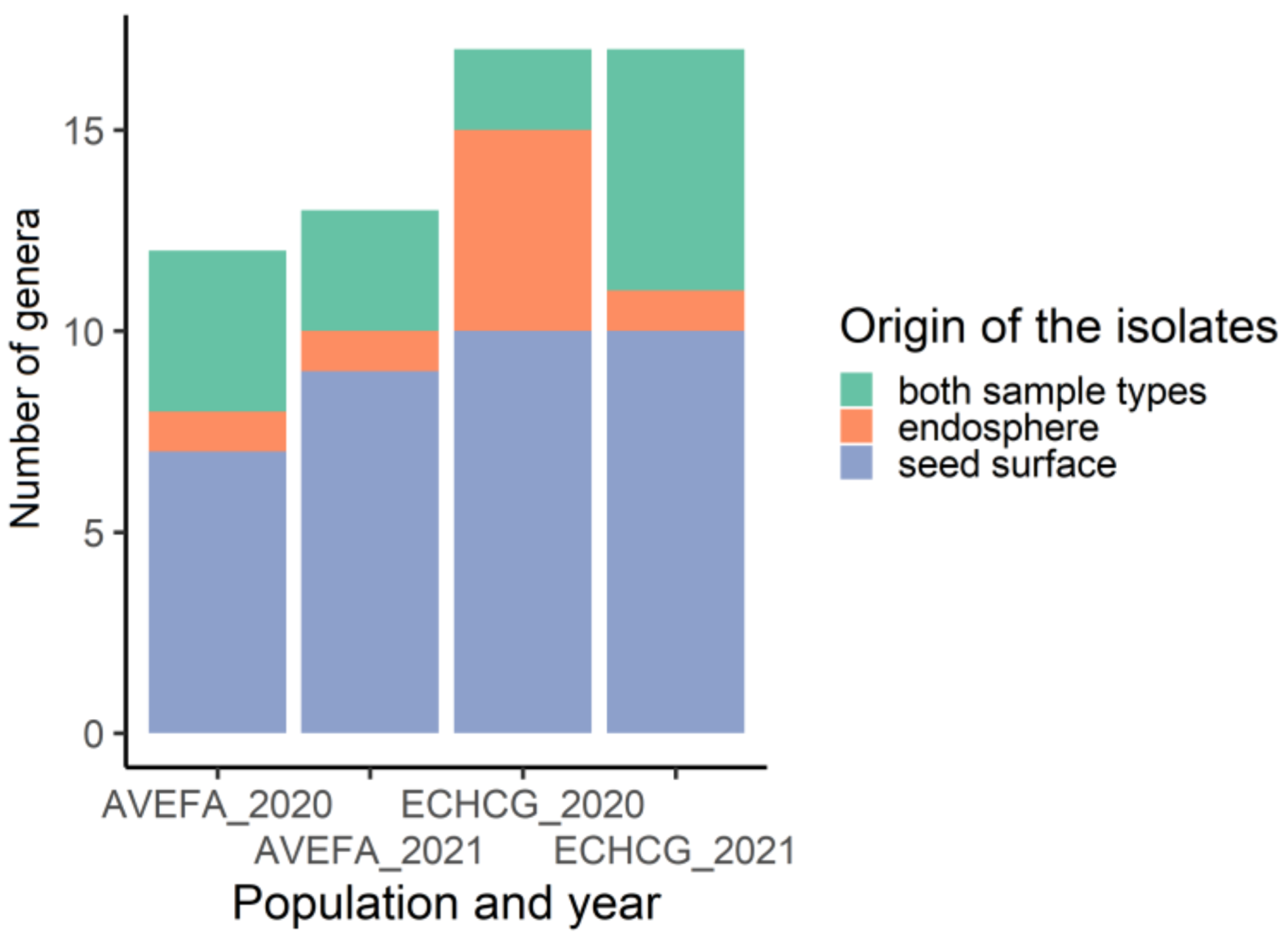
3.1. The Diversity of Epiphytic Seed-Borne Fungi Is Greater Than That of the Endophytic Seed-Borne Fungi

In total, 72 morphologically unique fungal isolates were recovered in 2020, and 96 were recovered in 2020 and 2021 from seeds of both A. fatua and E. crus-galli. Overall richness was higher in samples isolated from E. crus-galli (Figure 1).
The most frequent genera, found in more than one population of each species, were Alternaria, Fusarium, Cladosporium, and Sarocladium, followed by Sporobolomyces, Epicoccum, Vishniacozyma, and Aureobasidium. Several isolates were identified at the species level. The list of genera and species (identified at the species level) is given in Table 2.
Alternaria, Cladosporium, Fusarium, and Sarocladium were present in the seed endosphere, as well as on the seed surface of both plant species. However, most fungal isolates were either uniquely present in one plant species or one of the years (Figure 2). Different collection sites yielded a similar number of fungal isolates, and a rough proportion of 1:2 in the number of endophytic to epiphytic isolates was maintained in both years.
Three to six genera were isolated exclusively from the seeds of one species collected in one of the years. The number of genera isolated from the endosphere only was three in the case of E. crus-galli, while in A. fatua, only one genus was isolated from the endosphere only. Five genera were isolated only from the A. fatua seed surface, while nine were isolated from the E. crus-galli seed surface, and four more genera were isolated from the surface of both species. Comparing the number of common genera in seeds collected from different locations, it was found that more were common in 2021 in both species and either isolated from the seed surface or the endosphere, while in 2020, no common genera were found in A. fatua from different locations, and only one was common to all three locations among the isolates from the E. crus-galli seed surface. The highest number of genera exclusive to one population was isolated from the E. crus-galli seed surface in 2020, where five were unique to seeds from the Salgale location (Figure 2). These results indicate that the seed mycobiota of the studied species differ between plant populations.

3.2. Taxonomic Composition of Seed-Borne Fungi Differs between the Two Species

E. crus-galli mycobiota exhibited higher year-to-year variability compared to A. fatua. The hierarchical clustering based on Jaccard distances revealed the grouping of the A. fatua samples from both collection years, while E. crus-galli samples also clustered together, except for samples from seeds collected in 2021, revealing some overlap between the species (Figure 3).
The results of cluster analysis were supported by the analysis of similarities. Dissimilarity was not significant between the collection years, but there was a significant dissimilarity (p = 0.013) between the two species, although a great degree of overlap (R = 0.218) was also observed.

3.3. F. Sporotrichioides and F. Culmorum Had the Highest Potential to Reduce the Number of Intact E. Crus-Galli Seeds

Ten Fusarium isolates for which sporulation was attained were chosen for inoculation experiments using E. crus-galli seeds (Table 3).
Although all collected seeds were initially dormant, dormancy was partially lost in the drying process, and germination was stimulated by surface sterilization. The number of germinated seeds varied from 1.7% to 94.8% between the combinations of the collection year and plant population and between seeds infected with different isolates. Similarly, the proportion of infected and decayed seeds, as well as seedlings infected after germination, was variable between the treatments: 0–73.3% (infected seeds), 0–31.1% (decayed seeds), and 0–34.9% (infected seedlings) (Figure 4).
Inoculation of seeds with spores of the isolates 20s11, 21s19, 21s28, 21s61, and 21s72 resulted in the lowest number of intact seeds (25% or less of all the seeds remained intact) in all combinations of the collection year and plant population (Figure 4). The number of infected and decayed seeds was inconsistent between the seed-collection years and populations, except for isolates 21s19 and 21s72. Although the number of intact seeds was not significantly different from the control in the Stelpe population, including in 2020 and 2021, there was a tendency towards a lower number of intact seeds in samples inoculated with 21s19 and 21s72 (in 2021, the difference between 21s72 and the control was close to significant, p = 0.0622). Infected seeds were present in the control treatment, indicating that either the sterilization method was insufficient to eliminate all fungi from the seed surface or that Fusarium fungi were present deeper in the seed coat tissues.
Two 20s11 and 21s72 isolates identified as F. sporotrichioides could infect seeds already at 10 °C. On average, 5% of the total number of seeds were infected at low temperatures (Figure 5).
We observed competitive interaction between Fusarium isolates applied for seed inoculation and fungal isolates emerging from seeds in seed decay tests. Most antagonistic isolates were identified morphologically as Alternaria.

4. Discussion

4.1. Taxonomic Composition of Seed Fungal Communities

In this study, a culture-based taxonomic assessment of mycobiota of seeds from two noxious weed species, A. fatua and E. crus-galli, was performed. Fungal communities were compared with regard to plant species, seed-collection years, and locations, as well as the habitats (seed surface or seed endosphere). More seed-borne fungi were isolated from the surface of non-sterilized seeds of both weed species compared to the seed endosphere. Although the surface-sterilization method used in this study may not have eliminated 100% of the surface-borne microorganisms, there was a clear reduction in the number of isolated genera.
Most isolated fungal genera and species were typical for the mycobiota of seeds [5]. Two genera, Fusarium and Alternaria, were ubiquitously represented in endophytic and epiphytic samples from both weed species. Alternaria is a common seed colonizer [36,37]. Certain isolates of Alternaria have previously been reported to suppress the germination of E. crus-galli and A. fatua [38]. Thus, Alternaria alternata and A. tenuissima strains have been proposed as biocontrol agents for this weed species. In addition to its seed-degrading capacity, Alternaria sp. effectively infects E. crus-galli seedlings, precluding further development [39]. Fusarium is one of the most widespread seed-associated fungal genera [36] and is a genus represented by numerous species of common cereal pathogens. Fusarium is often isolated from Poaceae seeds [40]. Due to pathogenic properties, members of this genus are often viewed as a target rather than an agent of biocontrol. However, specific strains of F. avenaceum [41] and F. culmorum [16] have been shown to successfully colonize and degrade caryopsis of A. fatua, suggesting that certain strains of Fusarium are potentially interesting for application in weed seed biocontrol.
Cladosporium is another widespread seed-associated fungal genus [36] often implicated in competitive interactions with Fusarium [42]. Although Cladosporium was one of the most abundant epiphytic fungal genera isolated from the seed surface of both weed species in our study, it was much less frequently encountered among endophytic mycobiota samples. In fact, only seeds collected at a single site in 2020 for A. fatua and in 2021 for E. crus-galli contained Cladosporium in the endosphere, whereas the majority of samples from the seed surface of both weed species in both years contained representatives from this genus. A similar distinction was also observed in the case of Vishniacozyma, abundant on the surface of E. crus-galli seeds but absent from the endosphere. Such distinction between seed endosphere and surface concerning Cladosporium and Vishniacozyma colonization could be caused by the lower ability of these fungi to penetrate the seed coat or by lower competitiveness in relation to endophytes. However, further studies are required to test these speculations. Both Cladosporium and Vishniacozyma have also previously been reported as potential antagonists of Fusarium [42].
Epicoccum, a fungal genus previously described for its biocidal potential against seeds of E. crus-galli [43], was also among the most abundant genera in our study. Contrary to Cladosporium and Vishniacozyma, which were predominantly isolated from the seed surface, Epicoccum was much more frequently isolated from the endosphere, suggesting that different compartments of the seed provide a specific ecological niche for these fungi.
Sarocladium isolates identified as S. strictum (previously known as Acremonium strictum) [44] were isolated from the seeds of both plant species in both years, while those identified as S. bacillisporum were isolated only from E. crus-galli in 2021. In this study, Sarocladium was mostly isolated from the seed surface, but it also occurred in the endosphere. S. strictum is a pathogenic species causing Acremonium wilt in plants. However, its role as an endophyte needs to be studied better. S. strictum isolates obtained from maize kernels could produce mycotoxins, as well as antimicrobial and insecticidal compounds [45].
Isolates from the genus Aureobasidium were identified as A. pullulans, but in two cases, Protax-fungi search identified the isolates as Selenophoma sp., a genus that can be grouped in the same clade as Aureobasidium [46]. Aureobasidium was isolated from the seeds of both plant species in both years, although in 2020, only from A. fatua. This species is described as a floral pathogen [47] and an antagonist to Fusarium culmorum [48].
The isolates from the genus Bipolaris were identified as B. sorokiniana (synonym Cochliobolus sativus) by NCBI search and as Drechslera by Protax-fungi analysis. D. sorokiniana is considered a synonym of B. sorokiniana, and the latter is known as the causal agent of black point disease, which reduces seed quality and germination [49], as well as wheat seedling blight [50]. Other species from this genus, Drechslera avenacea and B. sorokiniana, were isolated from A. fatua seeds [21], and both fungi were pathogenic to A. fatua, but only D. avenacea was species-specific. Since B. sorokiniana, other Bipolaris species, and Drechslera species were isolated from the seeds of cultivated grasses [37], it is plausible that there is no species-specificity. In this study, B. sorokiniana/Drechslera was isolated only from E. crus-galli and not from A. fatua in both collection years, indicating that the applied methodology was suitable for isolating this fungal species. However, its absence in our sample pool might mean that B. sorokiniana/Drechslera is either rare or absent from A. fatua in Latvia. In future studies, it will be necessary to test the pathogenicity of this isolate to E. crus-galli.
Microdochium spp. can be hosted by different cereal crops and grasses, but in this study, it was isolated only from E. crus-galli. M. nivale is a causal agent of snow mold in Poaceae [51]. Colonization of seeds by Microdochium usually does not affect germination or seedling health, and the fungus only affects weakened plants [52]. Taxonomic designation as M. bolleyi was assigned to our isolates by both search methods, but low genetic diversity was reported for the ITS region, so RPB2 and the β-tubulin sequences would be preferable for species identification [51]. M. bolleyi was reported as an endophyte in herbaceous plant Fagonia cretica, capable of producing compounds with antibacterial and antifungal properties [53].
Moesziomyces is a genus of smut fungi that infects Poaceae plants. We isolated it in 2021 from seeds of both plant species. NCBI search identified the isolate from E. crus-galli as Pseudozyma aphidis, a synonym for Moesziomyces aphidis [54], while that isolated from A. fatua was Moesziomyces bullatus. Protax-fungi search identified all isolates as M. bullatus. The asexual (yeast) morph of this genus (previously known as Pseudozyma) is non-pathogenic, can be isolated from different plants and environmental samples, and can even be antagonistic to pathogenic fungi. In contrast, the sexual morph is parasitic, and E. crus-galli is considered the type of host of M. bullatus [54]. This distinction can explain the occurrence of this fungus in the seeds of both plant species. Contrary to a few widespread and omnipresent fungal genera, most fungal isolates were present in only specific collection sites or isolated from a very limited number of samples (Table 2). Among these less-frequent fungi were several yeast genera, Rhodotorula, Sampaiozyma, Sporobolomyces, Filobasidium, and Papiliotrema that, due to their seed-colonizing ability and low pathogenicity towards crops [55], have also been suggested as possible weed biocontrol agents. The ability to produce an antifungal toxin was reported in Bullera alba (basionym Sporobolomyces albus) [56]. The presence of many species with varying properties suggests that the fungal community is assembled on the surface and in the endosphere of the seeds after attaining equilibrium between different antagonistic fungi and, potentially, other microorganisms. The interactions of these fungi should be further studied to understand the potential of shifting this equilibrium to achieve the desired effect on seed fate in soil. Identification at the species level and distinction between strains are necessary to understand what determines the presence of certain fungi on seed surfaces or in the endosphere and the implications for seed germination and persistence in soil.
The advantages of the culture-based approach are that only viable microorganisms are isolated and identified, and living cultures can be stored for future reference. However, more comprehensive taxonomic profiles of seed-borne microorganisms can be obtained by NGS-based analysis of the DNA isolated from seed samples, which will be attempted in further studies.

4.2. Seed-Inoculation Experiment

Seeds of E. crus-galli were inoculated with a spore suspension of 10 different Fusarium isolates obtained from both E. crus-galli and A. fatua seeds to test the potential of these isolates to infect and degrade E. crus-galli seeds. To the best of our knowledge, Fusarium has not previously been tested for E. crus-galli seed degradation. Successful seed colonization by some of the tested isolates of Fusarium and subsequent decay of colonized E. crus-galli seeds that were observed in our experiments suggest that this widespread fungal genus holds potential for the biocontrol of E. crus-galli. However, fungal growth observed on the outer structures of the seeds may not result in seed decay if the hyphae do not penetrate inner seed structures, as observed in the case of F. graminearum infecting barley caryopses [57]. In contrast, the Fusarium isolate that infects seeds of Bromus tectorum is able to penetrate the embryo and endosperm and degrade the seed [15]. Hyphal growth was triggered by seed exudates, proposing an interesting question: do different Poaceae seeds produce similar compounds that can trigger infection by Fusarium or other fungi?
Seeds can escape the degrading effect of fungi by rapid germination. Therefore, an important property of F. sporotrichioides is its ability to infect seeds at low temperatures [58]. This allows the fungus to infect seeds in soil conditions early in the season before seeds have the potential to germinate. Seeds of E. crus-galli do not germinate at temperatures below 13 °C [26] and, consequently, can be more susceptible to fungal attack at low temperatures. F. sporotrichioides isolates could be further explored as a potential seed-degrading pathogen, but the host-species specificity should also be tested.
When considering the effect of microorganisms on the soil seed bank dynamic, it is difficult to separate seed fate from seedling fate because both decay and fatal germination can deplete the seed bank. Soil-borne and seed-borne fungi can infect the seedlings. For example, certain Fusarium strains reduce the emergence of E. crus-galli [17], and F. tricinctum increases seedling mortality in Stipa bungeana and Lespedeza davurica [59]. The ability to infect seeds and the effect of different Fusarium isolates on the seeds was very variable in the current study, in line with the results of other studies [17,22]. Interestingly, one of the isolates that caused the highest reduction in the number of intact E. crus-galli seeds (21s72) was isolated from A. fatua seeds, suggesting that the seed-degrading capacity is not host-species-specific. One of the possible causes of the between-year variability is the difference in the colonization rate of the seeds and the composition of the seed-borne microorganisms that can act as antagonists to the pathogens. Although the majority of analyzed seed samples were co-inhabited by Fusarium and Alternaria, both species can also exhibit competitive interactions, likely due to antagonistic toxin production [60]. The observed competitive interaction between Fusarium and Alternaria suggests that native endophytic mycobiota is a significant factor that needs to be considered when predicting the outcome of seed infections by potentially seed-degrading fungi.

5. Conclusions

In this study, we characterized the composition of culturable fungi from the surface and the endosphere of seeds of two commercially important weed species. Mature seeds harbored more fungi on the surface compared to the endosphere, and the described fungal genera included both potential seed pathogens and their antagonists. The artificial seed-infection experiment showed that some of the seed-borne Fusarium strains have the potential to degrade seeds of E. crus-galli, thereby influencing seed fate in a soil seed bank.

Author Contributions

Conceptualization, J.Ņ. and A.B.; methodology, J.Ņ., V.N., A.B., D.F. and I.K.; formal analysis, J.Ņ.; investigation, J.Ņ., A.B., A.R., E.S. and M.S.; writing—original draft preparation, J.Ņ., A.B., V.N. and D.F.; writing—J.Ņ., A.B., V.N., D.F. and I.K. All authors have read and agreed to the published version of the manuscript.

Funding

This research was funded by the Latvian Council of Science in the project Characterization and dynamics of seed-associated microbiome in weed soil seedbank, no. lzp-2020/1-0163.

Data Availability Statement

Data sharing not applicable.

Acknowledgments

We thank the owners of the fields for permission to collect seeds and Elvis Garais for help with seed collection and microorganism isolation.

Conflicts of Interest

The authors declare no conflict of interest.

References

  1. Oerke, E.C. Crop losses to pests. J. Agric. Sci. 2006, 144, 31–43. [Google Scholar] [CrossRef]
  2. Kudsk, P. Advances in Integrated Weed Management; Burleigh Dodds Science Publishing: Cambridge, UK, 2022; pp. 1–452. [Google Scholar]
  3. Klaedtke, S.; Jacques, M.A.; Raggi, L.; Préveaux, A.; Bonneau, S.; Negri, V.; Chable, V.; Barret, M. Terroir is a key driver of seed-associated microbial assemblages. Environ. Microbiol. 2016, 18, 1792–1804. [Google Scholar] [CrossRef] [PubMed] [Green Version]
  4. Hodgson, S.; de Cates, C.; Hodgson, J.; Morley, N.J.; Sutton, B.C.; Gange, A.C. Vertical transmission of fungal endophytes is widespread in forbs. Ecol. Evol. 2014, 4, 1199–1208. [Google Scholar] [CrossRef] [PubMed] [Green Version]
  5. Nelson, E.B. The seed microbiome: Origins, interactions, and impacts. Plant Soil 2018, 422, 7–34. [Google Scholar] [CrossRef]
  6. Pollard, A.T. Seeds vs fungi: An enzymatic battle in the soil seedbank. Seed Sci. Res. 2018, 28, 197–214. [Google Scholar] [CrossRef]
  7. Dalling, J.W.; Davis, A.S.; Schutte, B.J.; Arnold, A.E. Seed survival in soil: Interacting effects of predation, dormancy and the soil microbial community. J. Ecol. 2011, 99, 89–95. [Google Scholar] [CrossRef]
  8. Davis, A.S.; Fu, X.; Schutte, B.J.; Berhow, M.A.; Dalling, J.W. Interspecific variation in persistence of buried weed seeds follows trade-offs among physiological, chemical, and physical seed defenses. Ecol. Evol. 2016, 6, 6836–6845. [Google Scholar] [CrossRef]
  9. Hendry, G.A.F.; Thompson, K.; Moss, C.J.; Edwards, E.; Thorpe, P.C. Seed persistence: A correlation between seed longevity in the soil and ortho-dihydroxyphenol concentration. Funct. Ecol. 1994, 8, 658–664. [Google Scholar] [CrossRef]
  10. Wagner, M.; Mitschunas, N. Fungal effects on seed bank persistence and potential applications in weed biocontrol: A review. Basic Appl. Ecol. 2008, 9, 191–203. [Google Scholar] [CrossRef]
  11. Gardarin, A.; Duerr, C.; Mannino, M.R.; Busset, H.; Colbach, N. Seed mortality in the soil is related to seed coat thickness. Seed Sci. Res. 2010, 20, 243–256. [Google Scholar] [CrossRef]
  12. Nolde, S.B.; Vassilevski, A.A.; Rogozhin, E.A.; Barinov, N.A.; Balashova, T.A.; Samsonova, O.V.; Baranov, Y.V.; Feofanov, A.V.; Egorov, T.A.; Arseniev, A.S.; et al. Disulfide-stabilized helical hairpin structure and activity of a novel antifungal peptide EcAMP1 from seeds of barnyard grass (Echinochloa crus-galli). J. Biol. Chem. 2011, 286, 25145–25153. [Google Scholar] [CrossRef] [PubMed] [Green Version]
  13. Hoefnagels, M.H.; Linderman, R.G. Biological suppression of seedborne Fusarium spp. during cold stratification of Douglas fir seeds. Plant Dis. 1999, 83, 845–852. [Google Scholar] [CrossRef] [PubMed] [Green Version]
  14. Suárez, J.O.; Villada, D.; Oria de Rueda, J.A.; Alves-Santos, F.M.; Diez, J.J. Effects of Lactarius deliciosus and Rhizopogon roseolus ectomycorrhizal fungi on seeds and seedlings of Scots and stone pines inoculated with Fusarium oxysporum and Fusarium verticillioides. For. Chron. 2018, 94, 126–134. [Google Scholar] [CrossRef] [Green Version]
  15. Meyer, S.E.; Franke, J.L.; Baughman, O.W.; Beckstead, J.; Geary, B. Does Fusarium-caused seed mortality contribute to Bromus tectorum stand failure in the Great Basin? Weed Res. 2014, 54, 511–519. [Google Scholar] [CrossRef]
  16. De Luna, L.Z.; Kennedy, A.C.; Hansen, J.C.; Paulitz, T.C.; Gallagher, R.S.; Fuerst, E.P. Mycobiota on wild oat (Avena fatua L.) seed and their caryopsis decay potential. Plant Health Prog. 2011, 12, 20. [Google Scholar] [CrossRef] [Green Version]
  17. Mohler, C.L.; Dykeman, C.; Nelson, E.B.; Ditommaso, A. Reduction in weed seedling emergence by pathogens following the incorporation of green crop residue. Weed Res. 2012, 52, 467–477. [Google Scholar] [CrossRef]
  18. Garibaldi, A.; Gilardi, G.; Pasquali, M.; Kejji, S.; Gullino, M.L. Seed transmission of Fusarium oxysporum of Eruca vesicaria and Diplotaxis muralis. Z. Pflanzenk. Pflanzen.—J. Plant Dis. Protect. 2004, 4, 345–350. [Google Scholar]
  19. Masi, M.; Meyer, S.; Pescitelli, G.; Cimmino, A.; Clement, S.; Peacock, B.; Evidente, A. Phytotoxic activity against Bromus tectorum for secondary metabolites of a seed-pathogenic Fusarium strain belonging to the F. tricinctum species complex. Nat. Prod. Res. 2017, 31, 2768–2777. [Google Scholar] [CrossRef]
  20. Mancini, V.; Murolo, S.; Romanazzi, G. Diagnostic methods for detecting fungal pathogens on vegetable seeds. Plant Pathol. 2016, 65, 691–703. [Google Scholar] [CrossRef]
  21. Mortensen, K.; Hsiao, A.I. Fungal infestation of seeds from seven populations of wild oats (Avena fatua L.) with different dormancy and viability characteristics. Weed Res. 1987, 27, 297–304. [Google Scholar] [CrossRef]
  22. Donayre, D.K.M.; Dalisay, T.U.; Bayot, R.G.; Baltazar, A.M. Diversity and tissue specificity of endophytic fungi in barnyard grass (Echinochloa glabrescens Munro ex Hook. F.). Asia Life Sci. 2014, 23, 725–741. [Google Scholar]
  23. Heap, I. The International Herbicide-Resistant Weed Database. Available online: www.weedscience.org (accessed on 25 July 2022).
  24. Gallandt, E.R. How can we target the weed seedbank? Weed Sci. 2006, 54, 588–596. [Google Scholar] [CrossRef]
  25. Haring, S.C.; Flessner, M.L. Improving soil seed bank management. Pest Manag. Sci. 2018, 74, 2412–2418. [Google Scholar] [CrossRef]
  26. Maun, M.A.; Barrett, S.C.H. The biology of Canadian weeds. 77. Echinochloa crus-galli (L.) Beauv. Can. J. Plant Sci. 1986, 66, 739–759. [Google Scholar] [CrossRef]
  27. Collado, J.; Platas, G.; Paulus, B.; Bills, G.F. High-throughput culturing of fungi from plant litter by a dilution-to-extinction technique. FEMS Microbiol. Ecol. 2007, 60, 521–533. [Google Scholar] [CrossRef] [PubMed] [Green Version]
  28. White, T.J.; Bruns, T.; Lee, S.; Taylor, J. Amplification and direct sequencing of fungal ribosomal RNA genes for phylogenetics. In PCR Protocols. A Guide to Methods and Applications; Innis, M.A., Gelfand, D.H., Sninsky, J.J., White, T.J., Eds.; Academic Press: San Diego, CA, USA, 1990; pp. 315–322. [Google Scholar]
  29. Abarenkov, K.; Somervuo, P.; Nilsson, R.H.; Kirk, P.M.; Huotari, T.; Abrego, N.; Ovaskainen, O. Protax-fungi: A web-based tool for probabilistic taxonomic placement of fungal internal transcribed spacer sequences. New Phytol. 2018, 220, 517–525. [Google Scholar] [CrossRef] [PubMed] [Green Version]
  30. Sampietro, D.A.; Marín, P.; Iglesias, J.; Presello, D.A.; Vattuone, M.A.; Catalán, C.A.N.; Jaen, M.G. A molecular based strategy for rapid diagnosis of toxigenic Fusarium species associated to cereal grains from Argentina. Fungal Biol. 2010, 114, 74–81. [Google Scholar] [CrossRef] [PubMed]
  31. Purahong, W.; Alkadri, D.; Nipoti, P.; Pisi, A.; Lemmens, M.; Prodi, A. Validation of a modified Petri-dish test to quantify aggressiveness of Fusarium graminearum in durum wheat. Eur. J. Plant Pathol. 2012, 132, 381–391. [Google Scholar] [CrossRef]
  32. Oksanen, J.; Blanchet, F.G.; Friendly, M.; Kindt, R.; Legendre, P.; McGlinn, D.; Minchin, P.R.; O’hara, R.B.; Simpson, G.L.; Solymos, P.; et al. Package ‘Vegan’. Community Ecology Package; 2020. Available online: https://CRAN.R-project.org/package=vegan (accessed on 19 December 2022).
  33. Kolde, R.; Kolde, M.R. Package ‘Pheatmap’. R Package 1; R Package, 2018. Available online: https://CRAN.R-project.org/package=pheatmap (accessed on 19 December 2022).
  34. Hothorn, T.; Bretz, F.; Westfall, P.; Heiberger, R.M.; Schuetzenmeister, A.; Scheibe, S.; Hothorn, M.T. Package ‘Multcomp’, Simultaneous Inference in General Parametric Models; Project for Statistical Computing: Vienna, Austria, 2016. [Google Scholar]
  35. Chen, H. VennDiagram: Generate High-Resolution Venn and Euler Plots, R package version 1.7.3; R package, 2022. Available online: https://CRAN.R-project.org/package=VennDiagram (accessed on 19 December 2022).
  36. Simonin, M.; Briand, M.; Chesneau, G.; Rochefort, A.; Marais, C.; Sarniguet, A.; Barret, M. Seed microbiota revealed by a large-scale meta-analysis including 50 plant species. New Phytol. 2022, 234, 1448–1463. [Google Scholar] [CrossRef] [PubMed]
  37. Varga, Z.; Kreutzer, B.; Graiss, W. Mycological investigations of seeds of cultivated grasses. Bodenkultur 2008, 59, 95–100. [Google Scholar]
  38. Mohammed, Y.M.; Badawy, M.E. Potential of phytopathogenic fungal isolates as a biocontrol agent against some weeds. Egypt. J. Biol. Pest Control 2020, 30, 92. [Google Scholar] [CrossRef]
  39. Mohammad, R.S.M. Evaluation of Alternaria alternata causing leaf spot of barnyardgrass grown in rice fields. Afr. J. Microbiol. Res. 2012, 6, 4481–4488. [Google Scholar]
  40. Hietaniemi, V.; Rämö, S.; Yli-Mattila, T.; Jestoi, M.; Peltonen, S.; Kartio, M.; Sieviläinen, E.; Koivisto, T.; Parikka, P. Updated survey of Fusarium species and toxins in Finnish cereal grains. Food Addit. Contam. A Chem. Anal. Control. Expo. Risk Assess 2016, 33, 831–848. [Google Scholar] [CrossRef] [PubMed]
  41. Lewis, R.W.; Okubara, P.A.; Sullivan, T.S.; Madden, B.J.; Johnson, K.L.; Charlesworth, M.C.; Fuerst, E.P. Proteome-wide response of dormant caryopses of the weed, Avena fatua, after colonization by a seed-decay isolate of Fusarium avenaceum. Phytopathology 2022, 112, 1103–1117. [Google Scholar] [CrossRef]
  42. Cobo-Diaz, J.F.; Legrand, F.; Le Floch, G.; Picot, A. Influence of maize residues in shaping soil microbiota and Fusarium spp. communities. Microb. Ecol. 2022, 83, 702–713. [Google Scholar] [CrossRef]
  43. Motlagh, M.R.S. Evaluation of Epicoccum purpurascens as biological control agent of Echinochloa spp. in rice fields. J. Food Agric. Environ. 2011, 9, 394–397. [Google Scholar]
  44. Giraldo, A.; Gené, J.; Sutton, D.A.; Madrid, H.; De Hoog, G.S.; Cano, J.; Decock, C.; Crow, P.W.; Guarro, J. Phylogeny of Sarocladium (Hypocreales). Pers. Mol. Phylogeny Evol. Fungi 2015, 34, 10–24. [Google Scholar] [CrossRef] [Green Version]
  45. Błaszczyk, L.; Waśkiewicz, A.; Gromadzka, K.; Mikołajczak, K.; Chełkowski, J. Sarocladium and Lecanicillium associated with maize seeds and their potential to form selected secondary metabolites. Biomolecules 2021, 11, 98. [Google Scholar] [CrossRef]
  46. Zalar, P.; Gostinčar, C.; De Hoog, G.S.; Uršič, V.; Sudhadham, M.; Gunde-Cimerman, N. Redefinition of Aureobasidium pullulans and its varieties. Stud. Mycol. 2008, 61, 21–38. [Google Scholar] [CrossRef] [Green Version]
  47. Prashanthi, S.K.; Kulkarni, S. Aureobasidium pullulans, a potential mycoherbicide for biocontrol of eupatorium [Chromolaena odorata (L.) King and Robinson] weed. Curr. Sci. 2005, 88, 18–21. [Google Scholar]
  48. Wachowska, U.; Głowacka, K. Antagonistic interactions between Aureobasidium pullulans and Fusarium culmorum, a fungal pathogen of winter wheat. Biocontrol 2014, 59, 635–645. [Google Scholar] [CrossRef] [Green Version]
  49. Al-Sadi, A.M. Bipolaris sorokiniana-induced black point, common root rot, and spot blotch diseases of wheat: A review. Front. Cell. Infect. Microbiol. 2021, 11, 584899. [Google Scholar] [CrossRef] [PubMed]
  50. Liljeroth, E.; Bryngelsson, T. Seed treatment of barley with Idriella bolleyi causes systemically enhanced defence against root and leaf infection by Bipolaris sorokiniana. Biocontrol. Sci. Technol. 2002, 12, 235–249. [Google Scholar] [CrossRef]
  51. Abdelhalim, M.; Brurberg, M.B.; Hofgaard, I.S.; Rognli, O.A.; Tronsmo, A.M. Pathogenicity, host specificity and genetic diversity in Norwegian isolates of Microdochium nivale and Microdochium majus. Eur. J. Plant Pathol. 2020, 156, 885–895. [Google Scholar] [CrossRef] [Green Version]
  52. Gavrilova, O.P.; Orina, A.S.; Kessenikh, E.D.; Gustyleva, L.K.; Savelieva, E.I.; Gogina, N.N.; Gagkaeva, T.Y. Diversity of physiological and biochemical characters of Microdochium fungi. Chem. Biodivers. 2020, 17, e2000294. [Google Scholar] [CrossRef]
  53. Zhang, W.; Krohn, K.; Draeger, S.; Schulz, B. Bioactive isocoumarins isolated from the endophytic fungus Microdochium bolleyi. J. Nat. Prod. 2008, 71, 1078–1081. [Google Scholar] [CrossRef] [PubMed]
  54. Kruse, J.; Doehlemann, G.; Kemen, E.; Thines, M. Asexual and sexual morphs of Moesziomyces revisited. IMA Fungus 2017, 8, 117–129. [Google Scholar] [CrossRef] [PubMed] [Green Version]
  55. Triolet, M.; Edel-Hermann, V.; Gautheron, N.; Mondy, S.; Reibel, C.; André, O.; Guillemin, J.P.; Steinberg, C. Weeds harbor an impressive diversity of fungi, which offers possibilities for biocontrol. Appl. Environ. Microbiol. 2022, 88, e0217721. [Google Scholar] [CrossRef]
  56. Golubev, W.; Ikeda, R.; Shinoda, T.; Nakase, T. Antifungal activity of Bullera alba (Hanna) Derx. Mycoscience 1997, 38, 25–29. [Google Scholar] [CrossRef]
  57. Skadsen, R.W.; Hohn, T.M. Use of Fusarium graminearum transformed with gfp to follow infection patterns in barley and Arabidopsis. Physiol. Mol. Plant Pathol. 2004, 64, 45–53. [Google Scholar] [CrossRef]
  58. Gianinetti, A.; Finocchiaro, F.; Maisenti, F.; Satsap, D.K.; Morcia, C.; Ghizzoni, R.; Terzi, V. The caryopsis of red-grained rice has enhanced resistance to fungal attack. J. Fungi 2018, 4, 71. [Google Scholar] [CrossRef] [PubMed] [Green Version]
  59. Chen, T.; Nan, Z.; Zhang, X.; Hou, F.; Christensen, M.; Baskin, C. Does dormancy protect seeds against attack by the pathogenic fungus Fusarium tricinctum in a semiarid grassland of Northwest China? Plant Soil 2018, 422, 155–168. [Google Scholar] [CrossRef]
  60. Müller, M.E.H.; Urban, K.; Köppen, R.; Siegel, D.; Korn, U.; Koch, M. Mycotoxins as antagonistic or supporting agents in the interaction between phytopathogenic Fusarium and Alternaria fungi. World Mycotoxin J. 2015, 8, 311–321. [Google Scholar] [CrossRef]
Figure 1. Number of fungal genera isolated from the seed surface, seed endosphere, as well as from both endosphere and seed surface of A. fatua (AVEFA) and E. crus-galli (ECHCG) in 2020 and 2021.
Microorganisms 11 00184 g001
Figure 2. Venn diagrams showing the numbers of genera overlapping between different seed samples and habitats (epiphytes/endophytes).
Microorganisms 11 00184 g002
Figure 3. Hierarchical cluster diagram and heatmap based on Jaccard distances between fungal communities isolated from A. fatua (AVEFA) and E. crus-galli (ECHCG) seed samples collected in 2020 and 2021 in different locations (indicated by letters).
Microorganisms 11 00184 g003
Figure 4. Seed fate after inoculating surface-sterilized seeds with spore suspension of different Fusarium isolates and subsequent incubation at 10 °C for 14 days and at room temperature for 28 days. Non-inoculated seeds were used as a control. Seeds collected in different years and from different plant populations are shown separately. Asterisk (*) denotes treatments where the number of intact seeds at the end of experiment was significantly different from the control treatment.
Microorganisms 11 00184 g004
Figure 5. Average number of seeds with signs of Fusarium infection after 14 days at 10 °C. Non-inoculated seeds were used as a control. Asterisk (*) denotes treatments where the number of intact seeds at the end of experiment was significantly different from the control treatment.
Microorganisms 11 00184 g005
Table 1. Seed-Collection Dates and Location Details.
Plant SpeciesLocation NameCoordinatesDateCrop
2020202120202021
A. fatuaBebri56°44′ N
25°27′ E
05.08.01.08.Spring wheatSpring wheat
A. fatuaGaigalava56°43′ N
26°07′ E
05.08.01.08.Spring wheatSpring wheat
A. fatuaKrimunas56°32′ N
23°27′ E
13.08.04.08.Peas/spring barleyWinter wheat
A. fatuaZiras57°10′ N
21°35′ E
12.08.n/aSpring barleyn/a
E. crus-galliIecava56°59′ N
24°25′ E
05.09.23.08.ZucchiniMaize
E. crus-galliSalgale56°37′ N
23°58′ E
01.09.25.08.Spring oatsPotato
E. crus-galliStelpe56°52′ N
24°55′ E
05.09.23.08.Spring wheatSpring wheat
Table 2. Fungi isolated from A. fatua and E. crus-galli seeds and identified at the genus and species level using NCBI Blast search and Protax-fungi algorithm. No species is given where NCBI search resulted in multiple species with the same match for the query sequence. Frequency of occurrence was calculated as the summary incidence in seed samples from each species population in each year divided by the total number of seed samples, seven in A. fatua and six in E. crus-galli. A value of 1 means that the genus/species was isolated from each species population in both years.
Frequency of OccurrenceFungal Genera/Species
A. fatuaE. crus-galli
Endosphere Seed Surface Endosphere Seed Surface Protax-Fungi NCBI
0.000.140.000.17AcremoniumAcremonium sclerotigenum
0.430.430.830.83AlternariaAlternaria
0.000.430.000.33Aureobasidium pullulansAureobasidium melanogenum
0.000.000.000.50Bullera albaBullera alba
0.140.710.170.83CladosporiumCladosporium
0.000.140.000.00ClavicepsClaviceps
0.000.000.330.00DrechsleraBipolaris sorokiniana
0.000.000.170.00DavidiellaDavidiella
0.290.000.000.00DidymellaDidymella pomorum
0.000.140.000.00DioszegiaDioszegia hungarica
0.140.000.830.17EpicoccumEpicoccum nigrum
0.000.000.000.33Filobasidium magnum/oeirenseFilobasidium magnum/oeirense
0.710.430.500.83FusariumFusarium
0.000.000.000.17HoltermanniaHoltermanniella
0.000.000.000.50Microdochium bolleyiMicrodochium bolleyi
0.00.140.170.17Moeszyomyces bullatusMoeszyomyces bullatus/Pseudozyma aphidis
0.000.000.000.17MycosphaerellaMycosphaerella
0.000.000.000.33PapiliotremaPapiliotrema baii
0.000.000.000.33PenicilliumPenicillium
0.000.000.000.17PeniophoraPeniophora
0.000.140.000.00PhaeoisariopsisCladosporium
0.000.000.170.00PhaeosphaeriaParastagonospora
0.000.000.000.17PseudocercosporaCladosporium
0.140.430.000.33RhodotorulaRhodotorula glutinis/mucilaginosa/babjevae
0.000.000.000.17Sampaiozyma ingeniosa *Rhodotorula ingeniosa
0.140.570.330.50Sarocladium strictumSarocladium strictum
0.000.140.000.00SelenophomaAureobasidium pullulans/subglaciale
0.000.430.000.00SporisoriumSporisorium graminicola
0.000.710.000.17SporobolomycesSporobolomyces roseus/ruberrimus
0.000.570.000.67Vishniacozyma tephrensis/unknown/carnescensVishniacozyma tephrensis/victoriae/carnescens
* Until 2015—Rhodotorula ingeniosa.
Table 3. Fusarium isolates used for inoculating seeds of E. crus-galli in laboratory conditions.
IsolatesYear of IsolationPlant SpeciesPlant PopulationHabitatSpecies
20s022020A. fatuaBebriEpiphyteF. avenaceum
20s112020A. fatuaKrimunasEpiphyteF. sporotrichioides
20s162020E. crus-galliIecavaEpiphyteFusarium sp.
20s232020E. crus-galliSalgaleEpiphyteFusarium sp.
21s192021E. crus-galliStelpeEndophyteF. culmorum
21s232021E. crus-galliIecavaEndophyteF. avenaceum
21s282021E. crus-galliStelpeEndophyteF. avenaceum
21s412021E. crus-galliStelpeEpiphyteF. avenaceum
21s612021A. fatuaGaigalavaEndophyteF. sporotrichioides
21s722021A. fatuaBebriEpiphyteF. sporotrichioides
Disclaimer/Publisher’s Note: The statements, opinions and data contained in all publications are solely those of the individual author(s) and contributor(s) and not of MDPI and/or the editor(s). MDPI and/or the editor(s) disclaim responsibility for any injury to people or property resulting from any ideas, methods, instructions or products referred to in the content.

