Article

Antibacterial Activity of a Natural Clay Mineral against Burkholderia cepacia Complex and Other Bacterial Pathogens Isolated from People with Cystic Fibrosis

1
Department of Chemical and Biological Engineering, University of British Columbia, 2360 E Mall, Vancouver, BC V6T 1Z3, Canada
2
Centre for Understanding and Preventing Infection in Children, Division of Infectious Diseases, Department of Pediatrics, BC Children’s Hospital Research Institute, University of British Columbia, Vancouver, BC V5Z 4H4, Canada
3
Department of Civil Engineering, University of British Columbia, 6250 Applied Science Ln, Vancouver, BC V6T 1Z3, Canada
4
Department of Microbiology and Immunology, University of British Columbia, 2350 Health Sciences Mall, Vancouver, BC V6T 1Z3, Canada
*
Authors to whom correspondence should be addressed.
Current address: BC Centre for Disease Control, Provincial Health Services Authority, BC and Department of Pathology and Laboratory Medicine, Faculty of Medicine, University of British Columbia, Vancouver, BC V5Z 4R4, Canada.
Microorganisms 2023, 11(1), 150; https://doi.org/10.3390/microorganisms11010150
Submission received: 14 November 2022 / Revised: 15 December 2022 / Accepted: 28 December 2022 / Published: 6 January 2023
(This article belongs to the Special Issue Antimicrobial Activities of Natural Products)

Abstract

:
There is an impending crisis in healthcare brought about by a new era of untreatable infections caused by bacteria resistant to all available antibiotics. Thus, there is an urgent need to identify novel antimicrobial agents to counter the continuing threat posed by formerly treatable infections. We previously reported that a natural mineral clay known as Kisameet clay (KC) is a potent inhibitor of the organisms responsible for acute infections. Chronic bacterial infections present another major challenge to treatment by antimicrobials, due to their prolonged nature, which results in repeated exposure to antibiotics and a constant selection for antimicrobial resistance. A prime example is bacteria belonging to the Burkholderia cepacia complex (Bcc), which particularly causes some of the most serious chronic lung infections in patients with cystic fibrosis (CF) associated with unpredictable clinical outcomes, poor prognosis, and high mortality rates. Eradication of these organisms from CF patients with limited effective antimicrobial options is a major challenge. Novel therapeutic approaches are urgently required. Here, we report the in vitro antibacterial activity of KC aqueous suspensions (1–10% w/v) and its aqueous extract (L100) against a collection of extensively and multi-drug resistant clinical isolates of Bcc, Pseudomonas aeruginosa, and Stenotrophomonas maltophilia isolated from patients with CF. These findings present a potential novel therapy for further investigation in the clinic.

1. Introduction

Recalcitrant chronic bacterial infections in humans represent a significant therapeutic problem worldwide whereby repeated challenges with antibiotics promote the acquisition of drug-resistant bacteria by constant selection for new infections with intrinsically resistant organisms and selective pressures on existing organisms, further elevating their resistance [1]. This situation is intensified by the rapid spread of resistance genes, particularly among Gram-negative bacteria [2]. Cystic fibrosis (CF) is one such condition that renders affected individuals susceptible throughout their lives to chronic and ultimately deteriorating multi-year lung infections, which account for over 90% of the morbidity and mortality associated with the disease [1,3,4,5,6]. Originally described in 1938 [7], CF is a life-limiting inherited multi-organ disease due to one of over 2000 gene mutations of cystic fibrosis transmembrane conductance regulator (CFTR) gene leading to the dysfunction or absence of the CFTR protein, a membrane anion channel that regulates the transepithelial ion flow vital to maintaining the proper ion and water transport and epithelial surface hydration [4,8,9,10,11]. Abnormally viscous secretions in organ systems containing epithelial -most crucially in the lungs, pancreas, liver, and gastrointestinal tract- cause obstructions that lead to further inflammation and tissue damage [12,13]. Reportedly, dysfunctional or absent CFTR causes a wide disease spectrum in people with CF (pwCF) among which chronic endobronchial infections and exocrine pancreatic insufficiency remain the main clinical manifestation of the disease [9,14,15]. Although earlier publications emphasize that CF was typical in Caucasian populations by primarily affecting those of European descent, increasing worldwide awareness and reports illustrate a changing demography in which CF is not an uncommon genetic disorder in other races and ethnicities around the globe [11,16].
It has long been demonstrated that pwCF are prone to complex, polymicrobial pulmonary infections caused by a range of multi-drug resistant (MDR) opportunistic Gram-negative bacteria including Pseudomonas aeruginosa, Burkholderia cepacia complex (Bcc), Achromobacter species, and Stenotrophomonas maltophilia, among which Bcc members are particularly the most threatening and virulent pathogens isolated from pwCF [17,18,19,20,21].
The Bcc group comprises at least 20 phenotypically similar, phylogenetically closely related, diverse, and highly adaptable bacterial species [20,21]. Prior to the 1990s, Bcc was known as one species, Burkholderia cepacia [22]. Initially, B. cepacia was named Pseudomonas cepacia when first isolated from pwCF in 1977 [23]. It took until the mid-1990s when researchers found that B. cepacia was, indeed, composed of multiple distinct subgroups, and initially, five genomovars were identified as B. cepacia (genomovar I), B. multivorans (II), B. cenocepacia (III), B. stabilis (IV), B. vietnamiensis (V) [24]. The Bcc bacteria have emerged since the 1980s [25] as highly problematic opportunistic pathogens in immunocompromised individuals, most notably among pwCF, as well as patients who suffer from chronic granulomatous disease (CGD) [20,22,26,27,28,29,30].
Persistent pulmonary infections caused by Bcc bacteria remain a threat to pwCF due to their unpredictable infection trajectory ranging from a chronic asymptomatic phenotype to an uncertain, rapid fulminant respiratory failure, and septicaemic fatal cepacia syndrome [30,31,32]. These infections are associated with unforeseeable rates of progressive decline in lung function, poor prognosis, prolonged hospitalization, and elevated morbidity and mortality rates, especially among patients with more advanced pulmonary exacerbation or lung transplantation [28,31,33]. In fact, many transplantation centers refuse CF patients harboring Bcc as these pathogens cause death and disparity in post-transplant outcomes through immediate invasive disease and cepacia syndrome [34,35]. Outbreaks of different Bcc members are often reported, and a large body of evidence indicates their spread in a patient-to-patient manner [36].
The pathogenic mechanisms employed by Bcc members in pwCF are not fully understood but are likely due to multiple factors including high levels of both intrinsic and acquired mechanisms of resistance to diverse classes of antimicrobial agents [37,38]. Bcc species, particularly B. cenocepacia, exhibit heterogeneous resistance to a broad range of antibacterial agents due to periplasmic or membrane-bound β-lactamases, efflux pump-mediated MDR, restrictive porins, outer membrane (OM) permeability barriers, and alteration in drug targets [38,39,40]. Their ability to evade the inhibitory action of multiple classes of antibiotics together with their in vivo biofilm formation account for the serious nature of persistent infections and extreme difficulty in their elimination [40,41,42,43,44]. Moreover, the known high risk of inter-patient transmissibility is associated with adverse clinical courses, ranging from mild asymptomatic carriage to a fulminant decline in pulmonary function, and cepacia syndrome [29,30,32,41]. Noteworthy, these characteristics coupled with their high adaptability to environmental changes, make Bcc infections extremely challenging [40]. As there are few effective treatments for these deadly pathogens, novel therapeutic strategies are urgently needed [38,40,45].
Given the scientific and economic challenges facing the discovery of novel antimicrobial agents, relatively few active candidates are currently being developed in the pipeline [46,47]. Thus, exploring untapped natural sources may yield novel therapies in the battle against the escalating emergence of untreatable MDR pathogens [2,33]. Complementary and alternative medicines which exhibit antimicrobial properties could address this issue [47]. Recently, “historical” agents such as natural clay minerals with demonstrated curative applications and reported antibacterial activities have been of increasing interest [48,49].
Extending back to prehistory, natural clay minerals have been used by humans for medicinal, nutritional, and protective purposes [48,50,51,52]. Ancient evidence illustrates their successful applications for healing wounds, alleviating irritations, and cleaning skin (as anti-inflammatory or antiseptic agents) [48]. Clay minerals are the most abundant chemically-active constituents of the Earth’s surface with a defined nanostructure of geological origin [50,53]. They consist of mainly microcrystalline particles of hydrous charged sheet silicates (phyllosilicates) and of aluminum or magnesium silicates [52,54]. They possess specific physicochemical properties such as ultra-fine grain size (one dimension < 2.0 μm), vast specific surface area (~100′s m2/g), and ion exchange capacity via the intercalation of ions and retaining them in an exchangeable state [54,55].
The potential of natural clay minerals as antimicrobial agents received specific attention when, recently, a successful application of hydrated French green clay poultices for the treatment of advanced Buruli ulcer, caused by a pathogenic bacterium Mycobacterium ulcerans, was reported [56]. This prompted interest in investigating the physicochemical features and antibacterial properties of clay minerals in vitro [57,58,59]. A decade of studies since then has revealed that few deposits among natural healing clays worldwide exhibited antibacterial properties [60,61]. Notably, despite their different mineralogical and physicochemical characteristics, they all originated from hydrothermally altered volcanic clastic environments, containing nanoscale expandable clay minerals and iron-rich phases, and generating pH levels of either <5 or >10 by hydration [59,61].
Kisameet clay (KC), a naturally occurring glacial clay mineral from Kisameet Bay on the central coast of British Columbia, Canada [62], has been long recognized as a healing clay by the local Heiltsuk First Nations people due to its exceptional curative properties for treating various types of skin irritation and internal maladies [62,63]. In the 1940s KC aqueous suspensions were sold as a natural remedy for topical or oral administrations and numerous anecdotal reports suggest the effective therapeutic application of KC against diverse ailments, including topical applications for wounds, burns, arthritis, and skin irritation, or by oral consumption of clay suspensions for treating internal maladies such as duodenal ulcer and ulcerative colitis [63]. Despite those observations, there was limited experimental information concerning the biological, and physicochemical composition of KC related to its antimicrobial properties and spectrum of activity [62,63].
We recently performed a series of integrated studies of microbiological, mineralogical, and physicochemical properties of KC samples collected from different sites at the deposit on Kisameet Bay [64,65]. Our studies provided a better insight into what might make this clay active and highlighted the diversity and complexity of the KC deposit [64]. Moreover, we showed that KC is the first natural clay mineral found to be active against two major fungal pathogens (Candida albicans and Cryptococcus neoformans), as well as bacterial biofilms in vitro [65,66]. Through further investigation aiming to unravel the mechanism(s) of its antibacterial properties, we found that the broad-spectrum antibacterial features of KC can be extracted in water (aqueous leachates) [65,66].
Our previous studies demonstrated that aqueous suspensions of KC have a potent broad-spectrum antibacterial activity in vitro against the major nosocomial pathogens, the ESKAPE organisms (Enterococcus faecium, Staphylococcus aureus, Klebsiella pneumoniae, Acinetobacter baumannii, P. aeruginosa, and Enterobacter species) responsible for acute infections in hospitals [67]. Here, to investigate whether KC and its aqueous extract may have value in the treatment of the range of serious bacterial pathogens involved in chronic pulmonary infections in CF patients, we conducted a study with a collection of clinical isolates of Bcc, P. aeruginosa, and S. maltophilia collected from pwCF.

2. Materials and Methods

2.1. Bacterial Strains and Growth Conditions

This study was conducted on 17 clinical isolates consisting of twelve Bcc, four P. aeruginosa, and one S. maltophilia deposited at the Canadian Burkholderia cepacia Complex Research and Referral Repository (CBCCRRR) at the University of British Columbia, Vancouver, Canada. All the bacteria were isolated from collected clinical specimens from the respiratory tract (either sputum, throat, or cough swabs) or the blood of pwCF attending pediatric or adult CF clinics in Vancouver, BC between 1990 and 2015. Initial identification of bacterial isolates was carried out in the diagnostic microbiology labs at those clinics. Isolates were then transferred to CBCCRRR for identification at the species level as described previously [41]. Briefly, this was performed through a polyphasic approach using both phenotypic and genetic assays, recA polymerase chain reaction (PCR) analysis, and sequencing of this multilocus sequence typing allele [41]. Typing at the strain level was carried out using random amplified polymorphic DNA (RAPD) analysis [41]. All the isolates were then frozen and stored before experimental testing. The Bcc isolates include six B. cenocepacia, two B. multivorans, and one of each B. cepacia, B. stabilis, B. dolosa, and B. vientnamiensis. Among Bcc isolates, B. cenocepacia and B. multivorans strains were sequential isolates collected from three patients at different times. Out of four P. aeruginosa isolates, VC8263 and VC17829 were epidemic Type strains of A002 and A097, respectively [68,69]. All isolates were grown in Lysogeny broth (LB) (Miller) or on LB agar at 37 °C.

2.2. Clay Mineral Sample

The unprocessed natural Kisameet clay (KC-35) mineral used in this investigation was supplied by Kisameet Glacial Clay Inc. (West Vancouver, BC, Canada) in its original wet form. Following transport to the University of British Columbia, the clay sample was stored and sealed at 4 °C under normal atmospheric conditions in the dark. This clay sample previously exhibited potent antibacterial activity [67]. The clay sample was dried in a vacuum desiccator at room temperature. Dry KC samples were ground using a mortar and pestle, autoclaved at 121 °C for 1 h, and stored at room temperature before experimental testing. Measurement of pH was performed on equilibrated suspensions of 1 g KC mineral in 10 mL deionized water (dH2O) or aqueous leachate using a VWR-SB20 pH meter.

2.3. Antimicrobial Susceptibility Assay

Antimicrobial resistance profiles of isolates were characterized by standard agar disk diffusion susceptibility assays based on the Bauer-Kirby method and updated protocol provided by the American Society for Microbiology [70,71]. Susceptibility assays were carried out using cation-adjusted Mueller Hinton-II (MH) broth and agar media and a panel of 34 antibiotic disks (Oxoid, BBL) representing antibacterial agents from 14 different classes. In brief, an overnight culture of each isolate in MH broth was diluted and incubated with gentle shaking to reach the mid-exponential phase of growth. Then, MH agar plates were inoculated with the bacterial cultures and antibiotic disks were placed on the inoculated plates and incubated at 37 °C for 20–24 h before the zones of inhibition (ZOI) were measured. Experiments were performed at least three times.

2.4. Preparation of Aqueous Suspensions and Leachate of Clay

Aqueous suspensions of clay with 1 or 10% KC (w/v) concentrations were prepared by suspending 10 or 100 mg of dry, ground, autoclaved clay in 1 mL of sterile dH2O, respectively. To study the soluble fraction of KC suspensions, aqueous leachate (L100) preparations were first obtained by adding 2 g of autoclaved KC to 20 mL of sterile dH2O resulting in 10% (w/v) aqueous suspensions. After continuous stirring for 24 h at room temperature, KC suspensions were then centrifuged at 25,000 revolutions per minute (rpm) for 2 h at 4 °C to separate insoluble minerals. The supernatants were sterilized and clarified by passage through 0.22 μm (Millipore) filters. The obtained aqueous leachate (L100) was the sterile, clear, soluble fraction of KC suspensions after the removal of clay particles through ultracentrifugation and filter sterilization.

2.5. Antibacterial Assay with Clay Suspensions or Aqueous Leachates

An in vitro assay was used to examine the effect of KC on these bacterial isolates as described previously [67]. Briefly, overnight cultures of bacteria were diluted into the fresh LB broth to an approximate concentration of ~107 colony forming units (CFU) mL−1 and incubated at 37 °C with gentle mixing on an orbital rotating platform (200 rpm) to reach the mid-logarithmic phase of growth. Aqueous suspensions of 1 or 10% KC (w/v) were prepared by suspending dried, ground, autoclaved clay in sterile dH2O. Bacterial cells were collected by centrifugation, rinsed once with sterile phosphate-buffered saline (PBS pH 7.4), and re-suspended at ~107 CFU mL−1 in either 1% (w/v) KC suspensions (for P. aeruginosa and S. maltophilia isolates), or 10% (for all Bcc isolates), or in dH2O (as the viability control in the absence of KC). Then, suspensions were incubated with gentle shaking (200 rpm) at 37 °C to provide proper contact with clay particles and prevent sedimentation. Bacterial viability was determined using 10-fold serial dilution plating of aliquots removed at the start of experiments (time 0) and after 5, 24, and 48 h following exposure to clay suspensions. Antibacterial assays with KC leachate (L100) were performed similarly. The washed pellets of bacterial isolates were resuspended at ~107 CFU mL−1 in KC leachate samples and incubated at the same condition as described above. Viability counts were performed by removing aliquots at the start of the experiment and three time points within 48 h of exposure to L100.

3. Results

3.1. All the CF Isolates Exhibited Extensively Drug-Resistant (XDR) or MDR Profiles

Antimicrobial resistance profiles of isolates showed that all the B. cenocepacia, B. cepacia, and B. stabilis isolates presented XDR phenotypes [72]; widespread MDR was observed among the other isolates (Table 1). All isolates exhibited resistance to first- and second-generation cephalosporins, ertapenem, meropenem, amoxicillin-clavulanic acid, ampicillin, and nitrofurantoin. In addition, all B. cenocepacia, B. cepacia, B. multivorans VC5602, and S. maltophilia isolates were resistant to all six aminoglycosides and also spectinomycin tested, while P. aeruginosa strains were resistant to cefixime, cefpodoxime, sulfamethoxazole-trimethoprim, and trimethoprim. Moreover, resistance to sulfonamides, tetracycline, and trimethoprim was observed among all six B. cenocepacia isolates and B. cepacia strain. All the Bcc isolates, except for B. cenocepacia C9343 and B. stabilis, exhibited resistance to polypeptides (colistin and polymyxin B). Collectively, a few isolates were resistant to ceftazidime and piperacillin. Furthermore, sequential isolates of B. cenocepacia and B. multivorans showed some differences in their resistance profiles.

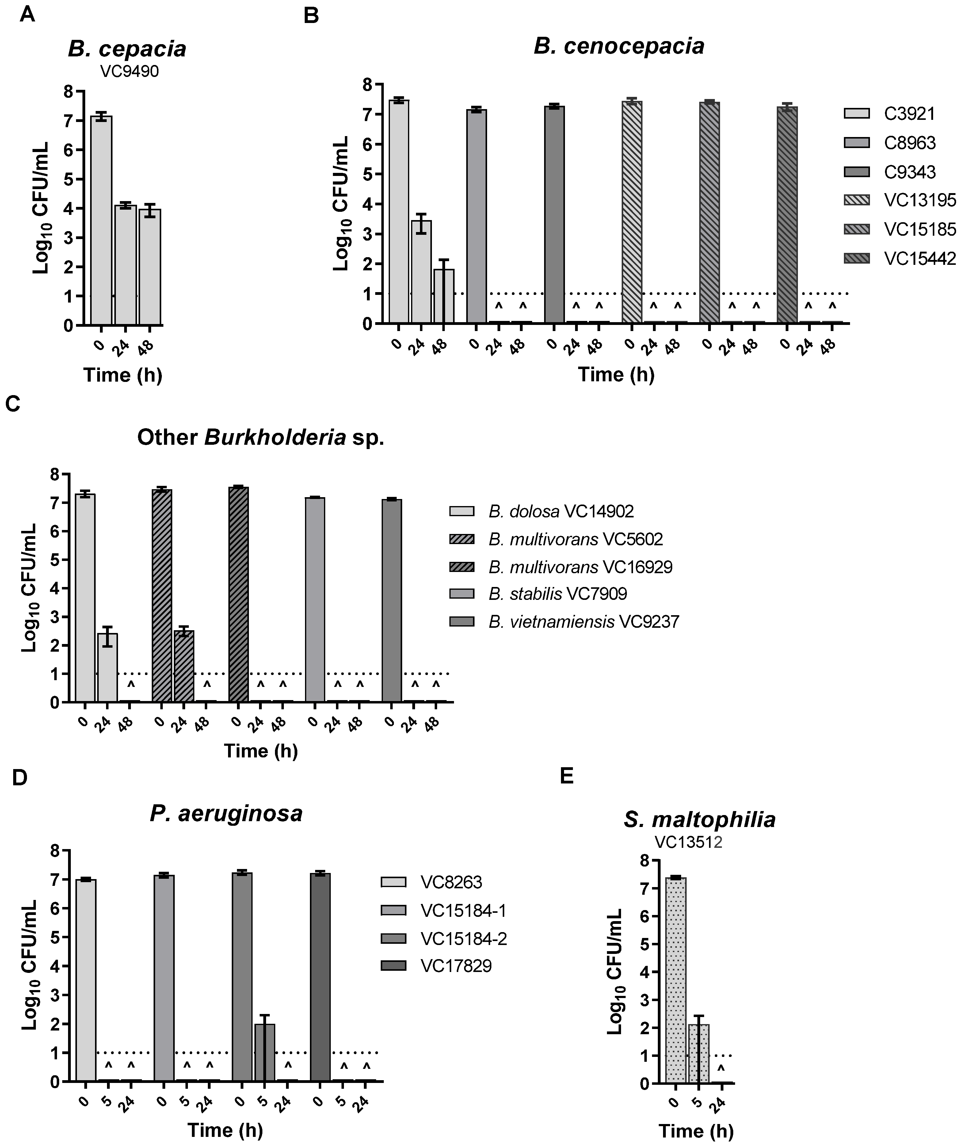
3.2. KC Aqueous Suspensions and Leachate Showed Potent Antibacterial Activity against All the Isolates

To examine the effect of KC on the isolates, an in vitro assay using 1 or 10% (w/v) aqueous suspensions of KC (pH 4.3–4.5) was performed as described previously [67]. As shown in Figure 1 exposure to KC reduced, and in most cases eliminated, the viability of all isolates tested. After 24 h treatment of Bcc strains with 10% (w/v) KC suspensions, no viable cells could be recovered except for B. dolosa and B. multivorans VC5602, which required up to 48 h (Figure 1B,C) and B. cepacia and B. cenocepacia C3921 that showed a 3–5 log10 decline in CFUs in the same period of treatment (Figure 1A,B). A 1% (w/v) suspension of KC did not inhibit Bcc isolates (data not shown) but caused a loss of the viability of P. aeruginosa VC8263, VC15184-1, and VC17829 in 5 h and P. aeruginosa VC15184-2 and S. maltophilia within 24 h of treatment (Figure 1D,E). In contrast, the viability controls without KC exhibited less than one log10 decline in CFU counts during the same periods of incubation (data not shown).
Here, to test if the antibacterial effect was due to soluble components released from KC particles, a water-leachable fraction of the KC suspension was prepared and assayed for activity against these isolates. Figure 2 illustrates that the KC aqueous leachate (L100, pH 3.9) was bactericidal to most of the isolates. While B. dolosa, B. stabilis, B. vietnamiensis, all P. aeruginosa, and S. maltophilia strains exhibited loss of viability after 24 h of treatment with L100 (Figure 2C–E), the same bactericidal effect took 48 h for B. cenocepacia C9343 and B. multivorans VC5602 (Figure 2B,C). B. cepacia exhibited more than a 4 log10 decline in CFU during the period of treatment and all B. cenocepacia strains, other than C9343, and also B. multivorans VC16929 showed a ~1–3 log10 decline in the viability (Figure 2A–C).

4. Discussion

CF once known as an untreatable uniformly fatal disease in early childhood among the Caucasian population is now recognized as a globally distributed disease of a new face with prolonged survival to adulthood [11,33]. Over six decades, there has been a remarkable improvement in health outcomes as well as a substantial increase in the life expectancy of pwCF [33]. The basis for the improvement in clinical and health outcomes is multifactorial, much of which is associated with advancements in antimicrobial treatments [45]. Despite such substantial advances, CF-associated morbidity is still dominated by recurrent pulmonary infections as the most severe manifestation of the disease [3,11,33]. Thus, continued development and optimal application of novel antimicrobial agents (including antibiotic combinations) are vital to improving the survival and life quality of pwCF. As the hallmark of CF pulmonary disease, an early and persistent bacterial infection is the major determinant of life span in affected patients [3,15,44]. The most challenging problem in the management of CF is the early development of chronic infections, which requires successful colonization followed by long-term organism survival and, typically, additional antimicrobial resistance [15,16]. In spite of continuous changes in the epidemiology of CF pathogens, Bcc still remains the most feared threat to pwCF [30,32].
The Bcc species of bacteria are responsible for the most challenging of all pulmonary infections in pwCF due to their remarkable resistance to most available therapeutic agents making them virtually difficult to treat [28,32,40,41]. As current antimicrobial options for Bcc are limited and antimicrobial resistance evolves rapidly, the development of novel therapeutic strategies aimed at disarming Bcc bacteria and other MDR infections from pwCF needs ongoing investigation [38,39]. While Bcc-infected CF patients are in dire need of effective therapeutics, some studies have bridged the gap by exploring novel approaches including antibiotic combination therapies [79,80,81], screening natural antimicrobial compounds such as medical plant-derived small molecules [82,83], and novel aerosolized antibiotic formulations [45,84,85]. One of the most significant advances in CF therapeutics has been the development of CFTR modulators, (both potentiators and correctors) as a corrective strategy, while bacteriophage therapy, vaccine strategies, and immunotherapy remain mostly experimental [3,80,86,87,88].
This study demonstrates the in vitro antibacterial effect of KC and its aqueous leachate (L100) against all the clinical isolates tested, including sequential isolates from chronic infections of B. cenocepacia and B. multivorans, the two most common Bcc species that account for around 85–97% of all Bcc infections [36,38,89], and P. aeruginosa isolates as the most common pathogen in pwCF [17,75]. These data confirmed our previous observations on the potent bactericidal effect of KC against MDR clinical isolates of P. aeruginosa [67]. Notably, two epidemic Type strains of P. aeruginosa (VC8263 and VC17829) were completely eradicated in our study. In addition, P. aeruginosa VC15184-1, a mucoid strain, and its non-mucoid derivative VC15184-2 were similarly affected by KC, indicating that a mucoid phenotype in P. aeruginosa is not likely to be a major factor in resistance to KC. In addition, we could not find any correlation between the mucoidy of Bcc isolates and their sensitivity toward KC suspension and its aqueous leachate (Supplementary Table S1). Noteworthy, our study revealed the high sensitivity of P. aeruginosa isolates toward KC and its leachate. As P. aeruginosa is a major opportunistic pathogen responsible for life-threatening infections, these findings will be a guide to the use of alternative metal-based antimicrobial compounds in the battle against this major recalcitrant group of pathogens. Elucidating the mode of action of KC leachates together with the potent growth inhibitory action of KC provides a specific direction to assess the therapeutic potential of KC and its derivatives for the inhibition of these major bacterial pathogens. Our studies further expand the spectrum of the activity of KC to include isolates of some of the most challenging bacterial pathogens from CF and suggest further studies of other globally important Burkholderia pathogens such as B. pseudomallei and B. mallei. Notably, B. pseudomallei, formerly known as Pseudomonas pseudomallei is the causative agent of melioidosis, a life-threatening infection in humans with a high fatality rate and a wide range of clinical manifestations [90,91].
To the best of our knowledge, this is the first study summarizing the antibacterial activity of a natural clay mineral and its derivatives against CF-related MDR Bcc and P. aeruginosa clinical pathogens. Recently, inhaled antibiotic therapy through aerosolized drug delivery has been explored as an effective method to deliver high concentrations of therapeutic agents to the lungs of patients suffering from respiratory illnesses [45,85]. Moreover, as the cornerstones of treatment for CF pulmonary infections consist of antimicrobial agents together with airway clearance therapy and treatments for affecting mucus rheology [3], KC leachate may be a potential therapeutic option, as a complementary or a suppressive antimicrobial treatment, for pathogenic colonization and chronic pulmonary infections in pwCF and in cases of CGD. Further detailed cytotoxicity investigations as well as in vivo studies in animal models remain to be carried out.
We previously characterized the geochemical and microbiological features across the KC deposit from different depths to obtain more insight into the characteristics that contribute to its antibacterial activity [64,65]. Supplementary Table S2 represents the results of the quantitative phase analysis of the KC clay sample using an X-ray diffraction method. The KC clay sample used in this study can be classified as a mixture of framework silicates and illite/chlorite type phyllosilicates, composed of silicate minerals (96.7%), mainly of tectosilicates (65.4%) known as framework silicates [64,65]. KC sample contains phyllosilicates (24.3%) including biotite, illite-type mica, and chlorite-type clinochlore [64]. Interestingly, while all the natural clay minerals with potent antibacterial activity were reported to contain smectite as the dominant mineral group [57,59,61], KC contains biotite as the major clay mineral [64,65].
We also performed elemental analyses of bulk KC samples and their aqueous leachates which characterized KC as an iron- and aluminum-rich clay mineral [64,65,66]. In addition, integrating physicochemical characterization with microbiological studies, dissected the complex antibacterial activities of KC, suggesting a multi-target mechanism of action [65,66]. Our mechanistic investigations, using Escherichia coli, Staphylococcus aureus, and P. aeruginosa (as representative Gram-negative, Gram-positive, and clinically important organisms, respectively), revealed that the antibacterial features of KC aqueous leachates were influenced by the presence of divalent and trivalent metal ions, in a pH-dependant manner [66]. Together these suggest a critical role for transition metal ions and aluminum in the potent antibacterial activity of KC leachates [65,66]. In fact, a low-pH buffered environment, rich in a combination of released metal ions, plays a key role and challenges treated bacteria synergistically through permeabilization of OM, destabilization of the cell membrane, and induction of oxidative stress [65]. As the OM permeability barrier is a critical contributor to Bcc antimicrobial resistance [32,38], further studies build on our current knowledge through KC active components and bacterial targets should aim to elucidate the lethal mode(s) of action of KC derivatives against Bcc and P. aeruginosa, particularly the role of OM. Moreover, Nunvar et al., reported recently that oxidative stress response- and transition metal metabolism-related genes in B. cenocepacia were affected substantially during CF chronic infections [92], so further investigations exploring these two aspects can yield more insight into the sensitivity of Bcc toward KC derivatives. Altogether, these studies can ultimately facilitate the development of more defined and consistent preparations of KC as potential suppressive or therapeutic options. Such natural mineral-based agents may offer novel weapons in our battle against MDR pathogens in the post-antibiotic era.

Supplementary Materials

The following are available online at https://www.mdpi.com/article/10.3390/microorganisms11010150/s1, Table S1. Mucoidy phenotype of the collection of CF isolates, Text S1. Mineralogical composition of Kisameet clay (KC) by X-ray diffraction, Table S2. Mineralogical composition of KC clay mineral using X-ray diffraction. Reference [93] is cited in the supplementary materials.

Author Contributions

Conceptualization, S.B. and J.E.D.; methodology, S.B., W.X., L.Y.L. and J.E.D.; software, S.B; visualization, S.B.; validation, S.B., J.E.A.Z. and J.E.D.; supervision, J.E.D. and L.Y.L.; formal analysis, S.B.; investigation, S.B.; resources, J.E.A.Z., L.Y.L. and J.E.D.; data curation, S.B.; writing—original draft preparation, S.B.; writing—review and editing, S.B., J.E.A.Z. and J.E.D.; project administration, J.E.D.; funding acquisition, S.B., W.X., J.E.A.Z., L.Y.L. and J.E.D. All authors have read and agreed to the published version of the manuscript.

Funding

These studies were supported by a MITACS Canada (UBC) student fellowship (grant 22R07416 to S.B.), a MITACS Elevate fellowship (grant IT01787 to W.X.), Kisameet Glacial Clay Inc. (Collaborative Research Agreement, UBC F13-03522, grant 11R17370 to L.Y.L.), and the Tally Fund (J.E.D.). They were also funded by an operating grant from Cystic Fibrosis Canada (20R67183) (J.E.A.Z.).

Data Availability Statement

Data are presented in the paper as well as the supporting information.

Acknowledgments

We acknowledge the Heiltsuk First Nation People and Kisameet Glacial Clay Inc. for providing the clay samples. We are grateful to John Grace for his contribution, coordination, and advice. We also thank Vivian Miao for helpful discussions in editing the manuscript and Ivan Villanueva for technical support.

Conflicts of Interest

The authors declare no conflict of interest.

References

  1. Grant, S.S.; Hung, D.T. Persistent bacterial infections, antibiotic tolerance, and the oxidative stress response. Virulence 2013, 4, 273–283. [Google Scholar] [CrossRef] [Green Version]
  2. Exner, M.; Bhattacharya, S.; Christiansen, B.; Gebel, J.; Goroncy-Bermes, P.; Hartemann, P.; Heeg, P.; Ilschner, C.; Kramer, A.; Larson, E.; et al. Antibiotic resistance: What is so special about multidrug-resistant Gram-negative bacteria? GMS Hyg. Infect. Control 2017, 12, Doc05. [Google Scholar] [CrossRef] [PubMed]
  3. Goetz, D.; Ren, C.L. Review of Cystic Fibrosis. Pediatr. Ann. 2019, 48, e154–e161. [Google Scholar] [CrossRef] [PubMed]
  4. Ratjen, F.; Bell, S.C.; Rowe, S.M.; Goss, C.H.; Quittner, A.L.; Bush, A. Cystic fibrosis. Nat. Rev. Dis. Prim. 2015, 1, 15010. [Google Scholar] [CrossRef] [PubMed]
  5. Lyczak, J.B.; Cannon, C.L.; Pier, G.B. Lung infections associated with cystic fibrosis. Clin. Microbiol. Rev. 2002, 15, 194–222. [Google Scholar] [CrossRef] [PubMed] [Green Version]
  6. Gibson, R.L.; Burns, J.L.; Ramsey, B.W. Pathophysiology and management of pulmonary infections in cystic fibrosis. Am. J. Respir. Crit. Care Med. 2003, 168, 918–951. [Google Scholar] [CrossRef] [PubMed]
  7. Andersen, D.H. Cystic fibrosis of the pancreas and its relation to celiac disease. Am. J. Dis. Child. 1938, 56, 344–399. [Google Scholar] [CrossRef]
  8. Kerem, B.S.; Zielenski, J.; Markiewicz, D.; Bozon, D.; Gazit, E.; Yahav, J.; Kennedy, D.; Riordan, J.R.; Collins, F.S.; Rommens, J.M. Identification of mutations in regions corresponding to the two putative nucleotide (ATP)-binding folds of the cystic fibrosis gene. Proc. Natl. Acad. Sci. USA 1990, 87, 8447–8451. [Google Scholar] [CrossRef] [Green Version]
  9. Spoonhower, K.A.; Davis, P.B. Epidemiology of Cystic Fibrosis. Clin. Chest Med. 2016, 37, 1–8. [Google Scholar] [CrossRef]
  10. Scotet, V.; L’Hostis, C.; Férec, C. The Changing Epidemiology of Cystic Fibrosis: Incidence, Survival and Impact of the CFTR Gene Discovery. Genes 2020, 11, 589. [Google Scholar] [CrossRef]
  11. De Boeck, K. Cystic fibrosis in the year 2020: A disease with a new face. Acta Paediatr. 2020, 109, 893–899. [Google Scholar] [CrossRef] [PubMed]
  12. Chen, Q.; Shen, Y.; Zheng, J. A review of cystic fibrosis: Basic and clinical aspects. Anim. Model. Exp. Med. 2021, 4, 220–232. [Google Scholar] [CrossRef] [PubMed]
  13. Cutting, G.R. Cystic fibrosis genetics: From molecular understanding to clinical application. Nat. Rev. Genet. 2015, 16, 45–56. [Google Scholar] [CrossRef] [Green Version]
  14. Regard, L.; Martin, C.; Chassagnon, G.; Burgel, P.R. Acute and chronic non-pulmonary complications in adults with cystic fibrosis. Expert Rev. Respir. Med. 2019, 13, 23–38. [Google Scholar] [CrossRef] [PubMed]
  15. Garcia, B.; Flume, P.A. Pulmonary Complications of Cystic Fibrosis. Semin. Respir. Crit. Care Med. 2019, 40, 804–809. [Google Scholar] [CrossRef]
  16. Filkins, L.M.; O’Toole, G.A. Cystic Fibrosis Lung Infections: Polymicrobial, Complex, and Hard to Treat. PLoS Pathog. 2015, 11, e1005258. [Google Scholar] [CrossRef] [Green Version]
  17. Davies, J.C. Pseudomonas aeruginosa in cystic fibrosis: Pathogenesis and persistence. Paediatr. Resp. Rev. 2002, 3, 128–134. [Google Scholar] [CrossRef]
  18. Lipuma, J.J. The changing microbial epidemiology in cystic fibrosis. Clin. Microbiol. Rev. 2010, 23, 299–323. [Google Scholar] [CrossRef] [Green Version]
  19. Leitão, J.H.; Sousa, S.A.; Ferreira, A.S.; Ramos, C.G.; Silva, I.N.; Moreira, L.M. Pathogenicity, virulence factors, and strategies to fight against Burkholderia cepacia complex pathogens and related species. Appl. Microbiol. Biotechnol. 2010, 87, 31–40. [Google Scholar] [CrossRef]
  20. Sfeir, M.M. Burkholderia cepacia complex infections: More complex than the bacterium name suggest. J. Infect. 2018, 77, 166–170. [Google Scholar] [CrossRef]
  21. Ragupathi, N.K.D.; Veeraraghavan, B. Accurate identification and epidemiological characterization of Burkholderia cepacia complex: An update. Ann. Clin. Microbiol. Antimicrob. 2019, 18, 7. [Google Scholar] [CrossRef] [Green Version]
  22. Jin, Y.; Zhou, J.; Zhou, J.; Hu, M.; Zhang, Q.; Kong, N.; Ren, H.; Liang, L.; Yue, J. Genome-based classification of Burkholderia cepacia complex provides new insight into its taxonomic status. Biol. Direct 2020, 15, 6. [Google Scholar] [CrossRef] [PubMed]
  23. Laraya-Cuasay, L.; Lipstein, M.; Huang, N. Pseudomonas cepacia in the respiratory flora of patients with cystic fibrosis (CF). Pediatr. Res. 1977, 11, 502. [Google Scholar] [CrossRef] [Green Version]
  24. Vandamme, P.; Holmes, B.; Vancanneyt, M.; Coenye, T.; Hoste, B.; Coopman, R.; Revets, H.; Lauwers, S.; Gillis, M.; Kersters, K.; et al. Occurrence of multiple genomovars of Burkholderia cepacia in cystic fibrosis patients and proposal of Burkholderia multivorans sp. nov. Int. J. Syst. Evol. Bacteriol. 1997, 47, 1188–1200. [Google Scholar] [CrossRef] [Green Version]
  25. Tablan, O.C.; Chorba, T.L.; Schidlow, D.V.; White, J.W.; Hardy, K.A.; Gilligan, P.H.; Morgan, W.M.; Carson, L.A.; Martone, W.J.; Jason, J.M. Pseudomonas cepacia colonization in patients with cystic fibrosis: Risk factors and clinical outcome. J. Pediatr. 1985, 107, 382–387. [Google Scholar] [CrossRef] [PubMed]
  26. Eberl, L.; Vandamme, P. Members of the genus Burkholderia: Good and bad guy. F1000Research 2016, 5, 1007. [Google Scholar] [CrossRef] [PubMed]
  27. Lipuma, J.J. Update on the Burkholderia cepacia complex. Curr. Opin. Pulm. Med. 2005, 11, 528–533. [Google Scholar] [CrossRef]
  28. Mahenthiralingam, E.; Urban, T.A.; Goldberg, J.B. The multifarious, multireplicon Burkholderia cepacia complex. Nat. Rev. Microbiol. 2005, 3, 144–156. [Google Scholar] [CrossRef]
  29. Speert, D.P. Advances in Burkholderia cepacia complex. Paediatr. Respir. Rev. 2002, 3, 230–235. [Google Scholar] [CrossRef]
  30. Leitão, J.H.; Feliciano, J.R.; Sousa, S.A.; Guerreiro, T.P.A.I. Burkholderia cepacia Complex Infections Among Cystic Fibrosis Patients: Perspectives and Challenges. In Progress in Understanding Cystic Fibrosis; IntechOpen: London, UK, 2017; pp. 73–99. [Google Scholar] [CrossRef]
  31. LiPuma, J.J. Burkholderia cepacia. Management issues and new insights. Clin. Chest Med. 1998, 19, 473–486. [Google Scholar] [CrossRef]
  32. Scoffone, V.C.; Chiarelli, L.R.; Trespidi, G.; Mentasti, M.; Riccardi, G.; Buroni, S. Burkholderia cenocepacia Infections in Cystic Fibrosis Patients: Drug Resistance and Therapeutic Approaches. Front. Microbiol. 2017, 8, 1592. [Google Scholar] [CrossRef] [PubMed] [Green Version]
  33. Bell, S.C.; Mall, M.A.; Gutierrez, H.; Macek, M.; Madge, S.; Davies, J.C.; Burgel, P.R.; Tullis, E.; Castaños, C.; Castellani, C.; et al. The future of cystic fibrosis care: A global perspective. Lancet Respir. Med. 2020, 8, 65–124. [Google Scholar] [CrossRef] [Green Version]
  34. Hirche, T.O.; Knoop, C.; Hebestreit, H.; Shimmin, D.; Solé, A.; Elborn, J.S.; Ellemunter, H.; Aurora, P.; Hogardt, M.; Wagner, T.O.; et al. Practical guidelines: Lung transplantation in patients with cystic fibrosis. Pulm. Med. 2014, 2014, 621342. [Google Scholar] [CrossRef] [Green Version]
  35. De Soyza, A.; McDowell, A.; Archer, L.; Dark, J.H.; Elborn, S.J.; Mahenthiralingam, E.; Gould, K.; Corris, P.A. Burkholderia cepacia complex genomovars and pulmonary transplantation outcomes in patients with cystic fibrosis. Lancet 2001, 358, 1780–1781. [Google Scholar] [CrossRef]
  36. Mahenthiralingam, E.; Baldwin, A.; Dowson, C.G. Burkholderia cepacia complex bacteria: Opportunistic pathogens with important natural biology. J. Appl. Microbiol. 2008, 104, 1539–1551. [Google Scholar] [CrossRef]
  37. Leitão, J.H.; Sousa, S.A.; Cunha, M.V.; Salgado, M.J.; Melo-Cristino, J.; Barreto, M.C.; Sá-Correia, I. Variation of the antimicrobial susceptibility profiles of Burkholderia cepacia complex clonal isolates obtained from chronically infected cystic fibrosis patients: A five-year survey in the major Portuguese treatment center. Eur. J. Clin. Microbiol. Infect. Dis. 2008, 27, 1101–1111. [Google Scholar] [CrossRef]
  38. Rhodes, K.A.; Schweizer, H.P. Antibiotic resistance in Burkholderia species. Drug Resist. Update 2016, 28, 82–90. [Google Scholar] [CrossRef] [Green Version]
  39. Lord, R.; Jones, A.M.; Horsley, A. Antibiotic treatment for Burkholderia cepacia complex in people with cystic fibrosis experiencing a pulmonary exacerbation. Cochrane Database Syst. Rev. 2020, 4, CD009529. [Google Scholar] [CrossRef]
  40. Regan, K.H.; Bhatt, J. Eradication therapy for Burkholderia cepacia complex in people with cystic fibrosis. Cochrane Database Syst. Rev. 2019, 4, CD009876. [Google Scholar] [CrossRef]
  41. Zlosnik, J.E.; Zhou, G.; Brant, R.; Henry, D.A.; Hird, T.J.; Mahenthiralingam, E.; Chilvers, M.A.; Wilcox, P.; Speert, D.P. Burkholderia species infections in patients with cystic fibrosis in British Columbia, Canada. 30 years’ experience. Ann. Am. Thorac. Soc. 2015, 12, 70–78. [Google Scholar] [CrossRef]
  42. Conway, B.A.; Venu, V.; Speert, D.P. Biofilm formation and acyl homoserine lactone production in the Burkholderia cepacia complex. J. Bacteriol. 2002, 184, 5678–5685. [Google Scholar] [CrossRef] [Green Version]
  43. Davies, J.C.; Bilton, D. Bugs, biofilms, and resistance in cystic fibrosis. Respir. Care 2009, 54, 628–640. [Google Scholar] [CrossRef]
  44. Ciofu, O.; Tolker-Nielsen, T.; Jensen, P.Ø.; Wang, H.; Høiby, N. Antimicrobial resistance, respiratory tract infections and role of biofilms in lung infections in cystic fibrosis patients. Adv. Drug Deliv. Rev. 2015, 85, 7–23. [Google Scholar] [CrossRef]
  45. Waters, V.; Smyth, A. Cystic fibrosis microbiology: Advances in antimicrobial therapy. J. Cyst. Fibros. 2015, 14, 551–560. [Google Scholar] [CrossRef] [PubMed]
  46. Silver, L.L. Challenges of antibacterial discovery. Clin. Microbiol. Rev. 2011, 24, 71–109. [Google Scholar] [CrossRef] [PubMed] [Green Version]
  47. Davies, J.E.; Behroozian, S. An ancient solution to a modern problem. Mol. Microbiol. 2020, 113, 546–549. [Google Scholar] [CrossRef] [PubMed] [Green Version]
  48. Carretero, M.I. Clay minerals and their beneficial effect upon human health. A review. Appl. Clay Sci. 2002, 21, 155–163. [Google Scholar] [CrossRef]
  49. Williams, L.B.; Holland, M.; Eberl, D.D.; Brunet, T.; de Courrsou, L.B. Killer clays! Natural antibacterial clay minerals. Miner. Soc. Bull. 2004, 139, 3–8. [Google Scholar]
  50. Gomes, C.S.F.; Silva, J.B.P. Minerals and clay minerals in medical geology. Appl. Clay Sci. 2007, 36, 4–21. [Google Scholar] [CrossRef]
  51. Carretero, M.I.; Gomes, C.S.F.; Tateo, F. Clays and human health. In Handbook of Clay Science, Developments in Clay Science; Bergaya, F., Theng, B.K.G., Lagaly, G., Eds.; Elsevier: Amsterdam, The Netherlands, 2006; Volume 1, pp. 717–741. ISBN 978-0-08-044183-2. [Google Scholar]
  52. Gomes, C. Healing and edible clays: A review of basic concepts, benefits and risks. Environ. Geochem. Health 2018, 40, 1739–1765. [Google Scholar] [CrossRef]
  53. Velde, B. Geology of clays. In Origin and Mineralogy of Clays, Clays and the Environment; Velde, B., Ed.; Springer: Berlin, Germany, 1995; pp. 1–7. [Google Scholar]
  54. Brigatti, M.F.; Galán, E.; Theng, B.K.G. Structure and Mineralogy of Clay Minerals. In Handbook of Clay Science, Developments in Clay Science; Bergaya, F., Theng, B.K.G., Lagaly, G., Eds.; Elsevier: Amsterdam, The Netherlands, 2006; Volume 1, pp. 21–81. ISBN 978-0-08-044183-2. [Google Scholar]
  55. Uddin, F. Clays, nanoclays, and montmorillonite minerals. Met. Mater. Trans. A 2008, 39, 2804–2814. [Google Scholar] [CrossRef]
  56. World Health Organization. Draft Report of the 5th WHO Advisory Group Meeting on Buruli Ulcer; Study group report on Buruli Ulcer treatment with clay; World Health Organization: Geneva, Switzerland, 2002.
  57. Williams, L.B.; Haydel, S.E.; Giese, R.F.; Eberl, D.D. Chemical and mineralogical characteristics of French green clays used for healing. Clays Clay Miner. 2008, 56, 437–452. [Google Scholar] [CrossRef] [PubMed]
  58. Haydel, S.E.; Remenih, C.M.; Williams, L.B. Broad-spectrum in vitro antibacterial activities of clay minerals against antibiotic-susceptible and antibiotic-resistant bacterial pathogens. J. Antimicrob. Chemother. 2008, 61, 353–361. [Google Scholar] [CrossRef] [PubMed] [Green Version]
  59. Williams, L.B.; Metge, D.W.; Eberl, D.D.; Harvey, R.W.; Turner, A.G.; Prapaipong, P.; Poret-Peterson, A.T. What makes a natural clay antibacterial? Environ. Sci. Technol. 2011, 45, 3768–3773. [Google Scholar] [CrossRef] [Green Version]
  60. Mpuchane, S.F.; Ekosse, G.I.E.; Gashe, B.A.; Morobe, I.; Coetzee, S.H. Mineralogy of southern Africa medicinal and cosmetic clays and their effects on the growth of selected test microorganisms. Fresenius Environ. Bull. 2008, 17, 547–557. [Google Scholar] [CrossRef]
  61. Morrison, K.D.; Misra, R.; Williams, L.B. Unearthing the antibacterial mechanism of medicinal clay: A geochemical approach to combating antibiotic resistance. Sci. Rep. 2016, 6, 19043. [Google Scholar] [CrossRef] [PubMed] [Green Version]
  62. Hauser, E.A. Kisameet Bay Clay Deposit. In Problems of Clay and Laterite Genesis Symposium at Annual Meeting of the American Institute of Mining and Metallurgical Engineers, St. Louis, MO, USA, 19–22 February 1951; The American Institute of Mining and Metallurgical Engineers: St. Louis, MO, USA, 1952; pp. 178–190. [Google Scholar]
  63. Ure, W.; Harris, J.A. Curative properties of rare earths found in B.C. peloid deposits. Bull. Vanc. Med. Assoc. 1946, 22, 230–237. [Google Scholar] [PubMed]
  64. Svensson, S.L.; Behroozian, S.; Xu, W.; Surette, M.G.; Li, L.; Davies, J. Kisameet Glacial Clay: An Unexpected Source of Bacterial Diversity. mBio 2017, 8, e00590-17. [Google Scholar] [CrossRef] [PubMed] [Green Version]
  65. Behroozian, S. Antimicrobial Properties of Kisameet Clay, A Natural Clay Mineral from British Columbia, Canada. Ph.D. Thesis, University of British Columbia, Vancouver, BC, Canada, 2019. Available online: http://open.library.ubc.ca/collections/ubctheses/24/items/1.0380811 (accessed on 4 September 2019).
  66. Behroozian, S.; Svensson, S.L.; Li, L.Y.; Davies, J.E. Broad-Spectrum Antimicrobial and Antibiofilm Activity of a Natural Clay Mineral from British Columbia, Canada. mBio 2020, 11, e02350-20. [Google Scholar] [CrossRef]
  67. Behroozian, S.; Svensson, S.L.; Davies, J. Kisameet clay exhibits potent antibacterial activity against the ESKAPE pathogens. mBio 2016, 7, e01842-15. [Google Scholar] [CrossRef]
  68. Speert, D.P.; Campbell, M.E.; Henry, D.A.; Milner, R.; Taha, F.; Gravelle, A.; Davidson, A.G.; Wong, L.T.; Mahenthiralingam, E. Epidemiology of Pseudomonas aeruginosa in cystic fibrosis in British Columbia, Canada. Am. J. Respir. Crit. Care Med. 2002, 166, 988–993. [Google Scholar] [CrossRef]
  69. Speert, D.P.; Henry, D.; Vandamme, P.; Corey, M.; Mahenthiralingam, E. Epidemiology of Burkholderia cepacia complex in patients with cystic fibrosis, Canada. Emerg. Infect. Dis. 2002, 8, 181–187. [Google Scholar] [CrossRef] [Green Version]
  70. Bauer, A.W.; Kirby, W.M.; Sherris, J.C.; Turck, M. Antibiotic susceptibility testing by a standardized single disk method. Am. J. Clin. Pathol. 1966, 45, 493–496. [Google Scholar] [CrossRef]
  71. Hudzicki, J. Kirby-Bauer Disk Diffusion Susceptibility Test Protocol. American Society for Microbiology. 2009. Available online: https://asm.org/Protocols/Kirby-Bauer-Disk-Diffusion-Susceptibility-Test-Pro. (accessed on 19 June 2016).
  72. Magiorakos, A.P.; Srinivasan, A.; Carey, R.B.; Carmeli, Y.; Falagas, M.E.; Giske, C.G.; Harbarth, S.; Hindler, J.F.; Kahlmeter, G.; Olsson-Liljequist, B.; et al. Multidrug-resistant, extensively drug-resistant and pandrug-resistant bacteria: An international expert proposal for interim standard definitions for acquired resistance. Clin. Microbiol. Infect. 2012, 18, 268–281. [Google Scholar] [CrossRef] [Green Version]
  73. Miller, R.R.; Hird, T.J.; Tang, P.; Zlosnik, J.E. Whole-Genome Sequencing of Three Clonal Clinical Isolates of B. cenocepacia from a Patient with Cystic Fibrosis. PLoS ONE 2015, 10, e0143472. [Google Scholar] [CrossRef] [PubMed] [Green Version]
  74. Zlosnik, J.E.; Speert, D.P. The role of mucoidy in virulence of bacteria from the Burkholderia cepacia complex: A systematic proteomic and transcriptomic analysis. J. Infect. Dis. 2010, 202, 770–781. [Google Scholar] [CrossRef] [PubMed] [Green Version]
  75. Silva, I.N.; Santos, P.M.; Santos, M.R.; Zlosnik, J.E.; Speert, D.P.; Buskirk, S.W.; Bruger, E.L.; Waters, C.M.; Cooper, V.S.; Moreira, L.M. Long-Term Evolution of Burkholderia multivorans during a Chronic Cystic Fibrosis Infection Reveals Shifting Forces of Selection. mSystems 2016, 1, e00029-16. [Google Scholar] [CrossRef] [PubMed] [Green Version]
  76. Kennedy, S.; Beaudoin, T.; Yau, Y.C.; Caraher, E.; Zlosnik, J.E.; Speert, D.P.; LiPuma, J.J.; Tullis, E.; Waters, V. Activity of Tobramycin against Cystic Fibrosis Isolates of Burkholderia cepacia Complex Grown as Biofilms. Antimicrob. Agents Chemother. 2016, 60, 348–355. [Google Scholar] [CrossRef] [Green Version]
  77. Veyssier, P.; Bryskier, A. Aminocyclitol aminoglycosides. In Antimicrobial Agents; Bryskier, A., Ed.; ASM Press: Washington, DC, USA, 2005; pp. 453–469. [Google Scholar]
  78. Ramirez, M.S.; Tolmasky, M.E. Aminoglycoside modifying enzymes. Drug Resist. Update 2010, 13, 151–171. [Google Scholar] [CrossRef] [PubMed] [Green Version]
  79. Van Dalem, A.; Herpol, M.; Echahidi, F.; Peeters, C.; Wybo, I.; De Wachter, E.; Vandamme, P.; Piérard, D. In Vitro Susceptibility of Burkholderia cepacia Complex Isolated from Cystic Fibrosis Patients to Ceftazidime-Avibactam and Ceftolozane-Tazobactam. Antimicrob. Agents Chemother. 2018, 62, e00590-18. [Google Scholar] [CrossRef]
  80. Zeiser, E.T.; Becka, S.A.; Wilson, B.M.; Barnes, M.D.; LiPuma, J.J.; Papp-Wallace, K.M. “Switching Partners”: Piperacillin-Avibactam Is a Highly Potent Combination against Multidrug-Resistant Burkholderia cepacia Complex and Burkholderia gladioli Cystic Fibrosis Isolates. J. Clin. Microbiol. 2019, 57, e00181-19. [Google Scholar] [CrossRef] [PubMed] [Green Version]
  81. El-Halfawy, O.M.; Naguib, M.M.; Valvano, M.A. Novel antibiotic combinations proposed for treatment of Burkholderia cepacia complex infections. Antimicrob. Resist. Infect. Control 2017, 6, 120. [Google Scholar] [CrossRef] [Green Version]
  82. Vasireddy, L.; Bingle, L.; Davies, M.S. Antimicrobial activity of essential oils against multidrug-resistant clinical isolates of the Burkholderia cepacia complex. PLoS ONE 2018, 13, e0201835. [Google Scholar] [CrossRef] [Green Version]
  83. Maida, I.; Lo Nostro, A.; Pesavento, G.; Barnabei, M.; Calonico, C.; Perrin, E.; Chiellini, C.; Fondi, M.; Mengoni, A.; Maggini, V.; et al. Exploring the Anti-Burkholderia cepacia Complex Activity of Essential Oils: A Preliminary Analysis. Evid.-Based Complement. Altern. Med. 2014, 2014, 573518. [Google Scholar] [CrossRef] [PubMed] [Green Version]
  84. Waters, V.; Yau, Y.; Beaudoin, T.; Wettlaufer, J.; Tom, S.K.; McDonald, N.; Rizvi, L.; Klingel, M.; Ratjen, F.; Tullis, E. Pilot trial of tobramycin inhalation powder in cystic fibrosis patients with chronic Burkholderia cepacia complex infection. J. Cyst. Fibros. 2017, 16, 492–495. [Google Scholar] [CrossRef] [Green Version]
  85. Nichols, D.P.; Durmowicz, A.G.; Field, A.; Flume, P.A.; VanDevanter, D.R.; Mayer-Hamblett, N. Developing Inhaled Antibiotics in Cystic Fibrosis: Current Challenges and Opportunities. Ann. Am. Thorac. Soc. 2019, 16, 534–539. [Google Scholar] [CrossRef] [PubMed]
  86. Semler, D.D.; Goudie, A.D.; Finlay, W.H.; Dennis, J.J. Aerosol phage therapy efficacy in Burkholderia cepacia complex respiratory infections. Antimicrob. Agents Chemother. 2014, 58, 4005–4013. [Google Scholar] [CrossRef] [PubMed] [Green Version]
  87. Pradenas, G.A.; Ross, B.N.; Torres, A.G. Burkholderia cepacia Complex Vaccines: Where Do We Go from here? Vaccines 2016, 4, 10. [Google Scholar] [CrossRef]
  88. Le Moigne, V.; Gaillard, J.L.; Herrmann, J.L. Vaccine strategies against bacterial pathogens in cystic fibrosis patients. Med. Mal. Infect. 2016, 46, 4–9. [Google Scholar] [CrossRef]
  89. Drevinek, P.; Mahenthiralingam, E. Burkholderia cenocepacia in cystic fibrosis: Epidemiology and molecular mechanisms of virulence. Clin. Microbiol. Infect. 2010, 16, 821–830. [Google Scholar] [CrossRef]
  90. Limmathurotsakul, D.; Golding, N.; Dance, D.A.; Messina, J.P.; Pigott, D.M.; Moyes, C.L.; Rolim, D.B.; Bertherat, E.; Day, N.P.; Peacock, S.J.; et al. Predicted global distribution of Burkholderia pseudomallei and burden of melioidosis. Nat. Microbiol. 2016, 1, 15008. [Google Scholar] [CrossRef] [PubMed] [Green Version]
  91. Gassiep, I.; Armstrong, M.; Norton, R. Human Melioidosis. Clin. Microbiol. Rev. 2020, 33, e00006-19. [Google Scholar] [CrossRef] [PubMed]
  92. Nunvar, J.; Capek, V.; Fiser, K.; Fila, L.; Drevinek, P. What matters in chronic Burkholderia cenocepacia infection in cystic fibrosis: Insights from comparative genomics. PLoS Pathog. 2017, 13, e1006762. [Google Scholar] [CrossRef] [PubMed]
  93. Bish, D.L. Studies of Clays and Clay Minerals Using X-ray Powder Diffraction and the Rietveld Method. United States. 1993. Available online: http://www.osti.gov/servlets/purl/10192067 (accessed on 26 June 2015).
Figure 1. Effect of aqueous suspensions of KC on the viability of isolates: 10% (w/v) against B. cepacia complex isolates (AC) and 1% (w/v) against P. aeruginosa isolates (D) and S. maltophilia isolate (E). CFUs have been determined at 0 h, 5 h, 24 h, and 48 h of incubation. ^ indicates that no viable cell could be recovered at that time point. Error bars represent the standard error (SE) of the mean of three independent replicates of each strain. The dotted line at log10 = 1 of the Y axis represents the limit of detection for CFUs.
Microorganisms 11 00150 g001
Figure 2. Effect of aqueous leachate of KC (L100) on the viability of B. cepacia complex isolates (AC), P. aeruginosa isolates (D), and S. maltophilia isolate (E). CFUs have been determined at 0 h, 5 h, 24 h, and 48 h of incubation. ^ indicates that no viable cell could be recovered at that time point. Error bars represent the standard error (SE) of the mean of three independent replicates of each strain. The dotted line at log10 = 1 of the Y axis represents the limit of detection for CFUs.
Microorganisms 11 00150 g002
Table 1. Resistance patterns of isolates to different classes of antimicrobial agents.
Amikacin (10)Gentamicin (30)Kanamycin (30)Neomycin (30)Spectinomycin (100) *Streptomycin (10)Tobramycin (10)Ertapenem (10)Imipenem (10)Meropenem (10)Cephalothin (30)Cephazolin (30)Cefotetan (30)Cefoxitin (30)Ceftazidime (30)Cefixime (5)Cefpodoxime (10)Ceftriaxone (30)Cefotaxime (30)Amoxicillin-clavulanic acid (30)Ampicillin (10)Piperacilin (100)Colistin (10)Polymyxin B (300) Ciprofloxacin (5) Levofloxacin (5) Nalidixic acid (30)Sulfamethoxazole- trimethoprim (25)Sulfadiazine (250)Trimethoprim (5)Doxycycline (30)Tetracycline (30)Chloramphenicol (30)Nitrofurantoin (300)
No.IsolateStrainYearSourceAminoglycosidesCarbapenems1st, 2nd, 3rd Generation CephalosporinsPenicillinsPolypeptidesQuinolonesSulfonamides Tetracyclines
1Burkholderia cepaciaVC94901999Sputum
2Burkholderia cenocepaciaC3921 a [73,74]1990Sputum
3Burkholderia cenocepaciaC8963 a [73,74]2000Resp e
4Burkholderia cenocepaciaC9343 a [73,74]2000Resp
5Burkholderia cenocepaciaVC13195 b2006Resp
6Burkholderia cenocepaciaVC15185 b2010Resp
7Burkholderia cenocepaciaVC15442 b2010Blood f
8Burkholderia dolosaVC149022009Sputum
9Burkholderia multivoransVC5602 c [75]1993Sputum
10Burkholderia multivoransVC16929 c2013Sputum
11Burkholderia stabilisVC79091993Sputum
12Burkholderia vietnamiensisVC9237 [76]1998Resp
13Pseudomonas aeruginosaVC8263 [68]1997Resp
14Pseudomonas aeruginosaVC15184-1 d2010Resp
15Pseudomonas aeruginosaVC15184-2 d2010Resp
16Pseudomonas aeruginosaVC17829 [68]2015Sputum
17Stenotrophomonas maltophiliaVC135122006Sputum
Filled black circles indicate resistance (inhibition ≤ 2 mm from the edge of the disks of antibiotic); no mark indicates the wider zone of inhibition (at least three replicates for each antibiotic). The amount (micrograms) per disk of antibiotics (Oxoid, BBL) is indicated in parentheses. * Spectinomycin is an aminocyclitol antibiotic, but as it is structurally related to the aminoglycosides, often considered alongside this group of antibiotics Refs [77,78]. a,b,c sequential isolates from the same patients that were previously evaluated for strain type by random amplified polymorphic DNA (RAPD) analysis Ref [78]. d two morphotypes of the strain VC15184: (−1, mucoid, and −2, non-mucoid strain which was recovered during the course of this study and confirmed to have the same RAPD phenotype). The remaining strains were independent isolates from one patient. e Resp indicates a sample from the respiratory tract, either sputum or throat/cough swab. f This strain was isolated from a patient’s blood sample during “cepacia” syndrome.
Disclaimer/Publisher’s Note: The statements, opinions and data contained in all publications are solely those of the individual author(s) and contributor(s) and not of MDPI and/or the editor(s). MDPI and/or the editor(s) disclaim responsibility for any injury to people or property resulting from any ideas, methods, instructions or products referred to in the content.

