
Congenital Syphilis Presenting with Brain Abnormalities at Neuroscan: A Case Report and a Brief Literature Review

 , ,
, ,  and
and 
Abstract
:1. Introduction
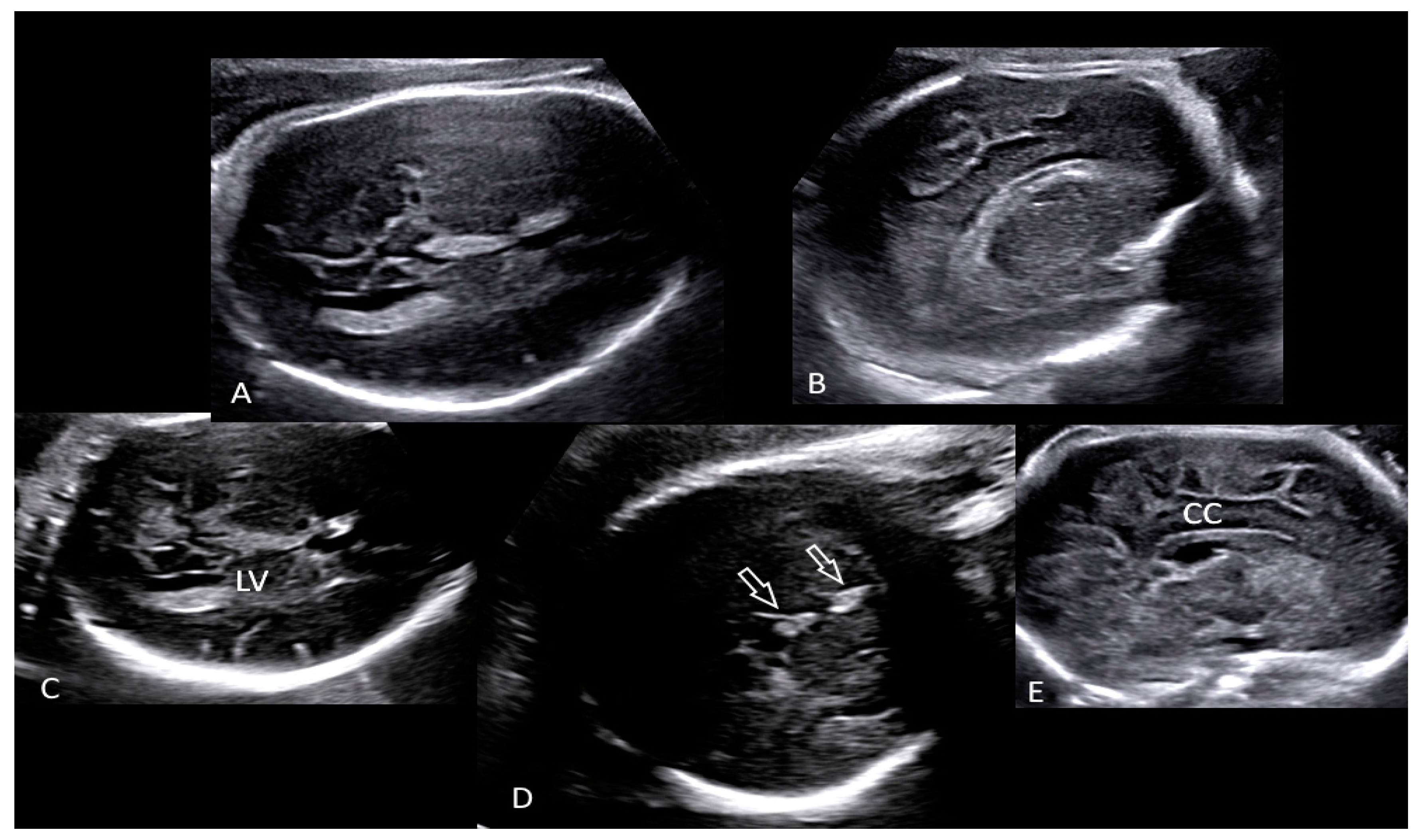
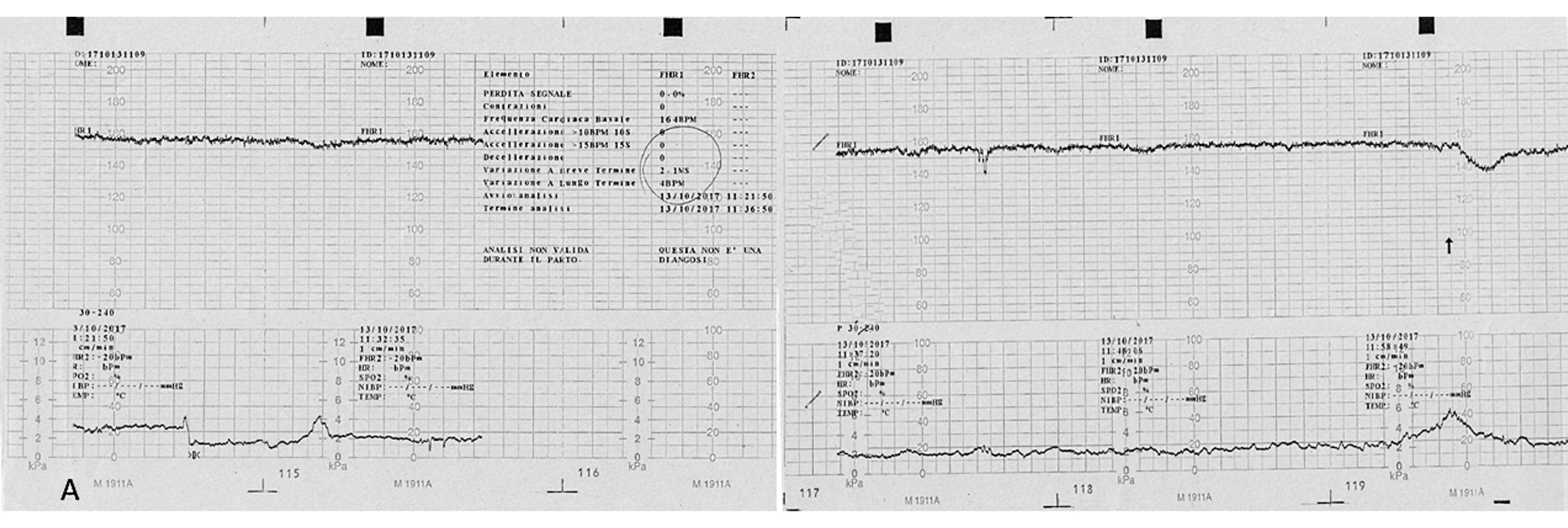
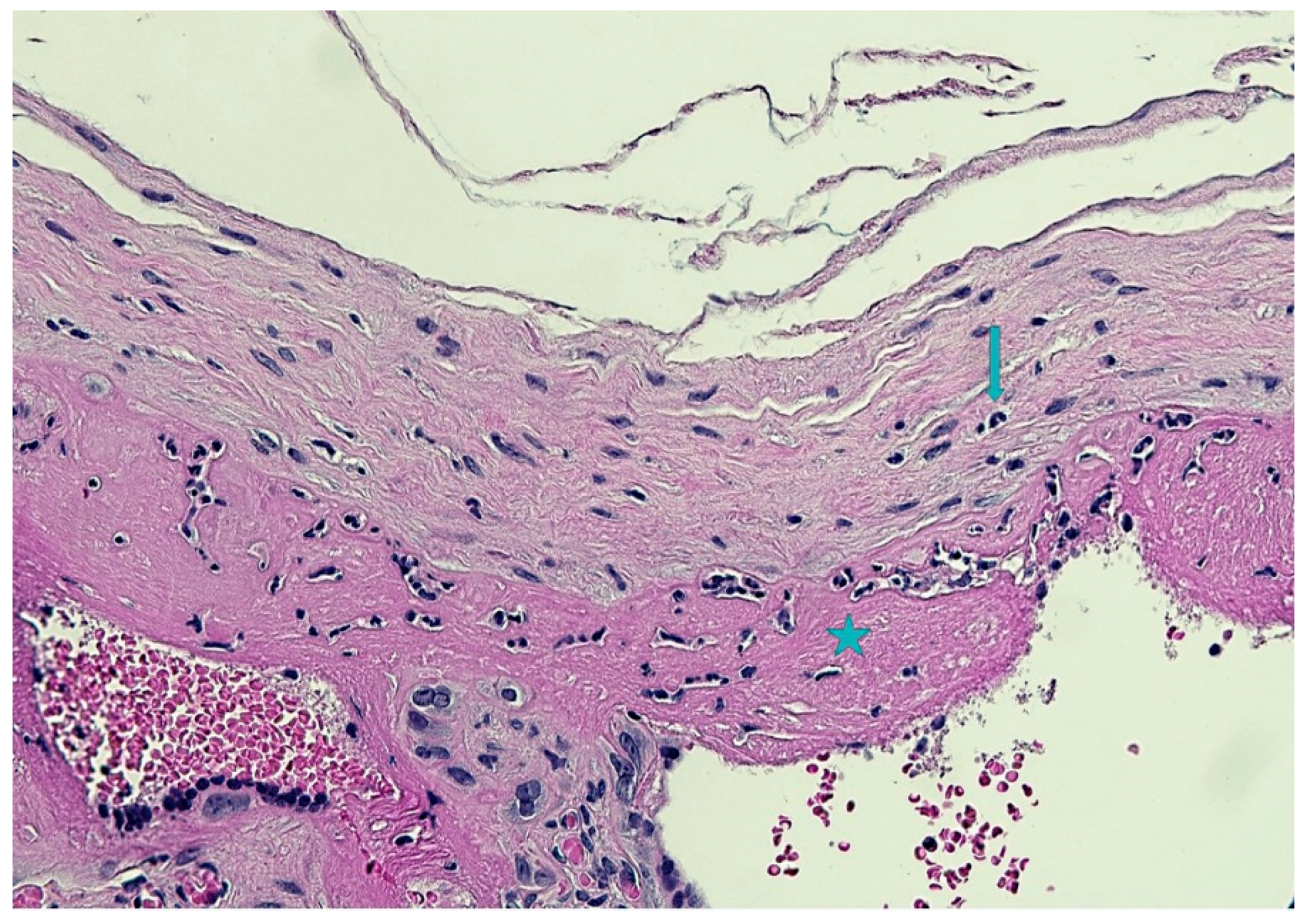
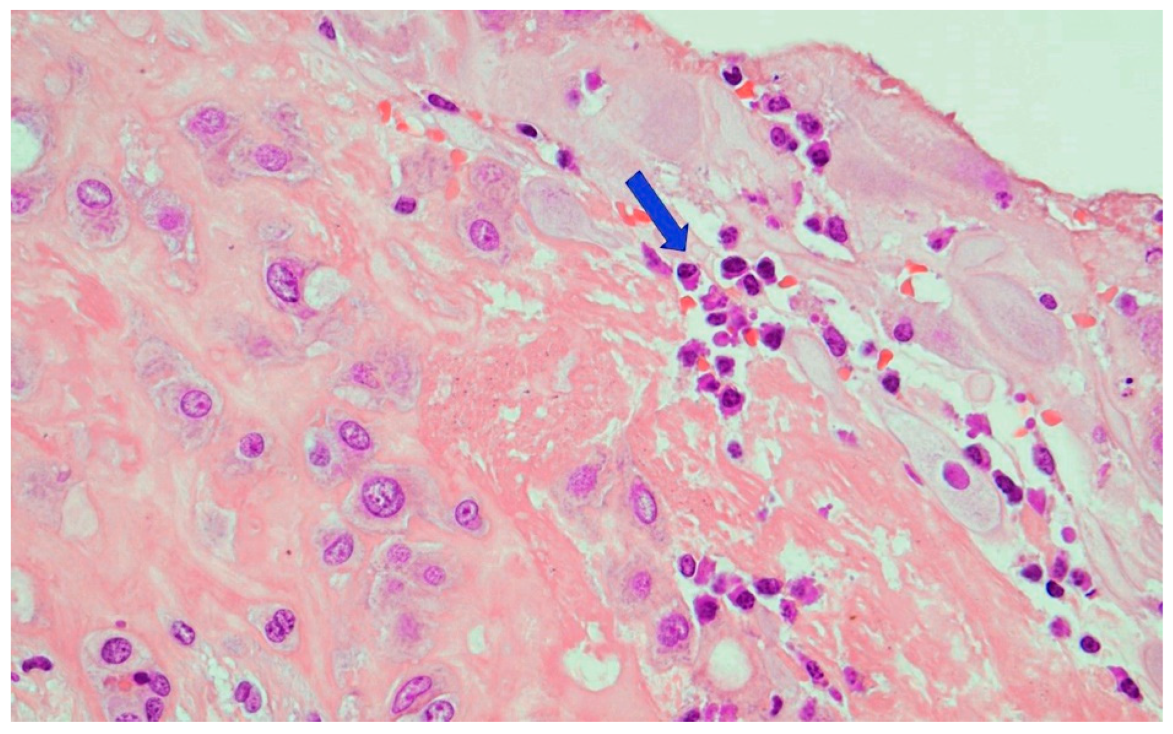
2. The Case
3. Discussion
4. Conclusions
Author Contributions
Funding
Institutional Review Board Statement
Informed Consent Statement
Data Availability Statement
Conflicts of Interest
References
- Peeling, R.W.; Mabey, D.; Kamb, M.L.; Chen, X.S.; Radolf, J.D.; Benzaken, A.S. Syphilis. Nat. Rev. Dis. Primers 2017, 3, 17073. [Google Scholar] [CrossRef] [PubMed]
- Hook, E.W., 3rd. Syphilis. Lancet 2017, 389, 1550–1557, Erratum in Lancet 2019, 393, 986. [Google Scholar] [CrossRef]
- Kojima, N.; Klausner, J.D. An Update on the Global Epidemiology of Syphilis. Curr. Epidemiol. Rep. 2018, 1, 24–38. [Google Scholar] [CrossRef] [PubMed]
- Tsuboi, M.; Evans, J.; Davies, E.P.; Rowley, J.; Korenromp, E.L.; Clayton, T.; Taylor, M.M.; Mabey, D.; Chico, R.M. Prevalence of syphilis among men who have sex with men: A global systematic review and meta-analysis from 2000–2020. Lancet Glob. Health 2021, 8, e1110–e1118. [Google Scholar] [CrossRef]
- Medoro, A.K.; Sánchez, P.J. Syphilis in Neonates and Infants. Clin. Perinatol. 2021, 2, 293–309. [Google Scholar] [CrossRef]
- Spicknall, I.H.; Kreisel, K.M.; Weinstock, H.S. Estimates of the Prevalence and Incidence of Syphilis in the United States, 2018. Sex Transm. Dis. 2021, 48, 247–252. [Google Scholar] [CrossRef]
- Stary, A. The Changing Spectrum of Sexually Transmitted Infections in Europe. Acta Derm. Venereol. 2020, 100, adv00114. [Google Scholar] [CrossRef]
- Korenromp, E.L.; Rowley, J.; Alonso, M.; Mello, M.B.; Wijesooriya, N.S.; Mahiané, S.G.; Ishikawa, N.; Le, L.V.; Newman-Owiredu, M.; Nagelkerke, N.; et al. Global burden of maternal and congenital syphilis and associated adverse birth outcomes-Estimates for 2016 and progress since 2012. PLoS ONE 2019, 14, e0211720. [Google Scholar]
- European Centre for Disease Prevention and Control. Congenital Syphilis; ECDC Annual Epidemiological Report for 2016; ECDC: Solna, Sweden, 2018.
- Thomas, D.D.; Navab, M.; Haake, D.A.; Fogelman, A.M.; Miller, J.N.; Lovett, M.A. Treponema pallidum invades intercellular junction of endothelial cell monolayers. Proc. Natl. Acad. Sci. USA 1988, 85, 3608–3612. [Google Scholar] [CrossRef] [Green Version]
- Organização Mundial de Saúde. Eliminação Mundial da Sífilis Congénita: Fundamento Lógico e Estratégia para Acção; Organização Mundial de Saúde: Geneva, Switzerland, 2008.
- Lukehart, S.A. Syphilis. In Harrison’s Principles of Internal Medicine; McGraw-Hill: New York, NY, USA, 2012. [Google Scholar]
- Hollier, L.M.; Cox, S. Syphilis. Semin. Perinatol. 1998, 22, 323–331. [Google Scholar] [CrossRef]
- Salooje, H.; Velaphi, S.; Goga, Y.; Afadapa, N.; Steen, R.; Lincetto, O. The prevention and management of congenital syphilis: An overview and recommendations. Bull. World Health Organ. 2004, 82, 424–430. [Google Scholar]
- Lubiganon, P.; Piaggio, G.; Villar, J.; Pinol, A.; Bakketeig, L.; Bergsjo, P.; Al-Mazrou, Y.; Ba’aqeel, H.; Beilzan, J.M.; Farnot, U.; et al. WHO Antenatal Care Trial Research Group. The epidemiology of syphilis in pregnancy. Int. J. STD AIDS 2001, 13, 486–494. [Google Scholar] [CrossRef]
- Qin, J.; Yang, T.; Xiao, S.; Tan, H.; Feng, T.; Fu, H. Reported estimates of adverse pregnancy outcomes among women with and without syphilis: A systematic review and meta-analysis. PLoS ONE 2014, 9, e102203. [Google Scholar] [CrossRef] [PubMed] [Green Version]
- Klausner, J.D. The sound of silence: Missing the opportunity to save lives at birth. Bull. World Health Organ. 2013, 91, 158–158A. [Google Scholar] [CrossRef] [PubMed]
- World Health Organization. Department of Reproductive Health and Research. Investment Case for Eliminating Mother-to-Child Transmission of Syphilis: Promoting Better Maternal and Child Health and Stronger Health Systems; WHO: Geneva, Switzerland, 2012.
- Newman, L.; Kamb, M.; Hawkes, S.; Gomez, G.; Say, L.; Seuc, A.; Broutet, N. Global estimates of syphilis in pregnancy and associated adverse outcomes: Analysis of multinational antenatal surveillance data. PLoS Med. 2013, 10, e1001396. [Google Scholar] [CrossRef]
- World Health Organization, Department of Reproductive Health and Research. The Global Elimination of Congenital Syphilis: Rationale and Strategy for Action. 2007. Available online: http://apps.who.int/iris/bitstream/10665/43782/1/9789241595858_eng.pdf (accessed on 20 August 2016).
- I Nuovi Livelli Essenziali di Assistenza (LEA). Available online: https://temi.camera.it (accessed on 19 July 2022).
- SIEOG. Linee Guida per Gli Screening Ecografici in Ostetricia e Ginecologia; CIC Edizioni Internazionali: Roma, Italy, 1996. [Google Scholar]
- Fetal Heart Rate Monitoring During Labor|ACOG. Available online: https://www.acog.org (accessed on 19 July 2022).
- Papile, L.A.; Burstein, J.; Burnstein, R.; Koffer, H. Incidence and evolution of subependymal and intraventricular henmorrhage: A study of infants with birth weights less than 1500 gm. J. Pediatr. 1978, 92, 529–534. [Google Scholar] [CrossRef]
- Thompson, J.M.; Irgens, L.M.; Skjaerven, R.; Rasmussen, S. Placenta weight percentile curves for singleton deliveries. BJOG 2007, 114, 715–720. [Google Scholar] [CrossRef]
- Kraus, F.T.; Redline, R.W.; Gersell, D.J.; Nelson, D.M.; Dicke, J.M. Chapter 5 Inflammation and Infection. In Placental Pathology; Armed Forces Institute of Pathology (AFIP): Washington, DC, USA, 2004; pp. 75–86. [Google Scholar]
- Hollier, L.M.; Harstad, T.W.; Sanchez, P.J.; Twickler, D.M.; Wendel, G.D., Jr. Fetal Syphilis: Clinical and Laboratory Characteristics. Obstet. Gynecol. 2001, 97, 947–953. [Google Scholar] [CrossRef]
- Rac, M.W.F.; Bryant, S.N.; McIntire, D.D.; Cantey, J.B.; Twickler, D.M.; Wendel, G.D., Jr.; Sheffield, J.S. Progression of ultrasound findings of fetal syphilis after maternal treatment. Am. J. Obstet. Gynecol. 2014, 211, 426.e1–426.e6. [Google Scholar] [CrossRef]
- Lawrence, N.L.; Twickler, D.M.; Peters, M.T.; Sanchez, P.J.; Wendel, G.D. Fetal Syphilis: Correlation of Sonographic Findings and Rabbit Infectivity Testing of Amniotic Fluid. J. Ultrasound Med. 1993, 2, 97–101. [Google Scholar]
- Park, I.U.; Tran, A.; Pereira, L.; Fakile, Y. Sensitivity and Specificity of Treponemal-specific Tests for the Diagnosis of Syphilis. Clin. Infect. Dis. 2020, 71, S13–S20. [Google Scholar] [CrossRef] [PubMed]
- Centers for Disease Control and Prevention. Sexually Transmitted Infections Treatment Guidelines. 2021. Available online: https://www.cdc.gov/std/treatment-guidelines/toc.htm (accessed on 10 July 2022).
- Lawrence, N.L.; Bohoman, V.R.; Sanchez, P.J.; Leos, N.K.; Twickler, D.M.; Wendel, G.D. In Utero Infection with Treponema Pallidium in Early Pregnancy. Prenat. Diagn. 1997, 17, 119–123. [Google Scholar]
- Wendel, G.D., Jr.; Sanchez, P.J.; Peters, M.T.; Harstad, T.W.; Potter, L.L.; Norgard, M.V. Identification of Treponema Pallidum in Amniotic Fluid and Fetal Blood from Pregnancies Complicated by Congenital Syphilis. Obstet. Gynecol. 1991, 78, 890–894. [Google Scholar]
- Wendel, G.D.; Maberry, M.C.; Christmas, J.T.; Goldberg, M.S.; Norgard, M.V. Examination of amniotic fluid in diagnosis congenital syphilis with fetal death. Obstet. Gynecol. 1989, 60, 967–970. [Google Scholar]
- Wendel, G.D., Jr.; Sheffield, J.S.; Hollier, L.M.; Hill, J.B.; Ramsey, P.S.; Sanchez, P.J. Treatment of syphilis in pregnancy and prevention of congenital syphilis. Clin. Infect. Dis. 2002, 35, S200–S209. [Google Scholar] [CrossRef]
- Araujo Júnior, E.; Santana, E.F.M.; Rolo, L.C.; Machado Nardozza, L.M.; Moron, A.F. Prenatal diagnosis of congenital syphilis using two- and three-dimensional ultrasonography: Case report. Case. Rep. Infect. Dis. 2012, 2012, 478436. [Google Scholar] [CrossRef]
- David, M.; Hcini, N.; Mandelbrot, L.; Sibiude, J.; Picone, O. Fetal and neonatal abnormalities due to congenital syphilis: A literature review. Prenat. Diagn. 2022, 42, 643–655. [Google Scholar] [CrossRef] [PubMed]
- Hira, S.K.; Bhat, G.J.; Patel, J.B.; Din, S.N.; Atilli, R.V.; Patel, M.I.; Baskamathan, S.; Hira, R.S.; Andu, N.N. Early Congenital Syphilis: Clinico-Radiologic Features in 202 Patients. Sex. Trans. Dis. 1985, 12, 177–183. [Google Scholar] [CrossRef]
- Long, W.A.; Ulshen, M.H.; Lawson, E.E. Clinical Manifestations of Congenital Syphilitic Hepatitis: Implications for Pathogenesis. J. Pediatr. Gastroenterol. Nutr. 1984, 3, 551–5555. [Google Scholar] [CrossRef]
- Anand, N.K.; Chellani, H.K.; Wadhwa, A.; Prasanna, S.B.; Mohan, M. Congenital Syphilitic Hepatitis. Indian Pediatrics 1991, 28, 157–159. [Google Scholar]
- Sheffield, J.S.; Sanchez, P.J.; Wendel, G.D., Jr.; Fong, D.W.I.; Margraf, L.R.; Zeray, F.; McIntire, D.D.; Rogers, B.B. Placental histopathology of congenital syphilis. Obstet. Gynecol. 2002, 100, 126–133. [Google Scholar] [PubMed]
- Qureshi, F.; Jacques, S.M.; Reyes, M.P. Placental histopathology in syphilis. Hum. Pathol. 1993, 24, 779–784. [Google Scholar] [CrossRef]
- Genest, D.R.; Choi-Hong, S.R.; Tate, J.E.; Qureshi, F.; Jacques, S.M.; Crum, C. Diagnosis of congenital syphilis from placental examination: Comparison of histopathology, Steiner stain, and polymerase chain reaction for Treponema pallidum DNA. Hum. Pathol. 1996, 27, 366–372. [Google Scholar] [CrossRef]
- Lucas, M.J.; Theriot, S.K.; Wendel, G.D., Jr. Doppler systolic-diastolic ratios in pregnancies complicated by syphilis. Obstet. Gynecol. 1991, 77, 217–222. [Google Scholar] [CrossRef] [PubMed]
- Kittipornpechdee, N.; Hanamornroongruang, S.; Lekmak, D.; Treetipsatit, J. Fetal and Placental Pathology in Congenital Syphilis: A Comprehensive Study in Perinatal Autopsy. Fetal Pediatr. Pathol. 2018, 37, 231–242. [Google Scholar] [CrossRef] [PubMed]
- Rawstron, S.A.; Vetrano, J.; Tannis, G.; Bromberg, K. Congenital syphilis: Detection of Treponema pallidum in stillborns. Clin. Infect. Dis. 1997, 24, 24–27. [Google Scholar] [CrossRef]
- Gomez, G.B.; Kamb, M.L.; Newman, L.M.; Mark, J.; Broutet, N.; Hawkes, S.J. Untreated maternal syphilis and adverse outcomes of pregnancy: A systematic review and meta-analysis. Bull. World Health Organ. 2013, 91, 217–226. [Google Scholar] [CrossRef]
- Carles, G.; Lochet, S.; Youssef, M.; El Guindi, W.; Helou, G.; Alassas, N.; Lambert, V. Syphilis et grossesse. Etude de 85 cas en Guyane française [Syphilis and pregnancy]. J. Gynecol. Obstet. Biol. Reprod. 2008, 37, 353–357. [Google Scholar] [CrossRef] [PubMed]


Publisher’s Note: MDPI stays neutral with regard to jurisdictional claims in published maps and institutional affiliations. |
© 2022 by the authors. Licensee MDPI, Basel, Switzerland. This article is an open access article distributed under the terms and conditions of the Creative Commons Attribution (CC BY) license (https://creativecommons.org/licenses/by/4.0/).
Share and Cite
Tonni, G.; Grisolia, G.; Pisello, M.; Zampriolo, P.; Fasolato, V.; Sindico, P.; Araújo Junior, E.; Bonasoni, M.P. Congenital Syphilis Presenting with Brain Abnormalities at Neuroscan: A Case Report and a Brief Literature Review. Microorganisms 2022, 10, 1497. https://doi.org/10.3390/microorganisms10081497
Tonni G, Grisolia G, Pisello M, Zampriolo P, Fasolato V, Sindico P, Araújo Junior E, Bonasoni MP. Congenital Syphilis Presenting with Brain Abnormalities at Neuroscan: A Case Report and a Brief Literature Review. Microorganisms. 2022; 10(8):1497. https://doi.org/10.3390/microorganisms10081497
Chicago/Turabian StyleTonni, Gabriele, Gianpaolo Grisolia, Marlene Pisello, Paolo Zampriolo, Valeria Fasolato, Paola Sindico, Edward Araújo Junior, and Maria Paola Bonasoni. 2022. "Congenital Syphilis Presenting with Brain Abnormalities at Neuroscan: A Case Report and a Brief Literature Review" Microorganisms 10, no. 8: 1497. https://doi.org/10.3390/microorganisms10081497
APA StyleTonni, G., Grisolia, G., Pisello, M., Zampriolo, P., Fasolato, V., Sindico, P., Araújo Junior, E., & Bonasoni, M. P. (2022). Congenital Syphilis Presenting with Brain Abnormalities at Neuroscan: A Case Report and a Brief Literature Review. Microorganisms, 10(8), 1497. https://doi.org/10.3390/microorganisms10081497