Article

Whole Genome Sequence Analysis of a Novel Apilactobacillus Species from Giant Honeybee (Apis dorsata) Gut Reveals Occurrence of Genetic Elements Coding Prebiotic and Probiotic Traits

1
Industrial Biotechnology Division, National Institute for Biotechnology and Genetic Engineering College, Pakistan Institute of Engineering and Applied Sciences (NIBGE-C, PIEAS), Faisalabad 38000, Pakistan
2
Biomedical Technology and Cell Therapy Research Laboratory, Department of Biomedical Engineering, Faculty of Medicine, McGill University, 3775 University Street, Montreal, QC H3A 2B4, Canada
3
Department of Chemistry, College of Sciences, United Arab Emirates University, Al-Ain 15551, United Arab Emirates
*
Authors to whom correspondence should be addressed.
Permanent Address: Department of Microbiology, Abbottabad University of Science and Technology, Havelian, Abbottabad 22500, Pakistan.
Microorganisms 2022, 10(5), 904; https://doi.org/10.3390/microorganisms10050904
Submission received: 30 March 2022 / Revised: 20 April 2022 / Accepted: 21 April 2022 / Published: 26 April 2022
(This article belongs to the Special Issue Microorganisms in Pollinators: Interactions with Other Factors 2.0)

Abstract

:
Apilactobacillus spp. are classified as obligate fructophilic lactic acid bacteria (FLAB) that inhabit fructose-rich niches such as honeybee gut. Lactic acid bacteria are an important component of the gut microbiome and play a crucial role in maintaining gut health. In this study, a new FLAB strain HBW1, capable of producing glucan-type exopolysaccharide, was isolated from giant honeybee (Apis dorsata) gut and subjected to whole genome sequencing (WHS) to determine its health-beneficial traits. The genome size of the isolate was 1.49 Mb with a GC content of 37.2%. For species level identity, 16S rDNA sequence similarity, genome to genome distance calculator (dDDH), and average nucleotide identity (ANI) values were calculated. Phylogenetic analysis showed that the isolate HBW1 belongs to the Apilactobacillus genus. The dDDH and ANI values in comparison with closely clustered Apilactobacillus kunkeei species were 52% and 93.10%, respectively. Based on these values, we concluded that HBW1 is a novel species of Apilactobacillus, and we propose the name Apilactobacillus waqarii HBW1 for it. Further, WHS data mining of HBW1 revealed that it harbors two glucosyltransferase genes for prebiotic glucan-type exopolysaccharide synthesis. Moreover, chaperon (clp) and methionine sulfoxide reductase (msrA, msrB, and msrC) genes as well as nutritional marker genes for folic acid (folD) and riboflavin biosynthesis (rib operon), important for conferring probiotic properties, were also detected. Occurrence of these genetic traits make HBW1 an excellent candidate for application to improve gut function.

1. Introduction

Studies during the last few years have shown that the gut and brain are interconnected both physically and chemically through a communication network called the gut–brain axis [1,2,3]. This implies that the brain and gut may reciprocally affect each other’s health. Diet and gut microbes play an important role in maintaining the gut health. Diet is one of the major factors that modulates the composition of gut microbiota [4] thus affecting brain health. Therefore, modulating the gut microbiome may help in improving brain health. Certain diet ingredients, such as probiotics and prebiotics, have the potential to play a crucial role in this respect. Indeed, recently reviewed research data have shown that these components are helpful in treatment of brain-related and other disorders [5,6,7].
Probiotics are defined as “live microorganisms that, when administered in adequate amounts, confer a health benefit to the host” [8]. These include mostly certain lactic acid bacteria (LAB) and bifidobacteria [9]. A prebiotic has been defined as “a substrate that is selectively utilized by host microorganisms conferring a health benefit” [10]. Prebiotics are fibrous substrates that resist enzymatic activity in the upper part of human gastrointestinal tract; thus, they are not digested in the small intestine. Consequently, they reach the colon unmetabolized and are fermented by the intestinal microbiota. This process positively regulates the activity of the specific intestinal microbiome that beneficially interacts with the host by the producing short chain fatty acids (SCFAs) and vitamins [11,12]. Chemically, prebiotics are oligo/polymers of carbohydrates such as fructooligosaccharides (FOS), galactooligosaccharides (GOS), xylooligosaccharides (XOS), etc., [12,13].
In addition to their probiotic effects, LAB are also of particular interest for the production of exopolysaccharides (EPSs) that have potential prebiotic functionality. For this purpose, LAB produce sucrase enzymes, which synthesize glucose or fructose polysaccharides called as glucans and fructans, respectively, using sucrose as a substrate [14,15]. The enzymes involved in glucan synthesis are known as glucansucrases and belong to the glycoside hydrolase family 70 (GH70) at carbohydrate-active enzymes database (http://www.cazy.org) (accessed on 10 March 2022) [16]. Depending on the type of linkages they introduce between glucose residues in the polysaccharide, glucansucrases are categorized as dextransucrases—synthesizing dextran (containing mainly α-(1-6) linkages); mutansucrases—synthesizing mutan (α-(1-3 linkages); reuteransucrases—synthesizing reuteran (α-(1-4) linkages); and alternansucrases that synthesize alternan (having alternating α-(1-6) and α-(1-3)-linkages) [15,17,18].
Fructophilic lactic acid bacteria (FLAB) is a specific subgroup of LAB that has been described and characterized recently [19]. FLAB have gained considerable attention due to their potential human health beneficial effects and for their proximity to lactic acid bacteria that are accepted as safe [20]. They live in symbiosis with insects such as honeybees that have special diets [21,22]. FLAB preferably use fructose as a substrate for their growth and inhabit only those niches that are rich in fructose content. They are also found in different fruit and vegetable food matrices such as banana, grapes, figs, durian fruit, legumes, and cocoa beans [23].
Among FLAB, there are some other members of the genus Lactobacillus that have recently been reclassified as Apilactobacillus to underline their specific adaptation to bees [24]. The genus Apilactobacillus includes different bacterial species; among them, Apilactobacillus kunkeei (basonym Lactobacillus kunkeei) and Apilactobacillus apinorum (basonym Lactobacillus apinorum) have adapted to bees [25]. These lactic acid bacteria constitute an important component of human gut microbiota as well and could play crucial role in maintaining gut health due to their prebiotic synthesizing potential or probiotics traits. In the present study, a new Apilactobacillus spp. strain, designated as HBW1, was isolated from giant honeybee (Apis dorsata) gut and subjected to whole genome sequencing with an aim to identify its prebiotic EPS synthesizing gene(s) and the genetic elements important for conferring probiotic properties to the isolate. We have proposed the name Apilactobacillus waqarii HBW1 for this isolate.

2. Materials and Methods

2.1. Culturing of Bacteria

A wild type giant honeybee (Apis dorsata), found in Southeast Asia, was sampled from a botanical garden in Faisalabad region of Pakistan. The outer surface of the bee was sterilized with 70% ethanol, and its gut was removed under sterile condition. The gut was transferred to sterilized De Man-Rogosa-Sharpe (MRS)-sucrose medium having the composition described previously [26] and incubated at 30 °C for 24 h. After visual appearance of growth, serial dilutions of the cultured broth were made and streaked on agar plates of the same medium to obtain single colonies. A single colony having a slimy mucoid appearance, indicating the production of EPS, was selected and transferred to sterilized fresh MRS-sucrose medium for growth. The process of transferring to solid and liquid media was repeated twice further to assure the isolation of pure culture, which was designated as HBW1.

2.2. Exopolysaccharide Production by the Isolate

To confirm the EPS production HBW1, MRS medium, supplemented with sucrose (20% w/v), was inoculated with HBW1 and incubated for 48 h in shaking incubator. To check for EPS production, a small aliquot of culture was run on thin layer chromatography (TLC) plate (Silica gel 60 F254; Merck, Darmstadt, Germany) for 6 h, after which it was air dried, and the sugar spots were visualized by developing with a solution containing 5% sulfuric acid in methanol, as described previously [27].

2.3. Genome Sequencing

The Genomic DNA of the isolate was extracted using a Thermo Scientific Gene JET Genomic DNA extraction kit (#K0721). The whole genome sequencing was carried out commercially by MicrobesNG (Birmingham, United Kingdom) using Illumina next generation sequencing at minimum coverage of 30×. The whole-genome sequencing data and bio-project/bio-sample information of the Apilactobacillus species (HBW1) reported in the present study are available at NCBI database (Accession numbers given in Table 1).

2.4. Genome Analysis

The genome annotation was carried out using NCBI Prokaryotic Genome Annotation Pipeline (PGAP) and Rapid Annotation using Subsystem Technology (RAST) server version 2.0 (Classic RAST default settings) [28]. The genomic digital DNA Hybridization (dDDH) values were calculated using the Genome-to-Genome Distance Calculator (GGDC) DSMZ (https://www.dsmz.de/services/online-tools/genome-to-genome-distance-calculator-ggdc) (accessed on 12 April 2022) [29]. Average Nucleotide Identity (ANI) values were determined using the Kostas Lab server (http://enve-omics.ce.gatech.edu/ani/) (accessed on 12 April 2022) [30] with default parameters.

2.5. Taxonomic Evaluation

For taxonomic studies, 16S rRNA gene sequences of the closely related species were taken from EzBioCloud and the National Center for Biotechnology Information (NCBI) databases [31]. The phylogenetic analysis and trees were constructed on the basis of whole genome sequences using the Type (Strain) Genome Server (TYGS) [29]. Trees were inferred with FastME 2.1.6.1 [32] from the Genome BLAST Distance Phylogeny (GBDP) distances calculated from genome sequences. The branch lengths were scaled in terms of GBDP distance formula d5. The numbers above branches were GBDP pseudo-bootstrap support values >60% from 100 replications, with an average branch support of 89.9 and 71.4% for 16S rRNA and genome sequences, respectively. The trees were rooted at the midpoint [33].

2.6. EPS Producing Genes

The genome sequence of HBW1 was also analyzed for the presence of genes specific for EPS synthesis and probiotic traits. Moreover, the EPS synthesizing gene was compared with top related strains obtained from EzBioCloud [31]. The phylogenetic tree was constructed using MEGA7 software by the neighbor joining method.

2.7. Submission Information

The whole genome of HBW1 was submitted to the NCBI database. The details are in Table 1.

3. Results and Discussion

3.1. EPS Production

Thin layer chromatographic analysis showed that the isolate Apilactobacillus waqarii HBW1 produced glucan type EPS using sucrose as a substrate (Figure 1). Previously, the presence of glucansucrase enzymes responsible for glucan type EPS synthesis has only been reported in Apilactobacillus kunkeei species among apilactobacilli [14]. Therefore, A. waqarii is only the second species among Apilactobacillus genus that produces glucan type EPS.

3.2. Genome Sequence and Annotation

The genome assembly was classified as undecided for the potential contaminations as determined by ContEst16S (Contamination Estimator by 16S) algorithm. As per NCBI PGAP analysis, the draft genome sequence of the isolate HBW1 comprised a total length of (1.49 Mb); contig count (38); N50 (229,491 bp), L50 (3), and G + C content (37.2%). Moreover, a total of 1340 protein-coding sequences and 75 RNAs were found in the genome (Table 2). Further ClassicRAST based functional gene subsystem clustering analysis revealed that 247 subsystems were present in the genomic island of the HBW1 (Table 2). The subsystems representing the amino acids and derivatives (79 ORFs); cofactors, vitamins, prosthetic groups, pigments (69 ORFs); carbohydrate metabolism (70 ORFs); protein metabolism (194 ORFs); fatty acids, lipids, and isoprenoids (56 ORFs) were present in the genome of HBW1. Furthermore, the subsystems connected with membrane transport (31 ORFs), stress response (33 ORFs), and sulfur metabolism (3 ORFs) were also identified (Figure 2).

3.3. Phylogenetic and Genome Based Classification at Species Level

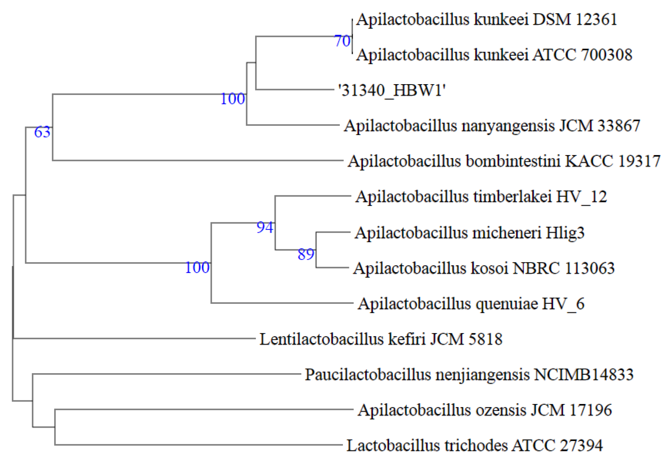
Taxonomic evaluation of the isolate HBW1 was carried out by systematically using a combination of 16S rRNA gene similarity and Overall Genome-Related Index (OGRI) that included ANI and dDDH. In this regard, the first step was to determine the strains that were closely related to HBW1. For this purpose, the 16S rRNA gene sequence of HBW1 was submitted to the NCBI (for BLASTn search) and EzBioCloud databases [31,34]. The results of the NCBI BLASTn and EzBioCloud showed a sequence similarity of 16S rRNA gene of HBW1 with other congener species of the genus Apilactobacillus (Kunkeei, Apinorum, Bombintestini, Timberlakei, Micheneri, Quenuiae, and Ozensis)—maximum similarity (100%) of the HBW1 was found with Apilactobacillus Kunkeei DSM 12,361 YH-15T [JXDB01000004], followed by Apilactobacillus apinorum Fhon13NT [JX099541], 98.82% (Table 3). However, genome-based phylogenetic analysis of the closely related whole genome sequences showed that the HBW1 formed a separate branch from Apilactobacillus kunkeei species (Figure 3). Further, to establish a more specific taxonomic position at the species level, a comparison of the genome of the HBW1 was carried out to its closely-related type strains using ANI and dDDH values (Table 3). According to the current bacterial taxonomy, the projected and generally accepted dDDH and ANI values are 70% and 95–96%, respectively, between genomes of the same species [32]. A comparison of the HBW1 and the close neighbor Apilactobacillus kunkeei DSM 12,361 strain YH15T revealed dDDH value of 52.0 and the ANI value 93.10 supporting HBW1 as a potential new species (Table 3). Taking together the 16S rRNA gene, dDDH, and ANI, the isolate HBW1 described here is a new species of Apilactobacillus genus, and we propose the name Apilactobacillus waqarii HBW1 for it. Previous studies showed that geographical location and even developmental stages can influence the diversity in composition of honeybee gut microbiota [35,36,37]. Among other factors, exposure to synthetic chemicals such as pesticides also determine the type of microbiota inhabiting honeybee gut [38]. Owing to distinct geographical and environmental conditions at the habitat of the host giant honeybee A. dorsata, some or all of these factors could have contributed to the occurrence of this novel Apilactobacillus species.

3.4. Genetic Traits of the Isolate HBW1 Important for Gut Function

3.4.1. Putative Genes for EPS Synthesis

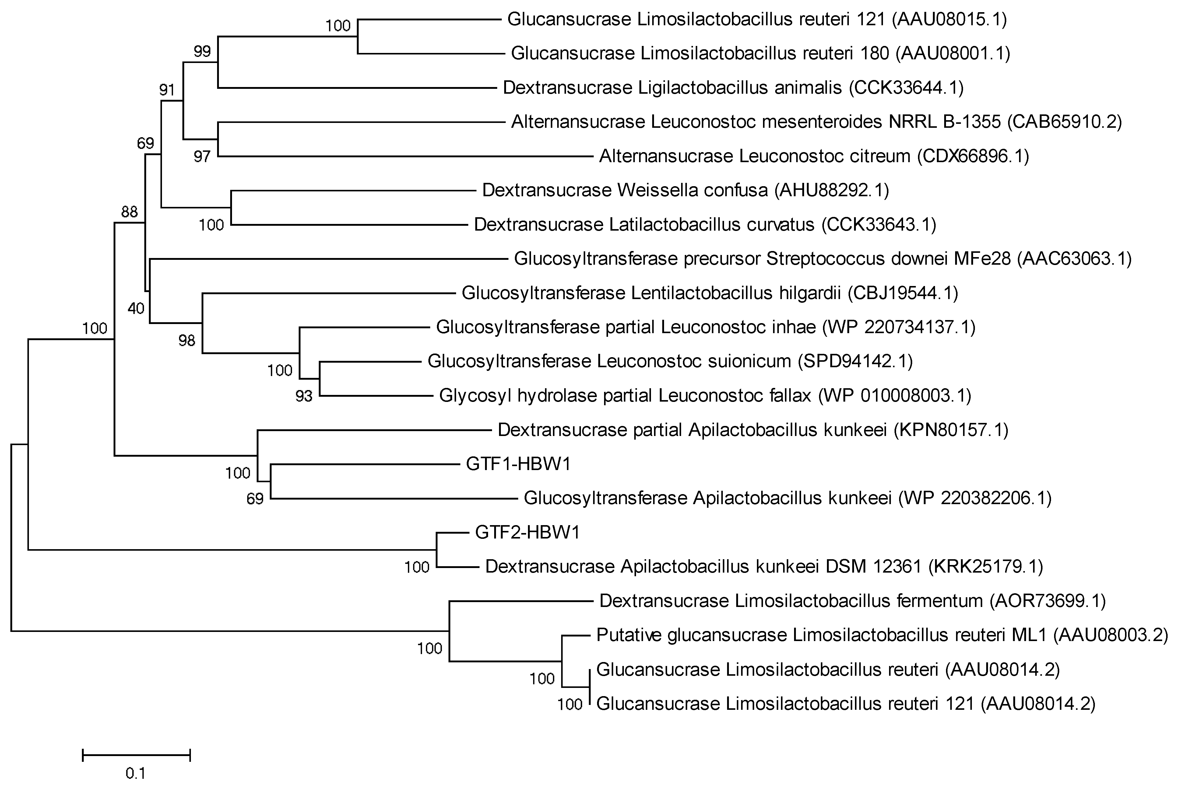
Mining of the whole genome sequencing data of HBW1 revealed that it harbored two genes coding for putative glucosyltransterase proteins, designated as GTF1-HBW1 and GTF2-HBW1, responsible for EPS synthesis. In the phylogenetic tree, the GTF1-HBW1 and GTF2-HBW1 clustered with glucosyltransferase of closely related species of Apilactobacillus kunkeei (WP-220382206.1) and dextransucrase of Apilactobacillus kunkeei (KPN80157.1), respectively (Figure 4). Interestingly, the GTF1-HBW1 and GTF2-HBW1 fell in highly divergent clades depicting that these proteins would be responsible for very different types of glucans, one of which is likely to be a dextran.
Several species of Apilactobacillus (basonym Lactobacillus) genus have been isolated from honeybee Apis mellifera gut [39]. Studies based on metagenomic analysis have shown that these FLAB are also widely distributed among honeybee genera A. mellifera [40] (Nowak et al. 2021), A. florea [36], and Apis dorsata [35]. However, EPS synthesizing enzymes have only been characterized from Apilactobacillus kunkeei [41,42]. In contrast, among LAB, production of dextran type EPS is commonly known in Leuconostoc and Weissella species [14,43,44]. Similar to A. waqarii HBW1, some of them were also reported to have multiple genes responsible for the production of glucan-type of EPSs [45].
In addition to some fructans and fructooligosaccharides (FOS), the prebiotic potential of dextran has also been documented. Using a batch-culture fermentation system designed to simulate transit through the large intestine, dextran has been demonstrated to elicit a bifidogenic effect similar to the well-known prebiotic fructooligosaccharides (FOS), which also resulted in decreased levels of undesirable bacteria such as clostridia and bacteroides [46]. Similarly, some linear and branched dextrans have been found to increase Bifidobacterium populations significantly during fermentation by human fecal microbiota [47].

3.4.2. Genetic Elements Conferring Potential Probiotic Characteristics

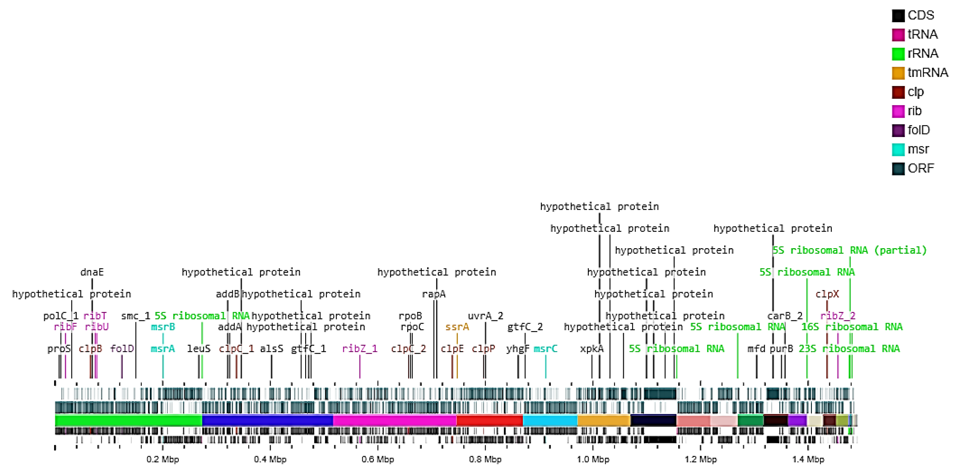
Mining of the whole genome sequence data of A. waqarii HBW1 revealed that it also harbored certain genes that are important for conferring probiotic properties to bacteria exerting beneficial effects on host health. Genes that are responsible for the active removal of stressors including DNA and the protein protection and repair system, e.g., clpATPase (chaperon) reported to have bile salt hydrolase and acid tolerance activity [48] were detected in the genome of HBW1 as shown in Table 4. The location of these genes on the HBW1 genome is shown in Figure 5. The CLP chaperone protein is a mimetic of the anorexigenic α-melanocyte stimulating hormone (α-MSH). Several recent studies on mice model have shown that the prevalence of this gene in enterobacteria resulted in reduced adiposity and weight gain in obesity rodent models [49,50,51]. This fact is further strengthened by the finding that low enterobacterial Clp B gene abundance was observed in the microbiota of obese humans [52]. Other probiotic genes such as methionine sulfoxide reductase genes msrA, msrB, and msrC are also very important, because reactive oxygen species (ROS) oxidize the methionine residues in proteins, resulting in the production of methionine-S-sulfoxides [Met-S-(O)] and methionine-R-sulfoxides [Met-R-(O]. These oxidized methionine residues can be repaired by the antioxidant enzymes, Met-S-(O) reductase (MsrA) and Met-R-(O) reductase (MsrB) [53]. We have also identified the nutritional marker gene genes for folic acid (folD) and riboflavin biosynthesis (rib operon). These nutritional marker genes have the potential ability to synthesize and transport B vitamins-riboflavin [54]. Synthesis of B vitamins is a desirable trait of probiotic bacteria since the human body does not synthesize these vitamins [55].

4. Conclusions

A novel species of fructophilic lactic acid bacteria Apilactobacillus, with the proposed name Apilactobacillus waqarii HBW1, was isolated from giant honeybee (Apis dorsata) gut. The genome of HBW1 harbors two EPS synthesis genes and genetic elements important for conferring probiotic properties, making it an excellent candidate for application to improve gut function. In future studies, the composition and glcosydic linkage analysis of the purified EPS products synthesized by heterologously expressed GTF proteins of HBW1 would resolve the exact chemical structure of these putative glucans.

Author Contributions

Conceptualization: M.A.A. and N.M.; methodology: W.A., S.K., N.A. and M.A.A.; investigation: W.A., S.K., N.A., J.E.A., K.A. and M.A.A.; resources: M.A.A., K.A. and N.M.; data curation: S.K., N.A., J.E.A. and M.A.A.; writing—original draft preparation: W.A. and M.A.A.; writing—review and editing: W.A., S.K., N.M., S.P. and M.A.A.; visualization: S.K., N.M. and M.A.A.; supervision: N.M. and M.A.A. All authors have read and agreed to the published version of the manuscript.

Funding

N.M.’s research is funded by United Arab Emirates University (UAEU) research grant number 31S414 and UPAR grant 2022, 12S094. Moreover, the study was supported by grant No. NRPU-14953 from the Higher Education Commission of Pakistan (MAA).

Institutional Review Board Statement

Not applicable.

Informed Consent Statement

Not applicable.

Data Availability Statement

The whole genome data presented in this study are openly available in NCBI database at https://www.ncbi.nlm.nih.gov/search/all/?term=JAHQYH000000000 (accessed on 23 June 2021) under the accession number JAHQYH000000000.

Conflicts of Interest

The authors declare no conflict of interest.

References

  1. Mayer, E.A.; Tillisch, K.; Gupta, A. Gut/brain axis and the microbiota. J. Clin. Investig. 2015, 125, 926–938. [Google Scholar] [CrossRef]
  2. Osadchiy, V.; Martin, C.R.; Mayer, E.A. The gut–brain axis and the microbiome: Mechanisms and clinical implications. Clin. Gastroenterol. Hepatol. 2019, 17, 322–332. [Google Scholar] [CrossRef]
  3. Mayer, E.A.; Nance, K.; Chen, S. The Gut–Brain Axis. Annu. Rev. Med. 2022, 73, 439–453. [Google Scholar] [CrossRef]
  4. Berding, K.; Vlckova, K.; Marx, W.; Schellekens, H.; Stanton, C.; Clarke, G.; Jacka, F.; Dinan, T.G.; Cryan, J. Diet and the microbiota–gut–brain Axis: Sowing the seeds of good mental health. Adv. Nutr. 2021, 12, 1239–1285. [Google Scholar] [CrossRef]
  5. Munawar, N.; Ahsan, K.; Muhammad, K.; Ahmad, A.; Anwar, M.A.; Shah, I.; Al Ameri, A.K.; Al Mughairbi, F. Hidden role of gut microbiome dysbiosis in schizophrenia: Antipsychotics or psychobiotics as therapeutics? Int. J. Mol. Sci. 2021, 22, 7671. [Google Scholar] [CrossRef]
  6. Kesika, P.; Suganthy, N.; Sivamaruthi, B.S.; Chaiyasut, C. Role of gut-brain axis, gut microbial composition, and probiotic intervention in Alzheimer’s disease. Life Sci. 2021, 264, 118627. [Google Scholar] [CrossRef]
  7. Munawar, N.; Ahmad, A.; Anwar, M.A.; Muhammad, K. Modulation of Gut Microbial Diversity through Non-Pharmaceutical Approaches to Treat Schizophrenia. Int. J. Mol. Sci. 2022, 23, 2625. [Google Scholar] [CrossRef]
  8. Hill, C.; Guarner, F.; Reid, G.; Gibson, G.R.; Merenstein, D.J.; Pot, B.; Morelli, L.; Canani, R.B.; Flint, H.J.; Salminen, S.; et al. Expert consensus document: The International Scientific Association for Probiotics and Prebiotics consensus statement on the scope and appropriate use of the term probiotic. Nat. Rev. Gastroenterol. Hepatol. 2014, 11, 506–514. [Google Scholar] [CrossRef] [Green Version]
  9. Roberfroid, M.B. Prebiotics and probiotics: Are they functional foods? Am. J. Clin. Nutr. 2000, 71, 1682S–1687S. [Google Scholar] [CrossRef]
  10. Gibson, G.; Hutkins, R.; Sanders, M.; Prescott, S.L.; Reimer, R.A.; Salminen, S.J.; Scott, K.; Stanton, C.; Swanson, K.S.; Cani, P.D.; et al. Expert consensus document: The International Scientific Association for Probiotics and Prebiotics (ISAPP) consensus statement on the definition and scope of prebiotics. Nat. Rev. Gastroenterol. Hepatol. 2017, 14, 491–502. [Google Scholar] [CrossRef] [Green Version]
  11. Mohanty, D.; Misra, S.; Mohapatra, S.; Sahu, P.S. Prebiotics and synbiotics: Recent concepts in nutrition. Food Biosci. 2018, 26, 152–160. [Google Scholar] [CrossRef]
  12. Davani-Davari, D.; Negahdaripour, M.; Karimzadeh, I.; Seifan, M.; Mohkam, M.; Masoumi, S.J.; Berenjian, A.; Ghasemi, Y. Prebiotics: Definition, types, sources, mechanisms, and clinical applications. Foods 2019, 8, 92. [Google Scholar] [CrossRef] [PubMed] [Green Version]
  13. Quigley, E.M.M. Prebiotics and probiotics in digestive health. Clin. Gastroenterol. Hepatol. 2019, 17, 333–344. [Google Scholar] [CrossRef] [PubMed]
  14. Korakli, M.; Vogel, R.F. Structure/function relationship of homopolysaccharide producing glycansucrases and therapeutic potential of their synthesised glycans. Appl. Microbiol. Biotechnol. 2006, 71, 790–803. [Google Scholar] [CrossRef] [PubMed]
  15. Van Hijum, S.A.; Kralj, S.; Ozimek, L.K.; Dijkhuizen, L.; van Geel-Schutten, I.G. Structure-function relationships of glucansucrase and fructansucrase enzymes from lactic acid bacteria. Microbiol. Mol. Biol. Rev. 2006, 70, 157–176. [Google Scholar] [CrossRef] [Green Version]
  16. Drula, E.; Garron, M.-L.; Dogan, S.; Lombard, V.; Henrissat, B.; Terrapon, N. The carbohydrate-active enzyme database: Functions and literature. Nucleic Acids Res. 2022, 50, D571–D577. [Google Scholar] [CrossRef]
  17. Monsan, P.; Bozonnet, S.; Albenne, C.; Joucla, G.; Willemot, R.-M.; Remaud-Siméon, M. Homopolysaccharides from lactic acid bacteria. Int. Dairy J. 2001, 11, 675–685. [Google Scholar] [CrossRef]
  18. Monsan, P.; Remaud-Siméon, M.; André, I. Transglucosidases as efficient tools for oligosaccharide and glucoconjugate synthesis. Curr. Opin. Microbiol. 2010, 13, 293–300. [Google Scholar] [CrossRef]
  19. Endo, A.; Salminen, S. Honeybees and beehives are rich sources for fructophilic lactic acid bacteria. Syst. Appl. Microbiol. 2013, 36, 444–448. [Google Scholar] [CrossRef]
  20. Brodmann, T.; Endo, A.; Gueimonde, M.; Vinderola, G.; Kneifel, W.; de Vos, W.M.; Salminen, S.; Gómez-Gallego, C. Safety of novel microbes for human consumption: Practical examples of assessment in the European Union. Front. Microbiol. 2017, 8, 1725. [Google Scholar] [CrossRef]
  21. Kwong, W.K.; Moran, N.A. Gut microbial communities of social bees. Nat. Rev. Microbiol. 2016, 14, 374–384. [Google Scholar] [CrossRef] [PubMed]
  22. Vásquez, A.; Forsgren, E.; Fries, I.; Paxton, R.J.; Flaberg, E.; Szekely, L.; Olofsson, T.C. Symbionts as major modulators of insect health: Lactic acid bacteria and honeybees. PLoS ONE 2012, 7, e33188. [Google Scholar]
  23. Filannino, P.; Di Cagno, R.; Tlais, A.Z.A.; Cantatore, V.; Gobbetti, M. Fructose-rich niches traced the evolution of lactic acid bacteria toward fructophilic species. Crit. Rev. Microbiol. 2019, 45, 65–81. [Google Scholar] [CrossRef] [PubMed]
  24. Zheng, J.; Wittouck, S.; Salvetti, E.; Franz, C.M.; Harris, H.; Mattarelli, P.; O’Toole, P.W.; Pot, B.; Vandamme, P.; Walter, J. A taxonomic note on the genus Lactobacillus: Description of 23 novel genera, emended description of the genus Lactobacillus Beijerinck 1901, and union of Lactobacillaceae and Leuconostocaceae. Int. J. Syst. Evol. Microbiol. 2020, 70, 2782–2858. [Google Scholar] [CrossRef] [PubMed]
  25. Vergalito, F.; Testa, B.; Cozzolino, A.; Letizia, F.; Succi, M.; Lombardi, S.J.; Tremonte, P.; Pannella, G.; Di Marco, R.; Sorrentino, E. Potential application of Apilactobacillus kunkeei for human use: Evaluation of probiotic and functional properties. Foods 2020, 9, 1535. [Google Scholar] [CrossRef]
  26. Ashfaq, I.; Amjad, H.; Ahmad, W.; Nasir, A.; Ali, A.; Ali, W.R.; Khaliq, S.; Hayat, A.; Ali, H.; Sattar, F. Growth Inhibition of Common Enteric Pathogens in the Intestine of Broilers by Microbially Produced Dextran and Levan Exopolysaccharides. Curr. Microbiol. 2020, 77, 2128–2136. [Google Scholar] [CrossRef]
  27. Roset, M.S.; Ciocchini, A.E.; Ugalde, R.A.; de Iannino, N.I. The Brucella abortus cyclic β-1, 2-glucan virulence factor is substituted with O-ester-linked succinyl residues. J. Bacteriol. 2006, 188, 5003–5013. [Google Scholar] [CrossRef] [PubMed] [Green Version]
  28. Aziz, R.K.; Bartels, D.; Best, A.A.; DeJongh, M.; Disz, T.; Edwards, R.A.; Formsma, K.; Gerdes, S.; Glass, E.M.; Kubal, M. The RAST Server: Rapid annotations using subsystems technology. BMC Genom. 2008, 9, 75. [Google Scholar] [CrossRef] [Green Version]
  29. Meier-Kolthoff, J.P.; Göker, M. TYGS is an automated high-throughput platform for state-of-the-art genome-based taxonomy. Nat. Commun. 2019, 10, 2182. [Google Scholar] [CrossRef] [PubMed]
  30. Goris, J.; Konstantinidis, K.T.; Klappenbach, J.A.; Coenye, T.; Vandamme, P.; Tiedje, J.M. DNA–DNA hybridization values and their relationship to whole-genome sequence similarities. Int. J. Syst. Evol. Microbiol. 2007, 57, 81–91. [Google Scholar] [CrossRef] [Green Version]
  31. Yoon, S.-H.; Ha, S.-M.; Kwon, S.; Lim, J.; Kim, Y.; Seo, H.; Chun, J. Introducing EzBioCloud: A taxonomically united database of 16S rRNA gene sequences and whole-genome assemblies. Int. J. Syst. Evol. Microbiol. 2017, 67, 1613. [Google Scholar] [CrossRef] [PubMed]
  32. Lefort, V.; Desper, R.; Gascuel, O. FastME 2.0: A comprehensive, accurate, and fast distance-based phylogeny inference program. Mol. Biol. Evol. 2015, 32, 2798–2800. [Google Scholar] [CrossRef] [PubMed] [Green Version]
  33. Farris, J.S. Estimating phylogenetic trees from distance matrices. Am. Nat. 1972, 106, 645–668. [Google Scholar] [CrossRef]
  34. Chun, J.; Oren, A.; Ventosa, A.; Christensen, H.; Arahal, D.R.; da Costa, M.S.; Rooney, A.P.; Yi, H.; Xu, X.-W.; De Meyer, S. Proposed minimal standards for the use of genome data for the taxonomy of prokaryotes. Int. J. Syst. Evol. Microbiol. 2018, 68, 461–466. [Google Scholar] [CrossRef] [PubMed]
  35. Wolter, A.; Hager, A.-S.; Zannini, E.; Galle, S.; Gänzle, M.; Waters, D.M.; Arendt, E.K. Evaluation of exopolysaccharide producing Weissella cibaria MG1 strain for the production of sourdough from various flours. Food Microbiol. 2014, 37, 44–50. [Google Scholar] [CrossRef] [PubMed]
  36. Saraithong, P.; Li, Y.; Saenphet, K.; Chen, Z.; Chantawannakul, P. Midgut bacterial communities in the giant Asian honeybee (Apis dorsata) across 4 developmental stages: A comparative study. Insect Sci. 2017, 24, 81–92. [Google Scholar] [CrossRef] [PubMed]
  37. Gruneck, L.; Khongphinitbunjong, K.; Popluechai, S. Gut microbiota associated with two species of domesticated honey bees from Thailand. Symbiosis 2021, 83, 335–345. [Google Scholar] [CrossRef]
  38. Niode, N.J.; Adji, A.; Rimbing, J.; Tulung, M.; Tallei, T.E. Composition and diversity of bacteria from giant Asian honeybee Apis dorsata gut. Biodiversitas 2021, 22, 906–912. [Google Scholar] [CrossRef]
  39. Paris, L.; Peghaire, E.; Mone, A.; Diogon, M.; Debroas, D.; Delbac, F.; El Alaoui, H. Honeybee gut microbiota dysbiosis in pesticide/parasite co-exposures is mainly induced by Nosema ceranae. J. Invertebr. Pathol. 2020, 172, 107348. [Google Scholar] [CrossRef]
  40. Olofsson, T.C.; Vásquez, A. Detection and identification of a novel lactic acid bacterial flora within the honey stomach of the honey bee Apis mellifera. Curr. Micriobiol. 2008, 57, 356–363. [Google Scholar] [CrossRef]
  41. Nowak, A.; Szczuka, D.; Górczyńska, A.; Motyl, I.; Kręgiel, D. Characterization of Apis mellifera gastrointestinal microbiota and lactic acid bacteria for honeybee protection—A review. Cells 2021, 10, 701. [Google Scholar] [CrossRef] [PubMed]
  42. Meng, X.; Gangoiti, J.; Wang, X.; Grijpstra, P.; van Leeuwen, S.S.; Pijning, T.; Dijkhuizen, L. Biochemical characterization of a GH70 protein from Lactobacillus kunkeei DSM 12361 with two catalytic domains involving branching sucrase activity. Appl. Microbiol. Biotechnol. 2018, 102, 7935–7950. [Google Scholar] [CrossRef] [PubMed]
  43. Bivolarski, V.; Iliev, I.; Ivanova, I.; Nikolova, M.; Salim, A.; Mihaylova, G.; Vasileva, T. Characterization of structure/prebiotic potential correlation of glucans and oligosaccharides synthetized by glucansucrases from fructophilic lactic acid bacteria from honey bee Apis mellifera. Biotechnol. Biotechnol. Equip. 2021, 35, 674–687. [Google Scholar] [CrossRef]
  44. Malang, S.K.; Maina, N.H.; Schwab, C.; Tenkanen, M.; Lacroix, C. Characterization of exopolysaccharide and ropy capsular polysaccharide formation by Weissella. Food Microbiol. 2015, 46, 418–427. [Google Scholar] [CrossRef]
  45. Molina, M.; Cioci, G.; Moulis, C.; Séverac, E.; Remaud-Siméon, M. Bacterial α-Glucan and Branching Sucrases from GH70 Family: Discovery, Structure–Function Relationship Studies and Engineering. Microorganisms 2021, 9, 1607. [Google Scholar] [CrossRef]
  46. Olano-Martin, E.; Mountzouris, K.C.; Gibson, G.R.; Rastall, R.A. In vitro fermentability of dextran, oligodextran and maltodextrin by human gut bacteria. Br. J. Nutr. 2000, 83, 247–255. [Google Scholar] [CrossRef] [PubMed] [Green Version]
  47. Sarbini, S.R.; Kolida, S.; Naeye, T.; Einerhand, A.; Brison, Y.; Remaud-Simeon, M.; Monsan, P.; Gibson, G.R.; Rastall, R.A.; microbiology, E. In vitro fermentation of linear and α-1, 2-branched dextrans by the human fecal microbiota. Appl. Environ. Microbiol. 2011, 77, 5307–5315. [Google Scholar] [CrossRef] [Green Version]
  48. Goel, A.; Halami, P.M.; Tamang, J.P. Genome analysis of Lactobacillus plantarum isolated from some Indian fermented foods for bacteriocin production and probiotic marker genes. Front. Microbiol. 2020, 11, 40. [Google Scholar] [CrossRef]
  49. Arnoriaga-Rodríguez, M.; Mayneris-Perxachs, J.; Burokas, A.; Pérez-Brocal, V.; Moya, A.; Portero-Otin, M.; Ricart, W.; Maldonado, R.; Fernández-Real, J.M. Gut bacterial ClpB-like gene function is associated with decreased body weight and a characteristic microbiota profile. Microbiome 2020, 8, 59. [Google Scholar] [CrossRef]
  50. Legrand, R.; Lucas, N.; Dominique, M.; Azhar, S.; Deroissart, C.; Le Solliec, M.-A.; Rondeaux, J.; Nobis, S.; Guérin, C.; Léon, F.; et al. Commensal Hafnia alvei strain reduces food intake and fat mass in obese mice—A new potential probiotic for appetite and body weight management. Int. J. Obes. 2020, 44, 1041–1051. [Google Scholar] [CrossRef] [Green Version]
  51. Déchelotte, P.; Breton, J.; Trotin-Picolo, C.; Grube, B.; Erlenbeck, C.; Bothe, G.; Fetissov, S.O.; Lambert, G. The probiotic strain H. alvei HA4597® improves weight loss in overweight subjects under moderate hypocaloric diet: A proof-of-concept, multicenter randomized, double-blind placebo-controlled study. Nutrients 2021, 13, 1902. [Google Scholar] [CrossRef] [PubMed]
  52. Million, M.; Angelakis, E.; Maraninchi, M.; Henry, M.; Giorgi, R.; Valero, R.; Vialettes, B.; Raoult, D. Correlation between body mass index and gut concentrations of Lactobacillus reuteri, Bifidobacterium animalis, Methanobrevibacter smithii and Escherichia coli. Int. J. Obes. 2013, 37, 1460–1466. [Google Scholar] [CrossRef] [PubMed] [Green Version]
  53. Madeira, J.-P.; Alpha-Bazin, B.M.; Armengaud, J.; Duport, C. Methionine residues in exoproteins and their recycling by methionine sulfoxide reductase AB serve as an antioxidant strategy in Bacillus cereus. Front. Microbiol. 2017, 8, 1342. [Google Scholar] [CrossRef] [PubMed]
  54. De Crécy-Lagard, V.; El Yacoubi, B.; de la Garza, R.D.; Noiriel, A.; Hanson, A.D. Comparative genomics of bacterial and plant folate synthesis and salvage: Predictions and validations. BMC Genom. 2007, 8, 245. [Google Scholar] [CrossRef] [PubMed] [Green Version]
  55. Gu, Q.; Li, P. Biosynthesis of vitamins by probiotic bacteria. In Probiotics and Prebiotics in Human Nutrition and Health; InTech: Rijeka, Croatia, 2016; pp. 135–148. [Google Scholar]
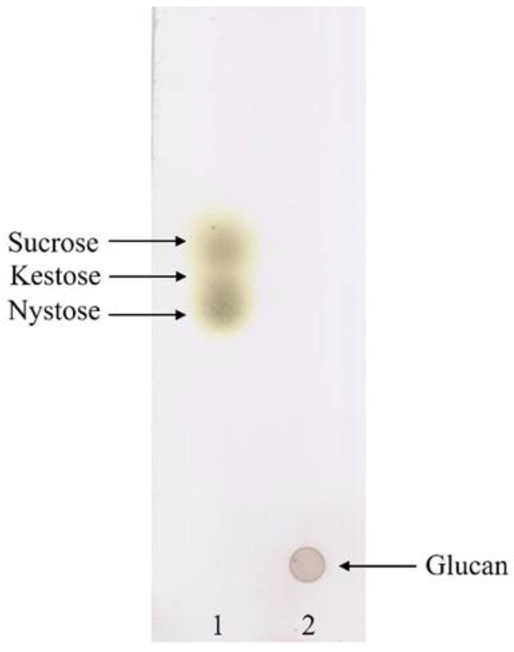
Figure 1. Thin layer chromatographic analysis for glucan synthesis by A. waqarii HBW1. 1: Standard; 2: Sample of HBW1 culture grown on MRS-sucrose medium.
Microorganisms 10 00904 g001
Figure 2. Genome annotation of Apilactobacillus waqarii HBW1 using the ClassicRAST online annotation server showing different types of subsystems.
Microorganisms 10 00904 g002
Figure 3. Whole genome based phylogenetic tree of Aplilactobacillus waqarii HBW1 generated by species tree generated by Type (Strain) Genome Server (TYGS). The numbers above branches are GBDP pseudo-bootstrap support values > 60% from 100 replications.
Microorganisms 10 00904 g003
Figure 4. Phylogenetic tree showing the relationship of two glycosyltransferase genes, i.e., GTF1-HBW1 and GTF2-HBW1 of the isolate Apilactobacillus waqarii HBW1 with related genes from other bacteria. The tree was constructed with MEGA7 using the neighbor joining method.
Microorganisms 10 00904 g004
Figure 5. Linear map of genomic DNA of Apilactobacillus waqarii HBW1showing the position of probiotic traits of related genes in different colors generated through the online CGview server.
Microorganisms 10 00904 g005
Table 1. NCBI Submission details of HBW1 Whole Genome Sequencing Data.
DescriptionInformation
Submission IDSUB9890030
Bio project IDPRJNA740110
Bio SampleSAMN19820174
Accession NoJAHQYH000000000
OrganismApilactobacillus sp. HBW1
Table 2. General genomic attributes of Apilactobacillus waqarii strain HBW1 predicted by the NCBI genome annotation pipeline.
FeatureValue
Genome size1.49 Mb
Genes (total)1419
G + C content37.2%
N50229,491
L503
Number of contigs38
CDSs (total)1344
Genes (coding)1340
CDSs (with protein)1340
Genes (RNA)75
rRNAs5, 4, 1 (5S, 16S, 23S)
Complete rRNAs4, 1, 1 (5S, 16S, 23S)
Partial rRNAs1, 3 (5S, 16S)
tRNAs62
ncRNAs3
Pseudo genes (total)4
CDSs (without protein)4
Pseudo genes (ambiguous residues)0 of 4
Pseudo genes (frameshifted)1 of 4
Pseudo genes (incomplete)1 of 4
Pseudo genes (internal stop)2 of 4
Subsystems247
Carbohydrate metabolism70
Table 3. Taxonomic evaluation of HBW1 on the basis of comparison of its 16S rRNA gene sequence similarity and overall genome-related index values with related type strains.
Strain Name and Accession No.16S% IdentitydDDH (%)ANI (%)
Apilactobacillus kunkeei DSM 12,361 YH15T100.0052.0093.10
[JXDB01000001]
Apilactobacillus apinorum Fhon13N98.8224.5082.44
[KQ440395]
Apilactobacillus bombintestini BHWM-497.6722.7078.18
[NZCP03262.1]
Apilactobacillus timberlakei HV-1296.8619.8074.67
[QUAP00000000.1]
Apilactobacillus micheneri Hlig395.45NANA
[KT833121]
Apilactobacillus ozensis JCM1719694.3822.0065.38
[AYYQ00000000.1]
NA: Genome sequence is not available.
Table 4. Probiotic related genes present in Apilactobacillus waqarii HBW1 genome detected by the RAST annotation server.
GenesLengthStrandPutative FunctionResponse
DNA and protein protection and repair clpATPase (chaperon) Acid and bile tolerance
clpB2583 bp+ATP-binding subunit clpB
clpC2097 bp+ATP-binding subunit clpC
clPE2172 bp ATP-binding subunit clpE
clpP594 bp +ATP-binding subunit clpP
clpX1236 bp+ATP-binding subunit clpX
Methionine sulfoxide reductase Persistence capacity in vivo
msrA522 bpMethionine sulfoxide reductase A
msrB483 bpMethionine sulfoxide reductase A
msrC465 bpMethionine sulfoxide reductase A
Folic acid biosynthesis Folic acid biosynthesis
folD858 bp+Methylenetetrahydrofolate dehydrogenase
Riboflavin biosynthesis operon Riboflavin biosynthesis
Ribf939 bp+Riboflavin kinase/FMN adenylyltransferase
Ribu582 bp+Riboflavin transporter
Ribt369 bp+Riboflavin biosynthesis RibT protein
Ribz11407 bp
Ribz21446 bp
Publisher’s Note: MDPI stays neutral with regard to jurisdictional claims in published maps and institutional affiliations.