Article

An Evaluation of Aluminum Tolerant Pseudomonas aeruginosa A7 for In Vivo Suppression of Fusarium Wilt of Chickpea Caused by Fusarium oxysporum f. sp. ciceris and Growth Promotion of Chickpea

by
Atifa Begum Mozumder
,
Kakoli Chanda
,
Ringhoilal Chorei
and
Himanshu Kishore Prasad
*
Department of Life Science and Bioinformatics, Assam University, Silchar 788011, India
*
Author to whom correspondence should be addressed.
Microorganisms 2022, 10(3), 568; https://doi.org/10.3390/microorganisms10030568
Submission received: 31 December 2021 / Revised: 30 January 2022 / Accepted: 7 February 2022 / Published: 5 March 2022
(This article belongs to the Section Plant Microbe Interactions)

Abstract

:
Chickpea wilt, caused by Fusarium oxysporum f. sp. ciceris, is a disease that decreases chickpea productivity and quality and can reduce its yield by as much as 15%. A newly isolated, moss rhizoid-associated Pseudomonas aeruginosa strain A7, demonstrated strong inhibition of Fusarium oxysporum f. sp. ciceris growth. An in vitro antimicrobial assay revealed A7 to suppress the growth of several fungal and bacterial plant pathogens by secreting secondary metabolites and by producing volatile compounds. In an in vivo pot experiment with Fusarium wilt infection in chickpea, the antagonist A7 exhibited a disease reduction by 77 ± 1.5%, and significantly reduced the disease incidence and severity indexes. Furthermore, A7 promoted chickpea growth in terms of root and shoot length and dry biomass during pot assay. The strain exhibited several traits associated with plant growth promotion, extracellular enzymatic production, and stress tolerance. Under aluminum stress conditions, in vitro growth of chickpea plants by A7 resulted in a significant increase in root length and plant biomass production. Additionally, hallmark genes for antibiotics production were identified in A7. The methanol extract of strain A7 demonstrated antimicrobial activity, leading to the identification of various antimicrobial compounds based on retention time and molecular weight. These findings strongly suggest that the strain’s significant biocontrol potential and plant growth enhancement could be a potential environmentally friendly process in agricultural crop production.

1. Introduction

Chickpeas (Cicer arietinum L.) are considered to be one of the oldest legumes known from ancient times. Most likely, it originated in southeastern Turkey and Syria’s adjacent areas [1]. Globally, it is a valuable crop and about 2.3 million tons enter the world market annually [2]. The major chickpea legume grain-producing regions are in southern and South-Eastern Asia, Africa, and Australia, [2].
It is estimated that India produces 9.075 million tons of chickpeas annually, accounting for 65% of the total production of chickpeas. The remarkable growth of chickpea pulse has been documented in India, comprising the most significant production (47%; 10.90 MT) among all pulse crops in 2019–2020 [2]. Chickpeas are sustainable legumes with nutritional value for human and animal consumption as well as soil fertility. Chickpeas seeds and leaves are an excellent source of carbohydrates, vitamins, fibers, and proteins. Chickpeas play a leading role in food safety in the world by supplying the protein deficit of daily food rations [3]. Chickpeas-based diets are recommended to combat lifestyle disease since they contain several essential minerals such as calcium, phosphorus, iron, and zinc among other substances, including phenolics and oligosaccharides [3]. There are numerous living and non-living stressors that affect chickpea production globally [4,5]. Among them, Fusarium wilt is the most severe biotic stressor of chickpeas, caused by Fusarium oxysporum f. sp. ciceris (Foc) in chickpea-growing nations all around the world [5,6,7]. Foc is a significant limiting factor in chickpea production, causing yield losses ranging from 10 to 100% depending on variety and climatic conditions [8,9]. This plant disease exhibits symptoms such as wilting, drooping, discoloration, yellowing, browning of the xylem vessels, and leads to the eventual collapse of the whole plant [6,9]. In chickpea, several sources of resistance have been identified [10,11], which have been successfully used in resistance-breeding programs at the Indian National Agricultural Research System (NARS) and International Crops Research Institute for the Semi-Arid Tropics (ICRISAT), increasing chickpea production [12]. A total of 8 Foc pathogenic race variants have been identified in the world so far (races 0, 1A, 1B/C, 2, 3, 4, and 6) [11,13]. Biological control of Foc using rhizobacteria is an ecologically sustainable approach since the pathogen is soil-borne and the indiscriminate chemical control measures lead to increased pesticide tolerance [13,14,15,16]. Several pseudomonas species found in the rhizosphere have proven to be effective biocontrol agents, both due to their widespread distribution in soil and their production of heat-resistant spores [17]. In agriculture, plant pathogenic fungi are responsible for severe damage and the loss of agricultural products [18,19]. The antagonist Plant Growth-Promoting Rhizobacteria (PGPR) act as biocontrol agents (BCAs) and secrete active ingredients such as natural products [20]. Natural products secreted by bacteria may suppress pathogen growth either directly by producing volatile or non-volatile secondary metabolites or indirectly by participating in the competition for nutrients [21,22,23]. Plant growth-promoting biocontrol agents produce natural products such as pyrrolnitrin, phenazine, phloroglucinol, hydrogen cyanides, siderophores, hydrolytic enzymes that inhibit and suppress the plant pathogen Fusarium oxysporum [23,24,25]. Additionally, Trichoderma harzianum and Bacillus subtillis secrete β-1,3 glucanase enzymes that have an inhibitory effect on chickpea wilt by inhibiting the pathogen’s growth [23,26]. Volatile organic compounds (VOCs) produced by PGPBs suppress phytopathogenic growth and produce morphological and physiological changes to plants, such as an increase in chlorophyll content, which further improves plant growth [27].
Plant growth-promoting bacteria (PGPB) that can withstand metals are being used to develop the growth of numerous crops in metal-polluted environments [28]. Microbes can be used in the agricultural field to protect natural habitats and ensure healthy crop production. Burkholderia ginsengiterrae N11-2, Chryseobacterium polytrichastri N10, Pseudomonas fluorescens, Pseudomonas fragi N8 and Pseudomonas simiae N3, have all been shown to grow in aluminum-stress conditions [28,29]. Plant Growth-Promoting Rhizobacteria (PGPR), which can produce phytohormones that are essential for plant growth, interact with the flora by increasing nutrient accessibility [21,23,30]. The pseudomonas strains can promote plant growth, as well as advance plant responses to harsh conditions, and allocate all the important characteristics to the host plant to promote growth [31,32,33,34,35]. North-Eastern India has various agro-climatic zones that demand a range of cultivation methods. The excessive rainfall in this region is causing basic cations in the soil in the region to leach out over time, destroying soil texture and bringing the thriving of metals like aluminum to bear on crop health [36]. Chemical forms of aluminum exhibit the highest degree of toxicity to plants when exposed to pH < 5 [37]. The metal has gained attention due to its toxic effects on acidifying soil, which limits crop production worldwide and the growth of most living organisms [38,39]. Aluminum’s toxicity has become a major concern as this metal is usually formed into complexes with silicates liberated because of acid rain [40]. Aluminum (Al) levels, about 1ppm in soil, reduce root growth through limiting water and nutrient uptake, which affects plant health and crop productivity [36,41]. By increasing the expression of aluminum stress-related genes, PGPBs are shown to improve the plant’s tolerance mechanisms [28,42]. Pseudomonas fluorescens (ICA/BECa 141), Kosakoniar adicincitans (IAC/BECa 95), Paraburkholderia tropica (IAC/BECa 135), and Herbaspirillium frisigense (IAC/BECa 152) have been shown to increase sugarcane growth on high Al bioavailability and nutrient as well as Al uptake in crop plants under acid soil conditions [43]. Pseudomonas plecoglossicida Pp20 strain ameliorates damages on maize roots induced by aluminum chloride (AlCl3) at a concentration of 90 µM by producing the plant growth-promoting hormone indole-3-acetic acid (IAA) and ethylene degrading enzyme, 1-aminocyclopropane-1-carboxylic acid (ACC) deaminase [44]. Apart from the production of phytohormones, the PGPB may evolve diverse mechanisms such as mineral solubilization, the development of siderophores (metal-chelating compounds), inhibiting metal transport in the roots, reducing lipid peroxidation and hydrogen-peroxide production, and sustained enhancement of antioxidant enzymes in the plants [41,42,43,44,45]. Few reports are available for chickpea metal tolerance using PGPB under salinity and metal (Mn) toxicity conditions [46,47]. Combining endophytic bacteria (Stenotrophomonas maltophilia), mycorrhizal fungi, and ferrimagnetic magnetite (Fe3O4) nanoparticles induced Rhizobium improved chickpea (Cicer arietinum L.) nodulation, leg hemoglobin content, nitrogenase activity, and growth by mitigating salt and metal stress and improving nutritional status of plants at salinity levels 75 and 150 mM [47]. However, no attempt has been made to employ PGPR inoculation to mitigate Al stress in chickpea plants growing in acid soil with high Al levels. The current study reports on the identification and characterization of a Pseudomonas aeruginosa strain A7 isolated from rhizoids of Polytrichum sp. from a waste disposal area. We report its biocontrol potential against phytopathogens and characterize its abilities to produce plant-growth promotion-related compounds such as indole-3-acetic (IAA), ammonia, siderophores, hydrogen cyanide (HCN), cellulase, chitinase, pectinase, proteases, the ability to solubilize tri-calcium phosphate, potassium, zinc oxide and can tolerate different abiotic stress conditions. The strain A7 enhanced the chickpea growth in aluminum stressed conditions and acts as a potential biocontrol agent to Fusarium wilt disease of chickpea caused by Fusarium oxysporum f. sp. ciceris (Foc).

2. Materials and Methods

2.1. The Isolation, Morphological and Biochemical Characterization of Aluminum-Tolerant Bacteria from the Rhizoids of Polytrichum sp.

Adult moss plants identified as Polytrichum sp. were collected from a waste disposal area at Cachar Paper Mill Panchgram, Hailakandi, (Assam), India. Approximately 10 g of tiny rhizoids of the moss adhering with soil were cut out and mixed with 90 mL phosphate-buffered saline (PBS) (HiMedia, Mumbai, India) in a conical flask. The flask was kept in a rotary shaker incubator at 150 rpm at 30 ± 2 °C for 60 min. Following shaking, the suspension was serially diluted to a factor of 10−6 with Phosphate-buffer-saline (PBS) buffer and plated on King’s Base agar medium (KB) (HiMedia, Mumbai, India) plates supplemented with aluminum (AlCl3, SD Fine Chemicals, Mumbai, India) and incubated for 2 days at 30 ± 2 °C [48,49]. On the plates, bacteriologically distinct colonies were selected for further purification. Pure strains were preserved at −80 °C in 20% glycerol [50]. A strain designated A7 was studied for its morphological characteristics, such as color, shape, size, elevation, and surface. The fluorescent activity of the colonies that appeared under ultra-violet (UV) light on the plates was evaluated after 24 h by re-inoculating them on the KB plate. The strain was processed through different biochemical characterizations such as catalase, citrate utilization, indole synthesis, methyl red generation, nitrate reduction, starch hydrolysis, urease, Voges-Proskauer, and hydrogen sulfide (H2S) production was achieved using KB003 Hi25TM Enterobacteriaceae identification Kit (HiMedia, Mumbai, India) [51].

2.2. Molecular Identification of the Strain

The 16S rRNA-encoding gene sequencing-based molecular identification of the strain was performed using total genomic DNA [52]. For the amplification of the 16S rRNA gene fragment, the fD1 (5′-GAGTTTGATCCTGGCTCA-3′) and rP2 (5′-ACGGCTACCTTGTTACGACTT-3′) primer set was used [53]. Polymerase chain reaction (PCR) mixture (50 µL) contained 50 ng of genomic DNA template, 0.5 µM of each of the primers, 200 µM of dNTPs, 1X-High-Fidelity buffer, 1U of Phusion High-Fidelity DNA polymerase enzyme (Thermo Scientific, Waltham, MA, USA). The polymerase chain reaction amplification was performed using a C1000 Touch Thermal Cycler (Bio-Rad, Hercules, CA, USA) with cycle conditions of initial denaturation at 98 °C for 30 s, 35 cycles of initial denaturation at 98 °C for 10 s, 50 °C (annealing) for 30 s, 72 °C (extension) for 1 min 30 s, and a final extension of 72 °C for 7 min. Gel electrophoresis was performed on the amplified products using 1.5% agarose and Tris-Acetate-EDTA (TAE). The PCR product was purified using a QIAquick PCR purification kit (Qiagen, Hilden, Germany). The restriction enzyme EcoRV (NEB, Ipswitch, MA, USA) cut plasmid vector pLitmus28 (NEB, Ipswitch, MA, USA) and 16S rRNA DNA was blunt ligated with T4 DNA ligase (Thermo Scientific, Waltham, MA, USA) to clone the PCR product. Electrocompetent E. coli TOP10 cells (Thermo Scientific, Waltham, MA, USA) were prepared and transformed with a ligation mix using a Gene Pulser (Bio-Rad, Hercules, CA, USA), following the standard protocol [54]. Using the corresponding primer set, colonies on Luria Bertani (LB) agar fortified with Ampicillin 100 µg mL−1 were selected and confirmed by colony PCR and plasmid DNA was isolated. An EcoRI restriction digestion (Thermo Scientific, Waltham, MA, USA) was performed to obtain the product of the desired size of 4.3 kb. The recombinant DNA products were purified further using PEG 8000 (Sigma Aldrich, Bangalore, India) and sequenced commercially. Two reads of the amplicon sequencing were assembled and analyzed using Chromas Pro version 2 (http://technelysium.com.au/wp/chromaspro/, accessed on 25 July 2020). To identify the strain, a blastn analysis was performed by comparing the derived gene sequence to known bacterial 16S ribosomal RNA gene sequences obtained from the NCBI GenBank website (https://www.ncbi.nlm.nih.gov/, accessed on 25 July 2020). A phylogenetic analysis was performed with the Maximum Likelihood method with 1000 bootstrap replications using MEGA version 6.0 software (https://www.megasoftware.net/, accessed on 25 July 2020) [55]. This nucleotide sequence was submitted for inclusion in GenBank under accession number MN197639.

2.3. In Vitro Antagonistic Activity of the Strain A7

To assess the in vitro antimicrobial activity, plant pathogenic fungal strains Rosellinia arcuata Petch (Rap) (ITCC 4143), Poria hypobrunnea Patch (Php) (ITCC 4141), Ustillinia zonata (Lev.) Sacco (Uzs) (ITCC 4144), Phellinus lamaensis (Murrill) Pat. (Plp) (ITCC 4140), Fusarium oxysporum (Fo) (ITCC 7093), Fusarium oxysporum f. sp. ciceris (Foc) (ITCC 3636) Fusarium oxysporum f. sp. lycopersici (Sacc.) (Fol) (ITCC 1322), Fusarium oxysporum f. sp. pisi (Fop) (ITCC 4814), Fusarium solani (Fs) (ITCC 6953), Curvularia eragostidies (Ce) (ITCC 5540) and Curvularia lunata (Wakker) Boedijn var. (Clb) (ITCC 5193) were obtained from Indian Type Culture Collection (ITCC), in the Division of Plant Pathology, Indian Agricultural Research Institute, New Delhi. The fungal pathogen Rhizoctonia solani (Rs) and bacterial pathogens Ralstonia solanacearum (Ras), Xanthomonas campestris (Xc) were kindly supplied from Assam Agricultural University, Jorhat, India.
The in vitro antifungal activity of strain A7 was performed using a concentric ring bioassay [56]. In the center of the Potato-dextrose Agar (PDA) plate, a 6 mm diameter mycelium disc was cut from the margin of a 4-day-old PDA grown fungal mycelium and placed. The bacterial strain was inoculated surrounding the fungal disc in a concentric ring manner with the help of a sterile funnel. As a control, a fungal disc without a concentric ring of bacteria was employed. The percentage of inhibition was calculated after 2 to 7 days of growth suppression at 30 °C, depending on the pace of growth. Using the formula below, the percentage of growth inhibition (PGI) of the fungal pathogen by the strain was calculated.
The dual-culture assay was used to produce antifungal volatile organic compounds (VOCs) [57]. A King’s B agar plate was streaked with strain A7, and a fungal mycelium disc was placed onto the center of the PDA plate. Parafilm was used to seal both inoculation plates [58]. However, it should be emphasized that the Petri dishes create a gap in the growth media, preventing the nonvolatile exudates secreted by A7 from reaching the fungus. KB plates spread with sterile distilled water and the fungal disc on a PDA plate sealed with parafilm was considered a control. The growth inhibition was determined after 2 to 7 days at 30 ± 2 °C, depending on their growth rate and the percentage of growth inhibition was recorded. The percentage of growth inhibition (PGI) of the fungal pathogen by the strain was calculated as:
PGI   ( % ) = ( D c   D t ) / ( D c ) × 100
where Dc is the pathogen’s growth rate in the absence of strain A7, and Dt is the pathogen’s growth rate in the presence of strain A7.
In vitro antifungal and antibacterial activities by the strain A7 with the cell-free supernatant (CFS) were completed by Agar-well diffusion and the Agar disk-diffusion method [59]. To establish the stationary phase cultures, A7 was cultured on KB broth for 2 days at 30 °C. The cell-free supernatant of the strain A7 was collected by centrifugation at 13,000 rpm for 10 min at 28 °C. To eliminate any debris, CFS was filtered using a 0.2 µm membrane filter (Millipore) and was ready to use [60]. Antifungal activity by using CFS was performed by inoculating a fungal disc at one edge of the PDA plate. A rectangular well was prepared at another edge using a sterile scalpel and a volume of 100 µL of CFS was introduced into it. The Petri dishes were incubated for 48 h at room temperature. The CFS was diffused into the medium and the growth of the fungal pathogen was inhibited and measured as above.
Antibacterial activity using the CFS of the strain A7 on KB agar plates was performed by inoculating freshly grown bacterial pathogens (0.1 optical density at 600 nm, OD600) by streaking. Then, on both ends of the agar surface, filter paper discs (6 mm in diameter) were inserted and labeled accordingly (i) filter paper disc with 50 µL CFS, (ii) filter paper disc with 50 µL sterile double distilled water as control. The Petri dishes were incubated at 30 ± 2 °C for a day. CFS was diffused into the medium and the germination and growth suppression of the respective pathogen was calculated by measuring the diameters of the zone of inhibition [61]. All assays were performed in three biological replicates.

2.4. Plant Growth-Promoting (PGP) Activities of the Strain A7

The strain’s plant-growth-promoting activities were assessed qualitatively using a plate-based assay and quantitatively using a liquid assay. Luria-Bertani (LB) agar plates amended with 5 mM L-tryptophan (SRL, Mumbai, India), Sodium Dodecyl Sulfate (SDS, 0.06%), and glycerol (1%), was used to detect Indole-3-Acetic acid (IAA) production. To obtain a colony with a diameter of 0.2–5 mm, the plates were incubated for 24 h. After that, the Petri dishes were covered using Whatman no.1, filter paper, and incubated for another 24 h. The filter papers were removed after the incubation period, saturated with Salkowski reagent (4 mL prepared using 2% of 0.5 M Ferric III Chloride in 35% of Sodium hypochlorite), and studied at 30 °C for 1 to 2 h until appropriate color development. The development of the red halo zone around the colony on the filter paper was considered a positive IAA producer [62]. IAA production was also performed quantitatively by inoculating the strain on the LB broth using Salkowski reagent with a 48 h time interval of 196 h. The absorbance was measured at 530 nm after the development of the pink color. Using the IAA standard (LobaChemi, Mumbai, India), the concentration of the generated IAA was measured [62]. Phosphate was solubilized on Pikovskaya’s (Pkv) agar (HiMedia, Mumbai, India), using the method outlined in [63]. Quantitative estimation was performed on the Pkv broth medium by the method described [64]. Briefly, 25 mL Pkv broth was inoculated with A7 and incubated at room temperature for 4 days. Additionally, 1 mL of the bacterial cell-free supernatant prepared by centrifuging at 30 min at 12,000 rpm, (Eppendorf Centrifuge 5804R, Eppendorf, Hamburg, Germany) was mixed with 10 mL chloromolybdic acid, and 35 mL of distilled water was added. The volume was then increased by adding chloro-stannous acid (0.25 mL). The total volume of 50 mL was maintained by adding distilled water. The absorbance was measured at 600 nm of the developing blue solution. The quantity of soluble phosphorus was determined using the KH2PO4 standard curve (Sigma Aldrich, St. Louis, MO, USA). A modified plate assay was used to determine the solubilization of the potassium [65] by inoculating the strain A7 on Aleksandrov agar medium mixed with Bromothymol Blue (an acid-base indicator dye) [66]. The Petri dishes were incubated at room temperature for 7 days. An improved visibility solubilization halo zone on the agar plates was positive for potassium solubilization. The formula for calculating the solubilization efficacy (SE) was as follows: SE = [the diameter of A7/the diameter of the halo zone] ∗ 100 [67]. By inoculating the strain on zinc oxide agar medium plates and incubating them at 30 °C for seven days, zinc oxide solubilization was measured qualitatively [68]. The above formula was used to compute the solubilization efficiency. The strain A7′s ability to fix atmospheric N2 was investigated using the nitrogen-free broth (Nfb) medium described [69]. At 30 °C, the plates were incubated for 24 h. After the incubation period, the medium’s color changed from pale green to blue, indicating the isolate’s ability to fix N2. Ammonia was produced by growing A7 on peptone water (10 mL) in each falcon tube and incubated for 2 to 3 days at 30 ± 2 °C. Following incubation, 500 µL of Nessler’s reagent was added to each tube. Brown to yellow color development was taken as a positive test for the production of ammonia [70]. Chrome Azurol S (CAS) assays were used for siderophores production [71]. The freshly prepared CAS solution was added to the KB agar medium as a 1:15 ratio and sterilized by autoclaving. Plates inoculated with A7, showing an orange halo zone surrounding the colony at 30 ± 2 °C for 3 days of incubation, were considered positive for siderophores production, and the diameter of the colored zone was recorded. The A7 was inoculated on a 20 mL KB medium and incubated at the above-mentioned parameters to determine the production of siderophores. Centrifugation at 3000 rpm for 5 min was used to remove the cells. Furthermore, 3 mL of cell-free supernatant, 300 µL of 5N hydrochloric acid solution, 1.5 mL of Arnow’s reagent, and 300 µL of 10N NaOH were mixed. The formation of a pink color was noticed after 10 min and recorded. The KB medium amended with 4.4 g of glycine/L was used to test the production of HCN using the strain A7. The socked filter paper (Whatman no. 1, Ankleshwar, India) on 2% Na2CO3 in the picric acid solution (0.5%) was kept on top of the Petri dishes. The dishes were packed with black paper after sealing with parafilm and incubated at 30 ± 2 °C for 4 days. HCN production was detected by the hue changing from yellow to red brown [72]. The chitinolytic activity was assayed on the chitin agar plate, as described by [73] with some modification. In Petri dishes, the lukewarm medium was poured and was allowed to harden. The A7 was inoculated onto the medium and incubated at 30 °C, where it was monitored for the formation of a purple halo zone surrounding the colony, and the measurement of the purple halo was recorded. The polyamine production using A7 was tested by streaking on a Long Ashton Decarboxylase (LAD) agar [74] for 7 days, the Petri dishes were incubated in the dark at room temperature. The strain’s arginine decarboxylation was represented by red halos on a yellow backdrop. The extracellular enzymatic activity of strain A7 was using different modified plate-based methods. Cellulase and xylanase activity was estimated by inoculating the strain on Congo-red gelatin agar medium supplemented with 0.5% Carboxymethylcellulose (Na-CMC, Sigma Aldrich, St. Louis, MO, USA) and xylan (Beechwood Xylan, Sigma Aldrich, St. Louis, MO, USA), respectively [75]. The Petri dishes were incubated for 3 to 7 days at room temperature. Bacterial colonies stained with congo-red were thought to be cellulose and xylan degraders [76]. The positive strain’s degrading potential was also measured qualitatively by equating hydrolysis capacity (HC). HC = Ratio of the halo zone diameter to the colony diameter [77]. The ligninolytic activity was observed using a Minimal Salt Medium-Kraft Lignin (MSM-KL) medium containing 0.5% Alkali lignin (Sigma Aldrich, St. Louis, MO, USA) supplemented with Methylene blue as the lignin polymeric dye and incubated at 30 ± 2 °C for 3 to 7 days. The strain possesses ligninolytic enzymes that undergo the oxidation of indicator dye. The plates were checked daily for bacterial growth and Methylene blue dye decolorization [78]. Pectinase production was estimated by inoculating A7 on an MM Pectin agar medium supplemented with 0.5% Citrus Peel Pectin (Sigma Aldrich, St. Louis, MO, USA) [79]. The Petri dishes were incubated for 2 days at room temperature and flooded for 15 min with a 5.0% (w/v) iodine solution (LabChem, India) to detect the halo zone around the colony, which shows the strain’s ability to produce pectinase. Proteases activity was performed by inoculating A7 on a Skim milk agar medium supplemented with 0.5% skim milk (Sigma Aldrich, St. Louis, MO, USA) and tested positive by the development of a distinct halo zone around the colony [80]. The above-mentioned formula was used to calculate the semi-quantitative enzyme production.

2.5. Growth Profile of Strain A7 on Different Concentrations of Aluminum, Iron, and Other Stressed Conditions

The growth profile of strain A7 was checked to tolerate an increasing concentration of aluminum (AlCl3) (0–4 mM) and other metal compounds using the Single Plate Serial Dilution Spotting (SP-SDS) method on modified M9 agar plates [80]. The liquid culture was in the M9 broth supplemented with 0.1% proteases peptone. Similarly, the effect of Fe (III) from 0–5 mM, Fe (II) from 0–3 mM, NaCl (0–8%) (Sigma Aldrich, St. Louis, MO, USA) [81], pH (1–14) and temperature (4–50 °C) was checked in three biological replicates [82,83]. Freshly grown bacterial cells (0.1 OD600) were serially diluted from 10−1 to 10−6, and 3 µL from each dilution was inoculated into the demarcated areas of SP-SDP and in a 10−1 dilution in a 50 mL broth. The Petri dishes were incubated for 18–48 h, and the flask was held in an orbital shaker at 160 rpm at 30 ± 2 °C for 48 h. The optical density of the cultures was taken at 600 nm (PerkinElmer UV/VIS spectrometer Lamda 25, USA). The relative growth (RG) was calculated using the formula [84]. Relative Growth (%) = At/Ac ∗ 100, where Ac = optical density of the average of three biological replicates of controls, and At = optical density of average three biological replicates of the treatments. Bacterial 20% and 50% growth inhibitory concentration (IC) IC20 and IC50 values were calculated using the dose–response data.

2.6. In Vitro Protective Effect of Strain A7 on Aluminum Toxicity in Chickpea Growth

For the in vitro protective effect of strain A7 against aluminum toxicity in chickpea, growth was verified using the jar method [36]. An adequate quantity of chickpea seeds (Pusa-362) was surface-sterilized for 3–5 min in a 0.1% HgCl2 solution followed by 6–7 washing with sterilized distilled water. The seeds were saturated in water for 12 h after which they were kept on moisten filter paper placed within a Petri dish for germination at 28 ± 2 °C for 72 h. The healthy germinated seeds of equal root and shoot length were transferred into a plastic jar filled with 400 mL of Hoagland nutritive solution (HiMedia, Mumbai, India). Over a 14 h photoperiod, plantlets were cultivated in a plant-growing room in white light with a photon flux intensity of 220 mol m−2 s1 (PAR). The Hoagland nutritive solution changed every 48 h to maintain the seedlings proper growth. These plantlets were then pretreated with 500 µM CaCl2 (pH 5) for a day. The growth of strain A7 was previously checked on different concentrations of CaCl2 (100, 200, 500, and 1000 µM). After one day, the pretreated seedlings were transferred to an experimental setup (1) T0 = Control (with no bacteria and no aluminum), (2) T1 = AlCl3 (only 100 µM of aluminum chloride), (3) T2 = A7 (only 0.1 OD600 cells of the bacterial strain) and (4) T3 = A7 + AlCl3 (0.1 OD600 cells of the bacterial strain + 100 µM of aluminum chloride) for 12, 24, 36, 48 h. The root length and shoot length, root fresh weight and dry weight, and relative root length rate (RRL) were all assessed to determine total growth and biomass. Four plants from each treatment were randomly selected and root shoot length was measured. For plant biomass measurements, four plants were grouped to measure the fresh weight and were dried at 80 °C for 48 h, weighing the dry biomass. Seed vigor index (SVI) [85] and relative root length (RRL) rate [36] were calculated using the formulae:
SVI   = ( Shoot   length   +   root   length ) ×   seed   germination %
RRL   = ( Root   length   in   aluminum   stress   conditions )   /   ( Root   length   in   control   conditions ) × 100

2.7. Control of Fusarium Wilt of Chickpea by Strain A7 in Pot Assay Experiment

The chickpea seeds of variety (Pusa-362) were collected from Krishi Vigyan Kendra, Hailakandi, Assam, India, and were used in the pot experiments. The variety Pusa-362 was tested for Fusarium wilt-disease control in pot and field experiments using bacterial biocontrol agents [86]. Fusarium oxysporum f. sp. ciceris (Foc) conidia were prepared by inoculating the pathogen onto the PDA medium incubated at room temperature for 8 days. For a healthier spore suspension, actively growing margins on the culture media plate was removed and 4 to 5 mL sterile dH2O was added. The mycelia were separated from the spore suspension, the conidial suspension was harvested, and the final concentration of the inoculum was prepared at 106 conidia/mL. Bacterial inoculum was prepared by growing the strain A7 in KB broth, 30 °C, 160 rpm, for a day. The cell culture was centrifuged for 10 min at 6000 rpm and a fresh pellet, the suspension was prepared in one-fourth strength of 1% sterile carboxymethylcellulose (Sigma Aldrich, St. Louis, MO, USA) to prepare a final concentration of 108 bacteria/mL [87].
Dry vermiculite soil collected from the vegetable field was crushed, sieved, and sterilized by autoclave. The seeds were surface-sterilized for 4–5 min with an HgCl2 (0.1%) solution, then washed 6–7 times with sterile dH2O. The seeds were saturated in sterile water for 12 h, transferred, and placed properly in a seedling bed prepared with the soil in a plastic tray for germinated and grown at 28 ± 2 °C for 7 days. Seven numbers of 7 days old seedlings were then transferred to a pot containing 2 kg of soil.
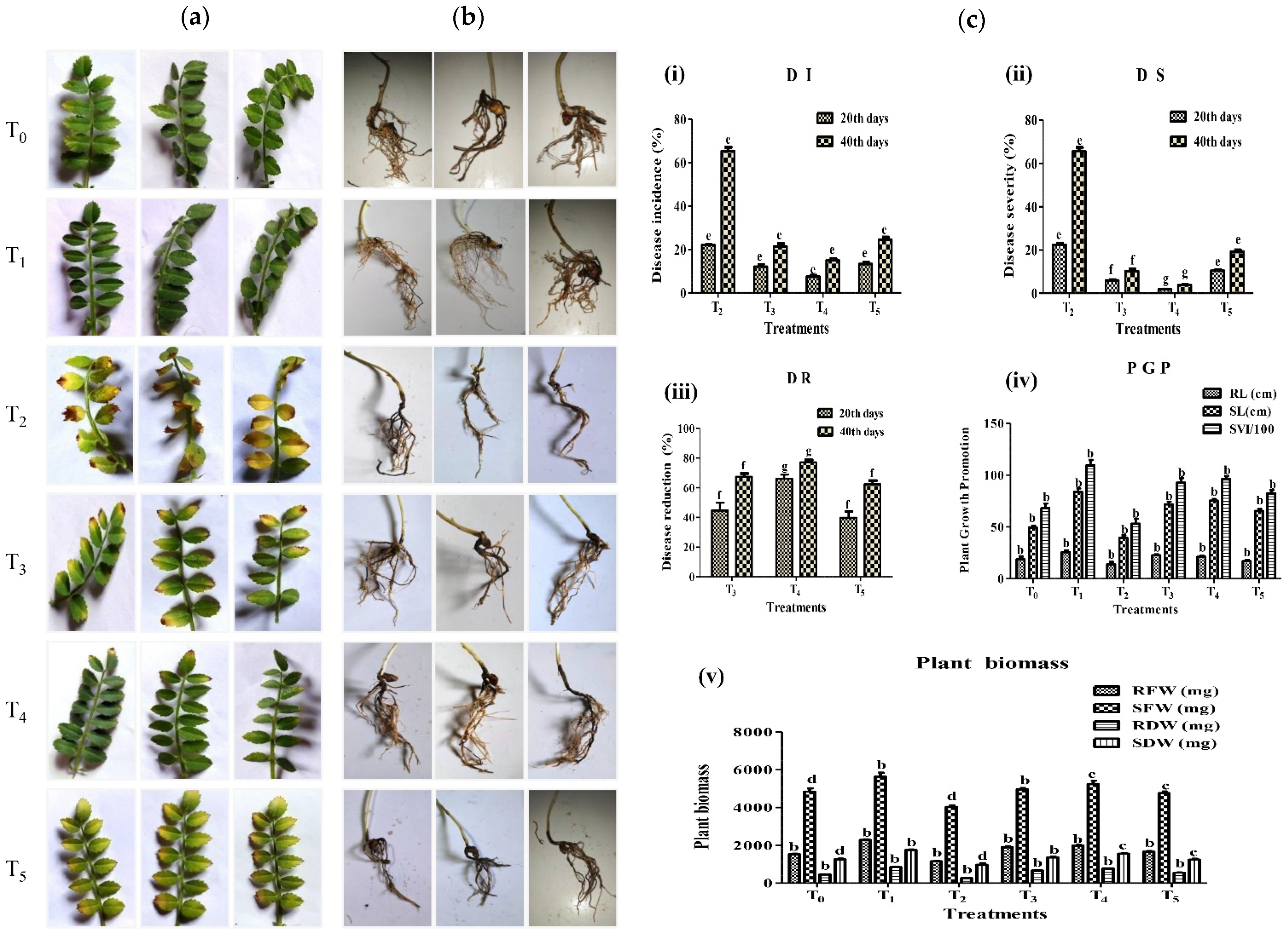
The seedlings were treated by inoculating 5 mL of bacterial cell suspension and 5 mL fungal spore suspension in the following treatments: (1) T0 = sterile distilled water as control (2) T1 = only bacterial cell suspension (3) T2 = only fungal spore suspension, (4) T3 = bacterial cell suspension and fungal spore suspension, (5) T4 = bacterial cell suspension two days before the fungal spore suspension, (6) T5 = fungal spore suspension two days before the bacterial cell suspension. The pots were observed for diseases such as a yellowing of the leaves after 40 days with a 20-day interval, and root rot and darkening of vascular tissues were observed after 40 days after sowing [88]. Fusarium wilt disease severity was visually assessed on a 0–5 rating. The severity was calculated using the percentage of foliage with visible yellowing or necrosis along with acropetal progression compared with the control pots. The scale of 0 = healthier plants, 1 = 1 to 20% wilt disease on one fourth leaves, 2 = 21 to 49% wilt disease on two fourth leaves, 3 = 50 to 70% wilt disease on three fourth leaves, 4 = 71 to 100% wilt disease on the whole plant and 5 = all leaves dead [89]. The percentage of disease incidence (DI) [90], disease severity (DS), and disease reduction (DR) [89] was scored 40 days (with 20 days interval) after sowing according to the formulas below:
DI   ( % ) = ( No .   of   attained   leaves / Total   no .   of   leaves ) × 100  
DS   ( % ) = ( Σ   n   ×   v / N   ×   V ) × 100
where n = number of leaves infected by wilt, v = scale of disease severity (0 to 5), N = number of leaves observed, V = value the highest scale [89].
DR   ( % ) = [ 1 ( Disease   Incidence   in   treatment / Disease   incidence   in   control ) ] × 100  
Plant growth parameters, i.e., root and shoot length, fresh and dry weight of root and shoot, and the seed vigor index was measured for each plant after the completion of the experiments.

2.8. Detection of Antifungal Antibiotic Genes Present in A7 by PCR Amplification

The PCR-based detection of biomarker genes for the biosynthesis of antifungal antibiotics was achieved using gene-specific primer sets and cycling conditions listed in Table 1. Primers were used for the detection of gene phlD for 2,4-diacetylphloroglucinol (2,4-DAPG) [91], hcnBC for hydrogen cyanide (HCN) [92], phzC, and phzD for phenazine-1-carboxylic acid (PCA) [91], phzH for phenazine-1-carboxamide (PCN) [93], prnD for pyrrolnitrin (PRN) and pltC for pyoluteorin (PLT) [94] and darS or darR for 2-hexyl-5-propyl resorcinol (DarA, DarB enzymes site removed) [95] (Table 1). For each gene, a 10 µL PCR reaction consisted of 0.5 µM primers (forward and reverse), 10 ng A7 genomic DNA, 10× PCR buffer, 200 µM each dNTP, and 0.1U Taq DNA polymerase (Qiagen, Hilden, Germany).

2.9. Characterization and Analysis of Bioactive Natural Compounds Extract Present in A7 by TLC, HPLC, and GC-MS

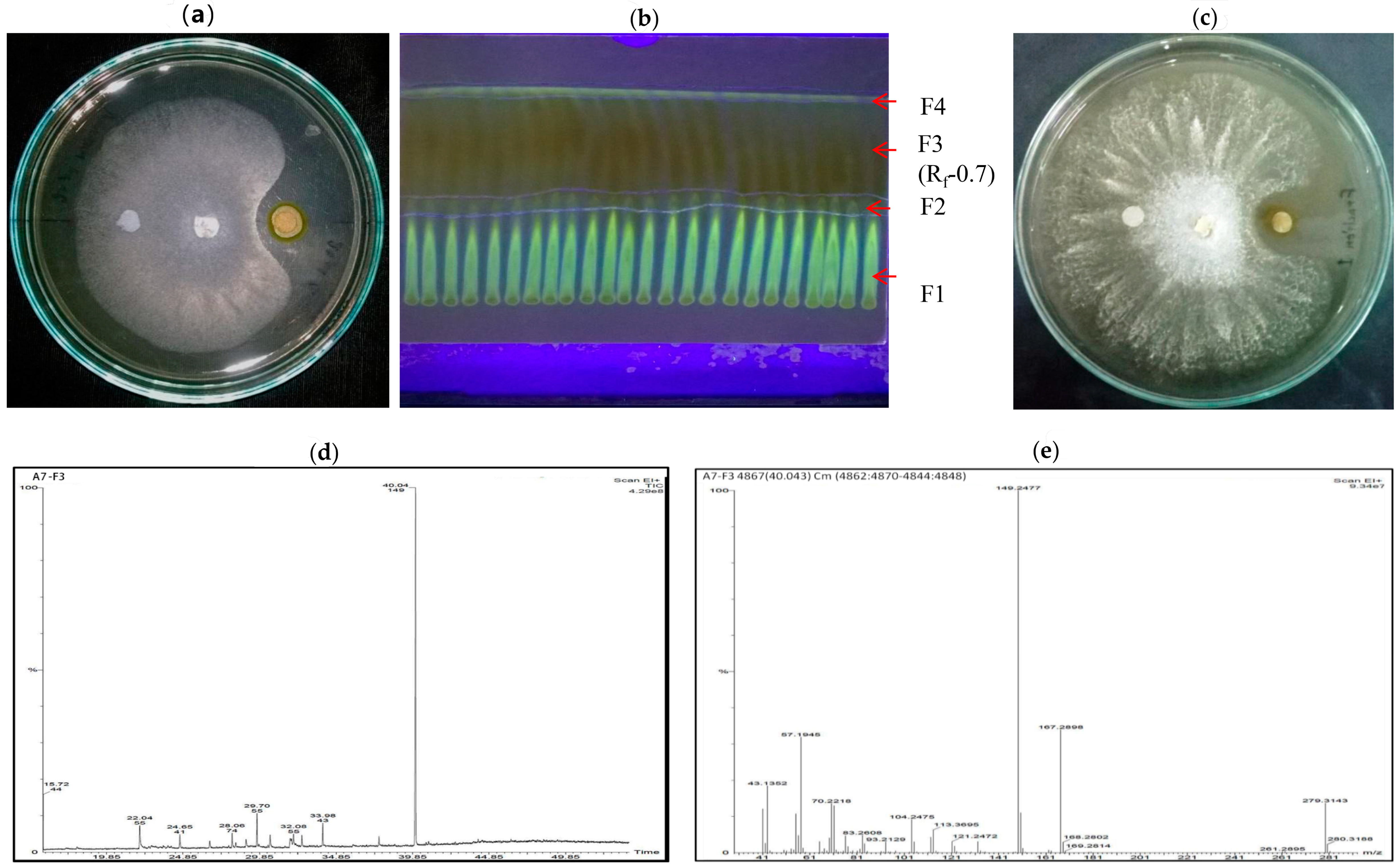
To determine the bioactive natural compounds responsible for the antifungal and antibacterial activities of A7, the strain was cultivated in 1 L KB broth and grown for 5 days at 30 ± 2 °C with shaking at 150 rpm [96]. Cells were centrifuged at 8000 rpm for 10 min, and the resulting cell-free supernatant (CFS) was acidified to pH-2 by adding concentrated hydrochloric acid. The acidified CFS was mixed with equal volume (1:1 v/v) of ethyl acetate and incubated until the separation of two layers. The organic layer was decanted and evaporated at room temperature. The dry residue obtained was dissolved in 250 µL of methanol. An amount of 10 µL was evaluated for antifungal activity along with the control (10 µL ddH2O) by the agar-disc diffusion method described earlier. Aliquots of extracts were filtered and analyzed by Thin-layer chromatography (TLC) [97] and High-Performance Liquid Chromatography (HPLC). TLC using silica-G plates activated at 110 °C for 30 min, were used to fractionate the extracted organic compound in chloroform/acetone (9:1 v/v). The chromatogram was visible under UV light (Bio Red) at 254 nm after the TLC plate had dried [98]. The Rf value was calculated after the separation of the fractions, marked, recovered again in methanol, and evaluated again for antifungal activity. The maximum antifungal activity showed by the fraction was analyzed by an HPLC system (1260 Infinity, Agilent Technologies, Santa Clara, CA, USA, with a liquid autosampler, at Indian Institute of Technology Guwahati, IITG). The mobile phase was a 45% CH3CN solution containing 0.1% H3PO4, with a speed of 1.0 mL/min. The wavelength detection was fixed at 270 nm [99]. HPLC elutes of the purified extract were examined via Gas Chromatography-Mass Spectroscopy (GC-MS) (Clarus 680 GC and Clarus 600C MS, PerkinElmer, Waltham, MA, USA; Liquid Autosampler, Library Software: Turbomass NIST 2008, at IITG) equipped with Capillary column: Elite-5MS Capillary Column: Length: 60 m, I.D:0.25 mm (Max. Program Temperature 350 °C), Phase Reference: 5% di-phenyl 95% dimethylpolysiloxane (low bleed) and a mass detector activated in EMV mode. The flow rate of the carrier gas (helium) was set at 1.0 mL min−1, the injection-port temperature was kept at 250 °C. The column oven was kept at 80 °C for 2 min, followed by 10 °C min−1 to 250 °C for 0 min, then at 50 °C min−1 to 280 °C for 9 min. Between m/z 40 and 450, electron impact spectra in a positive ionization mode was obtained [100].

2.10. Statistical Analysis

The results are expressed as mean ± standard error mean (SEM) from data collected across experimental reports (n = 3 per data point, a biological triplicate, and repeated experiment). Data were analyzed using an analysis of variance (ANOVA), two-way ANOVA with Bonferroni posttests, and one-way ANOVA with Tukey’s multiple comparisons means test to identify a significant difference with p-value < 0.05. All statistical analyses were carried out using GraphPad Prism 5 software version 5.01 GraphPad Software Inc., San Diego, CA, USA.

3. Results

3.1. Isolation of the Strain and Its Morphological and Biochemical Characterization

A total of 24 distinct bacterial strains were obtained from the rhizoids of the gametophyte of Polytrichum sp. on King’s Base agar medium supplemented with 100 µM of AlCl3. Among these isolates, A7 was selected based on its promising In vitro preliminary plant-growth-promoting, biocontrol activities against different plant pathogenic microbes, and other activities. Morphologically, A7 was rod shaped, smooth and green-pigmented. The green-pigmented, fluorescent activity of the strain was identified under UV light after 24 h of inoculation on KB agar medium. The strain reacted negatively but Gram-staining was found positive for the utilization of different carbohydrate sources, namely arabinose, xylose, glucose, trehalose, and melibiose but could not use adonitol, rhamnose, cellobiose, saccharose, raffinose, and lactose. Furthermore, A7 was also positive for the use of malonate, lysine decarboxylation, nitrate reduction, citrate and negative for ONPG, ornithine reduction, phenylalanine deamination, H2S production, Voges Proskauers, methyl red, and esculin hydrolysis (Table 2).

3.2. A7 Was Identified as Pseudomonas Aeruginosa Using Molecular Tools

The 16S rRNA gene fragment was amplified through PCR amplification with the universal primer set corresponding to a single band (approx. 1.5 kb) (Figure 1A) detected by gel electrophoresis. The transformation of E. coli TOP10 with a ligation mix by electroporation resulted in the appearance of small transparent colonies on Luria Bertani (LB) agar plates. The recombinant plasmid was verified by the colony PCR amplification of approx. 1.5 kb gene fragment. EcoRI digestion confirmed the recombinant plasmid of size 4.3 kb (Figure 1B). Molecular characterization of the strain A7 was performed based on 16S rRNA gene sequencing identity using blast searches. NCBI blastn analysis of the fragment exhibited 99% similarity with the 16S rRNA gene sequences of Pseudomonas aeruginosa DSM 50071, Pseudomonas aeruginosa ATCC 10145, and Pseudomonas aeruginosa NBRC 12689. The phylogenetic tree of the strain was constructed with the closest Pseudomonas aeruginosa strains using the maximum likelihood method with 1000 bootstrap (Figure 1D). The phylogenetic analysis showed that the A7 sequence clustered with several other Pseudomonas aeruginosa with high bootstrap support (Figure 1D). Based on this observation, the strain was named Pseudomonas aeruginosa A7 and its 16S rRNA gene sequence that was determined deposited in NCBI GenBank with the accession number MN197639.

3.3. A7 Showed Bioactivity against Different Phytopathogens

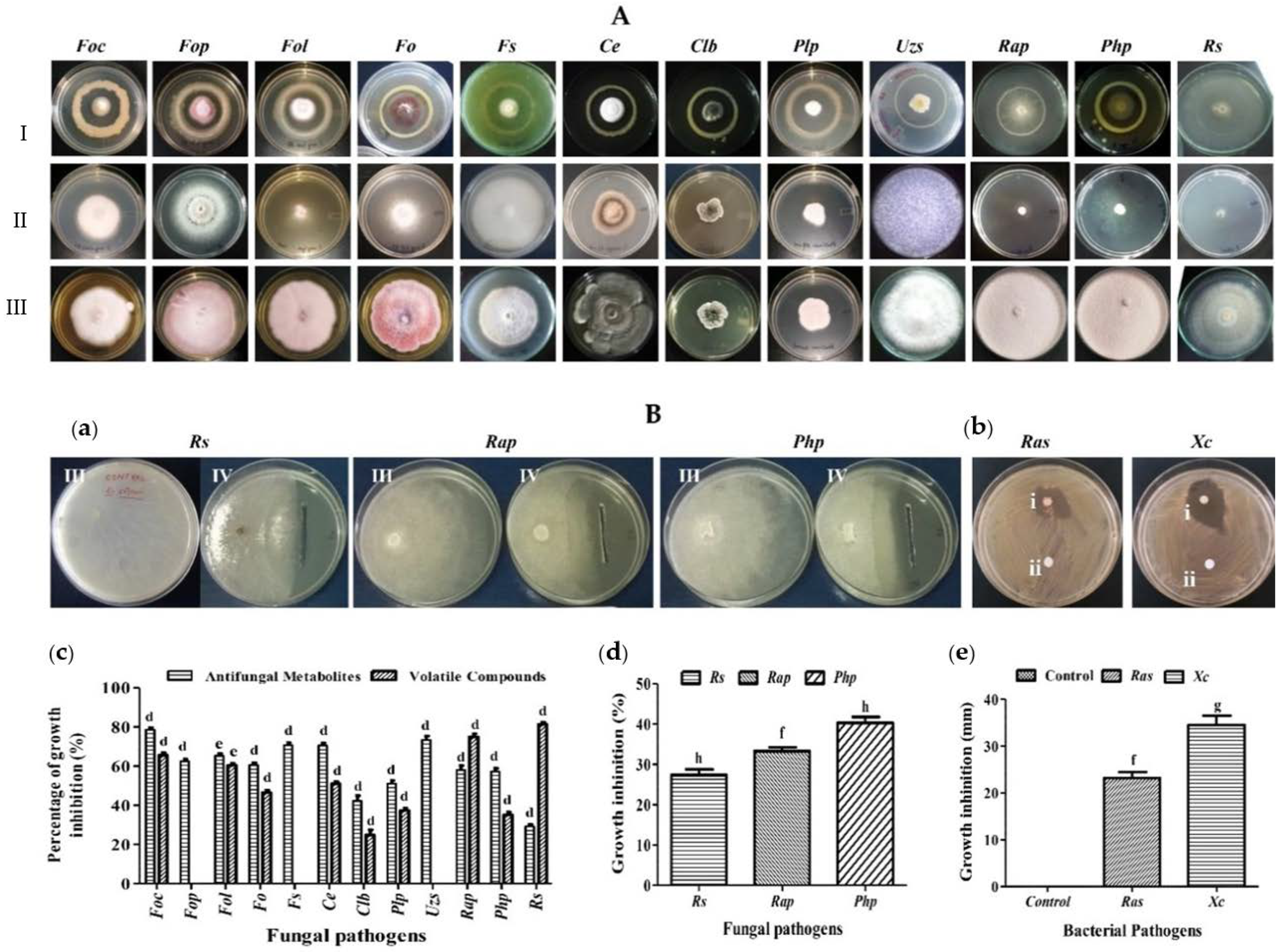
The strain was tested for antagonistic activity against different phytopathogenic fungi (12) and bacteria (2) using the dual-culture technique. The bacterial strain A7 exhibited significant (p < 0.001) antimicrobial activity against phytopathogens by producing secondary metabolites including volatile organic compounds (Figure 2, Table 3). The inhibition percentage for fungal pathogens ranged from 29 ± 0.03% to 82 ± 0.04%. The maximum fungal inhibition by A7 by secreting antifungal metabolite was against Curvularia lunata (Wakker) Boedijn var. (Clb, 82.53 ± 0.9%), followed by, Fusarium oxysporum f. sp. ciceris (Foc, 78.59 ± 1.1%), Ustillinia zonata (Lev.) Sacco (Uzs, 73.34 ± 2.0%) and the minimum inhibition were for Rhizoctonia solani (Rs, 29.20 ± 1.03%). The strain A7 also exhibited a moderate growth inhibition of Rosellinia arcuata Petch (Rap, 57.95 ± 2.3%), Poriahypobrunnea Patch (Php, 57.26 ± 1.5%), Phellinus lamaensis (Murrill) Pat. (Plp, 51.01 ± 1.7%), Fusarium oxysporum (Fo, 60.56 ± 1.2%), Fusarium oxysporum f. sp. lycopersici (Sacc.) (Fol, 65.26 ± 1.2%), Fusarium oxysporum f. sp. pisi (Fop, 62.51 ± 1.0%), Fusarium solani (Fs, 70.66 ± 1.2%) and Curvularia eragostidies (Ce, 43.54 ± 0.9% (Figure 2A(a)). The maximum growth inhibition caused by A7 using unidentified volatile organic compounds was against Rs (81.25 ± 1.1%), followed by Rap (75.99 ± 1.9%), Clb (75.95 ± 1.2%), Foc (65 ± 0.01%), Fol (60.51 ± 1.3%), Fo (46.44 ± 1.3%), Ce (43.70 ± 1.3%), Plp (37.36 ± 1.2%) and Php (35.14 ± 1.4%), and was unable to stop the growth of Clb, Fop, and Fs but the morphology of the pathogens altered in these dual cultures (Figure 2A(b),B(c)). The A7′s CFS inhibited the growth of the fungus Php (40.33 ± 1.4%), Rap (33.33 ± 0.8%), Rs (27.33 ± 1.4%). The bacterial pathogens’ growth inhibition measured in diameter was 23.16 ± 1.3 mm for Ralstonia solanacearum (Ras) and 34.5 ± 2.0 mm for Xanthomonas campestris (Xc), respectively, compared to the control (sterile de-ionized water) (p ˂ 0.05) (Figure 2B(a,b,d,e)) (Table 3).

3.4. A7 Is Endowed with Multiple Plant Growth-Promoting Traits

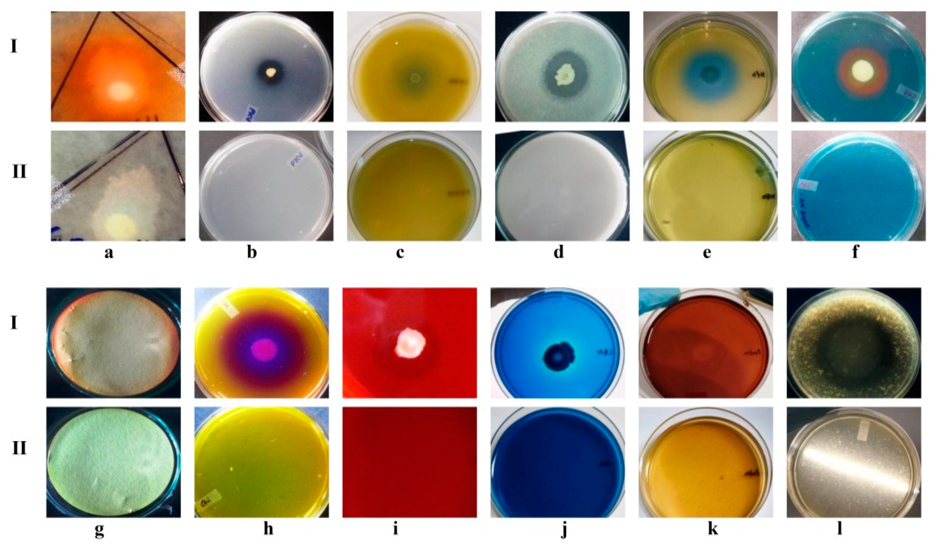
The A7 plant growth-promoting activities were assessed on different plate-based and liquid-medium-based assays (Figure 3, Table 4). Indole-3-acetic acid production was considered positive after the development of a red halo zone on the Whatman no-1 filter paper membrane around the colony. IAA production was also checked quantitatively with the addition of L-tryptophan at 48 h intervals of incubation. The IAA production increased from 3.3 ± 0.30 to 6.5 ± 0.30 µg/mL during 48 to 96 h of incubation. With a further increase in the incubation time, the production decreased from 5.4 ± 0.03 to 3.1 ± 0.10 µg/mL during 144 to 196 h of incubation (Table 4). The synthesis of IAA without the presence of L-tryptophan was also assessed (Figure S1). The qualitative and quantitative phosphate solubilization activity of A7 was evaluated to be positive, and the solubilization of tri-calcium phosphate was 146 ± 0.50 µg/mL. The A7 was found to be able to solubilize potassium and zinc oxide, and the solubilization efficacy (SE) was recorded at 33% and 29%, respectively (Table 4). The N2 fixation was evaluated positively for A7 by the alteration of the color of the medium from pale green to blue (Figure 3e). The diameter of the pale green halo zone was recorded at 52 ± 0.05 mm. The increase in pH related to the creation of ammonia and nitrates from atmospheric N2 fixation caused this color shift. Siderophores production was considered positive after the formation of an orange halo around the colony and was measured to be 45 ± 0.05 mm in diameter. Furthermore, the pink appearance in the supernatant indicated the production of siderophores (Figure 3f). The formation of a red-brown tint on filter paper indicated positive HCN synthesis by the strain. Positive ammonia production was also shown by the strain developing brown to yellow precipitate and the strain failed to produce polyamines (data not shown). Chitin degradation was considered positive after the development of the purple zone surrounding the strain from a yellow-colored chitin medium containing Bromocresol purple. In the region of chitin utilization, the pH of the medium shifts from acidic to alkaline due to the production of chitinase enzyme. The diameter of the degraded chitin chain was measured as a purple zone of 58 ± 0.05 mm in diameter. The other enzymatic activity of A7 was measured semi-quantitatively for hydrolytic capability (HC) of different nutrient substrates. The most common enzymatic activity in the bacterial strain A7 was for cellulase (2.4 HC), lignin (+), pectinase (2 HC), and proteases (1.3 HC), (Figure 3i–l and Table 4) and negative for xylanase activity (data not shown).

3.5. A7 Is Tolerant to Concentrations of Aluminum and Other Stress Conditions

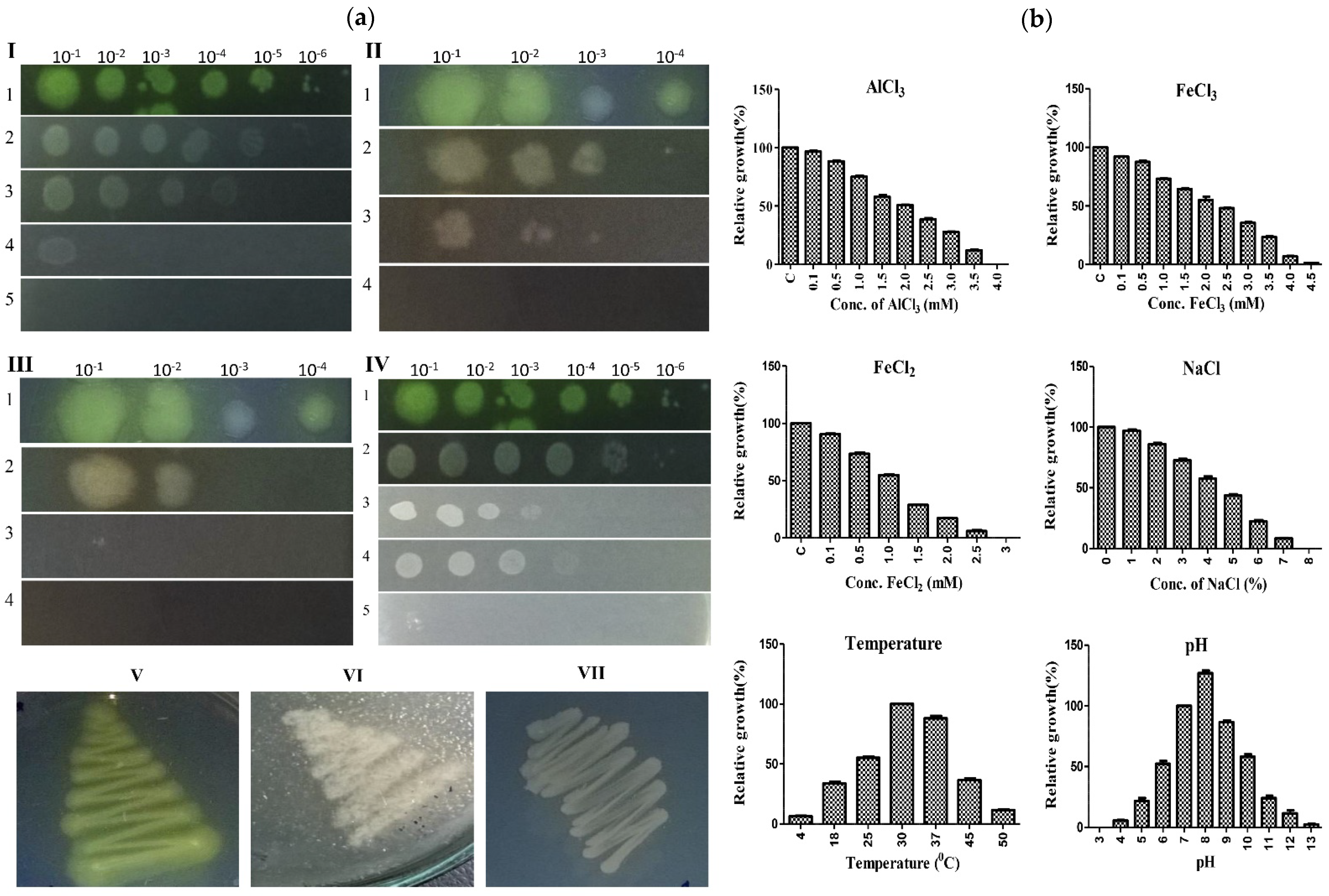
To assess the metal tolerance potential of A7, the strain was tested for its growth on high aluminum and iron amended solid and liquid medium. The A7 could grow and tolerate 0–3.5 mM of aluminum on both the SP-SDS and MM broth medium. The minimum growth inhibitory concentration of A7 for the metal aluminum, i.e., IC20 and IC50, was 0.76 ± 0.03 and 1.9 ± 0.05 mM. The strain could also tolerate and grow in the presence of FeCl3 (up to 4.5 mM) and FeCl2 (up to 2.5 mM). The IC20 and IC50 values were calculated for FeCl3 (1.00 ± 0.04, 2.51 ± 0.02 mM) and FeCl2 (0.47 ± 0.05, 1.18 ± 0.08 mM). Based on the different concentrations for the three metal compounds, the relative growth was equal, i.e., 73–96% at a concentration of 0.1–1.0 mM for AlCl3 and FeCl3. Beyond this concentration, the relative growth decreased, and stopped at a concentration of 4, 4.5, and 3 mM for the three metal compounds, respectively. The strain was also checked for temperature, salinity, and pH tolerance potentials. The optimum temperature for A7 growth was 25–40 °C, with diminished growth and reduced fluorescent activity below 10 or above 40 °C (Figure 4a, panel V–VII). The A7 could tolerate salinity stress by up to 7% of NaCl. The optimum pH range for A7 was from pH 6 to 9 with maximum growth at pH-8, as the relative growth was recorded as 126.97 ± 2.0% as compared to control (pH-7). (Figure 4b and Table 5).

3.6. In Vitro Protective Effect of A7 against Aluminum Toxicity in Chickpea

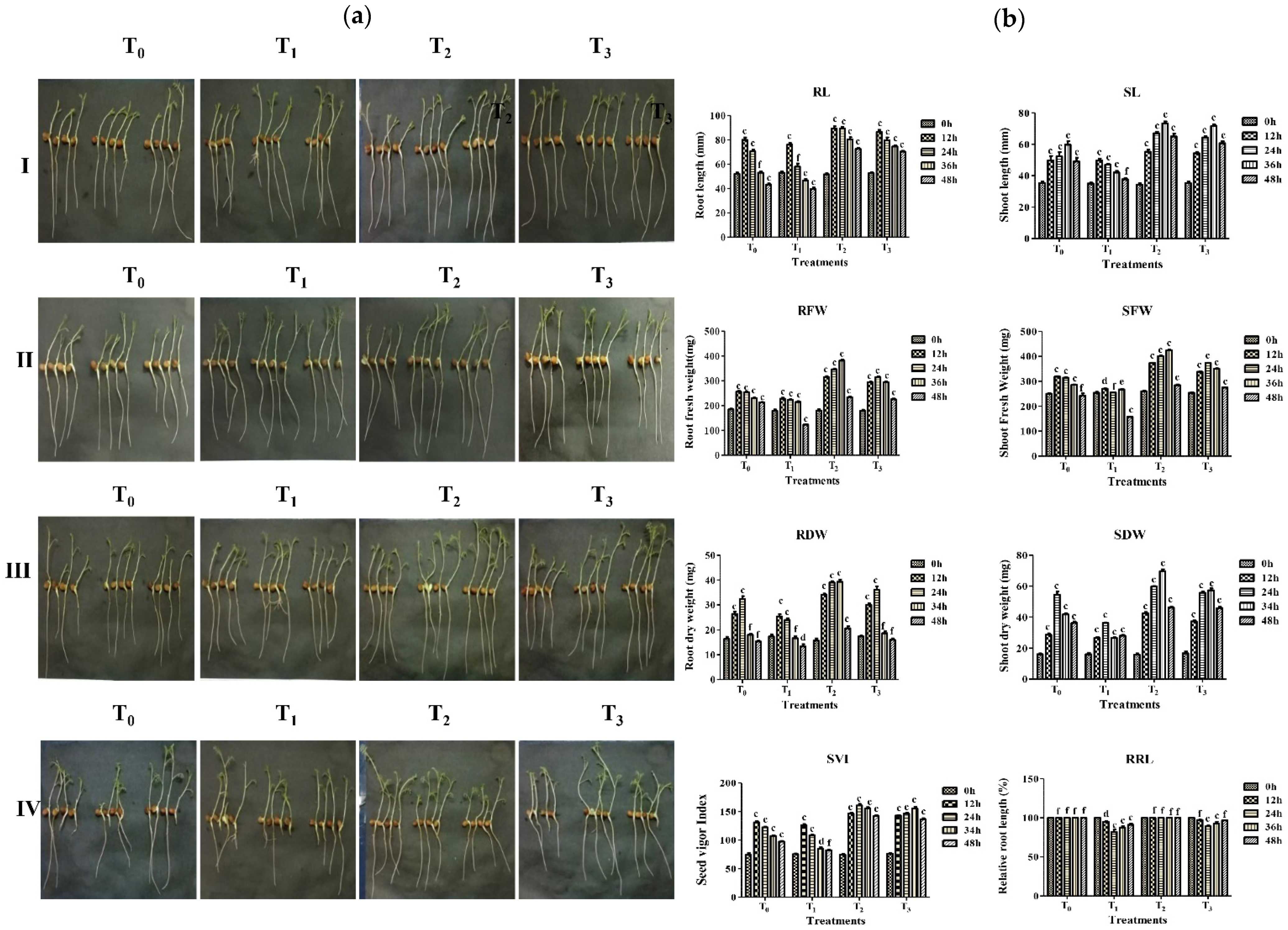
Since strain A7 was isolated on an aluminum enriched medium, it was further tested for its ability to grow chickpea seedlings in aluminum conditions. The impact of the four treatments on the overall growth of chickpea, i.e., the root, shoot length, root, shoot fresh and dry weight, relative root length rate, was observed with 12 h time intervals for 48 h. The treatment of chickpea seedling by strain A7 in the presence of 100 μM FeCl3 solution resulted in a significant (p ˂ 0.001) increase in root length, shoot length, root-shoot fresh and dry biomass, seedling vigor index, and the relative root-length rate compared to only aluminum treated seedlings (Figure 5a,b). When the seedlings were treated only with aluminum, morphological changes on the root and root-growth inhibition were observed. When the seedlings were treated with A7 in presence of aluminum, the root length, as well as the fresh and dry weight of the root, exhibited a similar trend to the treatment of seedlings with A7 alone. The protective effect of A7 on aluminum treated chickpea was measured in terms of root length (max. 89 ± 1.4 mm and min. 42 ± 1.7 mm) and root fresh (max. 382 ± 3.8 mg and min. 123 ± 1.7 mg) root dry weight (max. 40 ± 0.8 mg and min. 13 ± 0.8 mg) relative root length rate (max. 97 ± 0.3% and min 82 ± 3.0%). The values of seed vigor index, i.e., plant overall growth was recorded as max. 1530 ± 3.0 and min. 1345 ± 1.5, respectively, during the incubation period (Figure 5 and Table 6).

3.7. A7 Was a Potential Biocontrol Candidate against Fusarium Wilt of Chickpea

The in vivo control of Foc wilt disease of chickpea by strain A7 was evaluated in pot experiments using six treatments in which three treatments (T3–T5) involved A7 and Foc. The disease symptoms on the leaves of chickpea such as yellowing or necrosis from top to bottom were observed and recorded after the 20th and 40th days after inoculation while root rot was observed after 40th days of inoculation (Figure 6 and Figure S2A–C and Table 7). These symptoms led to the drying of leaves, drooping of petioles, and darkening of vascular tissues which are characteristic symptoms of Foc wilting. In comparison to the pathogen-inoculated positive control, all treatments, including A7, consistently suppressed the disease. When the chickpea seedlings were treated only with the spore suspension of Foc, severe wilt disease developed on the plants, and the percentage of disease incidence was 22.03 ± 0.4% to 65.43 ± 1.7%, and the disease severity was 22.43 ± 0.8% to 65.54 ± 1.7% (Table 7). The percentage of disease incidence was 12.12 ± 1.0 to 21.37 ± 1.3%, disease severity was 5.95 ± 0.5 to 10.29 ± 0.8%, and disease reduction was 44.70 ± 5.2 to 67.23 ± 2.3% when both A7 and Foc were inoculated in the soil simultaneously. Interestingly, when the soil was first treated with A7 before two days of the inoculation of Foc, the disease reduction was maximum 66.01 ± 2.9 to 77.00 ± 1.5% and minimum disease incidence 7.4 ± 0.6 to 14.99 ± 0.6 and disease severity 1.85 ± 0.1% to 3.92 ± 0.1% was observed from the plants compared to the soil treated only with Foc. Similarly, when the soil was treated with Foc prior two days to the inoculation of A7, the disease incidence was 13.20 ± 1.0 to 24.56 ± 1.0%, disease severity was 10.48 ± 0.5% to 19.38 ± 0.6%, and the disease reduction was 39.73 ± 1.6 to 62.26 ± 2.5%. From these different combinations of treatments, it was clear that strain A7 showed maximum disease protection when it was applied two days before inoculation of the pathogen and minimum when it was applied two days after the inoculation of the pathogen. From these experimental settings, the plant-growth-promoting activities of strain A7 on chickpea were also observed and recorded. The application of A7 on the plants resulted in the healthy growth of plants compared with the non-inoculated control at the end of 40th days of inoculation. The maximum root length, shoot length and plant biomass was recorded when treated with only A7 compared to control. The PGP parameters including root length, shoot length, root and shoot fresh and dry weight, and the seed vigor index were observed and recorded in different treatments (p < 0.05) (Figure 6, Table 8).

3.8. A7 Genome Had Antibiotics, DAPG, HCN, and PCN Biosynthesis Capabilities

The A7 genomic DNA was used for PCR amplification and verification of antibiotic marker genes, which suggested the presence of three gene segments encoding for, 2, 4-diacetylphloroglucinol (2, 4-DAPG), hydrogen cyanide production (HCN), and phenazine-1-carboxamide (PCN), respectively (Figure 1D). DNA fragments of sizes of 745 bp of phlD, 587 bp of hcnBC, and 2000 bp of phzH genes were amplified using gene-specific primer sets. These are the major genes engaged in the production of 2, 4-DAPG, HCN, and PCN, respectively. However, prnD for pyrrolnitrin (PRN), phzC and phzD for Phenazine-1-Carboxylic acid (PCA), pltC for pyoluteorin, and darS or darR for 2-hexyl-5-propyl resorcinol (DarA, DarB) genes were not detected by PCR using gene-specific primer sets mentioned in Table 1.

3.9. Characterization and Analysis of Bioactive Natural Compounds in A7 through TLC, HPLC, and GC-MS

The extracted methanol organic mixture from A7 showed a 35% growth inhibition of Rosellinia arcuata Petch compared to control. Four main fractions, namely, F1, F2, F3, and F4, were exhibited on the TLC plate when observed under UV light (Bio Red) at 254 nm. The fraction F3 was further purified, and a single discrete spot was obtained with a 0.70Rf value. The final purified methanol extract showed 30% growth inhibition of Rap (Figure 7). The compound’s retention time was 6.080 min, according to HPLC analysis (data not shown). Therefore, the partially purified fraction was selected for GC-MS for further characterization. The National Institute of Standards and Technology (NIST) library identified many chemical compounds. Among them, twenty-two compounds were selected dependent on their molecular formula, molecular mass, and retention time (RT). These compounds are Oleic acid; 4-fluoro-1-methyl-5-carboxylic acid; 2-tridecenoic acid and 6,10-dimethyl-4-undecanol (RT 22.04), 1-pentadecene; 1-tridecene; 10-heneicosene (C, T) and 1-hexadecene (RT 24.65); Tridecanoic acid; 12-methyl-ester; Hexadecanoic acid; methyl ester; Tridecanoic acid, methyl ester; Hexacosanoic acid methyl ester (RT-28.06); 5-eicosene; E-15-heptadecena; Cycloeicosane; and 1-heptadecene (RT 29.70), 9-octadecenoic acid; 12-octadecenoic acid; Trans-13-octadecenoic acid (RT 32.80); 1,2-benzenedicarboxylic acid; mono (2-ethylhexyl) ester; 6-ethyloct-3-yl-2-ethylhexyl ester; and 1,2-benzenedicarboxylic acid, Diisooctyl ester, Bis (2-ethylhexyl) phthalate (RT 40.04) (Table 9).

4. Discussion

In the current research, based on its 16S rRNA sequences and phylogenetic analyses, aluminum-tolerant A7 obtained from the rhizoids of Polytrichum sp. was identified as Pseudomonas aeruginosa. Morphologically, A7 was Gram-negative, coccus, round, smooth and green pigmented [105]. In this study, multiple phenotypes of A7 related to plant growth promotion (PGP) and biocontrol potentials are reported. The biological control of plant diseases using bacteria has evolved as a sustainable disease-management approach. Bacteria associated with the root or in the rhizosphere are capable of secreting a range of antibiotic compounds, making them a potential source of biocontrol agents [106]. Burkholderia sp., Pseudomonas sp., Flavobacterium sp., and Bacillus sp. are examples of antifungal and PGP root and rhizobacteria. They play an important role in plant-growth promotion by secreting phytohormones, most importantly Indole-3-acetic acid (IAA) [107,108]. The inability of P. putida GR12-2 deficient in IAA synthesis to increase root growth and lateral-root formation was indicative of the role of bacterial IAA in plant benefits [109]. Soil fertility and plant nutrition are critical for ensuring adequate levels of micronutrients such as potassium, phosphorus, zinc, and many others, necessary for plant growth and development [110]. A PGPR that solubilizes phosphate has great potential in agricultural applications. Richardson et al. [111] found that bacteria increased the amount of P available in the soil for plant uptake. Similarly, in this study, Pseudomonas aeruginosa A7 could solubilize tri-calcium phosphate in vitro. Pseudomonas sp. AF-54 showed the possibility of inorganic P-solubilization by reducing pH and releasing acetic, malic, and gluconic acid [112]. The rhizosphere typically contains P-solubilizing bacteria, which produce gluconic acids to liberate inorganic phosphate from the soil [113,114]. The action of PGPR is frequently linked to the regulation of phytohormones involved in root development and growth as well as phosphorus solubilization [115]. Potassium-solubilizing bacteria have been observed to produce potassium from decomposing organic matter, crop residues, aluminosilicates, kaolinites, and feldspars by acidosis, chelation, and complex formation and exchange reactions [65,116,117,118]. Bacillus sp., Burkholderia sp., and Pseudomonas sp. have been reported to efficiently solubilize potassium from K-feldspar from tea-planted soil [119]. According to [67,120] Rahnella aquatilis, Pseudomonas orientalis, Pantoea agglomerans, and Enterobacter isolated from the rhizosphere of paddy soil and rubber plant-soil, respectively, may solubilize potassium. Arthrobacter sp., Mesorhizobium sp., Paenibacillus sp., Klebsiella variicola and Enterobacter cloacae rape rhizobacteria and Klebsiella variicola, and Enterobacter cloacae tobacco rhizobacteria are reported to be able to solubilize potassium [121,122]. To reduce the use of chemicals, nitrogen fertilizer application in plants and atmospheric nitrogen fixation in soil by bacterial strains boost nitrogen availability in soil vital for plant growth [110]. Rhizospheres such as Aster gymnocephalus, Gringelia sp., Haplopappus sp., Juniperus sp. and Lygodesmia sp., are the major source of the nitrogen-fixing genus Paenibacillus. However, Azospirillum lipoferum and Bradyrhizobium japonicum were the reported rhizobacteria of Haplopappus sp., and Viguiera linearis [123]. It has also been suggested that root-associated Pseudomonas stutzeri A1501 fixes atmospheric nitrogen [124]. Diazotrophs, isolated from pepper vines, possess an enzyme that can convert N2 into NH4+ that plants can take up through the biological nitrogen fixation process [125,126]. Azospirillum brasilense sp. 245 and Burkholderia kururiensis M130 improved lateral-root development and ethylene signaling responses during rice-seedling cultivation through the biological nitrogen fixation process [127]. Interestingly, in this study, A7 exhibited nitrogen fixation in the nitrogen-free medium, which can be explored for the mechanism and future use. Agrobacterium tumefaciens, Rhizobium sp. [67]. Pseudomonas and Gluconacetobacter are capable of oxidizing zinc in a solid environment [68]. PGPR, including Pseudomonas aeruginosa A7, produces iron-chelating agents that bind insoluble ferric ions, thereby inhibiting the growth of phytopathogens and thus helping to control plant diseases. Siderophores are metal-chelating agents of low molecular weight that produced by bacteria and secreted as a means of facilitating iron uptake into their cells under Fe-limiting conditions [71,128]. Moreover, siderophores can bind a variety of metals, in addition to iron and several chemical structures and properties. The bacterial siderophores act as biocontrol agents and help in plant growth, thus applicable in mineral weathering, biosensors, bioremediation, and chelation agents [129]. Various physiologic and biochemical activities in plant growth and metabolism involve either root-based chelate breakdown or ligand exchange to provide iron and other metals from bound siderophores [130,131].
Multiple rhizo-Pseudomonas from diverse environments are known to decrease plant diseases through the secretion of natural product metabolites [105,132]. Extracellular enzyme-like cellulase, chitinase, and proteases produced by these bacteria attack the cell wall of plant pathogenic fungi, causing cell lysis and subsequent death [133]. Chitinase hydrolyzes the polysaccharide chitin, a linear homopolymer of β-1,4-linked N-acetyl-d-glucosamine (GlcNAc) residue found in the cell wall of fungi [134,135]. Antifungal protein degrading enzymes, Proteases, are known to be produced by biocontrol and PGP bacteria [136]. Several species of Arthrobacter, Pseudomonas, Sphingobium, Streptomyces, and Rhodococcus possess ligninolytic activity in vitro [137]. Fungal pathogens and PGPR enzymes, the most effective agents of biological control of plant diseases caused by fungal pathogens hydrolyze the 1,4-glycosidic bonds between the N-acetyl glucosamine in fungal chitin [73]. Pseudomonas aeruginosa A7 was found to degrade chitin and demonstrated wide spectrum fungal growth inhibition suggesting one such biocontrol mechanism. Hydrogen cyanide (HCN), a major inhibitor of cytochrome c oxidase and several other metalloenzymes, is produced and secreted by a few bacterial species as a secondary metabolite [138]. HCN synthase is a membrane-bound flavoenzyme present in proteobacteria (especially Fluorescent pseudomonads) that oxidizes glycine to generate HCN and CO2. Sequence similarities exist between the hcnABC structural genes of Pseudomonas fluorescens and Pseudomonas aeruginosa and genes encoding different amino acid dehydrogenases/oxidases, particularly nopaline oxidase of Agrobacterium tumefaciens [138]. PGPB produces hydrogen cyanide that interferes with essential enzymes leading to the degradation of phytopathogens cell walls and their control in rhizosphere soil [139,140]. HCN has been associated with the control of M. javanica disease in tomatoes and O. obesity, an agricultural pest in India [141,142]. Both NIA-2 and NIA-5, as well as Pseudomonas fluorescence, impart a beneficial effect on wheat and banana growth in studies [143,144].
The use of bacteria that promotes plant growth in metal-rich environments has been touted as one of the newest trends since metal-tolerant bacteria can support the diverse growth of crops in metal-rich environments [28]. Al can have a beneficial or toxic effect on plants and other organisms, depending on factors such as metal concentration, chemical form, and growth conditions. Al has largely been studied for its physiological, biochemical, and molecular effects on plants (increased nutrient uptake, root growth stimulation, and enzyme activity stimulation, among other things) [145]. Plant growth is adversely affected by metal in acid soil, characterized by a deficiency of nutrients and toxicity by aluminum is the main limiting factor [146]. It has been shown that aluminum inhibits the rapid expansion of roots in plants and the rate of relative root length provides a standard indicator of the toxicity of aluminum [36]. It was shown that metal-tolerant bacteria can advance plant development and increase metal uptake by hyper-and non-hyperaccumulator plants [147]. Bacillus sp. KGMDI and Bacillus sp. K25 tolerate their growth up to 75 mg/L, equal to 0.56 mM of aluminum, iron, and copper [148]. A7 could tolerate AlCl3 up to 3.5 mM, FeCl3 up to 4.5 mM and FeCl2 up to 2 mM. In this study, Pseudomonas aeruginosa A7 produced IAA in the presence of 100 µM AlCl3, which may be responsible for the growth of chickpea by increasing root and shoot elongation, leaf surface area, and dry biomass. Burkholderia ginsengiterrae N11-2, Chryseobacterium polytrichastri N10, Pseudomonas fluorescens, Pseudomonas fragi N8, and Pseudomonas simiae N3, have been reported to grow in aluminum stress conditions [28,29].
Several PGPR strains are reported to have been used in the integrated biological control of Fusarium wilt in chickpea [89,149,150]. Fusarium wilt, produced by the soil-borne fungus Fusarium oxysporum ciceris is a major issue restricting chickpea output around the world [9,151]. The disease symptoms such as yellowing of leaves, abnormal branching, drooping petioles, necrosis of foliage, and drying of roots are early symptoms [9,152]. Currently, chemical-control approaches have no effect on the Fusarium wilt of chickpea control in the field, so biological control is the most promising and effective method [153]. In this study, we demonstrated that strain A7 showed antagonistic activities against Foc under in vitro and in vivo conditions. Remarkably, the strain was able to promote chickpea growth and no negative impact on its growth was observed. PGPR genus Bacillus (63.14%), Burkholderia, and Pseudomonas (53.52%) have been reported to be effectively exploited as control agents for the pathogen by producing HCN pyrrolnitrin, phloroglucinol, phenazine, siderophores, cell-wall-degradation enzymes that suppress the Fusarium oxysporum f. sp. ciceris by inhibiting mycelial growth. As a result, they play a critical role in a variety of microbial interactions, resulting in abnormal hyphal swelling, becoming exceptionally lengthy and vacuolization, which manipulates physiological processes in other fungi and bacteria [26,150,154,155]. In our in vivo experiment, we found that the PGPR strain A7 consistently suppressed yellowing disease caused by Foc when applied to the soil. The A7 strain significantly reduced the yellowing disease and root rot in chickpea plants compared to the pathogen-inoculated control. So, this strain could be an excellent choice for further biological control research and chickpea-farming success.
The presence of antifungal antibiotic genes in the bacterial strain A7 was confirmed by a polymerase chain reaction for marker genes for the synthesis of 2,4-DAPG, hydrogen cyanide synthase, and phenazine-1-carboxamide. These antibiotics are known to target plasma-membrane integrity or electron transport in many fungal and bacterial pathogens and indirectly help plant growth [156,157,158]. Additionally, A7 was shown to produce several interesting bioactive compounds with known and novel activities. According to earlier research, lipid derivatives such as 9-octadecanoic acid, hexadecanoic acid methyl ester, octadecenoic acid, 1,2-benzene dicarboxylic acid, mono (2-Ethylhexyl) ester, 1,2-benzene dicarboxylic acid and diisooctyl ester [159] were found to be powerful antibacterial compounds [103]. They induced the autolysis of cell membranes, damaged cell walls, and inhibited transcription, and translation in S. aureus, E. coli, and Salmonella sp. [160]. Interestingly these compounds have been reported to have antimicrobial, anti-inflammatory, antioxidant, and anticancer properties [161]. The antimicrobial and antioxidant activity of 1-hexadecene and hexadecane extracted from Acanthophora spicifera was also reported [162]. It is reported that various indirect and direct mechanisms contribute to pseudomonas promoting plant development [163]. Similarly, in this study, we report multiple phenotypes and mechanisms of biocontrol and plant-growth promotion by the reported strain.
In our report, several isolates of Pseudomonas aeruginosa have been reported as plant growth-promoting rhizobacteria (PGPR). Pseudomonas aeruginosa RRALC3 isolated from degraded forest soil from the Indian state of Chennai, enhanced the growth, macro-micronutrient contents, along with enhanced carbon content of the native legume Pongamia pinnata seedlings [164]. Pseudomonas aeruginosa isolated from Korean mine soil was reported to have several plant-protection traits including antagonism against bacterial phytopathogens and the stimulation of defense-related genes in tomato plants [165]. PGPR Pseudomonas aeruginosa was reported to be the dominant bacteria from the rhizosphere of sorghum, paddy, tobacco, tea, and maize crops in India [166]. Recently, the Pseudomonas aeruginosa strain RTE4, isolated from the tea plant rhizosphere [167]; tomato plant rhizosphere associated Pseudomonas aeruginosa strain CQ-40 [168] and Pseudomonas aeruginosa FG106 [169]; and saffron rhizosphere Pseudomonas aeruginosa strain YY322 [170], have been reported as PGPR with multiple beneficial phenotypes and biocontrol activities in the plants.
Using culture-dependent and independent methods, it is suggested that many human pathogenic bacteria from the genus Burkholderia, Enterobacter, Pseudomonas, Ralstonia, Serratia, Staphylococcus, and Stenotrophomonas are prominent members of several rhizospheres [171,172]. Several species from these genera possess growth-promoting properties for plants, as well as excellent antagonistic properties against plant pathogens; as a result, they are used to prevent disease and promote plant growth [171]. To use rhizosphere, isolate as a successful biocontrol agent, the strains should be tested for safety and risk assessments should be performed [20]. Human isolates of Pseudomonas aeruginosa are recognized as an opportunistic, nosocomial human pathogen and thus the environmental isolates must be extensively researched before being considered as a biological control agent. Thus, the comparative virulence attributes of the reported Pseudomonas aeruginosa A7 will allow it to be used as a BCA and a safe biofertilizer. Additionally, since the strain is already adapted to local environmental conditions, it may be transformed into a biofertilizer in future research involving different chickpea varieties, environmental conditions, and field trials. Further research is required to fully understand the multiple biocontrol and chickpea plant growth mechanisms of strain A7. This study thus provides another biocontrol arsenal against wilt disease which can be useful in the control of Fusarium wilt in aluminum and metal-rich soils.

5. Conclusions

A Fusarium oxysporum f. sp. ciceris (Foc) antagonist bacterium was identified from the rhizoids of adult moss, identified as Pseudomonas aeruginosa A7 on a morphological and molecular basis. The strain produced antimicrobial metabolites with strong antagonistic activities against multiple plant pathogenic fungi and bacteria. It was shown to have significant inhibitory effects on the Fusarium wilt of chickpea and enhanced chickpea growth in a pot experiment. Chickpea seedlings under aluminum stress grew significantly better when treated with A7. Based on the PCR results, the strain carried several biocontrol markers related to the biosynthesis of antibiotics HCN, 2, 4-DAPG, and PCN. Furthermore, A7′s poly-extremophilic and extensive adaptations to metal stress can be useful in the bioremediation of metal-rich soils. The strain showed promising phenotypes with the potential of improving seedling growth and development while reducing losses caused by fungal plant diseases. Thus, the strain can be used in agriculture as a natural alternative to conventional PGPR and fungicides after the comprehensive biological safety assessment using the tools of whole-genome sequencing, transcriptomics, and metabolomics studies. Additional research is required to establish strain A7’s mechanism of plant-growth promotion in chickpea or other plants. Interestingly, the strains’ lignin degradation and production of bioactive compounds can be harnessed using other biotechnological interventions.

Supplementary Materials

The following supporting information can be downloaded at: https://www.mdpi.com/article/10.3390/microorganisms10030568/s1, Figure S1: Bar diagram showing the production of Indole-3-acetic acid in the presence (5 mM) and absence of L-tryptophan by the bacterial strain A7 with 48 h time interval for 196 h, Figure S2A: In vivo establishment and control of Fusarium wilt disease of chickpea caused by Fusarium oxysporum f. sp. ciceris by the bacterial strain A7 at the first day (Day-1) of inoculation, Figure S2B: In vivo establishment and control of Fusarium wilt disease of chickpea caused by Fusarium oxysporum f. sp. ciceris by the bacterial strain A7 at 20th days (Day-20) of inoculation (a,b), Figure S2C: In vivo establishment and control of Fusarium wilt disease of chickpea caused by Fusarium oxysporum f. sp. ciceris by the bacterial strain A7 at 40th days (Day 40) of inoculation (a,b).

Author Contributions

Conceptualization, A.B.M. and H.K.P.; methodology, A.B.M. and H.K.P.; validation, A.B.M., K.C. and R.C.; formal analysis, A.B.M. and H.K.P.; investigation, A.B.M.; resources, H.K.P.; data curation, A.B.M.; writing—original draft preparation, A.B.M.; writing—review and editing, A.B.M. and H.K.P.; visualization, A.B.M.; supervision, H.K.P.; funding acquisition, H.K.P. All authors have read and agreed to the published version of the manuscript.

Funding

This work was supported by the Department of Biotechnology (DBT), New Delhi, India (Sanction No. BT/PR14242/AGR/05/515/2010). A.B.M., acknowledge the Ph.D. funding of Maulana Azad National Fellowship for Minority Students (F1-17.1/2015-16/MANF-2015-17-ASS-62440/(SA-III/Website), University Grant Commission, Govt. of India, New Delhi.

Institutional Review Board Statement

Not applicable.

Informed Consent Statement

Not applicable.

Data Availability Statement

All data related to this manuscript are incorporated in the manuscript only.

Conflicts of Interest

The authors declare no conflict of interest.

References

  1. Van der Maesen, L. Origin, history and taxonomy of chickpea. In The Chickpea; 1987; pp. 11–34. Available online: https://edepot.wur.nl/304694 (accessed on 20 October 2021).
  2. Merga, B.; Haji, J. Economic importance of chickpea: Production, value, and world trade. Cogent Food Agric. 2019, 5, 1615718. [Google Scholar] [CrossRef]
  3. Jukanti, A.; Gaur, P.; Gowda, C.; Chibbar, R. Nutritional quality and health benefits of chickpea (Cicer arietinum L.): A review. Br. J. Nutr. 2012, 108, S11–S26. [Google Scholar] [CrossRef] [PubMed] [Green Version]
  4. Solh, M.; Halila, H.; Hernandez-Bravo, G.; Malik, B.; Mihov, M.; Sadri, B. Biotic and abiotic stresses constraining the productivity of cool season food legumes in different farming systems: Specific examples. In Expanding the Production and Use of Cool Season Food Legumes; Springer: Berlin/Heidelberg, Germany, 1994; pp. 219–230. [Google Scholar]
  5. Haware, M.P.; Nene, Y.L. Symptomless carriers of the chickpea wilt Fusarium. Plant Dis. 1982, 66, 250–251. [Google Scholar] [CrossRef]
  6. Jendoubi, W.; Bouhadida, M.; Boukteb, A.; Béji, M.; Kharrat, M. Fusarium Wilt Affecting Chickpea Crop. Agriculture 2017, 7, 23. [Google Scholar] [CrossRef] [Green Version]
  7. Srivastava, A.; Dixit, G.P.; Singh, N.P.; Saxena, D.R.; Saabale, P.R.; Raghuvanshi, K.S.; Anandani, V.P. Evaluation of chickpea (Cicer arietinum L.) accessions for Fusarium wilt resistance. J. Environ. Biol. 2021, 42, 82–89. [Google Scholar] [CrossRef]
  8. Dubey, S.C.; Suresh, M.; Singh, B. Evaluation of Trichoderma species against Fusarium oxysporum f. sp. ciceris for integrated management of chickpea wilt. Biol. Control 2007, 40, 118–127. [Google Scholar] [CrossRef]
  9. Jiménez-Díaz, R.M.; Castillo, P.; del Mar Jiménez-Gasco, M.; Landa, B.B.; Navas-Cortés, J.A. Fusarium wilt of chickpeas: Biology, ecology and management. Crop Prot. 2015, 73, 16–27. [Google Scholar] [CrossRef]
  10. Pande, S.; Kishore, G.K.; Upadhyaya, H.D.; Rao, J.N. Identification of Sources of Multiple Disease Resistance in Mini-core Collection of Chickpea. Plant Dis. 2006, 90, 1214–1218. [Google Scholar] [CrossRef] [Green Version]
  11. Chobe, D.R.; Om, G.; Maruti, P. Radiation induced mutation for resistance against races/pathotypes of Fusarium oxysporum f. sp. ciceris in chickpea (Cicer arietinum L.). Indian Phytopathol. 2016, 69, 699–701. [Google Scholar]
  12. Sharma, M.; Babu, T.K.; Gaur, P.; Ghosh, R.; Rameshwar, T.; Chaudhary, R.; Upadhyay, J.; Gupta, O.; Saxena, D.; Kaur, L. Identification and multi-environment validation of resistance to Fusarium oxysporum f. sp. ciceris in chickpea. Field Crops Res. 2012, 135, 82–88. [Google Scholar] [CrossRef] [Green Version]
  13. Bhar, A.; Jain, A.; Das, S. Soil pathogen, Fusarium oxysporum induced wilt disease inchickpea: A review on its dynamicity and possible control strategies. Proc. Indian Natl. Sci. Acad. 2021, 87, 260–274. [Google Scholar] [CrossRef]
  14. Kumar, B.D. Fusarial wilt suppression and crop improvement through two rhizobacterial strains in chick pea growing in soils infested with Fusarium oxysporum f. sp. ciceris. Biol. Fertil. Soils 1999, 29, 87–91. [Google Scholar] [CrossRef]
  15. Landa, B.B.; Navas-Cortés, J.A.; Jiménez-Díaz, R.M. Integrated management of Fusarium wilt of chickpea with sowing date, host resistance, and biological control. Phytopathology 2004, 94, 946–960. [Google Scholar] [CrossRef] [PubMed] [Green Version]
  16. He, D.-C.; He, M.-H.; Amalin, D.M.; Liu, W.; Alvindia, D.G.; Zhan, J. Biological Control of Plant Diseases: An Evolutionary and Eco-Economic Consideration. Pathogens 2021, 10, 1311. [Google Scholar] [CrossRef]
  17. Compant, S.; Duffy, B.; Nowak, J.; Clément, C.; Barka, E.A. Use of plant growth-promoting bacteria for biocontrol of plant diseases: Principles, mechanisms of action, and future prospects. Appl. Environ. Microbiol. 2005, 71, 4951–4959. [Google Scholar] [CrossRef] [Green Version]
  18. Thambugala, K.M.; Daranagama, D.A.; Phillips, A.J.; Kannangara, S.D.; Promputtha, I. Fungi vs. fungi in biocontrol: An overview of fungal antagonists applied against fungal plant pathogens. Front. Cell. Infect. Microbiol. 2020, 10, 10. [Google Scholar] [CrossRef]
  19. Almeida, F.; Rodrigues, M.L.; Coelho, C. The still underestimated problem of fungal diseases worldwide. Front. Microbiol. 2019, 10, 214. [Google Scholar] [CrossRef] [Green Version]
  20. Basu, A.; Prasad, P.; Das, S.N.; Kalam, S.; Sayyed, R.Z.; Reddy, M.S.; El Enshasy, H. Plant Growth Promoting Rhizobacteria (PGPR) as Green Bioinoculants: Recent Developments, Constraints, and Prospects. Sustainability 2021, 13, 1140. [Google Scholar] [CrossRef]
  21. Oleńska, E.; Małek, W.; Wójcik, M.; Swiecicka, I.; Thijs, S.; Vangronsveld, J. Beneficial features of plant growth-promoting rhizobacteria for improving plant growth and health in challenging conditions: A methodical review. Sci. Total Environ. 2020, 743, 140682. [Google Scholar] [CrossRef]
  22. Jiao, X.; Takishita, Y.; Zhou, G.; Smith, D.L. Plant Associated Rhizobacteria for Biocontrol and Plant Growth Enhancement. Front. Plant Sci. 2021, 12, 634796. [Google Scholar] [CrossRef]
  23. Olanrewaju, O.S.; Glick, B.R.; Babalola, O.O. Mechanisms of action of plant growth promoting bacteria. World J. Microbiol. Biotechnol. 2017, 33, 197. [Google Scholar] [CrossRef] [PubMed] [Green Version]
  24. Pereg, L.; McMillan, M. Scoping the potential uses of beneficial microorganisms for increasing productivity in cotton cropping systems. Soil Biol. Biochem. 2015, 80, 349–358. [Google Scholar] [CrossRef] [Green Version]
  25. Anjaiah, V.; Cornelis, P.; Koedam, N. Effect of genotype and root colonization in biological control of fusarium wilts in pigeonpea and chickpea by Pseudomonas aeruginosa PNA1. Can. J. Microbiol. 2003, 49, 85–91. [Google Scholar] [CrossRef]
  26. Moradi, H.; Bahramnejad, B.; Amini, J.; Siosemardeh, A.; Haji-Allahverdipoor, K. Suppression of chickpea (‘Cicer arietinum’ L.)’Fusariums’ wilt by ‘Bacillus subtillis’ and ‘Trichoderma harzianum’. Plant Omics 2012, 5, 68–74. [Google Scholar]
  27. Cappellari, L.d.R.; Chiappero, J.; Palermo, T.B.; Giordano, W.; Banchio, E. Volatile Organic Compounds from Rhizobacteria Increase the Biosynthesis of Secondary Metabolites and Improve the Antioxidant Status in Mentha piperita L. Grown under Salt Stress. Agronomy 2020, 10, 1094. [Google Scholar] [CrossRef]
  28. Farh, M.E.-A.; Kim, Y.-J.; Sukweenadhi, J.; Singh, P.; Yang, D.-C. Aluminium resistant, plant growth promoting bacteria induce overexpression of Aluminium stress related genes in Arabidopsis thaliana and increase the ginseng tolerance against Aluminium stress. Microbiol. Res. 2017, 200, 45–52. [Google Scholar] [CrossRef] [PubMed]
  29. Appanna, V.D.; St. Pierre, M. Influence of phosphate on aluminum tolerance in Pseudomonas fluorescens. FEMS Microbiol. Lett. 1994, 124, 327–332. [Google Scholar] [CrossRef]
  30. Maheshwari, D.K.; Dheeman, S. Field Crops: Sustainable Management by PGPR; Springer: Berlin/Heidelberg, Germany, 2019. [Google Scholar]
  31. Qessaoui, R.; Bouharroud, R.; Furze, J.N.; El Aalaoui, M.; Akroud, H.; Amarraque, A.; Van Vaerenbergh, J.; Tahzima, R.; Mayad, E.H.; Chebli, B. Applications of New Rhizobacteria Pseudomonas Isolates in Agroecology via Fundamental Processes Complementing Plant Growth. Sci. Rep. 2019, 9, 12832. [Google Scholar] [CrossRef] [Green Version]
  32. Niu, X.; Song, L.; Xiao, Y.; Ge, W.; Niu, X.; Song, L.; Xiao, Y.; Ge, W. Drought-tolerant plant growth-promoting rhizobacteria associated with foxtail millet in a semi-arid agroecosystem and their potential in alleviating drought stress. Front. Microbiol. 2018, 8, 2580. [Google Scholar] [CrossRef]
  33. Vurukonda, S.S.K.P.; Vardharajula, S.; Shrivastava, M.; SkZ, A. Enhancement of drought stress tolerance in crops by plant growth promoting rhizobacteria. Microbiol. Res. 2016, 184, 13–24. [Google Scholar] [CrossRef]
  34. David, B.V.; Chandrasehar, G.; Selvam, P.N. Pseudomonas fluorescens: A Plant-Growth-Promoting Rhizobacterium (PGPR) with Potential Role in Biocontrol of Pests of Crops. In Crop Improvement Through Microbial Biotechnology; Prasad, R., Gill, S.S., Tuteja, N., Eds.; International Institute of Biotechnology and Toxicology (IIBAT): Padappai, India, 2018; pp. 221–243. [Google Scholar]
  35. Ram Kumar, A.; Selvaraj, S.; Selvam, K.A.; Devanathan, J. Isolation and characterization of drought stress tolerant plant growth promoting rhizobacter from chilli crop. Bull. Sci. Res. 2020, 3, 1–12. [Google Scholar] [CrossRef]
  36. Awasthi, J.P.; Saha, B.; Regon, P.; Sahoo, S.; Chowra, U.; Pradhan, A.; Roy, A.; Panda, S.K. Morpho-physiological analysis of tolerance to aluminum toxicity in rice varieties of North East India. PLoS ONE 2017, 12, e0176357. [Google Scholar] [CrossRef] [Green Version]
  37. Rahman, M.A.; Lee, S.-H.; Ji, H.C.; Kabir, A.H.; Jones, C.S.; Lee, K.-W. Importance of Mineral Nutrition for Mitigating Aluminum Toxicity in Plants on Acidic Soils: Current Status and Opportunities. Int. J. Mol. Sci. 2018, 19, 3073. [Google Scholar] [CrossRef] [PubMed] [Green Version]
  38. Greger, J. Aluminum metabolism. Annu. Rev. Nutr. 1993, 13, 43–63. [Google Scholar] [CrossRef] [PubMed]
  39. Mora, M.; Alfaro, M.; Jarvis, S.; Demanet, R.; Cartes, P. Soil aluminium availability in And isolates of southern Chile and its effect on forage production and animal metabolism. Soil Use Manag. 2006, 22, 95–101. [Google Scholar] [CrossRef]
  40. Pellet, D.M.; Papernik, L.A.; Kochian, L.V. Multiple aluminum-resistance mechanisms in wheat (roles of root apical phosphate and malate exudation). Plant Physiol. 1996, 112, 591–597. [Google Scholar] [CrossRef] [Green Version]
  41. Silambarasan, S.; Logeswari, P.; Cornejo, P.; Kannan, V.R. Role of plant growth–promoting rhizobacterial consortium in improving the Vigna radiata growth and alleviation of aluminum and drought stresses. Environ. Sci. Pollut. Res. 2019, 26, 27647–27659. [Google Scholar] [CrossRef]
  42. Sukweenadhi, J.; Kim, Y.J.; Choi, E.S.; Koh, S.C.; Lee, S.W.; Kim, Y.J.; Yang, D.C. Paenibacillus yonginensis DCY84T induces changes in Arabidopsis thaliana gene expression against aluminum, drought, and salt stress. Microbiol. Res. 2015, 172, 7–15. [Google Scholar] [CrossRef]
  43. Labanca, E.R.G.; Andrade, S.A.L.; Kuramae, E.E.; Silveira, A.P.D. The modulation of sugarcane growth and nutritional profile under aluminum stress is dependent on beneficial endophytic bacteria and plantlet origin. Appl. Soil Ecol. 2020, 156, 103715. [Google Scholar] [CrossRef]
  44. Zerrouk, I.Z.; Rahmoune, B.; Khelifi, L.; Mounir, K.; Baluska, F.; Ludwig-Müller, J. Algerian Sahara PGPR confers maize root tolerance to salt and aluminum toxicity via ACC deaminase and IAA. Acta Physiol. Plant. 2019, 41, 91. [Google Scholar] [CrossRef]
  45. Tirry, N.; Joutey, N.T.; Sayel, H.; Kouchou, A.; Bahafid, W.; Asri, M.; El Ghachtouli, N. Screening of plant growth promoting traits in heavy metals resistant bacteria: Prospects in phytoremediation. J. Genet. Eng. Biotechnol. 2018, 16, 613–619. [Google Scholar] [CrossRef] [PubMed]
  46. Brígido, C.; Menéndez, E.; Paço, A.; Glick, B.R.; Belo, A.; Félix, M.R.; Oliveira, S.; Carvalho, M. Mediterranean Native Leguminous Plants: A Reservoir of Endophytic Bacteria with Potential to Enhance Chickpea Growth under Stress Conditions. Microorganisms 2019, 7, 392. [Google Scholar] [CrossRef] [PubMed] [Green Version]
  47. Abd-Alla, M.H.; Nafady, N.A.; Bashandy, S.R.; Hassan, A.A. Mitigation of effect of salt stress on the nodulation, nitrogen fixation and growth of chickpea (Cicer arietinum L.) by triple microbial inoculation. Rhizosphere 2019, 10, 100148. [Google Scholar] [CrossRef]
  48. King, E.O.; Ward, M.K.; Raney, D.E. Two simple media for the demonstration of pyocyanin and fluorescin. J. Lab. Clin. Med. 1954, 44, 301–307. [Google Scholar] [PubMed]
  49. Opelt, K.; Berg, G. Diversity and antagonistic potential of bacteria associated with bryophytes from nutrient-poor habitats of the Baltic Sea Coast. Appl. Environ. Microbiol. 2004, 70, 6569–6579. [Google Scholar] [CrossRef] [Green Version]
  50. Somasegaran, P.; Hoben, H.J. Quantifying the growth of rhizobia. In Handbook for Rhizobia; Springer: Berlin/Heidelberg, Germany, 1994; pp. 47–57. [Google Scholar]
  51. Bergey, D.H. Bergey’s Manual of Determinative Bacteriology. Lippincott Williams & Wilkins: Philadelphia, PA, USA, 1994. [Google Scholar]
  52. Maniatis, T.; Fritsch, E.; Sambrook, J. Molecular Cloning: A Laboratory Manual 3; Cold Spring Harbor Laboratory: New York, NY, USA, 1982. [Google Scholar]
  53. Weisburg, W.G.; Barns, S.M.; Pelletier, D.A.; Lane, D.J. 16S ribosomal DNA amplification for phylogenetic study. J. Bacteriol. 1991, 173, 697–703. [Google Scholar] [CrossRef] [Green Version]
  54. Russell, D.W.; Sambrook, J. Molecular Cloning: A Laboratory Manual; Cold Spring Harbor Laboratory Cold: New York, NY, USA, 2001; Volume 1. [Google Scholar]
  55. Tamura, K.; Stecher, G.; Peterson, D.; Filipski, A.; Kumar, S. MEGA6: Molecular Evolutionary Genetics Analysis version 6.0. Mol. Biol. Evol. 2013, 30, 2725–2729. [Google Scholar] [CrossRef] [Green Version]
  56. Adetuyi, F.; Cartwright, D. Studies of the antagonistic activities of bacteria endemic to cereal seeds. II. Quantification of antimycotic activity. Ann. Appl. Biol. 1985, 107, 33–43. [Google Scholar] [CrossRef]
  57. Stracquadanio, C.; Quiles, J.M.; Meca, G.; Cacciola, S.O. Antifungal activity of bioactive metabolites produced by Trichoderma asperellum and Trichoderma atroviride in liquid medium. J. Fungi 2020, 6, 263. [Google Scholar] [CrossRef]
  58. Rahmansyah, M. Endophytic fungi isolated from mangrove plant and have antagonism role against fusarium wilt. J. Agric. Biol. Sci. 2013, 8, 251–257. [Google Scholar]
  59. Heatley, N.G. A method for the assay of penicillin. Biochem. J. 1944, 38, 61–65. [Google Scholar] [CrossRef] [PubMed] [Green Version]
  60. Yang, K.M.; Kim, J.-S.; Kim, H.-S.; Kim, Y.-Y.; Oh, J.-K.; Jung, H.-W.; Park, D.-S.; Bae, K.-H. Lactobacillus reuteri AN417 cell-free culture supernatant as a novel antibacterial agent targeting oral pathogenic bacteria. Sci. Rep. 2021, 11, 1631. [Google Scholar] [CrossRef]
  61. Balouiri, M.; Sadiki, M.; Ibnsouda, S.K. Methods for in vitro evaluating antimicrobial activity: A review. J. Pharm. Anal. 2016, 6, 71–79. [Google Scholar] [CrossRef] [Green Version]
  62. Bric, J.M.; Bostock, R.M.; Silverstone, S.E. Rapid in situ assay for indoleacetic acid production by bacteria immobilized on a nitrocellulose membrane. Appl. Environ. Microbiol. 1991, 57, 535–538. [Google Scholar] [CrossRef] [PubMed] [Green Version]
  63. Gaur, A. Physiological Functions of Phosphate Solubilizing Micro-Organisms. In Phosphate Solubilizing Micro-organisms as Biofertilizers; Omega Scientific Publishers: New Delhi, India, 1990; pp. 16–72. [Google Scholar]
  64. King, E.J. The colorimetric determination of phosphorus. Biochem. J. 1932, 26, 292–297. [Google Scholar] [CrossRef] [PubMed]
  65. Meena, V.; Maurya, B.; Verma, J.; Aeron, A.; Kumar, A.; Kim, K.; Bajpai, V.K. Potassium solubilizing rhizobacteria (KSR): Isolation, identification, and K-release dynamics from waste mica. Ecol. Eng. 2015, 81, 340–347. [Google Scholar] [CrossRef]
  66. Rajawat, M.V.S.; Singh, S.; Tyagi, S.P.; Saxena, A.K. A modified plate assay for rapid screening of potassium-solubilizing bacteria. Pedosphere 2016, 26, 768–773. [Google Scholar] [CrossRef]
  67. Khanghahi, M.Y.; Pirdashti, H.; Rahimian, H.; Nematzadeh, G.; Sepanlou, M.G. Potassium solubilising bacteria (KSB) isolated from rice paddy soil: From isolation, identification to K use efficiency. Symbiosis 2018, 76, 13–23. [Google Scholar] [CrossRef]
  68. Rokhbakhsh-Zamin, F.; Sachdev, D.; Kazemi-Pour, N.; Engineer, A.; Pardesi, K.R.; Zinjarde, S.; Dhakephalkar, P.K.; Chopade, B.A. Characterization of plant-growth-promoting traits of Acinetobacter species isolated from rhizosphere of Pennisetum glaucum. J. Microbiol. Biotechnol. 2011, 21, 556–566. [Google Scholar] [CrossRef]
  69. Day, J.; Döbereiner, J. Physiological aspects of N2-fixation by a Spirillum from Digitaria roots. Soil Biol. Biochem. 1976, 8, 45–50. [Google Scholar] [CrossRef]
  70. Cappuccino, J.G.; Sherman, N. Microbiology: A Laboratory Manual; Pearson: Upper Saddle River, NJ, USA, 2005. [Google Scholar]
  71. Schwyn, B.; Neilands, J. Universal chemical assay for the detection and determination of siderophores. Anal. Biochem. 1987, 160, 47–56. [Google Scholar] [CrossRef]
  72. Lorck, H. Production of hydrocyanic acid by bacteria. Physiol. Plant. 1948, 1, 142–146. [Google Scholar] [CrossRef]
  73. Agrawal, T.; Kotasthane, A.S. Chitinolytic assay of indigenous Trichoderma isolates collected from different geographical locations of Chhattisgarh in Central India. SpringerPlus 2012, 1, 73. [Google Scholar] [CrossRef] [PubMed] [Green Version]
  74. Cloete, K.J.; Valentine, A.J.; Stander, M.A.; Blomerus, L.M.; Botha, A. Evidence of symbiosis between the soil yeast Cryptococcus laurentii and a sclerophyllous medicinal shrub, Agathosma betulina (Berg.) Pillans. Microb. Ecol. 2009, 57, 624–632. [Google Scholar] [CrossRef]
  75. Gupta, P.; Samant, K.; Sahu, A. Isolation of Cellulose-Degrading Bacteria and Determination of Their Cellulolytic Potential. Int. J. Microbiol. 2012, 2012, 578925. [Google Scholar] [CrossRef] [Green Version]
  76. Lu, W.-J.; Wang, H.-T.; Nie, Y.-F.; Wang, Z.-C.; Huang, D.-Y.; Qiu, X.-Y.; Chen, J.-C. Effect of inoculating flower stalks and vegetable waste with ligno-cellulolytic microorganisms on the composting process. J. Environ. Sci. Health Part B 2004, 39, 871–887. [Google Scholar] [CrossRef]
  77. Hendricks, C.W.; Doyle, J.D.; Hugley, B. A new solid medium for enumerating cellulose-utilizing bacteria in soil. Appl. Environ. Microbiol. 1995, 61, 2016–2019. [Google Scholar] [CrossRef] [Green Version]
  78. Bandounas, L.; Wierckx, N.J.; de Winde, J.H.; Ruijssenaars, H.J. Isolation and characterization of novel bacterial strains exhibiting ligninolytic potential. BMC Biotechnol. 2011, 11, 94. [Google Scholar] [CrossRef] [Green Version]
  79. Mercimek Takcı, H.A.; Turkmen, F.U. Extracellular pectinase production and purification from a newly isolated Bacillus subtilis strain. Int. J. Food Prop. 2016, 19, 2443–2450. [Google Scholar] [CrossRef] [Green Version]
  80. Thomas, P.; Sekhar, A.C.; Upreti, R.; Mujawar, M.M.; Pasha, S.S. Optimization of single plate-serial dilution spotting (SP-SDS) with sample anchoring as an assured method for bacterial and yeast cfu enumeration and single colony isolation from diverse samples. Biotechnol. Rep. 2015, 8, 45–55. [Google Scholar] [CrossRef] [Green Version]
  81. Tank, N.; Saraf, M. Salinity-resistant plant growth promoting rhizobacteria ameliorates sodium chloride stress on tomato plants. J. Plant Interact. 2010, 5, 51–58. [Google Scholar] [CrossRef]
  82. Miller, T.L.; Wolin, M. A serum bottle modification of the Hungate technique for cultivating obligate anaerobes. Appl. Microbiol. 1974, 27, 985–987. [Google Scholar] [CrossRef] [PubMed]
  83. Woolfrey, B.F.; Gresser-Burns, M.E.; Lally, R.T. Effect of temperature on inoculum as a potential source of error in agar dilution plate count bactericidal measurements. Antimicrob. Agents Chemother. 1988, 32, 513–517. [Google Scholar] [CrossRef] [PubMed] [Green Version]
  84. Rajkumar, B.; Sharma, G.; Paul, A. Isolation and characterization of heavy metal resistant bacteria from Barak River contaminated with pulp paper mill effluent, South Assam. Bull. Environ. Contam. Toxicol. 2012, 89, 263–268. [Google Scholar] [CrossRef]
  85. Abdul-Baki, A.A.; Anderson, J.D. Vigor Determination in Soybean Seed by Multiple Criteria1. Crop Sci. 1973, 13, 630–633. [Google Scholar] [CrossRef]
  86. Dubey, S.C.; Singh, V.; Priyanka, K.; Upadhyay, B.K.; Singh, B. Combined application of fungal and bacterial bio-agents, together with fungicide and Mesorhizobium for integrated management of Fusarium wilt of chickpea. Entomophaga 2015, 60, 413–424. [Google Scholar] [CrossRef]
  87. Idris, H.A.; Labuschagne, N.; Korsten, L. Screening rhizobacteria for biological control of Fusarium root and crown rot of sorghum in Ethiopia. Biol. Control 2007, 40, 97–106. [Google Scholar] [CrossRef] [Green Version]
  88. Zaim, S.; Bekkar, A.A.; Belabid, L. Efficacy of Bacillus subtilis and Trichoderma harzianum combination on chickpea Fusarium wilt caused by F. oxysporum f. sp. ciceris. Arch. Phytopathol. Plant Prot. 2018, 51, 217–226. [Google Scholar] [CrossRef]
  89. Landa, B.B.; Hervás, A.; Bettiol, W.; Jiménez-Díaz, R.M. Antagonistic activity of Bacteria from the chickpea rhizosphere against Fusarium Oxysporum f. sp. ciceris. Phytoparasitica 1997, 25, 305–318. [Google Scholar] [CrossRef]
  90. Sharma, K.D.; Chen, W.; Muehlbauer, F.J. Genetics of chickpea resistance to five races of Fusarium wilt and a concise set of race differentials for Fusarium oxysporum f. sp. ciceris. Plant Dis. 2005, 89, 385–390. [Google Scholar] [CrossRef] [Green Version]
  91. Raaijmakers, J.M.; Weller, D.M.; Thomashow, L.S. Frequency of antibiotic-producing Pseudomonas spp. in natural environments. Appl. Environ. Microbiol. 1997, 63, 881–887. [Google Scholar] [CrossRef] [PubMed] [Green Version]
  92. Ramette, A.; Frapolli, M.; Défago, G.; Moënne-Loccoz, Y. Phylogeny of HCN synthase-encoding hcnBC genes in biocontrol fluorescent pseudomonads and its relationship with host plant species and HCN synthesis ability. Mol. Plant-Microbe Interact. 2003, 16, 525–535. [Google Scholar] [CrossRef] [PubMed] [Green Version]
  93. Mavrodi, D.V.; Bonsall, R.F.; Delaney, S.M.; Soule, M.J.; Phillips, G.; Thomashow, L.S. Functional analysis of genes for biosynthesis of pyocyanin and phenazine-1-carboxamide from Pseudomonas aeruginosa PAO1. J. Bacteriol. 2001, 183, 6454–6465. [Google Scholar] [CrossRef] [PubMed] [Green Version]
  94. de Souza, J.T.; Weller, D.M.; Raaijmakers, J.M. Frequency, Diversity, and Activity of 2,4-Diacetylphloroglucinol-Producing Fluorescent Pseudomonas spp. in Dutch Take-all Decline Soils. Phytopathology 2003, 93, 54–63. [Google Scholar] [CrossRef] [Green Version]
  95. Calderón, C.E.; Pérez-García, A.; de Vicente, A.; Cazorla, F.M. The dar genes of Pseudomonas chlororaphis PCL1606 are crucial for biocontrol activity via production of the antifungal compound 2-hexyl, 5-propyl resorcinol. Mol. Plant-Microbe Interact. 2013, 26, 554–565. [Google Scholar] [CrossRef] [Green Version]
  96. Gurusiddaiah, S.; David, R.G.; Kennedy, A.C.; Ogg, A.G., Jr. Isolation and Characterization of Metabolites from Pseudomonas fluorescens-D7 for Control of Downy Brome (Bromus tectorum). Weed Sci. 1994, 42, 492–501. [Google Scholar] [CrossRef]
  97. Wang, J.; Yue, Y.-D.; Tang, F.; Sun, J. TLC screening for antioxidant activity of extracts from fifteen bamboo species and identification of antioxidant flavone glycosides from leaves of Bambusa textilis McClure. Molecules 2012, 17, 12297–12311. [Google Scholar] [CrossRef] [Green Version]
  98. Pyka, A. Detection Progress of Selected Drugs in TLC. BioMed Res. Int. 2014, 2014, 732078. [Google Scholar] [CrossRef]
  99. Zhou, T.; Chen, D.; Li, C.; Sun, Q.; Li, L.; Liu, F.; Shen, Q.; Shen, B. Isolation and characterization of Pseudomonas brassicacearum J12 as an antagonist against Ralstonia solanacearum and identification of its antimicrobial components. Microbiol. Res. 2012, 167, 388–394. [Google Scholar] [CrossRef]
  100. Karthikeyan, S.C.; Velmurugan, S.; Donio, M.B.S.; Michaelbabu, M.; Citarasu, T. Studies on the antimicrobial potential and structural characterization of fatty acids extracted from Sydney rock oyster Saccostrea glomerata. Ann. Clin. Microbiol. Antimicrob. 2014, 13, 332. [Google Scholar] [CrossRef] [Green Version]
  101. Kumar, V.; Bhatnagar, A.; Srivastava, J. Antibacterial activity of crude extracts of Spirulina platensis and its structural elucidation of bioactive compound. J. Med. Plants Res. 2011, 5, 7043–7048. [Google Scholar]
  102. Shaaban, M.T.; Ghaly, M.F.; Fahmi, S.M. Antibacterial activities of hexadecanoic acid methyl ester and green-synthesized silver nanoparticles against multidrug-resistant bacteria. J. Basic Microbiol. 2021, 61, 557–568. [Google Scholar] [CrossRef] [PubMed]
  103. Pu, Z.-H.; Zhang, Y.-Q.; Yin, Z.-Q.; Jiao, X.; Jia, R.-Y.; Yang, L.; Fan, Y. Antibacterial activity of 9-octadecanoic acid-hexadecanoic acid-tetrahydrofuran-3,4-diyl ester from neem oil. Agric. Sci. China 2010, 9, 1236–1240. [Google Scholar] [CrossRef]
  104. Rahman, M.; Anwar, M.N. Fungitoxic and cytotoxic activity of a novel compound 1,2-benzenedicarboxylic acid, diisooctyl ester of Plumbago zeylanica linn. Asian J. Microbiol. Biotechnol. Environ. Sci. 2006, 8, 461–464. [Google Scholar]
  105. Ramette, A.; Frapolli, M.; Fischer-Le Saux, M.; Gruffaz, C.; Meyer, J.-M.; Défago, G.; Sutra, L.; Moënne-Loccoz, Y. Pseudomonas protegens sp. nov., widespread plant-protecting bacteria producing the biocontrol compounds 2,4-diacetylphloroglucinol and pyoluteorin. Syst. Appl. Microbiol. 2011, 34, 180–188. [Google Scholar] [CrossRef]
  106. Ryan, R.P.; Germaine, K.; Franks, A.; Ryan, D.J.; Dowling, D.N. Bacterial endophytes: Recent developments and applications. FEMS Microbiol. Lett. 2008, 278, 1–9. [Google Scholar] [CrossRef] [Green Version]
  107. Sarwar, M.; Kremer, R.J. Enhanced suppression of plant growth through production of L-tryptophan-derived compounds by deleterious rhizobacteria. Plant Soil 1995, 172, 261–269. [Google Scholar] [CrossRef]
  108. Shao, J.; Xu, Z.; Zhang, N.; Shen, Q.; Zhang, R. Contribution of indole-3-acetic acid in the plant growth promotion by the rhizospheric strain Bacillus amyloliquefaciens SQR9. Biol. Fertil. Soils 2015, 51, 321–330. [Google Scholar] [CrossRef]
  109. Patten, C.L.; Glick, B.R. Role of Pseudomonas putida indoleacetic acid in development of the host plant root system. Appl. Environ. Microbiol. 2002, 68, 3795–3801. [Google Scholar] [CrossRef] [Green Version]
  110. Boubekri, K.; Soumare, A.; Mardad, I.; Lyamlouli, K.; Hafidi, M.; Ouhdouch, Y.; Kouisni, L. The Screening of potassium-and phosphate-solubilizing actinobacteria and the assessment of their ability to promote wheat growth parameters. Microorganisms 2021, 9, 470. [Google Scholar] [CrossRef]
  111. Richardson, A.E.; Simpson, R.J. Soil microorganisms mediating phosphorus availability update on microbial phosphorus. Plant Physiol. 2011, 156, 989–996. [Google Scholar] [CrossRef] [PubMed] [Green Version]
  112. Majeed, A.; Abbasi, M.K.; Hameed, S.; Yasmin, S.; Hanif, M.K.; Naqqash, T.; Imran, A. Pseudomonas sp. AF-54 containing multiple plant beneficial traits acts as growth enhancer of Helianthus annuus L. under reduced fertilizer input. Microbiol. Res. 2018, 216, 56–69. [Google Scholar] [CrossRef] [PubMed]
  113. de Werra, P.; Péchy-Tarr, M.; Keel, C.; Maurhofer, M. Role of gluconic acid production in the regulation of biocontrol traits of Pseudomonas fluorescens CHA0. Appl. Environ. Microbiol. 2009, 75, 4162–4174. [Google Scholar] [CrossRef] [PubMed] [Green Version]
  114. Elhaissoufi, W.; Khourchi, S.; Ibnyasser, A.; Ghoulam, C.; Rchiad, Z.; Zeroual, Y.; Lyamlouli, K.; Bargaz, A. Phosphate solubilizing rhizobacteria could have a stronger influence on wheat root traits and aboveground physiology than rhizosphere P solubilization. Front. Plant Sci. 2020, 11, 979. [Google Scholar] [CrossRef]
  115. Islam, S.; Akanda, A.M.; Prova, A.; Islam, M.T.; Hossain, M.M. Isolation and Identification of Plant Growth Promoting Rhizobacteria from Cucumber Rhizosphere and Their Effect on Plant Growth Promotion and Disease Suppression. Front. Microbiol. 2016, 6, 1360. [Google Scholar] [CrossRef] [Green Version]
  116. Etesami, H.; Emami, S.; Alikhani, H. Potassium solubilizing bacteria (KSB): Mechanisms, promotion of plant growth, and future prospects-A review. J. Soil Sci. Plant Nutr. 2017, 17. [Google Scholar] [CrossRef]
  117. Sattar, A.; Naveed, M.; Ali, M.; Zahir, Z.A.; Nadeem, S.M.; Yaseen, M.; Meena, V.S.; Farooq, M.; Singh, R.; Rahman, M. Perspectives of potassium solubilizing microbes in sustainable food production system: A review. Appl. Soil Ecol. 2019, 133, 146–159. [Google Scholar] [CrossRef]
  118. Sun, F.; Ou, Q.; Wang, N.; xuan Guo, Z.; Ou, Y.; Li, N.; Peng, C. Isolation and identification of potassium-solubilizing bacteria from Mikania micrantha rhizospheric soil and their effect on M. micrantha plants. Glob. Ecol. Conserv. 2020, 23, e01141. [Google Scholar] [CrossRef]
  119. Bagyalakshmi, B.; Ponmurugan, P.; Balamurugan, A. Potassium solubilization, plant growth promoting substances by potassium solubilizing bacteria (KSB) from southern Indian Tea plantation soil. Biocatal. Agric. Biotechnol. 2017, 12, 116–124. [Google Scholar] [CrossRef]
  120. Dong, X.; Lv, L.; Wang, W.; Liu, Y.; Yin, C.; Xu, Q.; Yan, H.; Fu, J.; Liu, X. Differences in Distribution of Potassium-Solubilizing Bacteria in Forest and Plantation Soils in Myanmar. Int. J. Environ. Res. Public Health 2019, 16, 700. [Google Scholar] [CrossRef] [Green Version]
  121. Zhang, C.; Kong, F. Isolation and identification of potassium-solubilizing bacteria from tobacco rhizospheric soil and their effect on tobacco plants. Appl. Soil Ecol. 2014, 82, 18–25. [Google Scholar] [CrossRef]
  122. Xiao, Y.; Wang, X.; Chen, W.; Huang, Q. Isolation and identification of three potassium-solubilizing bacteria from rape rhizospheric soil and their effects on ryegrass. Geomicrobiol. J. 2017, 34, 873–880. [Google Scholar] [CrossRef]
  123. Navarro-Noya, Y.E.; Hernández-Mendoza, E.; Morales-Jiménez, J.; Jan-Roblero, J.; Martínez-Romero, E.; Hernández-Rodríguez, C. Isolation and characterization of nitrogen fixing heterotrophic bacteria from the rhizosphere of pioneer plants growing on mine tailings. Appl. Soil Ecol. 2012, 62, 52–60. [Google Scholar] [CrossRef]
  124. Yan, Y.; Yang, J.; Dou, Y.; Chen, M.; Ping, S.; Peng, J.; Lu, W.; Zhang, W.; Yao, Z.; Li, H. Nitrogen fixation island and rhizosphere competence traits in the genome of root-associated Pseudomonas stutzeri A1501. Proc. Natl. Acad. Sci. USA 2008, 105, 7564–7569. [Google Scholar] [CrossRef] [Green Version]
  125. Boddey, R.M.; Dobereiner, J. Nitrogen fixation associated with grasses and cereals: Recent results and perspectives for future research. Plant Soil 1988, 108, 53–65. [Google Scholar] [CrossRef]
  126. Zakry, F.; Halimi, M.; Rahim, K.A.; Osumanu, H.; Wong, S.; Franklin, R.; Stephen, L.; Make, J. Isolation and plant growth-promoting properties of rhizobacterial diazotrophs from pepper vine (Piper nigrum L.). Malays. Appl. Biol. 2010, 39, 41–45. [Google Scholar]
  127. Vargas, L.; de Carvalho, T.L.G.; Ferreira, P.C.G.; Baldani, V.L.D.; Baldani, J.I.; Hemerly, A.S. Early responses of rice (Oryza sativa L.) seedlings to inoculation with beneficial diazotrophic bacteria are dependent on plant and bacterial genotypes. Plant Soil 2012, 356, 127–137. [Google Scholar] [CrossRef]
  128. Kotasthane, A.S.; Agrawal, T.; Zaidi, N.W.; Singh, U. Identification of siderophore producing and cynogenic fluorescent Pseudomonas and a simple confrontation assay to identify potential bio-control agent for collar rot of chickpea. 3 Biotech 2017, 7, 137. [Google Scholar] [CrossRef] [Green Version]
  129. Ahmed, E.; Holmström, S.J.M. Siderophores in environmental research: Roles and applications. Microb. Biotechnol. 2014, 7, 196–208. [Google Scholar] [CrossRef]
  130. Rajkumar, M.; Ae, N.; Prasad, M.N.V.; Freitas, H. Potential of siderophore-producing bacteria for improving heavy metal phytoextraction. Trends Biotechnol. 2010, 28, 142–149. [Google Scholar] [CrossRef]
  131. Rout, G.R.; Sahoo, S. Role Of Iron In Plant Growth And Metabolism. Rev. Agric. Sci. 2015, 3, 1–24. [Google Scholar] [CrossRef] [Green Version]
  132. Haas, D.; Keel, C. Regulation of antibiotic production in root-colonizing Pseudomonas spp. and relevance for biological control of plant disease. Annu. Rev. Phytopathol. 2003, 41, 117–153. [Google Scholar] [CrossRef] [PubMed]
  133. Zhang, X.; Huang, Y.; Harvey, P.R.; Ren, Y.; Zhang, G.; Zhou, H.; Yang, H. Enhancing plant disease suppression by Burkholderia vietnamiensis through chromosomal integration of Bacillus subtilis chitinase gene chi113. Biotechnol. Lett. 2012, 34, 287–293. [Google Scholar] [CrossRef] [PubMed]
  134. Khan, N.; Martínez-Hidalgo, P.; Ice, T.A.; Maymon, M.; Humm, E.A.; Nejat, N.; Sanders, E.R.; Kaplan, D.; Hirsch, A.M. Antifungal Activity of Bacillus Species Against Fusarium and Analysis of the Potential Mechanisms Used in Biocontrol. Front. Microbiol. 2018, 9, 2363. [Google Scholar] [CrossRef] [Green Version]
  135. Zarei, M.; Aminzadeh, S.; Zolgharnein, H.; Safahieh, A.; Daliri, M.; Noghabi, K.A.; Ghoroghi, A.; Motallebi, A. Characterization of a chitinase with antifungal activity from a native Serratia marcescens B4A. Braz. J. Microbiol. 2011, 42, 1017–1029. [Google Scholar] [CrossRef] [Green Version]
  136. Ji, Z.-L.; Peng, S.; Chen, L.-L.; Liu, Y.; Yan, C.; Zhu, F. Identification and characterization of a serine proteases from Bacillus licheniformis W10: A potential antifungal agent. Int. J. Biol. Macromol. 2020, 145, 594–603. [Google Scholar] [CrossRef]
  137. Bugg, T.D.; Ahmad, M.; Hardiman, E.M.; Singh, R. The emerging role for bacteria in lignin degradation and bio-product formation. Curr. Opin. Biotechnol. 2011, 22, 394–400. [Google Scholar] [CrossRef]
  138. Blumer, C.; Haas, D. Mechanism, regulation, and ecological role of bacterial cyanide biosynthesis. Arch. Microbiol. 2000, 173, 170–177. [Google Scholar] [CrossRef]
  139. Nandi, M.; Selin, C.; Brawerman, G.; Fernando, W.D.; de Kievit, T. Hydrogen cyanide, which contributes to Pseudomonas chlororaphis strain PA23 biocontrol, is upregulated in the presence of glycine. Biol. Control 2017, 108, 47–54. [Google Scholar] [CrossRef]
  140. Ramette, A.; Moënne-Loccoz, Y.; Défago, G. Genetic diversity and biocontrol potential of fluorescent pseudomonads producing phloroglucinols and hydrogen cyanide from Swiss soils naturally suppressive or conducive to Thielaviopsis basicola mediated black root rot of tobacco. FEMS Microbiol. Ecol. 2006, 55, 369–381. [Google Scholar] [CrossRef] [Green Version]
  141. Kumar, A.; Bahadur, I.; Maurya, B.; Raghuwanshi, R.; Meena, V.; Singh, D.; Dixit, J. Does a plant growth-promoting rhizobacteria enhance agricultural sustainability? J. Pure Appl. Microbiol. 2015, 9, 715–724. [Google Scholar]
  142. Siddiqui, I.A.; Shaukat, S.S.; Sheikh, I.H.; Khan, A. Role of cyanide production by Pseudomonas fluorescens CHA0 in the suppression of root-knot nematode, Meloidogyne javanica in tomato. World J. Microbiol. Biotechnol. 2006, 22, 641–650. [Google Scholar] [CrossRef]
  143. Gamez, R.M.; Rodríguez, F.; Vidal, N.M.; Ramirez, S.; Alvarez, R.V.; Landsman, D.; Mariño-Ramírez, L. Banana (Musa acuminata) transcriptome profiling in response to rhizobacteria: Bacillus amyloliquefaciens Bs006 and Pseudomonas fluorescens Ps006. BMC Genom. 2019, 20, 378. [Google Scholar] [CrossRef] [PubMed] [Green Version]
  144. Sarki, M.; Panhwar, Q.; Ali, A.; Jamali, M.; Rajpar, I.; Depar, N. Assessment of Potential Bacterial Isolates for Enhancing Plant Nutrient Uptake and Growth of Wheat. Malays. J. Soil Sci. 2021, 25, 107–124. [Google Scholar]
  145. Bojórquez-Quintal, E.; Escalante-Magaña, C.; Echevarría-Machado, I.; Martínez-Estévez, M. Aluminum, a Friend or Foe of Higher Plants in Acid Soils. Front. Plant Sci. 2017, 8, 1767. [Google Scholar] [CrossRef] [PubMed]
  146. Kochian, L.V.; Piñeros, M.A.; Liu, J.; Magalhaes, J.V. Plant Adaptation to Acid Soils: The Molecular Basis for Crop Aluminum Resistance. Annu. Rev. Plant Biol. 2015, 66, 571–598. [Google Scholar] [CrossRef]
  147. Zaidi, S.; Usmani, S.; Singh, B.R.; Musarrat, J. Significance of Bacillus subtilis strain SJ-101 as a bioinoculant for concurrent plant growth promotion and nickel accumulation in Brassica juncea. Chemosphere 2006, 64, 991–997. [Google Scholar] [CrossRef]
  148. Heera, S.; Rajor, A. Bacterial Treatment and Metal Characterization of Biomedical Waste Ash. J. Waste Manag. 2014, 2014, 956316. [Google Scholar] [CrossRef] [Green Version]
  149. Joseph, B.; Ranjan Patra, R.; Lawrence, R. Characterization of plant growth promoting rhizobacteria associated with chickpea (Cicer arietinum L.). Int. J. Plant Prod. 2012, 1, 141–152. [Google Scholar] [CrossRef]
  150. Schmidt, C.; Agostini, F.; Leifert, C.; Killham, K.; Mullins, C. Influence of soil temperature and matric potential on sugar beet seedling colonization and suppression of Pythium damping-off by the antagonistic bacteria Pseudomonas fluorescens and Bacillus subtilis. Phytopathology 2004, 94, 351–363. [Google Scholar] [CrossRef] [Green Version]
  151. Upasani, M.L.; Limaye, B.M.; Gurjar, G.S.; Kasibhatla, S.M.; Joshi, R.R.; Kadoo, N.Y.; Gupta, V.S. Chickpea-Fusarium oxysporum interaction transcriptome reveals differential modulation of plant defense strategies. Sci. Rep. 2017, 7, 7746. [Google Scholar] [CrossRef] [PubMed]
  152. Jimenez-Diaz, R.; Trapero-Casas, A.; De La Colina, J.C. Races of Fusarium oxysporum f. sp. ciceri infecting chickpeas in southern Spain. In Vascular Wilt Diseases of Plants; Springer: Berlin/Heidelberg, Germany, 1989; pp. 515–520. [Google Scholar]
  153. Bubici, G.; Kaushal, M.; Prigigallo, M.I.; Gómez-Lama Cabanás, C.; Mercado-Blanco, J. Biological control agents against Fusarium wilt of banana. Front. Microbiol. 2019, 10, 616. [Google Scholar] [CrossRef] [PubMed] [Green Version]
  154. Rani, S.R.; Mane, S. Effect of Different Organic Amendments on Fusarium oxysporum f. sp ciceri Causing Chick Pea Wilt. 2014. Available online: https://pesquisa.bvsalud.org/portal/resource/pt/sea-168214 (accessed on 22 November 2021).
  155. Verma, J.P.; Yadav, J.; Tiwari, K.N.; Jaiswal, D.K. Evaluation of plant growth promoting activities of microbial strains and their effect on growth and yield of chickpea (Cicer arietinum L.) in India. Soil Biol. Biochem. 2014, 70, 33–37. [Google Scholar] [CrossRef]
  156. Jousset, A.; Lara, E.; Wall, L.G.; Valverde, C. Secondary metabolites help biocontrol strain Pseudomonas fluorescens CHA0 to escape protozoan grazing. Appl. Environ. Microbiol. 2006, 72, 7083–7090. [Google Scholar] [CrossRef] [PubMed] [Green Version]
  157. Zhou, P.; Yan, H.; Gu, B. Competitive complexation of metal ions with humic substances. Chemosphere 2005, 58, 1327–1337. [Google Scholar] [CrossRef]
  158. Casares, D.; Escribá, P.V.; Rosselló, C.A. Membrane lipid composition: Effect on membrane and organelle structure, function and compartmentalization and therapeutic avenues. Int. J. Mol. Sci. 2019, 20, 2167. [Google Scholar] [CrossRef] [Green Version]
  159. Xu, Y.; Wang, X.; Zhang, K.; Li, G. Bioactive constituents from the bacteirium Alcaligenes faecalis YMF 3.175. Appl. Biochem. Microbiol. 2015, 51, 52–57. [Google Scholar] [CrossRef]
  160. Al-Marzoqi, A.H.; Hameed, I.H.; Idan, S.A. Analysis of bioactive chemical components of two medicinal plants (Coriandrum sativum and Melia azedarach) leaves using gas chromatography-mass spectrometry (GC-MS). Afr. J. Biotechnol. 2015, 14, 2812–2830. [Google Scholar]
  161. Johannes, E.; Litaay, M. The Bioactivity Of Hexadecanoic Acid Compound Isolated From Hydroid Aglaophenia cupressina Lamoureoux As Antibacterial Agent Against Salmonella typhi. Int. J. Biol. Med. Res. 2016, 7, 5469–5472. [Google Scholar]
  162. Zakaria, N.A.; Ibrahim, D.; Shaida, S.F.; Supardy, N.A. Phytochemical composition and antibacterial potential of hexane extract from Malaysian red algae, Acanthophora spicifera (Vahl) Borgesen. World Appl. Sci. J. 2011, 15, 496–501. [Google Scholar]
  163. Jain, R.; Pandey, A. A phenazine-1-carboxylic acid producing polyextremophilic Pseudomonas chlororaphis (MCC2693) strain, isolated from mountain ecosystem, possesses biocontrol and plant growth promotion abilities. Microbiol. Res. 2016, 190, 63–71. [Google Scholar] [CrossRef] [PubMed]
  164. Radhapriya, P.; Ramachandran, A.; Anandham, R.; Mahalingam, S. Pseudomonas aeruginosa RRALC3 enhances the biomass, nutrient and carbon contents of Pongamia pinnata seedlings in degraded forest soil. PLoS ONE 2015, 10, e0139881. [Google Scholar] [CrossRef] [PubMed]
  165. Durairaj, K.; Velmurugan, P.; Park, J.-H.; Chang, W.-S.; Park, Y.-J.; Senthilkumar, P.; Choi, K.-M.; Lee, J.-H.; Oh, B.-T. Potential for plant biocontrol activity of isolated Pseudomonas aeruginosa and Bacillus stratosphericus strains against bacterial pathogens acting through both induced plant resistance and direct antagonism. FEMS Microbiol. Lett. 2017, 364. [Google Scholar] [CrossRef] [PubMed] [Green Version]
  166. Gupta, V.; Buch, A. Pseudomonas aeruginosa Predominates as Multifaceted Rhizospheric Bacteria with Combined Abilities of P-solubilization and Biocontrol. J. Pure Appl. Microbiol. 2019, 13, 319–328. [Google Scholar] [CrossRef] [Green Version]
  167. Chopra, A.; Bobate, S.; Rahi, P.; Banpurkar, A.; Mazumder, P.B.; Satpute, S. Pseudomonas aeruginosa RTE4: A Tea Rhizobacterium With Potential for Plant Growth Promotion and Biosurfactant Production. Front. Bioeng. Biotechnol. 2020, 8, 861. [Google Scholar] [CrossRef]
  168. Wang, X.; Zhou, X.; Cai, Z.; Guo, L.; Chen, X.; Chen, X.; Liu, J.; Feng, M.; Qiu, Y.; Zhang, Y.; et al. A Biocontrol Strain of Pseudomonas aeruginosa CQ-40 Promote Growth and Control Botrytis cinerea in Tomato. Pathogens 2020, 10, 22. [Google Scholar] [CrossRef]
  169. Ghadamgahi, F.; Tarighi, S.; Taheri, P.; Saripella, G.V.; Anzalone, A.; Kalyandurg, P.B.; Catara, V.; Ortiz, R.; Vetukuri, R.R. Plant Growth-Promoting Activity of Pseudomonas aeruginosa FG106 and Its Ability to Act as a Biocontrol Agent against Potato, Tomato and Taro Pathogens. Biology 2022, 11, 140. [Google Scholar] [CrossRef]
  170. Hu, S.; Wang, X.; Sun, W.; Wang, L.; Li, W. In Vitro Study of Biocontrol Potential of Rhizospheric Pseudomonas aeruginosa against Pathogenic Fungi of Saffron (Crocus sativus L.). Pathogens 2021, 10, 1423. [Google Scholar] [CrossRef]
  171. Berg, G.; Eberl, L.; Hartmann, A. The rhizosphere as a reservoir for opportunistic human pathogenic bacteria. Environ. Microbiol. 2005, 7, 1673–1685. [Google Scholar] [CrossRef]
  172. Mendes, R.; Garbeva, P.; Raaijmakers, J.M. The rhizosphere microbiome: Significance of plant beneficial, plant pathogenic, and human pathogenic microorganisms. FEMS Microbiol. Rev. 2013, 37, 634–663. [Google Scholar] [CrossRef]
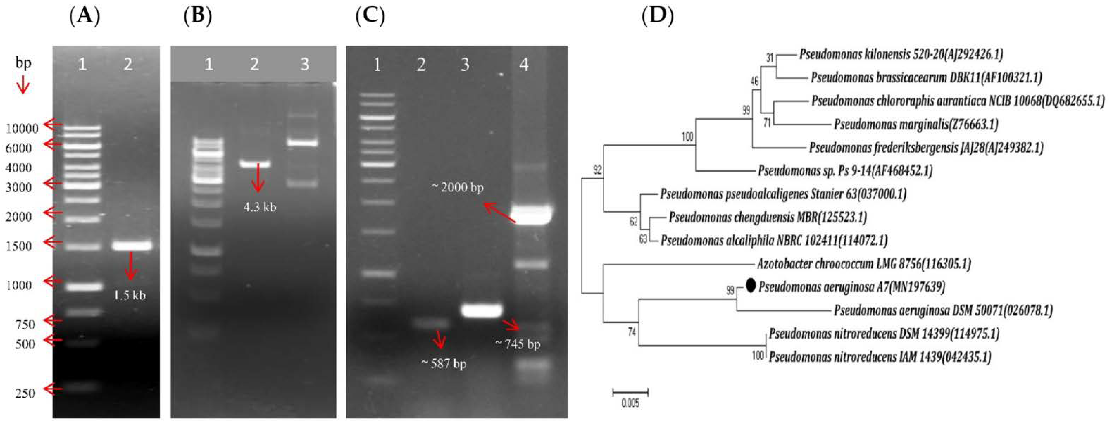
Figure 1. Molecular characterization of the bacterial strain A7. (A) Gel image of PCR amplified gene fragment [(lane 1 = Gene Ruler: 1 kb DNA Ladder; lane 2 = 16S rRNA gene fragment with 1.5 kb (kilo base)], (B) Gel image of EcoRI digested recombinant plasmid DNA fragment (lane 1 = Gene Ruler: 1kb DNA Ladder; lane 2 = digested 4.3 kb recombinant plasmid DNA fragment and lane 3 = undigested recombinant plasmid DNA, (C) Gel image of PCR amplified antifungal antibiotics gene fragments (lane 1 = Gene Ruler: 1 kb DNA Ladder; lane 2 = gene fragment hcnBC with approx. 587 bp (base pairs); lane 3 = gene fragment phlD with approx. 745 bp, lane 4 = gene fragment phzH with approx. 2000 bp) and (D) Phylogenetic tree analysis of the strain A7 (Pseudomonas aeruginosa A7) by Maximum Likelihood method based on the sequence of the 16S rRNA gene of the bacterial strain. Values are given at branch nodes and are based on 1000 replicates. The scale bar indicates 0.005 substitutions per nucleotide position. The accession numbers are enclosed in parenthesis.
Microorganisms 10 00568 g001
Figure 2. (A) A7 bacterial strain inhibits fungal plant pathogens in vitro. (I) By concentric ring dual culture bioassay method, (II) By dual plate bioassay method and (III) Control plates of all the fungal pathogens on potato dextrose agar medium at 30 ± 2 °C for a particular incubation period in three biological replicates. Control plates were the same for both experiments, performed for each fungus. Foc = Fusarium oxysporum f.sp. ciceris, Fop = Fusarium oxysporum f. sp. pisi, Fol = Fusarium oxysporum f. sp. lycopersici (Sacc.), Fo = Fusarium oxysporum, Fs = Fusarium solani (on King’s Base agar medium plate, Ce = Curvularia eragrostidis, Clb = Curvularia lunata (Wakker) Boedijn var., Plp = Phellinus lamaensis (Murrill) Pat, Uzs = Ustulina zonata (lev.) Sacc (after seven days of incubation), Rap =Rosellinia arcuata Petch, Php =Poria hypobrunnea Petch, and Rs =Rhizoctonia solani (after two days of incubation). (B) In vitro antifungal and antibacterial activity of cell-free supernatant (CFS) of A7 against plant fungal and bacterial pathogens. (a) By agar well diffusion method and (b) by agar disk diffusion method on Potato Dextrose Agar and King’s Base agar medium plates at 30 ± 2 °C in three biological replicates. III = Control and IV = Agar well containing 100 µL of CFS on each plate. Rs = Rhizoctonia solani, Rap =Rosellinia arcuata Petch and Php =Poria hypobrunnea Petch. Ras = Ralstonia solanacearum and Xc = Xanthomonas campestris (i = agar disc containing 50 µL of CFS and ii = control with 50 µL of sterile double distilled water) after two days of the incubation period. (c) Bar diagram showing the Percentage of growth inhibition by A7 against plant fungal pathogens by producing antifungal metabolites and volatile compounds. Values are expressed as means (n = 3) ± Standard Error Mean (SEM). Statistical differences were analyzed using a Two-way analysis of variances (ANOVA). Significant differences (d = p < 0.001, e = p > 0.05 followed by Bonferroni Posttests) compared with antifungal metabolites vs. volatile compounds. (d) Bar diagram showing the percentage of growth inhibition by the CFS of A7 against plant fungal pathogens and (e) Bar diagram showing growth inhibition of bacterial plant pathogens (calculated as millimeter-scale). Values are expressed as means (n = 3) ± Standard Error Mean (SEM). Statistical differences were analyzed using a One-way analysis of variances (ANOVA). Significant differences (f = p < 0.001, g = p < 0.01 and h = p < 0.05, followed by Tukey’s multiple comparison test) compared with the control.
Microorganisms 10 00568 g002
Figure 3. In vitro plant-growth-promoting activities of the bacterial strain A7 on different plate-based agar mediums at 30 ± 2 °C for a particular incubation period in three biological replicates. I = Different PGP activities as (a) Indole-3-acetic acid production on Whatman no.1 filter paper in Luria Bertani agar (after one day of incubation), (b) Phosphate solubilization on Pikovskaya’s agar, (c) Potassium solubilization on modified Aleksandrov agar, (d) Zinc solubilization on Zinc Oxide agar (after seven days of incubation period), (e) Nitrogen fixation on N2 free agar (after one day of incubation), (f) Siderophores production on CAS agar (after three days of incubation) and (g) Hydrogen cyanide production (HCN) on glycine agar (after four days of incubation), (h) Chitinase production on colloidal chitin agar (after four days of incubation), (i) Cellulase production on Congo—red gelatin agar (after four days of incubation), (j) Lignin degradation on Methylene blue indicator MSM—KL agar (after seven days of incubation period), (k) Pectinase production on M9 Citrus peel pectin agar (after two days of incubation period), (l) Proteases production on Skim milk agar (after seven days of incubation), and II = Control plate of the respective activities.
Microorganisms 10 00568 g003
Figure 4. Growth profile of the bacterial strain A7 at different abiotic stress conditions. (a) Growth of A7 at different concentrations of metal compounds (FeCl3, FeCl2, AlCl3), pH and temperature by Single Plate Serial Dilution Spotting (SP—SDS) and streaking plate inoculation method at 30 ± 2 °C for a particular incubation period in three biological replicates. (I) AlCl3 (1 = control, 2 = 1 mM, 3 = 2 mM, 4 = 3 mM 5 = 4 mM) (at a dilutions of 10−1 to 10−6), (II) FeCl3 (1 = control, 2 = 2 mM, 3 = 4 mM, 4 = 6 mM), (III) FeCl2 (1 = control, 2 = 2 mM, 3 = 4 mM, 4 = 6 mM) (at a dilutions of 10−1 to 10−4), (IV) pH (1 = control, pH—7, 2 = pH—6, 3 = pH—5.5, 4 = pH—5, 5 = pH—4.5) (at a dilutions of 10−1 to 10−6), (V) Growth of the bacterial strain A7 at 30 °C as control, (VI) Growth of the bacterial strain A7 at 4 °C, (VII) Growth of the bacterial strain A7 at 50 °C after two days of incubation period. (b) Bar diagram showing Relative growth of the bacterial strain A7 at different concentrations of metal compounds (FeCl3, FeCl2, and AlCl3), NaCl, Temperature and pH. Values are expressed as means (n = 3) ± Standard Error Mean (SEM).
Microorganisms 10 00568 g004
Figure 5. (a) Influence of the bacterial strain A7 against aluminum toxicity on the growth promotion of chickpea at 12 h interval of time for 48 h at 14 h photoperiod in white light with a photon flux intensity of 220 mol m−2 s1 (PAR) in three biological replicates. (I) at 12 h, (II) at 24 h, (III) at 36 h, and (IV) at 48 h. T0 = Control (with no bacteria and aluminum), T1 = AlCl3 (only 100 µM of aluminum chloride), T2 = A7 (only freshly grown 0.1 OD600 cells of A7) and (4) T3 = A7 + AlCl3 (freshly grown 0.1 OD600 cells of A7 + 100 µM of aluminum chloride). (b) Bar diagram showing different growth parameters of the chickpea plants with the influence of the bacterial strain A7 against aluminum toxicity after 12, 24, 36, and 48 h of incubation. RL = Root Length, SL = Shoot Length, RFW = Root Fresh Weight, SFW = Shoot Fresh Weight, RDW = Root Dry Weight, SDW = Shoot Dry Weight, SVI = Seed Vigor Index and RRL = Root Relative Length. Values are expressed as means (n = 3) ± Standard Error Mean (SEM). Each replicate consists of sixteen plants per jar. Statistical differences were analyzed by two-way analysis of variances (ANOVA). Significant differences (c = p < 0.001, d = p < 0.01, e = p < 0.05 and f = p > 0.05, followed by Bonferroni Posttests) compared with the control.
Microorganisms 10 00568 g005
Figure 6. In vivo establishment and reduction of chickpea wilt caused by Fusarium oxysporum f. sp. ciceris by the bacterial strain A7. The data in panels a and b is after 40th days of inoculation with the treatments as T0 = Sterile distilled water as control, T1 = Only bacterial cell suspension, T2 = Only fungal spore suspension, T3 = Bacterial cell suspension and fungal spore suspension, T4 = Bacterial cell suspension two days before the fungal spore suspension and T5 = Fungal spore suspension two days before the bacterial cell suspension at room temperature in three biological replicates. (a) Yellowing of leaves, (b) Root rot, and darkening of vascular tissues. (c) Bar diagram showing the percentage of (i) disease incidence (DI), (ii) disease severity (DS), and (iii) disease reduction (DR) of Fusarium wilt of chickpea from pathogen Fusarium oxysporum f. sp. ciceris by the strain A7 after 20th and 40th days of inoculation. Values are expressed as means (n = 3) ± Standard Error Mean (SEM). Statistical differences were analyzed by a Two-way analysis of variances (ANOVA). Significant differences (e = p < 0.001, f = p < 0.01 and g = p > 0.05, followed by Bonferroni Posttests) compared with the control. Bar diagram also shows (iv) plant-growth promotion (PGP) and (v) plant biomass of chickpea during the establishment of Fusarium wilt of chickpea caused by Fusarium oxysporum f. sp. ciceris after 40 days of inoculation. Values are expressed as means (n = 3) ± Standard Error Mean (SEM). Statistical differences were analyzed by two-way analysis of variances (ANOVA). Significant differences (b = p < 0.001, c = p < 0.01 and d = p > 0.05, followed by Bonferroni Posttests) compared with the control.
Microorganisms 10 00568 g006
Figure 7. Bio-active compounds produced by the bacterial strain A7 in three biological replicates. (a) Antifungal activity of the dried ethyl acetate extract of the strain A7 (before thin layer chromatographic separation) against Rosellinia arcuata Patch. (b) TLC plate exhibited four fractions of the dried ethyl acetate extract of the bacterial strain with a single discrete fraction (F3) with 0.70 Rf value, (c) Antifungal activity of the methanol extract of TLC purified fraction F3 against Rosellinia arcuata Patch, (d) Gas Chromatography (GC) analysis of the TLC purified fraction F3 of A7 for compound characterization and (e) Mass spectra (MS) analysis of the fraction F3 of A7 for compound identification.
Microorganisms 10 00568 g007
Table 1. List of Primers used for identification of the bacterial isolates and antibiotic genes.
Literature
Name
Primer SequencePrimer CharactersPCR Condition (°C)Ref.
fD1F-5′-GAGTTTGATCCTGGCTCA-3′16S rDNAID 98 °C for 30 s, FD 98 °C for 10 s, An 50 °C for 30 s,[53]
rP2R-5′-ACGGCTACCTTGTTACGACTT-3′Ex 72 °C at 1.30 min, FEx 72 °C for 7 min.
Phl2aF-5′-GAGGACGTCGAAGACCACCA-3′2,4-DAPGID 94 °C for 1.30 min, FD 94 °C for 35 s, An 53 °C for 30 s,[53]
Phl2bR-5′-ACCGCAGCATCGTGTATGAG-3′Ex 72 °C for 45 s, FEx 72 °C for 10 min.
AcaF-5′-ACTGCCAGGGGCGGATGTGC-3′HCNID 94 °C for 2.30 min, FD 94 °C for 30 s, An 63 °C for 30 s,[91,92]
AcbR-5′-ACGATGTGCTCGGCGTAC-3′Ex 72 °C for 60 s, FEx 72 °C for 10 min.
PCA2aF-5′-TTGCCAAGCCTCGCTCCAAC-3′PCAID 94 °C for 2 min, FD 94 °C for 60 s, An 55 °C for 45 s,[91]
PCA3bR-5′-CCGCGTTGTTCCTCGTTCAT-3′Ex 72 °C for 60 s, FEx 72 °C for 10 min.
PhzH-upF-5′-CGCACGGATCCTTTCAGAATGTTC-3′PCNID 98 °C for 30 s, FD 98 °C for 10 s, An 55 °C for 30 s,[93]
PhzH-lowR-5′-GCCACGCCAAGCTTCACGCTCA-3′Ex 70 °C for 7 min, FEx 70 °C for 10 min.
PLTC1F-5′-AACAGATCGCCCCGGTACAGAACG-3′PLTID 95 °C for 2 min, FD 95 °C for 2 min, An 57 °C for 1 min,[94]
PLTC2R-5′-AGGCCCGGACACTCAAGAAACTCG-3′ Ex 72 °C for 1 min, FEx 72 °C for 7 min.
PRND1F-5′-GGGGCGGGCCGTGGTGATGGA-3′PRNID 95 °C for 2 min, FD 95 °C for 1 min, An 68 °C for 1 min,[94]
PRND2R-5′-YCCCGCSGCCTGYCTGGTCTG-3′ Ex 72 °C for 1 min, FEx 72 °C for 7 min.
darAFF-5′-ATCGTCAATGATCTCTGGC-3′HPR,ID 94 °C for 2 min, FD 94 °C for 1 min, An 50 °C for 1 min,[95]
darARR-5′-TTATCCACTGCCTCTCCC-3′DARA, Ex 72 °C for 1 min, FEx 72 °C for 10 min.
darBFF-5′-GATACTCAGCGAGCGTGC-3′HPR,ID 94 °C for 2 min, FD 94 °C for 1 min, An 50 °C for 1 min,
darBRR-5′-CACCAGGTTATGGCGTCAG-3′DARB Ex 72 °C for 1 min, FEx 72 °C for 10 min.
HCN = Hydrogen cyanide synthase (E), PCA = Phenazine-1-carboxylic acid, PCN = Phenazine-1-carboxamide, PLT = Pyoluteorin, PRN = Pyrrolnitrin, HPR = 2-Hexyl,5-propyl resorcinol, DARA = Dialkylresorcinol gene A, DARB = Dialkylresorcinol gene B, ID = Initial Denaturation temperature, FD = Final Denaturation temperature, An = Annealing temperature, Ex = Extension temperature, FEx = Final Extension temperature. Ref. = References.
Table 2. Morphological and biochemical characteristics of the strain A7. + Positive, − Negative.
Morphological CharactersResponse
ShapeRound
Gram reactionNegative, rod shaped
ColorGreen pigmented
BiochemicalCarbohydrate utilization
ONGP productionArabinose+
Lysine Utilization+Xylose+
Ornithine utilizationAdonitol
Urease productionRhamnose
Phenylalanine DeaminationCellobiose
Nitrate reduction+Melibiose+
H2S productionSaccharose
Citrate utilization+Raffinose
Voges Proskauer’s testTrehalose+
Methyl red testGlucose+
Indole productionLactose
Malonate utilization+
Esculin hydrolysis
Oxidase production+
Table 3. Effect of the bacterial strain A7 on fungal and bacterial plant pathogen growth inhibition.
Fungal PathogensPGI (%) a
Plant Fungal PathogensSource of CollectionDiseased CausedITCC NoAntifungal MetabolitesVolatile
Compounds
Fusarium oxysporum f.sp. ciceris (Foc)Chick peaFusarium wilt363678.59 ± 1.1 d65.51 ± 1.3 d
Fusarium oxysporum f. sp. pisi (Fop)PeaWilt 481462.51 ± 1.0 dN/D c
Fusarium oxysporum f. sp. lycopersici (Sacc.) (Fol)TomatoWilt 132265.26 ± 1.2 e60.37 ± 1.0 e
Fusarium oxysporum (Fo)Maize StalkWilt 709360.56 ± 1.2 d46.44 ± 1.3 d
Fusarium solani (Fs)Paddy soilWilt 695370.66 ± 1.2 dN/D c
Rhizoctonia solani (Rs)CucumberCollar rotAAU29.20 ± 1.0 d81.25 ± 1.1 d
Curvularia eragrostidis (Ce)MusaLeaf spot554070.56 ± 1.3 d51.00 ± 1.3 d
Curvularia lunata (Wakker) Boedijn var. (Clb)RiceLeaf spot519342.24 ± 2.6 d24.88 ± 2.5 d
Rosellinia arcuata Petch (Rap)Tea bushRoot rot414357.95 ± 2.3 d74.99 ± 1.5 d
Poria hypobrunnea Petch (Php)Tea bushStem canker414157.26 ± 1.5 d35.14 ± 1.4 d
Phellinus lamaensis (Murrill) Pat.(Plp)Tea bushBrown root rot414051.01 ± 1.7 d37.36 ± 1.2 d
Ustulina zonata (Lev.) Sacco (Uzs)Tea bushCharcoal stump rot414473.34 ± 2.0 dN/D c
Plant fungal and bacterial pathogensPGI (%) b
Rhizoctonia solani (Rs)CucumberCollar rotAAU27.33 ± 1.4 h
Rosellinia arcuata Petch (Rap)Tea bushRoot rot414333.33 ± 0.8 f
Poria hypobrunnea Petch (Php)Tea bushStem canker414140.33 ± 1.4 h
Ralstonia solanacearum (Ras)-Brown rotAAU23.17 ± 1.3 mm f
Xanthomonas campestris (Xc)-Black rotAAU34.50 ± 2.0 mm g
Percentage of growth inhibition (PGI) of Pseudomonas aeruginosa A7 against plant fungal pathogens and growth inhibition of bacterial plant pathogens collected from Indian Type Culture Collection (ITCC) and Assam Agricultural University (AAU). a PGI (%) = (Dc − Dt)/(Dc) ∗ 100 in dual culture. Values are expressed as means (n = 3) ± Standard Error Mean (SEM). Statistical differences were analyzed by a two-way analysis of variances (ANOVA). Significant differences (d p < 0.001, e p > 0.05 followed by Bonferroni Posttests) compared with antifungal metabolites and volatile compounds are indicated by letters (d and e). b PGI (%) = (Dc − Dt)/(Dc) ∗ 100 in presence of Cell-Free Supernatant (CFS) of A7 and growth inhibition of bacterial plant pathogens (mm). Values are expressed as means (n = 3) ± Standard Error Mean (SEM). Statistical differences were analyzed by One-way analysis of variances (ANOVA). Significant differences (f p < 0.001, g p < 0.01, and h p < 0.05 followed by Tukey’s multiple comparison test) compared with the control. c N/D—No growth inhibition. mm = millimeter scale.
Table 4. Plant-growth-promoting activities of the strain A7 and its enzymatic activities.
Direct and Indirect PGP Traits of A7ResponseIndole-3-Acetic ACID Production
Time(h)With Tryptophan (µg/mL)Without Tryptophan (µg/mL)
Phosphate solubilization146.00 ± 0.5 µg/mL483.30 ± 0.3 d1.30 ± 0.3 d
Potassium solubilization33% (SE) a966.50 ± 0.3 c3.60 ± 0.1 c
ZnO solubilization29% (SE) a1445.40 ± 0.4 c2.50 ± 0.0 c
Nitrogen Fixation52.00 ± 0.05 mm1963.10 ± 0.1 e2.40 ± 0.2 e
Siderophores production45.00 ± 0.05 mm
HCN production+Quantitative production of Indole-3-acetic acid at 48 h interval incubation period. Statistical differences were analyzed by two-way analysis of variances (ANOVA).
Chitinase production58.00 ± 0.05 mm
Cellulase production2.4 (HC) b
Lignin degradation+
Pectinase production2 (HC) b
Proteases production1.3 (HC) b
Polyamine production-
Amylase production+
Xylanase production-
Catalase production+
Different plant-growth-promoting and extracellular enzymatic activities with different measurement units. a SE (Solubilization Efficacy) = (Diameter of A7/Diameter of the halo zone] ∗ 100, b HC (hydrolysis capacity) = (Ratio of the halo zone diameter to the colony diameter). mm = millimeter. Values are expressed as means n = 3 ± Standard Error Mean (SEM). Significant differences (c p < 0.001, d p < 0.01 and e p > 0.05, followed by Bonferroni Posttests) compared with and without tryptophan are indicated by letters c, d, and e.
Table 5. Relative growth (RG) of the bacterial strain A7 on different abiotic stress conditions.
AlCl3FeCl3FeCl2NaClTemperaturepH
Conc. (mM)RG (%) aConc. (mM)RG (%) aConc. (mM)RG (%) aConc. (%)RG (%) aTemp (°C)RG (%) apHRG (%) a
C100.00 ± 0.0C100.00 ± 0.0C100.00 ± 0.0C100.00 ± 0.046.35 ± 0.430.00 ± 0.0
0.196.55 ± 0.70.192.03 ± 0.40.190.54 ± 0.5196.98 ± 1.01833.66 ± 1.445.50 ± 0.5
0.588.25 ± 0.80.587.50 ± 1.00.573.38 ± 1.1285.93 ± 1.02555.22 ± 1.0521.59 ± 2.4
1.075.00 ± 1.11.073.20 ± 0.31.054.75 ± 0.6372.53 ± 1.230(C)100.00 ± 0.0652.31 ± 2.3
1.557.81 ± 1.61.564.34 ± 0.81.528.78 ± 0.1457.44 ± 1.83788.00 ± 2.07(C)100.00 ± 0.0
2.050.74 ± 0.32.055.07 ± 2.72.017.21 ± 0.3543.51 ± 1.14536.50 ± 1.58126.97 ± 2.0
2.538.70 ± 0.82.548.19 ± 0.42.56.08 ± 0.7622.33 ± 1.05011.56 ± 0.4986.50 ± 1.5
3.027.63 ± 0.43.035.70 ± 0.830.00 ± 0.078.31 ± 0.3 1058.00 ± 2.0
3.511.94 ± 0.73.523.62 ± 0.6 80.00 ± 0.0 1124.00 ± 2.0
4.00.00 ± 0.04.06.93 ± 0.5 1211.50 ± 2.5
4.51.36 ± 0.0 132.40 ± 0.5
Inhibitory Concentration (IC) (mM)
MetalIC20 bIC50 cMetalIC20 bIC50 cMetalIC20 bIC50 c
AlCl30.76 ± 0.031.9 ± 0.0FeCl31.00 ± 0.042.50 ± 0.02FeCl20.47 ± 0.051.18 ± 0.08
Percentage Relative Growth of the bacterial strain at different concentrations of metal compounds: AlCl3, FeCl3, FeCl2, at different concentrations of NaCl, at different temperature and at different pH range. a RG (%) = (Optical density the treatments/Optical density of controls) ∗ 100. mM = millimolar. b IC20 = 20% of growth inhibitory concentration of metals, c IC50 = 50% of growth inhibitory concentration of metals. Conc. = Concentrations, Temp. = Temperature, C = control. Values are expressed as means n = 3 ± Standard Error Mean (SEM) each biological replicate.
Table 6. Influence of strain A7 on the growth of chickpea in a stressed condition.
TimeTreatmentRL (mm)SL (mm)RFW (mg)SFW (mg)RDW (mg)SDW (mg)SVI aRRL (%) b
12 hT080 ± 2.0 c50 ± 2.9 c256 ± 3.0 c307 ± 3.6 c26 ± 0.8 c29 ± 0.8 c1290 ± 1.0 c100 ± 0.0 f
T176 ± 1.5 c50 ± 1.2 c227 ± 3.9 c292 ± 1.2 d25 ± 0.8 c27 ± 0.6 c1240 ± 2.0 c95 ± 1.0 d
T288 ± 1.4 c55 ± 1.4 c316 ± 3.4 c365 ± 2.6 c34 ± 0.5 c42 ± 0.8 c1445 ± 1.5 c100 ± 0.0 f
T386 ± 1.8 c54 ± 0.8 c294 ± 2.3 c342 ± 1.2 c30 ± 0.5 c37 ± 0.8 c1400 ± 1.0 c97 ± 0.3 f
24 hT071 ± 1.4 c52 ± 2.6 c254 ± 2.0 c334 ± 2.6 c32 ± 1.2 c54 ± 2.3 c1210 ± 1.0 c100 ± 0.0 f
T158 ± 2.6 f47 ± 0.5 c224 ± 2.0 c313 ± 1.7 f24 ± 1.5 c36 ± 0.3 c1077 ± 1.7 c82 ± 3.0 c
T289 ± 1.4 c67 ± 1.0 c346 ± 3.1 c431 ± 1.4 c39 ± 0.5 c60 ± 0.3 c1580 ± 2.0 c100 ± 0.0 f
T379 ± 2.3 c64 ± 0.8 c315 ± 2.7 c401 ± 2.4 c36 ± 3.3 c57 ± 0.8 c1445 ± 1.5 c89 ± 1.1 c
36 hT053 ± 1.1 f60 ± 2.4 c231 ± 1.7 c420 ± 1.2 c18 ± 0.4 f42 ± 3.3 c1060 ± 1.0 c100 ± 0.0 f
T147 ± 1.2 e42 ± 1.0 d215 ± 2.9 c242 ± 1.2 e16 ± 3.2 f27 ± 3.3 c825 ± 1.5 d88 ± 1.8 c
T278 ± 3.1c73 ± 1.2c382 ± 3.8 c565 ± 7.7 c40 ± 0.8 c70 ± 1.2 c1530 ± 1.0 c100 ± 0.0 f
T378 ± 3.1 c73 ± 1.2 c381 ± 3.8 c565 ± 7.7 c39 ± 0.8 f70 ± 1.2 c1530 ± 1.0 c100 ± 0.0 c
48 hT042 ± 1.7 c49 ± 2.5 c213 ± 1.5 c231 ± 1.2 f15 ± 0.9 f36 ± 0.3 c961.± 1.6 c100 ± 0.0 f
T140 ± 1.2 c38 ± 0.8 f123 ± 1.7 c143 ± 1.8 c13 ± 0.8 d27 ± 0.0 c815 ± 0.5 f91 ± 1.5 c
T272 ± 1.4 c65 ± 1.7 c234 ± 2.0 c292 ± 1.4 c20 ± 0.8 c46 ± 0.5 c1400 ± 1.0 c100 ± 0.0 f
T370 ± 0.6 c61 ± 1.2 c225 ± 2.9 c247 ± 4.7 c16 ± 0.5 f45 ± 0.8 c1345 ± 1.5 c97 ± 0.3 f
Plant-Growth Promotion of chickpea plant in the presence and absence of aluminum chloride with a 12 h interval of time for 48 h. T0 = Control, T1 = only AlCl3, T2 = only bacterial strain A7 and T3 = Bacterial strain A7 + AlCl3, RL = Root Length, SL = Shoot Length, RFW = Root Fresh Weight, SFW = Shoot Fresh Weight, RDW = Root Dry Weight, SDW = Shoot Dry Weight, a SVI (Seed Vigor Index) = (Shoot length + root length) ∗ seed germination %, b RRL (Relative Root Length) = (Root length in aluminum stress condition)/(Root length in control conditions) ∗ 100. Values are expressed as means (n = 3) ± Standard Error Mean (SEM). Statistical differences were analyzed by two-way analysis of variances (ANOVA). Significant differences (c p < 0.001, d p < 0.01, e p < 0.05 and f p > 0.05, followed by Bonferroni Posttests) compared with control.
Table 7. In vivo disease incidence, severity, and reduction of Fusarium wilt of chickpea by A7.
After 20th Days of InoculationAfter 40th Days of Inoculation
TreatmentsDI (%) aDS (%) bDR (%) cDI (%) aDS (%) bDR (%) c
T222.03 ± 0.4 e22.43 ± 0.8 eN/D d65.43 ± 1.7 e65.54 ± 1.7 eN/D d
T312.12 ± 1.0 e5.95 ± 0.5 f44.70 ± 5.2 f21.37 ± 1.3 e10.29 ± 0.8 f67.23 ± 2.3 f
T47.4 ± 0.6 e1.85 ± 0.1 g66.01 ± 2.9 g14.99 ± 0.6 e3.92 ± 0.1 g77.00 ± 1.5 g
T513.20 ± 1.0 e10.48 ± 0.5 e39.73 ± 1.6 f24.56 ± 1.0 e19.38 ± 0.6 e62.26 ± 2.5 f
Establishment of Fusarium wilt Disease incidence (DI), Disease severity (DS), caused by Fusarium oxysporum f. sp. ciceris and Disease reduction (DR) by the strain A7 after 20th and 40th days of inoculation with the following treatments: T2 = only fungal spore suspension, T3 = bacterial cell suspension, and fungal spore suspension, T4 = bacterial cell suspension two days before the fungal spore suspension and T5 = fungal spore suspension two days before the bacterial cell suspension. a DI (%) = (No. of attained leaves/Total no. of leaves) ∗ 100, b DS (%) = (Σ n × v/N × V) ∗ 100 and c DR (%) = [1 − (DI in treatment/DI in control)] ∗ 100 after 20th and 40th days of inoculation. d N/D = no disease reduction. Values are expressed as means (n = 3) ± Standard Error Mean (SEM). Statistical differences were analyzed by a Two-way analysis of variances (ANOVA). Significant differences (e p < 0.001, f p < 0.01 and g p > 0.05, followed by Bonferroni Posttests) compared with the control.
Table 8. In vivo growth promotion of chickpea plant by the strain A7 during the treatments with Fusarium oxysporum f. sp. ciceris.
Plant Growth PromotionPlant Biomass
TRL (cm)SL (cm)SVI/100 aRFW (mg)SFW (mg)RDW (mg)SDW(mg)
T018.66 ± 2.6 b49.33 ± 2.0 b68.00 ± 4.6 b1530.00 ± 26.4 d4840.00 ± 173.8 d456.00 ± 14.0 d1263.33 ± 31.7 d
T125.66 ± 1.7 b83.66 ± 3.4 b109.33 ± 5.2 b2280.00 ± 43.5 a5633.33 ± 210.7 a853.33 ± 26.0 a1756.66 ± 29.6 a
T214.00 ± 2.3 b39.33 ± 2.6 b53.00 ± 4.9 b1157.00 ± 29.8 d4030.00 ± 88.8 d270.00 ± 17.3 d966.66 ± 88.1 d
T322.66 ± 1.7 b71.66 ± 2.6 b92.66 ± 4.3 b1903.33 ± 53.6 b4963.33 ± 68.3 b665.66 ± 11.0 b1363.33 ± 31.7 b
T421.00 ± 1.1 b75.33 ± 1.4 b96.33 ± 2.6 b1984.66 ± 45.1 c5240.00 ± 183.3 c762.00 ± 14.7 c1556.66 ± 29.6 c
T517.00 ± 1.1 b65.33 ± 2.0 b82.33 ± 3.1 b1663.00 ± 31.6 c4763.33 ± 87.6 c555.66 ± 12.8 c1240.00 ± 50.3 c
Plant growth promotion of chickpea plant during the establishment of Fusarium wilt disease caused by Fusarium oxysporum f. sp. ciceris after 40th days of inoculation with the treatments: T0 = Sterile distilled water as control, T1 = Only bacterial cell suspension, T2 = Only fungal spore suspension, T3 = Bacterial cell suspension and fungal spore suspension, T4 = Bacterial cell suspension two days before the fungal spore suspension and T5 = Fungal spore suspension two days before the bacterial cell suspension. RL = Root Length, SL = Shoot Length, a Seed Vigor Index/100 = (Shoot length + root length) ∗ seed germination %, RFW = Root Fresh Weight, SFW = Shoot Fresh Weight, RDW = Root Dry Weight, SDW = Shoot Dry Weight. Values are expressed as means (n = 3) ± Standard Error Mean (SEM). Statistical differences were analyzed by two-way analysis of variances (ANOVA). Significant differences (b p < 0.001, c p < 0.01 and d p > 0.05, followed by Bonferroni Posttests) compared with the control.
Table 9. GC-MS analysis of bioactive compounds produced by Pseudomonas aeruginosa A7.
RTMajor CompoundsMMMFActivityRef.
22.04Oleic acid282C18H32O2Unknown
4-fluoro-1-methyl-5-carboxylic acid172C7H9FN2O2Unknown
2-tridecenoic acid212C7H9FN2O2Unknown
6,10-dimethyl-4-undecanol200C13H28OUnknown
24.651-pentadecene200C15H30Unknown
1-tridecene182C13H26Unknown
10-heneicosene (C, T)294C21H42Unknown
1-hexadecene224C16H32antibacterial activity[101]
28.06Tridecanoic acid, 12-methyl-ester242C15H30O2Unknown
Hexadecanoic acid, methyl ester270C17H34O2antioxidant, antibacterial[102]
Tridecanoic acid, methyl ester228C14H28O2Unknown
Hexacosanoic acid, methyl ester410C27H54O2Unknown
29.705-Eicosene280C20H40antimicrobial, anticancer[101]
E-15-heptadecenal252C17H32Oantibacterial activity
Cycloeicosane280C20H40antimicrobial, anticancer
1-heptadecene238C17H34Unknown
32.089-octadecenoic acid296C19H36O2antibacterial activity[103]
12-octadecenoic acid296C18H34O2Unknown
Trans-13-octadecenoic acid238C18H34O2Unknown
40.041,2-benzenedicarboxylic acid mono (2-ethylhexyl) ester278C16H22O4antimicrobial, anticancer[100]
6-ethyloct-3-yl 2-ethylhexyl ester418C26H42O4Unknown
1,2-benzenedicarboxylic acid diisooctyl ester, Bis (2-ethylhexyl) phthalate390C24H38O4antifungal[104]
Bacterial bioactive compounds produced by the bacterial strain were analyzed using Gas Chromatography–Mass Spectroscopy and the compounds were identified by the National Institute of Standards and Technology (NIST) library based on their retention time, molecular mass, and molecular formula. RT = Retention time, MM = Molecular mass, MF = Molecular formula. Ref. = References.
Publisher’s Note: MDPI stays neutral with regard to jurisdictional claims in published maps and institutional affiliations.