Article

Detection of mcr-1 Gene in Undefined Vibrio Species Isolated from Clams

by
Christian Valdez
1,†,
Cátia Costa
1,†,
Marco Simões
1,
Carla C. C. R. de Carvalho
2,
Teresa Baptista
1 and
Maria J. Campos
1,*
1
MARE-Marine and Environmental Sciences Centre, ESTM, Polytechnic of Leiria, 2520-630 Peniche, Portugal
2
iBB-Institute for Bioengineering and Biosciences, Department of Bioengineering, Associate Laboratory i4HB—Institute for Health and Bioeconomy, Instituto Superior Técnico, Universidade de Lisboa, Av. Rovisco Pais, 1049-001 Lisboa, Portugal
*
Author to whom correspondence should be addressed.
These authors contributed equally to the development of this study.
Microorganisms 2022, 10(2), 394; https://doi.org/10.3390/microorganisms10020394
Submission received: 23 December 2021 / Revised: 24 January 2022 / Accepted: 3 February 2022 / Published: 8 February 2022
(This article belongs to the Section Environmental Microbiology)

Abstract

:
The increase of antimicrobial resistant strains is leading to an emerging threat to public health. Pathogenic Vibrio are responsible for human and animal illness. The Enterobacteriaceae family includes microorganisms that affect humans, causing several infections. One of the main causes of human infection is related to the ingestion of undercooked seafood. Due to their filter-feeding habit, marine invertebrates, such as clams, are known to be a natural reservoir of specific microbial communities. In the present study, Vibrionaceae and coliforms microorganisms were isolated from clams. A microbial susceptibility test was performed using the disk diffusion method. From 43 presumptive Vibrio spp. and 17 coliforms, three Vibrio spp. with MICs to colistin >512 mg L−1 were found. From the 23 antimicrobial resistance genes investigated, only the three isolates that showed phenotypic resistance to colistin contained the mcr-1 gene. Genotypic analysis for virulence genes in EB07V indicated chiA gene presence. The results from the plasmid cure and transformation showed that the resistance is chromosomally mediated. Biochemical analysis and MLSA, on the basis of four protein-coding gene sequences (recA, rpoB, groEL and dnaJ), grouped the isolates into the genus Vibrio but distinguished them as different from any known Vibrio spp.

1. Introduction

The genus Vibrio contains more than 130 species [1,2] and includes both non-pathogenic and pathogenic species, being highly genetically diverse [3,4]. Vibrio spp. are Gram-negative motile bacteria, facultative anaerobic, and non-spore-forming. With cells shaped as curved or straight rods, and typically found in freshwater, estuarine and marine environments [1,5]. Pathogenic species are associated with several human diseases caused by the natural microbiota of aquatic environments [6] and are mostly linked to the ingestion of undercooked seafood [7]. Vibriosis is one of the most prevalent bacterial diseases associated with marine fish and shellfish [8]. Non-pathogenic Vibrio, on the other hand, are not associated with human illnesses, and are, therefore, not routinely identified [9]. These so-called “marine species” or “marine vibrios”, have nonetheless been frequently isolated, thus receiving more attention in recent years since they have an important role in specific ecological niches and belong to autochthonous marine microbial communities [4]. These niches are often fish farms, where antibiotics are frequently used, and Vibrio can become reservoirs of antimicrobial resistance genes [10,11].
The Enterobacteriaceae family comprises Gram-negative bacteria that affect humans, causing urinary tract infections, pneumoniae, gastroenteritis, meningitis, and sepsis [12]. Enterobacteriaceae possess a bacilli shape, are non-spore-forming, have variable motility, and are generally found inside other organisms. Usually, these microorganisms have the ability to mobilise their antimicrobial genetic determinants in plasmids via conjugation, which means that they can be resistant to almost every existing antibiotic [12]. Hospitals throughout the world have been reporting clinical cases of people that contract severe bacterial infections due to the presence of resistant lineages of major infectious agents such as Escherichia coli [13] and Klebsiella pneumoniae [14].
The abusive and erroneous use of antibiotics by human and veterinary medicine [15,16] promotes a selective pressure for antimicrobial resistant bacteria. The increase of resistant strains is leading to an emerging threat to public health [17,18]. It is estimated that 10 million lives could be lost due to antimicrobial resistant bacteria annually by 2050, if no action is taken [19].
For many years (1950–1970), polymyxin antibiotics were commonly employed in clinical use, until they were gradually withdrawn because of toxicity issues. However, nowadays, the available antibiotics are not enough to cope with the emergence of multidrug-resistance bacteria, resulting in the re-introduction of polymyxins as the last line of defence for clinical treatment [20]. Among polymyxins, the two most used are polymyxin E (colistin) and polymyxin B [21]. Until recently, all the findings about polymyxin-resistant mechanisms were chromosomally mediated [22,23,24]. However, the plasmid-mediated colistin resistance gene mcr has already been reported. The first report of mcr-1 was in E. coli and K. pneumoniae [25] in China. Since then, the mcr-1 gene has emerged in strains collected in many parts of the world (e.g., Europe—[26,27,28,29]; Asia—[30,31]; North America—[32]; Africa—[33]). A very large study of bacterial genomes concluded that almost all mobile colistin resistance genes (from mcr-1 to mcr-9) originated from environmental bacteria, mainly from water sources [34]. The origin of mcr-1 is believed to be from Moraxella species [34,35]. As far as the authors know, up to this date, the presence of mcr-1 has been mostly reported in the Enterobacteriaceae family and only once in a non-Enterobacteriaceae, Vibrio parahaemolyticus [36].
Bivalve molluscs such as mussels, clams, or oysters are commercially valuable as a food delicacy. It means that these organisms are regularly monitored for the presence of Vibrio spp. and enteric bacteria as indicative of pollution [37]. Furthermore, bivalves are filter-feeders and can filtrate between 20 and 100 L of water per day (species-specific), retaining several microorganisms [38,39].
In this report, environmental bacteria were isolated from two clam species, Ruditapes decussatus, and Ruditapes philippinarum, after selective isolation for Vibrio microorganisms and coliform bacteria. The aim of this study was to evaluate the profile of antimicrobial resistance to three important groups of antimicrobials (polymyxins, carbapenems, and quinolones) in the microbial isolates, and the evaluation of the presence of virulence genes. Three Vibrio spp. were found resistant to colistin, with the presence of mcr-1, and one of them also presented the virulent chiA gene. The phenotypic and phylogenetic analysis strongly support that the three isolates belong to the genus Vibrio and represent undescribed species.

2. Materials and Methods

2.1. Study Area, Sample Collection and Microbial Isolation

Individuals of R. decussatus and R. philippinarum were collected in the Óbidos Lagoon (Caldas da Rainha, Portugal, 39°24′44.1″ N 9°12′54.0″ W, in November of 2019). And other individuals of R. decussatus were collected in Ria Formosa (Algarve, Portugal, 37°01′05.4″ N 8°00′11.6″ W, in January of 2020) (Figure 1). The clams were transported, under controlled temperature (10 °C), to the laboratory and immediately processed upon arrival.
Enumeration of Vibrionaceae and coliforms was performed by serial dilutions, of the molluscs edible part and inter-valve liquid, in saline solution (0.85% w/v) and plating in thiosulfate citrate bile salts sucrose agar (TCBS; VWR, Radnor, PA, USA) and Chromocult® Coliform agar (Merck, Darmstadt, Germany), respectively.
Plates were incubated for 24 h at 28 °C for Vibrio spp. and at 37 °C for coliforms. Colony-Forming Units (CFUs) with distinct morphology on TCBS agar were inoculated onto Tryptic Soy Agar (TSA; Biolife, Milano, Italy) supplemented with NaCl (1.5% w/v), and incubated for 24 h at 28 °C. CFU that had distinct morphology on Chromocult® were inoculated onto Nutrient Agar (NA; VWR, Radnor, PA, USA), and incubated for 24 h at 37 °C. The isolates were conserved at −80 °C in cryovials (VWR, Radnor, PA, USA).

2.2. Phenotypic Characterisation of Isolates

2.2.1. Antimicrobial Susceptibility Test

Antimicrobial susceptibility tests were performed by the disk diffusion method, according to the EUCAST guidelines [40] against the antimicrobial: colistin (10 μg), cefotaxime (30 μg), imipenem (10 μg), ciprofloxacin (5 μg) and meropenem (10 μg). The disks were from Thermo Fisher Scientific. Briefly, a microbial suspension in 0.85% saline solution was adjusted to the 0.5 McFarland turbidity standard on a densitometer (VWR, Radnor, PA, USA). For coliforms, the colonies were grown in Mueller-Hinton agar (MHA) plates (Thermo Fisher Scientific, Waltham, MA, USA), at 37 °C for 24 h, and for Vibrionaceae in MHA with 1.5% (w/v) NaCl at 28 °C for 24 h. The antimicrobial inhibition halos were then measured after this period.
The minimum inhibition concentration (MIC) was determined by the microdilution method, according to the EUCAST guidelines [41], with the addition of 2% (w/v) NaCl. The plates were incubated at 28 °C for 24 h and the MIC was recorded as the lowest concentration of the colistin that completely inhibited the growth.

2.2.2. Biochemical Characterisation

The Gram staining procedure was performed according to Coico [42] together with the KOH test [43]. Oxidase reaction was made using Cytochrome Oxidase test (bioMérieux, Marcy l’Etoile, France). The biochemical analysis of the three isolates was conducted using API 20NE (bioMérieux, Marcy l’Etoile, France) at 28 °C. The inocula were prepared in saline solution (2% w/v), adjusted to the 0.5 McFarland turbidity standard. The galleries were filled according to the manufacturer’s instructions. Reads were carried out at 24 h and 48 h. The tests were performed in triplicate.

2.2.3. Lipid Profile

Each isolate was grown in TSA (BD, Sparks, MD, USA) at 30 °C for 24 ± 1 h. The fatty acids of the isolates were extracted and methylated to fatty acid methyl esters (FAMEs) using the Sherlock Instant FAMETM method (MIDI, Inc., Newark, DE, USA). FAMEs were analysed by gas chromatography on a gas chromatograph (GC Agilent Technologies 6890N, Santa Clara, CA, USA), with a flame detector and a 7683 B series injector, using a 25 m Agilent J&W Ultra 2 capillary column. The FAMEs profile of each isolate was determined by the Sherlock® software package, v. 6.2 (MIDI, Inc.).

2.3. Genotypic Characterisation of Isolates

2.3.1. DNA Extraction

The DNA from the bacterial isolates was extracted by a simple boiling method. Briefly, the isolates were grown overnight in Tryptic Soy Broth (TSB; Biolife, Milano, Italy) with a final concentration of 2% (w/v) NaCl, at 28 °C for Vibrionaceae, and at 37 °C for coliforms. Then, 1 mL of each bacteria suspension was centrifuged (10,000× g, 1 min) and the supernatant was removed. The pellet was resuspended in 100 µL of sterile Milli Q water and heated at 100 °C for 10 min, followed by centrifugation at 10,000× g for 5 min. The supernatant was collected and kept at −20 °C until further use.

2.3.2. Amplification of the 16S rRNA Gene and Housekeeping Genes, Search for Antimicrobial Resistance and Virulence Genes on the Isolates

The search for antimicrobial resistance genes was performed on all isolates (n = 60, 43 presumptive Vibrio spp. and 17 coliforms) and included three classes of antimicrobials: polymyxins (mcr-1, mcr-2, mcr-3, mcr-4, mcr-5, mcr-6, mcr-7, mcr-8, mcr-9), β-lactams (blaIMP, blaVIM, blaKPC, blaNDM, blaOXA, blaCTX-M, blaSHV and blaTEM), and quinolones (qnrA, qnrB, qnrC, qnrD, qnrS and qepA). The virulence genes searched for were chiA, vhpA, luxR, flaC, hlyA, tlh, tdh, trh, ctxA, ompU, zot, tcpI, tcpA, sno/sto, vvh, vpi, yrpl, ompk and vhh. The 16S rRNA gene, together with four house-keeping genes (recA, rpoB, groEl, dnaJ), was amplified and sequenced for phylogenetic analysis in the three Vibrio spp. where mcr-1 was detected. All primers are described in Table S1.
The PCR reactions consisted of NZYTaq II 2x Green Master Mix (Nzytech, Lisboa, Portugal), primers (0.5 μM), and 5 μL of DNA, for a total volume of 50 μL. The PCR was performed in thermal cycle (BioRad, T100, Hercules, CA, USA), and the conditions were the following: 1 cycle of denaturation at 95 °C for 5 min, followed by 35 cycles of denaturation at 95 °C for 30 s, annealing step between 50 °C and 68 °C for 30 s (primer-specific—see Table S1), an elongation step at 72 °C for 40 s, and a final cycle of elongation at 72 °C for 5 min. PCR products were purified using GeneJet PCR Kit (Thermo Fisher Scientific, Waltham, MA, USA), according to the manufacturer’s instructions and sequenced by Sanger sequencing (StabVida, Lisbon, Portugal). The Basic Local Alignment Search Tool (BLAST®; https://blast.ncbi.nlm.nih.gov/Blast.cgi, accessed on 10 May 2020) databases from the National Center for Biotechnology Information (NCBI) was used to identify the isolates to the highest taxonomic level possible. The gene sequences obtained for the three isolates, EB07V, NJ21V and NJ22V have been deposited in GenBank under the following accession numbers: (16S rRNA—OL756099; recA—OL828804; rpoB—OL828807; groEl—OL828810; dnaJ—OL828813 and mcr-1—OM333890) for EB07V; (16S rRNA—OL756100; recA—OL828805; rpoB—OL828808; groEl—OL828811; dnaJ—OL828814 and mcr-1—OM333891) for NJ21V and (16S rRNA—OL756101; recA—OL828806; rpoB—OL828809; groEl—OL828812; dnaJ—OL828815 mcr-1—OM333892) for NJ22V.

2.4. Phylogenetic Analysis

All phylogenetic analyses based on partial sequences of 16S rRNA, recA, rpoB, groEl and dnaJ were performed on Molecular Evolutionary Genetics Analysis (MEGA-X) software [44]. Multilocus Sequence Alignments (MLSA) with partial concatenated sequences of recA, rpoB, groEl and dnaJ from the isolates were aligned by ClustalW with full-length reference genes obtained from closely related species belonging to genus Vibrio and other genera (Photobacterium and Grimontia), available on GeneBank from NCBI (accession numbers are available in Table S2). Photobacterium damselae 9046-81 and Grimontia hollisae FDAARGOS 111 were used as the outgroup. A phylogenetic tree was constructed with the Maximum Likelihood (ML) method [45] and Kimura 2-parameter model. Initial trees for the heuristic search were obtained by applying the Neighbor-Joining method to a matrix of pairwise distances estimated using the Maximum Composite Likelihood (MCL) approach. A discrete Gamma distribution was used to model evolutionary rate differences among sites (5 categories; +G, parameter = 0.8508). The topologies of the phylogenetic tree were determined using bootstrap analyses based on 1000 replicates.

2.5. Evaluation of mcr-1 Mobility

2.5.1. Plasmid Extraction and Bacteria Transformation

The Vibrio spp. isolates were grown overnight in TSB supplemented with NaCl (1.5% w/v), at 28 °C. GeneJET Kit (Thermo Fisher Scientific, Waltham, MA, USA) was used for plasmid extraction, following the manufacturer’s instructions. Transformation by heat shock using TOP10 chemically competent E. coli (Thermo Fisher Scientific, Invitrogene, Waltham, MA, EUA) was performed. Briefly, the extracted plasmid was added to TOP10 competent E. coli and left on ice for 20 min. Then, the mixture was heated for 50 s at 42 °C and placed on ice for 2 min. 500 μL of Luria-Bertani (LB; VWR, Radnor, PA, USA) medium (at room temperature) was added and the cells were incubated at 37 °C for 1 h. The tubes were centrifuged (10,000× g, 1 min) and the supernatant was discarded. The pellet was resuspended and inoculated on LB agar supplemented with colistin (PanReac AppliChem, Barcelona, Spain; 2 mg L−1). The plates were incubated overnight at 37 °C.

2.5.2. Plasmid Curing

Bacteria plasmid curing involves the loss of plasmids when the growth occurs in the presence of a plasmid curing agent, like ethidium bromide, acridine orange, or sodium dodecyl sulphate in sublethal concentrations [46]. If bacteria lose antimicrobial resistance after curing, it is reasonable to believe that resistance genes are located in the lost plasmid.
The Vibrio spp. isolates were grown overnight at 28 °C, in MHA (Thermo Fisher Scientific, Waltham, MA, EUA) supplemented with 2% NaCl (w/v) and 2 mg L−1 colistin. The inoculum was prepared in a 0.85% (w/v) saline suspension and adjusted to the 0.5 McFarland turbidity standard. Then, sequential dilutions up to the 10−3 dilution were made, and ethidium bromide was added in the concentration of 0.025, 0.05, and 0.1 g L−1. The microorganisms were incubated at 28 °C (200 rpm) until their growth reached the end of the exponential phase. After that, the isolates were grown on both MHA with and without colistin (2 mg L−1), at 28 °C for 24 h. Curing efficiency was determined by comparing the number of growing colonies.

3. Results and Discussion

3.1. Enumeration of Vibrionaceae and Coliforms Isolates

The CFU per g of molluscs, of Vibrio spp. and coliforms were determined for each clam species isolated at different locations. The counts are available in Table 1.
The CFU with distinct morphology on TCBS and on ChromoCult® Coliform agar were isolated, totalising 38 presumptive Vibrio spp. and 17 coliforms. Among the 17 coliforms isolated, nine were E. coli (blue/violet colonies) and three were Citrobacter freundii (red colonies).
Food safety criteria regarding microorganisms are available at the European Commission regulation [47]. There is increased concern regarding food safety in shellfish products. For live bivalve molluscs, E. coli has been used as an indicator of hygiene quality. The method used to enumerate E. coli is the most probable number (MPN). The range between 230 and 700 MPN per 100 g of bivalve flesh and intra-valvular liquid, is acceptable for direct human consumption according to ISO TS 16649-3:2015 (https://www.iso.org/standard/56824.html, accessed on 12 December 2021). A standard bacterial analysis does not reveal the presence of enteroviruses or microorganisms of the genus Vibrio. Thus, there is a need for considering new sanitary quality indicators [48,49].

3.2. Antimicrobial Resistance of the Isolates

All 60 isolates (both Vibrio spp. and coliforms) were tested for susceptibility/resistance to colistin, cefotaxime, imipenem, ciprofloxacin, and meropenem (Table S3). Almost all isolates showed some degree of sensitivity since a halo was formed. However, three Vibrio spp. isolates, EB07V, NJ21V, and NJ22V, showed phenotypic resistance to colistin since an inhibitory halo was not formed (Table S3). The EUCAST does not have Epidemiological Cut off Values (ECOFF) or clinical breakpoint for Vibrio spp. against colistin. Still, considering the ECOFF for Escherichia coli [50], the isolates (EB07V, NJ21V and NJ22V) had a MIC of >512 mg L−1 and can be considered resistant to colistin. EB07V was isolated from R. decussatus collected at Óbidos Lagoon, and NJ21V plus NJ22V from R. philippinarum collected at Ria Formosa.
The presence of antimicrobial resistance genes in all 60 isolates was determined and from the 23 antimicrobial resistance genes investigated, only the three isolates that showed phenotypic resistance to colistin presented the mcr-1 gene (Tables S4–S6). The mcr-1 gene was confirmed by sequencing the three DNA fragments, and compared to those publicly available (BLAST, https://blast.ncbi.nlm.nih.gov/Blast.cgi, accessed on 21 January 2022). The three mcr-1 sequences shared 100% of identity with plasmid-mediated mcr-1 gene sequences published (E. coli, Salmonella enterica and Klebsiella pneumoniae).
Considering these results, the presence of virulence genes was assessed on the three isolates. Out of the 19 virulence genes tested, only the chiA gene (chitinase A) was found in EB07V. Chitin is the main structural polysaccharide of crustaceans [51], being a valuable carbon and nitrogen source in marine environments [52]. To use chitin, marine bacteria such as V. vulnificus [53], V. harveyi [54], or V. anguillarum, and V. parahaemolyticus [55], require a group of enzymes called chitinases and chitin-binding proteins. Chitinases are able to convert chitin into useful biomolecules [51]. The relationship of V. cholerae with live copepods (which possess chitin) has already been observed, resulting in an increase in bacteria survivability and culturability [56]. The associations between Vibrio spp. with copepods suggest that copepods may be an environmental reservoir for the pathogens and a source of their dissemination [57]. Studies regarding the consumption of copepod nauplii by clams in freshwater, marine, and estuarine environments have been published [58,59]. This grazing can be termed “incidental predation”. However, bivalves can devastate zooplankton populations [60]. Thus, the isolates from this study could have been transmitted through this interaction between copepods and clams.
Since the three Vibrio spp. isolates presented the mcr-1 gene, it was important to ascertain whether the resistance was plasmidic or genomic. Thus, an attempt to transform competent E. coli cells and cure the isolates was conducted. In the case of the transformation, no transformed cells were obtained. When attempting to cure the isolates, no differences were observed when the number of growing colonies in medium MHA with and without colistin were compared (Figure S1). These results indicate that the observed resistance to colistin in all three isolates is chromosomally mediated. Lei et al. [36] found the mcr-1 gene in V. parahaemolyticus isolates obtained from shrimp and tested its mobility by conjugation. Their results show that the mcr-1 gene is plasmid-mediated, and it is allocated in IncX4 plasmid. Indeed, IncX4 plasmids were previously found to be associated with the dissemination of mcr-1 in enterobacteria and probably were successfully mobilised to Vibrio spp. [36,61]. Through in silico analysis in the database (www.ncbi.nlm.nih.gov, accessed on 12 December 2021), it was possible to verify the presence of mcr-1 in the genome of two V. parahaemolyticus (accession number: NNEB01000260.1 and NNHN01000221.1) isolated in China in 2017 and 2019.
Until 2015, colistin resistance was exclusively related to chromosomic mutations in genes involved in lipid A decoration, i.e., pmrA/B, phoP/Q, ccrA/B, lpxACD, or mgrB genes that resulted in a modification of bacterial membranes by adding sugar to the lipid A moiety [62,63]. The colistin resistance processes can also involve genes encoding phosphoethanolamine transferase (PET) and/or glycosyltransferase proteins that are essential for membrane phospholipid biosynthesis [64,65,66]. It was in 2015 that a mobile colistin resistance mechanism was found for the first time: the mcr-1 variant encoding for a PET [25]. Khedher et al. [34] showed that PET genes are ubiquitous in bacteria since they were detected in 1047 species, which could not be explained by the abusive overuse of antibiotics for human and veterinary medicine, or agricultural purposes. These findings highlighted a question regarding the role of these enzymes and suggested a defense system in the biosphere against bacteriophages in aquatic environments, but also against cationic antimicrobial peptides secreted by vertebrates (e.g., humans) and invertebrates. The authors also found that bacteria that are intrinsically resistant to colistin, i.e., Serratia spp. and Proteus spp., also contain MCR variants. V. vulnificus and V. cholerae have chromosomally mediated colistin resistance [67]. In the case of the biotype EI Tor of Vibrio cholerae O1, for example, an eptA gene is present in addition to the almEFG operon [68,69,70]. Apparently, it is the simultaneous expression of the eptA gene [70] and almEFG operon [71,72] that trigger the appropriate lipopolysaccharide-lipid A modification, promoting polymyxin resistance [73]. In the case of V. vulnificus, it has been reported that the potassium uptake protein TrkA, was responsible for the resistance to polymyxin B [74]. Taking into account that the three isolates did not pass their colistin resistance gene through horizontal transfer models, the results clearly indicate that the isolates contain the mcr-1 variant in their chromosomes and not in a plasmid.

3.3. Biochemical Analysis of the Isolates

The three isolates, EB07V, NJ21V, and NJ22V were positive in the KOH test which indicates the isolates are Gram-negative. Vibrio spp. isolates were biochemically characterised, using API 20NE strips (Table 2).
All three isolates were positive for nitrate reduction and cytochrome oxidase activity, like most members of the genus Vibrio, with the exception of V. metschnikovii and V. gazogenes [75]. In addition, the isolates were positive for tryptophane production, esculin, gelatin hydrolysis, and β-galactosidase activity. Conversely, arginine dihydrolase and urease were negative in all isolates. The isolates were able to assimilate glucose, mannitol, N-acetyl-glucosamine, maltose, potassium gluconate, and malate. However, none of the isolates could assimilate arabinose, and capric and phenylacetic acids. Mannose was only assimilated by EB07V and NJ21V. EB07V and NJ22V were not capable of assimilating adipic acid and, in the case of NJ21V, the results were not conclusive. Only EB07V was able to ferment glucose and assimilate trisodium citrate. It is known that Vibrio strains have the ability to ferment glucose anaerobically [9,76]. However, NJ21V and NJ22V were unable of doing so. Nevertheless, NJ21V and NJ22V were able to grow on TCBS medium, which has an alkaline pH of 8.6, bile salt, and NaCl which suppress the growth of most interfering bacteria. Thus, due to the selectivity of the medium and subsequent genetic analysis, the isolates NJ21V and NJ22V were considered as Vibrio spp.
The lipid profile of microorganisms may be used for their identification [77,78]. The most prevalent fatty acids (Table 3) in the isolates EB07V, NJ21V and NJ22V were 16:0, 18:3 ω6c, summed feature 3 (16:1 ω6c/16:1 ω7c), and summed feature 8 (18:1 ω6c/18:1 ω7c). When compared to other studies that used the same system to analyse the fatty acid profiles of Vibrio spp. grown under the same growth conditions, similar profiles were obtained. V. alginolyticus, V. fischeri, V. harveyi, and V. natriegens present as major fatty acids 16:0 (13–36%), 18:1 ω7c (15–22%), and summed feature 3 (34–36%) [79]. The cellular fatty acid composition of V. inhibens sp. nov. contains similarly mainly 16:0 (14–30%), 18:1 ω7c (8–24%), and summed feature 3 (35–43%) [80]. It has been proposed that V. inhibens is a later heterotypic synonym of V. jasicida [81]. The most curious fatty acid in the three isolates of the present study is the polyunsaturated 18:3 w6c (Table 3). Nevertheless, the production of polyunsaturated fatty acids has been described in marine and psychrophilic Vibrio spp. [82,83].

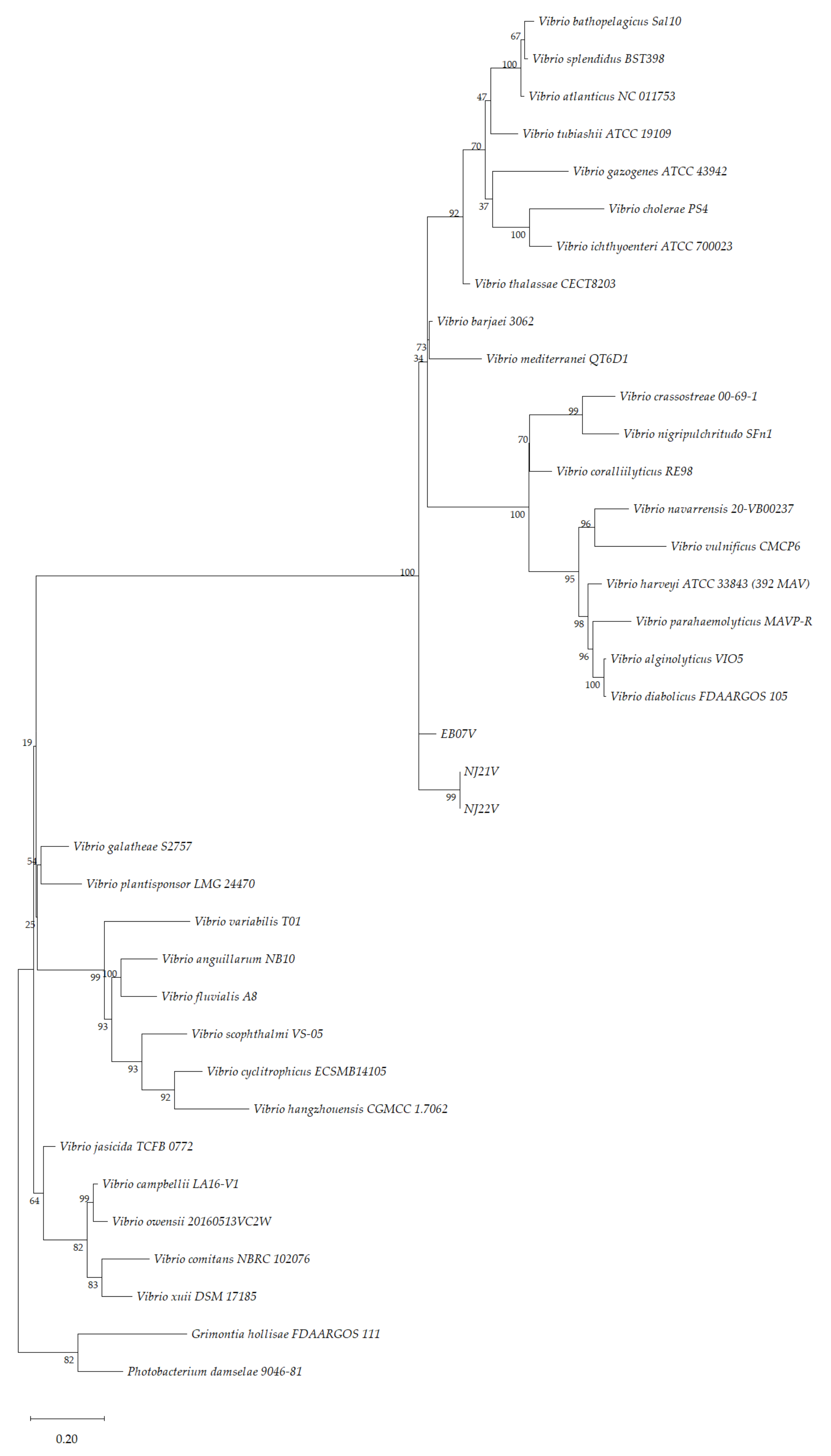
3.4. Phylogenetic Analysis

After comparing the 16S rRNA sequences of the three isolates to those publicly available (BLAST, https://blast.ncbi.nlm.nih.gov/Blast.cgi, accessed on 21 December 2021), the isolates were identified as belonging to the Vibrio genus, but it was not possible to identify them to the species level. The isolate EB7V shared 99.47% of similarity with V. hangzhouensis and V. mediterranei; NJ21V shared 98.68% of similarity with several Vibrio spp. (e.g., V. xuii, V. parahaemolyticus, V. galatheae); and NJ22V shared 98.71% of similarity with some Vibrio spp. (e.g., V. xuii, V. parahaemolyticus, V. scophthalmi). The genus Vibrio contains a large number of closely related species sharing a high level of sequence similarity with just 0.1 or 0.2% difference in the nucleotide sequence of 16S rRNA [84]. Based on that, the use of other genes (e.g., rpoA, recA, pyrH, and atpA) to discriminate closely related species has been proposed [85,86]. Four other house-keeping genes were chosen, recA, rpoB, groEL, and dnaJ that were sequenced and aligned. The gene groEL, for example, was reported to be extremely conserved in nature [87] and was already used to identify Vibrio isolates [88].
The phylogenetic tree for concatenated sequences for the house-keeping genes was constructed using the ML method (Figure 2). The MLSA is based on partial sequences of the four house-keeping genes for 32 Vibrio spp., Photobacterium damselae 9046-81 and Grimontia hollisae FDAARGOS 111 (used as outgroup) and the three isolates. MLSA has been proposed as a useful technique for the identification of Vibrio isolates and for studying the phylogeny in this genus [85,89]. Moreover, this technique has been utilised for the identification of some new Vibrio species, such as V. gigantis and V. crassostreae [90,91] and, more recently for V. barjaei and V. nitrifigilis [92,93].
However, the three isolates clustered in the genus Vibrio but do not cluster with any Vibrio clade, which suggests that the three isolates may represent new undescribed species of the genus Vibrio. In the case of NJ21V and NJ22V, it appears that they belong to the same species, although being different strains. Further studies using these isolates are needed to prove this statement.

4. Conclusions

In this study, the presence of the mobilisable colistin resistance gene mcr-1 in three Vibrio spp. (EB07V, NJ21V, and NJ22V), isolated in microbial communities from clams, highlights a potential threat to public health. Clams are an appreciated food delicacy and colistin is one of the last line of defence antibiotics used for clinical treatment [20]. One of the three mcr-1 positives isolates, EB07V, also carried the gene chiA, responsible for the invasion of tissues of chitin-containing organisms, which may increase its pathogenic potential. The location of the mcr-1 resistance gene appears to be chromosomal which indicates a low potential for mobilisation. Biochemical analysis and MLSA on the basis of four protein-coding gene sequences (recA, rpoB, groEL and dnaJ) grouped the isolates into the genus Vibrio but distinguished them as different from other species of Vibrio, suggesting we might be in the presence of new species. To corroborate this statement further investigation is required. However, the fact that the microbial communities in the environment possess the colistin mobilise resistant determinant mcr-1, highlights the importance of this study.

Supplementary Materials

The following are available online at https://www.mdpi.com/article/10.3390/microorganisms10020394/s1, Table S1: Primers sequences used to amplify house-keeping, antimicrobial resistances and virulence genes. Table S2: Accession numbers from NCBI of the species used to performed MLSA on MEGA X software. Table S3: Antimicrobial susceptibility test using colistin, imipenem, cefotaxime, meropenem and ciprofloxacin disks. The halo measurements displayed in mm. Table S4: Antimicrobial resistances genes search on all the isolates. The first class of antibiotics searched was polymyxin (mcr-1, mcr-2, mcr-3, mcr-4, mcr-5, mcr-6, mcr-7, mcr-8 and mcr-9). – indicates non detected gene and + indicates detected gene. Table S5: Antimicrobial resistances genes search on all the isolates. The second class was β-lactams (blaIMP, blaVIM, blaKPC, blaNDM, blaOXA, blaCTX-M, blaSHV and blaTEM). – indicates non detected gene and + indicates detected gene. Table S6: Antimicrobial resistances genes search on all the isolates. The third antimicrobial class searched was quinolone (qnrA, qnrB, qnrC, qnrD, qnrS and qepA). – indicates non detected gene and + indicates detected gene. Figure S1: Plasmid curing of the isolates EB07V, NJ21V and NJ22V. The number of CFU was determined in MHA plates with and without colistin. References [95,96,97,98,99,100,101,102,103,104,105,106,107,108,109,110,111,112,113,114,115,116,117,118] have been cited in the Supplementary materials.

Author Contributions

Conceptualization, T.B. and M.J.C.; Investigation, C.V., C.C., M.S., C.C.C.R.d.C., T.B. and M.J.C. Resources, T.B. and M.J.C.; Supervision, M.J.C.; Writing—original draft, C.C.; Writing—review and editing, C.C., C.C.C.R.d.C. and M.J.C. All authors have read and agreed to the published version of the manuscript.

Funding

This study had the support of Fundação para a Ciência e a Tecnologia (FCT) through the Strategic Project UID/MAR/04292/2020 granted to MARE and grant agreement nº POCI-01-0247-FEDER-035234; AlgaValor, Portugal 2020 program.

Conflicts of Interest

The authors declare no conflict of interest.

References

  1. Farmer, J.J. The Family Vibrionaceae. In The Prokaryotes; Dworkin, M., Falkow, S., Rosenberg, E., Schleifer, K.H., Stackebrandt, E., Eds.; Springer: New York, NY, USA, 2006. [Google Scholar] [CrossRef]
  2. Parte, A.C.; Carbasse, J.S.; Meier-Kolthoff, J.P.; Reimer, L.C.; Göker, M. List of Prokaryotic names with Standing in Nomenclature (LPSN) moves to the DSMZ. Int. J. Syst. Evol. Microbiol. 2020, 70, 5607–5612. [Google Scholar] [CrossRef] [PubMed]
  3. Le Roux, F. Environmental vibrios: «A walk on the wild side». Environ. Microbiol. Rep. 2017, 9, 27–29. [Google Scholar] [CrossRef] [PubMed] [Green Version]
  4. Gennari, M.; Ghidini, V.; Caburlotto, G.; Lleo, M.M. Virulence genes and pathogenicity islands in environmental Vibrio strains nonpathogenic to humans. FEMS Microbiol. Ecol. 2012, 82, 563–573. [Google Scholar] [CrossRef] [PubMed] [Green Version]
  5. Baker-Austin, C.; Trinanes, J.; Gonzalez-Escalona, N.; Martinez-Urtaza, J. Non-Cholera Vibrios: The Microbial Barometer of Climate Change. Trends Microbiol. 2017, 25, 76–84. [Google Scholar] [CrossRef] [PubMed] [Green Version]
  6. Faruque, M.S.; Nair, G.B. Epidemiology. In The Biology of Vibrios; Thompson, F.L., Austin, B., Swings, J., Eds.; ASM Press: Washington, DC, USA, 2006; pp. 385–400. [Google Scholar]
  7. Baker-Austin, C.; Oliver, J.D.; Alam, M.; Ali, A.; Waldor, M.K.; Qadri, F.; Martinez-Urtaza, J. Vibrio spp. infections. Nat. Rev. Dis. Primers 2018, 4, 1–19. [Google Scholar] [CrossRef] [PubMed]
  8. Ina-Salwany, M.Y.; Al-Saari, N.; Mohamad, A.; Mursidi, F.A.; Mohd-Aris, A.; Amal, M.N.A.; Kasai, H.; Mino, S.; Sawabe, T.; Zamri-Saad, M. Vibriosis in Fish: A Review on Disease Development and Prevention. J. Aquat. Anim. Health 2019, 31, 3–22. [Google Scholar] [CrossRef] [PubMed]
  9. CDC. Chapter 6-Isolation and Identification of Vibrio cholerae. In Laboratory Methods for the Diagnosis of Epidemic Dysentery and Cholera; CDC: Atlanta, Georgia, 1999. [Google Scholar]
  10. Alcaide, E.; Blasco, M.-D.; Esteve, C. Occurrence of drug-resistant bacteria in two European eel farms. Appl. Environ. Microbiol. 2005, 71, 3348–3350. [Google Scholar] [CrossRef] [Green Version]
  11. Pedersen, K.; Skall, H.F.; Lassen-Nielsen, A.M.; Nielsen, T.F.; Henriksen, N.H.; Olesen, N.J. Surveillance of health status on eight marine rainbow trout, Oncorhynchus mykiss (Walbaum), farms in Denmark in 2006. J. Fish. Dis. 2008, 31, 659–667. [Google Scholar] [CrossRef]
  12. Oliveira, J.; Reygaert, W.C. Gram Negative Bacteria; StatPearls Publishing: Treasure Island, FL, USA, 2019. [Google Scholar]
  13. Hertz, F.B.; Nielsen, J.B.; Schønning, K.; Littauer, P.; Knudsen, J.D.; Løbner-Olesen, A.; Frimodt-Møller, N. Population structure of Drug-Susceptible, -Resistant and ESBL-producing Escherichia coli from Community-Acquired Urinary Tract Infections. BMC Microbiol. 2016, 16, 63. [Google Scholar] [CrossRef] [Green Version]
  14. Kontopidou, F.; Giamarellou, H.; Katerelos, P.; Maragos, A.; Kioumis, I.; Trikka-Graphakos, E.; Valakis, C.; Maltezou, H.C. Infections caused by carbapenem-resistant Klebsiella pneumoniae among patients in intensive care units in Greece: A multi-centre study on clinical outcome and therapeutic options. CMI 2014, 20, O117–O123. [Google Scholar] [CrossRef] [PubMed] [Green Version]
  15. Awad, A.I.; Eltayeb, I.B. Self-medication practices with antibiotics and antimalarials among Sudanese undergraduate university students. Ann. Pharmacother. 2007, 41, 1249–1255. [Google Scholar] [CrossRef] [PubMed]
  16. Ruuskanen, M.; Muurinen, J.; Meierjohan, A.; Pärnänen, K.; Tamminen, M.; Lyra, C.; Kronberg, L.; Virta, M. Fertilizing with animal manure disseminates antibiotic resistance genes to the farm environment. J. Environ. Qual. 2016, 45, 488–493. [Google Scholar] [CrossRef] [PubMed]
  17. Levy, S.B.; Marshall, B. Antibacterial resistance worldwide: Causes, challenges and responses. Nat. Med. 2004, 10, S122–S129. [Google Scholar] [CrossRef] [PubMed]
  18. Paterson, D.L.; Harris, P.N.A. Colistin resistance: A major breach in our last line of defence. Lancet Infect. Dis. 2016, 16, 132–133. [Google Scholar] [CrossRef]
  19. O’Neill, J. Tackling Drug-Resistant Infections Globally: Final Report and Recommendations. AMR Rev. 2016. Available online: http://amr-review.org/sites/default/files/160518_Final%20paper_with%20cover.pdf (accessed on 22 December 2021).
  20. Jeannot, K.; Bolard, A.; Plesiat, P. Resistance to polymyxins in Gram-negative organisms. Int. J. Antimicrob. Agents 2017, 49, 526–535. [Google Scholar] [CrossRef] [PubMed]
  21. Rabanal, F.; Cajal, Y. Recent advances and perspectives in the design and development of polymyxins. Nat. Prod. Rep. 2017, 34, 886–908. [Google Scholar] [CrossRef] [PubMed]
  22. Kempf, I.; Fleury, M.A.; Drider, D.; Bruneau, M.; Sanders, P.; Chauvin, C.; Madec, J.-Y.; Jouy, E. What do we know about resistance to colistin in Enterobacteriaceae in avian and pig production in Europe? Int. J. Antimicrob. Agents 2013, 42, 379–383. [Google Scholar] [CrossRef] [PubMed]
  23. Lee, J.-Y.; Chung, E.S.; Na, I.Y.; Kim, H.; Shin, D.; Ko, K.S. Development of colistin resistance in pmrA-, phoP-, parR-and cprR-inactivated mutants of Pseudomonas aeruginosa. J. Antimicrob. Chemother. 2014, 69, 2966–2971. [Google Scholar] [CrossRef] [PubMed] [Green Version]
  24. Olaitan, A.O.; Morand, S.; Rolain, J.-M. Mechanisms of polymyxin resistance: Acquired and intrinsic resistance in bacteria. Front. Microbiol. 2014, 5, 643. [Google Scholar] [CrossRef] [PubMed] [Green Version]
  25. Liu, Y.-Y.; Wang, Y.; Walsh, T.R.; Yi, L.-X.; Zhang, R.; Spencer, J.; Doi, Y.; Tian, G.; Dong, B.; Huang, X. Emergence of plasmid-mediated colistin resistance mechanism MCR-1 in animals and human beings in China: A microbiological and molecular biological study. Lancet Infec. Dis. 2016, 16, 161–168. [Google Scholar] [CrossRef]
  26. Anjum, M.F.; Duggett, N.A.; AbuOun, M.; Randall, L.; Nunez-Garcia, J.; Ellis, R.J.; Rogers, J.; Horton, R.; Brena, C.; Williamson, S. Colistin resistance in Salmonella and Escherichia coli isolates from a pig farm in Great Britain. J. Antimicrob. Chemother. 2016, 71, 2306–2313. [Google Scholar] [CrossRef] [PubMed] [Green Version]
  27. Irrgang, A.; Roschanski, N.; Tenhagen, B.-A.; Grobbel, M.; Skladnikiewicz-Ziemer, T.; Thomas, K.; Roesler, U.; Kaesbohrer, A. Prevalence of mcr-1 in E. coli from livestock and food in Germany, 2010–2015. PLoS ONE 2016, 11, e0159863. [Google Scholar] [CrossRef] [PubMed]
  28. Sánchez-Benito, R.; Iglesias, M.R.; Quijada, N.M.; Campos, M.J.; Ugarte-Ruiz, M.; Hernández, M.; Pazos, C.; Rodríguez-Lázaro, D.; Garduño, E.; Domínguez, L.; et al. Escherichia coli ST167 carrying plasmid mobilisable mcr-1 and bla(CTX-M-15) resistance determinants isolated from a human respiratory infection. Int. J. Antimicrob. Agents. 2017, 50, 285–286. [Google Scholar] [CrossRef]
  29. Xavier, B.B.; Lammens, C.; Butaye, P.; Goossens, H.; Malhotra-Kumar, S. Complete sequence of an IncFII plasmid harbouring the colistin resistance gene mcr-1 isolated from Belgian pig farms. J. Antimicrob. Chemother. 2016, 71, 2342–2344. [Google Scholar] [CrossRef] [PubMed] [Green Version]
  30. Li, R.; Lu, X.; Munir, A.; Abdullah, S.; Liu, Y.; Xiao, X.; Wang, Z.; Mohsin, M. Widespread prevalence and molecular epidemiology of tet(X4) and mcr-1 harboring Escherichia coli isolated from chickens in Pakistan. Sci. Total Environ. 2021, 806, 150689. [Google Scholar] [CrossRef] [PubMed]
  31. Walsh, T.R.; Wu, Y. China bans colistin as a feed additive for animals. Lancet Infect. Dis. 2016, 16. [Google Scholar] [CrossRef]
  32. McGann, P.; Snesrud, E.; Maybank, R.; Corey, B.; Ong, A.C.; Clifford, R.; Hinkle, M.; Whitman, T.; Lesho, E.; Schaecher, K.E. Escherichia coli Harboring mcr-1 and blaCTX-M on a Novel IncF Plasmid: First Report of mcr-1 in the United States. Antimicrob. Agents Chemother. 2016, 60, 4420–4421. [Google Scholar] [CrossRef] [PubMed] [Green Version]
  33. Elnahriry, S.S.; Khalifa, H.O.; Soliman, A.M.; Ahmed, A.M.; Hussein, A.M.; Shimamoto, T.; Shimamoto, T. Emergence of Plasmid-Mediated Colistin Resistance Gene mcr-1 in a Clinical Escherichia coli Isolate from Egypt. Antimicrob. Agents. Chemother. 2016, 60, 3249–3250. [Google Scholar] [CrossRef] [Green Version]
  34. Khedher, M.B.; Baron, S.A.; Riziki, T.; Ruimy, R.; Raoult, D.; Diene, S.M.; Rolain, J.-M. Massive analysis of 64,628 bacterial genomes to decipher water reservoir and origin of mobile colistin resistance genes: Is there another role for these enzymes? Sci. Rep. 2020, 10, 1–10. [Google Scholar] [CrossRef] [PubMed] [Green Version]
  35. Kieffer, N.; Nordmann, P.; Poirel, L. Moraxella Species as Potential Sources of MCR-Like Polymyxin Resistance Determinants. Antimicrob. Agents. Chemother. 2017, 61, e00129-17. [Google Scholar] [CrossRef] [PubMed] [Green Version]
  36. Lei, T.; Zhang, J.; Jiang, F.; He, M.; Zeng, H.; Chen, M.; Wu, S.; Wang, J.; Ding, Y.; Wu, Q. First detection of the plasmid-mediated colistin resistance gene mcr-1 in virulent Vibrio parahaemolyticus. Int. J. Food Microbiol. 2019, 308, 108290. [Google Scholar] [CrossRef]
  37. Romanenko, L.A.; Uchino, M.; Kalinovskaya, N.I.; Mikhailov, V.V. Isolation, phylogenetic analysis and screening of marine mollusc-associated bacteria for antimicrobial, hemolytic and surface activities. Microbiol. Res. 2008, 163, 633–644. [Google Scholar] [CrossRef] [PubMed]
  38. Richards, G.P. Microbial purification of shellfish: A review of depuration and relaying. J. Food Prot. 1988, 51, 218–251. [Google Scholar] [CrossRef] [PubMed]
  39. Robertson, L. The potential for marine bivalve shellfish to act as transmission vehicles for outbreaks of protozoan infections in humans: A review. Int. J. Food Microbiol. 2007, 120, 201–216. [Google Scholar] [CrossRef] [PubMed]
  40. EUCAST. Antimicrobial Susceptibility Testing—EUCAST Disk Diffusion Method. Available online: https://www.eucast.org/fileadmin/src/media/PDFs/EUCAST_files/Disk_test_documents/2021_manuals/Manual_v_9.0_EUCAST_Disk_Test_2021.pdf (accessed on 21 January 2021).
  41. EUCAST. Broth microdilution—EUCAST Reading Guide v 4.0. Available online: https://www.eucast.org/ast_of_bacteria/mic_determination/?no_cache=1 (accessed on 14 January 2022).
  42. Coico, R. Gram staining. Curr. Protoc. 2006. [Google Scholar] [CrossRef]
  43. Suslow, T.V.; Schroth, M.N.; Isaka, M. Application of a rapid method for Gram differentiation of plant pathogenic and saprophytic bacteria without staining. Phytopathology 1982, 72, 917–918. [Google Scholar] [CrossRef]
  44. Kumar, S.; Stecher, G.; Li, M.; Knyaz, C.; Tamura, K. MEGA X: Molecular Evolutionary Genetics Analysis across Computing Platforms. Mol. Biol. Evol. 2018, 35, 1547–1549. [Google Scholar] [CrossRef]
  45. Felsenstein, J. Confidence limits on phylogenies: An approach using the bootstrap. Evolution 1985, 39, 783–791. [Google Scholar] [CrossRef]
  46. Letchumanan, V.; Chan, K.-G.; Lee, L.-H. An insight of traditional plasmid curing in Vibrio species. Front. Microbiol. 2015, 6. [Google Scholar] [CrossRef]
  47. Law, E.U. Microbiological Criteria for Foodstuffs. Available online: https://eur-lex.europa.eu/legal-content/EN/TXT/?uri=CELEX%3A02005R2073-20200308 (accessed on 10 August 2021).
  48. Gerba, C.P.; Goyal, S.M.; LaBelle, R.L.; Cech, I.; Bodgan, G.F. Failure of indicator bacteria to reflect the occurrence of enteroviruses in marine waters. Am. J. Public Health. 1979, 69, 1116–1119. [Google Scholar] [CrossRef] [Green Version]
  49. Formiga-Cruz, M.; Allard, A.K.; Conden-Hansson, A.-C.; Henshilwood, K.; Hernroth, B.E.; Jofre, J.; Lees, D.N.; Lucena, F.; Papapetropoulou, M.; Rangdale, R.E. Evaluation of potential indicators of viral contamination in shellfish and their applicability to diverse geographical areas. Appl. Environ. Microbiol. 2003, 69, 1556–1563. [Google Scholar] [CrossRef] [Green Version]
  50. Høi, L.; Dalsgaard, I.; Dalsgaard, A. Improved isolation of Vibrio vulnificus from seawater and sediment with cellobiose-colistin agar. Appl. Environ. Microbiol. 1998, 64, 1721–1724. [Google Scholar] [CrossRef] [Green Version]
  51. Mondal, M.; Nag, D.; Koley, H.; Saha, D.R.; Chatterjee, N.S. The Vibrio cholerae extracellular chitinase ChiA2 is important for survival and pathogenesis in the host intestine. PLoS ONE 2014, 9, e103119. [Google Scholar] [CrossRef] [PubMed] [Green Version]
  52. Zobell, C.E.; Rittenberg, S.C. The occurrence and characteristics of chtinoclastic bacteria in the sea. J. Bacteriol. 1938, 35, 275–287. [Google Scholar] [CrossRef] [Green Version]
  53. Wortman, A.T.; Somerville, C.C.; Colwell, R.R. Chitinase determinants of Vibrio vulnificus: Gene cloning and applications of a chitinase probe. Appl. Environ. Microbiol. 1986, 52, 142–145. [Google Scholar] [CrossRef] [PubMed] [Green Version]
  54. Soto-GiI, R.W.; Zyskind, J.W. Chitin, Chitosan, and Related Enzymes; Zikakis, J., Ed.; Academic Press Inc.: New York, NY, USA, 1984. [Google Scholar]
  55. Hirono, I.; Yamashita, M.; Aoki, T. Molecular cloning of chitinase genes from Vibrio anguillarum and V. parahaemolyticus. J. Appl. Microbiol. 1998, 84, 1175–1178. [Google Scholar] [CrossRef]
  56. Huq, A.; Small, E.B.; West, P.A.; Huq, M.I.; Rahman, R.; Colwell, R.R. Ecological relationships between Vibrio cholerae and planktonic crustacean copepods. Appl. Environ. Microbiol. 1983, 45, 275–283. [Google Scholar] [CrossRef] [Green Version]
  57. Pruzzo, C.; Vezzulli, L.; Colwell, R.R. Global impact of Vibrio cholerae interactions with chitin. Environ. Microbiol. 2008, 10, 1400–1410. [Google Scholar] [CrossRef] [PubMed]
  58. Kimmerer, W.J.; Gartside, E.; Orsi, J.J. Predation by an introduced clam as the likely cause of substantial declines in zooplankton of San Francisco Bay. Mar. Ecol. Prog. Ser. 1994, 113, 81–93. [Google Scholar] [CrossRef]
  59. Kimmerer, W.J.; Lougee, L. Bivalve grazing causes substantial mortality to an estuarine copepod population. J. Exp. Mar. Biol. Ecol. 2015, 473, 53–63. [Google Scholar] [CrossRef]
  60. Pace, M.L.; Findlay, S.E.G.; Fischer, D. Effects of an invasive bivalve on the zooplankton community of the Hudson River. Freshw. Biol. 1998, 39, 103–116. [Google Scholar] [CrossRef]
  61. Sun, J.; Fang, L.-X.; Wu, Z.; Deng, H.; Yang, R.-S.; Li, X.-P.; Li, S.-M.; Liao, X.-P.; Feng, Y.; Liu, Y.-H. Genetic analysis of the IncX4 plasmids: Implications for a unique pattern in the mcr-1 acquisition. Sci. Rep. 2017, 7, 424. [Google Scholar] [CrossRef] [PubMed]
  62. Yu, Z.; Qin, W.; Lin, J.; Fang, S.; Qiu, J. Antibacterial Mechanisms of Polymyxin and Bacterial Resistance. Biomed. Res. Int. 2015, 2015, 679109. [Google Scholar] [CrossRef] [PubMed]
  63. Bakour, S.; Olaitan, A.O.; Ammari, H.; Touati, A.; Saoudi, S.; Saoudi, K.; Rolain, J.M. Emergence of Colistin- and Carbapenem-Resistant Acinetobacter baumannii ST2 Clinical Isolate in Algeria: First Case Report. Microb. Drug. Resist. 2015, 21, 279–285. [Google Scholar] [CrossRef] [PubMed]
  64. Nishimura, Y.; Eguchi, T. Biosynthesis of archaeal membrane lipids: Digeranylgeranylglycerophospholipid reductase of the thermoacidophilic archaeon Thermoplasma acidophilum. J. Biochem. 2006, 139, 1073–1081. [Google Scholar] [CrossRef] [PubMed]
  65. Baron, S.; Hadjadj, L.; Rolain, J.M.; Olaitan, A.O. Molecular mechanisms of polymyxin resistance: Knowns and unknowns. Int. J. Antimicrob. Agents. 2016, 48, 583–591. [Google Scholar] [CrossRef]
  66. Mambelli, L.I.; Teixeira, S.F.; Jorge, S.D.; Kawamura, B.; Meneguelo, R.; Barbuto, J.A.M.; de Azevedo, R.A.; Ferreira, A.K. Phosphoethanolamine induces caspase-independent cell death by reducing the expression of C-RAF and inhibits tumor growth in human melanoma model. Biomed. Pharmacother. 2018, 103, 18–28. [Google Scholar] [CrossRef]
  67. Massad, G.; Oliver, J.D. New selective and differential medium for Vibrio cholerae and Vibrio vulnificus. Appl. Environ. Microbiol. 1987, 53, 2262–2264. [Google Scholar] [CrossRef] [Green Version]
  68. Hankins, J.V.; Madsen, J.A.; Giles, D.K.; Brodbelt, J.S.; Trent, M.S. Amino acid addition to Vibrio cholerae LPS establishes a link between surface remodeling in Gram-positive and Gram-negative bacteria. Proc. Natl. Acad. Sci. USA 2012, 109, 8722–8727. [Google Scholar] [CrossRef] [Green Version]
  69. Henderson, J.C.; Herrera, C.M.; Trent, M.S. AlmG, responsible for polymyxin resistance in pandemic Vibrio cholerae, is a glycyltransferase distantly related to lipid A late acyltransferases. J. Biol. Chem. 2017, 292, 21205–21215. [Google Scholar] [CrossRef] [PubMed] [Green Version]
  70. Herrera, C.M.; Henderson, J.C.; Crofts, A.A.; Trent, M.S. Novel coordination of lipopolysaccharide modifications in Vibrio cholerae promotes CAMP resistance. Mol. Microbiol. 2017, 106, 582–596. [Google Scholar] [CrossRef] [PubMed] [Green Version]
  71. Herrera, C.M.; Crofts, A.A.; Henderson, J.C.; Pingali, S.C.; Davies, B.W.; Trent, M.S. The Vibrio cholerae VprA-VprB two-component system controls virulence through endotoxin modification. mBio 2014, 5, e02283-14. [Google Scholar] [CrossRef] [PubMed] [Green Version]
  72. Bina, X.R.; Howard, M.F.; Ante, V.M.; Bina, J.E. Vibrio cholerae LeuO links the ToxR regulon to expression of lipid A remodeling genes. Infect. Immun. 2016, 84, 3161–3171. [Google Scholar] [CrossRef] [Green Version]
  73. Zhang, H.; Srinivas, S.; Xu, Y.; Wei, W.; Feng, Y. Genetic and Biochemical Mechanisms for Bacterial Lipid A Modifiers Associated with Polymyxin Resistance. Trends Biochem. Sci. 2019, 44, 973–988. [Google Scholar] [CrossRef]
  74. Chen, Y.C.; Chuang, Y.C.; Chang, C.C.; Jeang, C.L.; Chang, M.C. A K+ uptake protein, TrkA, is required for serum, protamine, and polymyxin B resistance in Vibrio vulnificus. Infect. Immun. 2004, 72, 629–636. [Google Scholar] [CrossRef] [Green Version]
  75. Tantillo, G.M.; Fontanarosa, M.; Di Pinto, A.; Musti, M. Updated perspectives on emerging vibrios associated with human infections. Lett. Appl. Microbiol. 2004, 39, 117–126. [Google Scholar] [CrossRef] [PubMed]
  76. Percival, S.L.; Williams, D.W. Chapter Twelve—Vibrio. In Microbiology of Waterborne Diseases, 2nd ed.; Percival, S.L., Yates, M.V., Williams, D.W., Chalmers, R.M., Gray, N.F., Eds.; Academic Press: London, UK, 2014; pp. 237–248. [Google Scholar]
  77. Kunitsky, C.; Osterhout, G.; Sasser, M. Identificatiom of microorganisms using fatty acid methyl ester (FAME) analysis and the MIDI Sherlock® microbial identification system. In Encyclopedia of Rapid Microbiological Methods; Miller, M.J., Ed.; PDA: Bethesda, MD, USA; DHI Publishing LLC: River Grove, IL, USA,, 2006; Volume 3. [Google Scholar]
  78. de Carvalho, C.C.C.R.; Caramujo, M.J. The various roles of fatty acids. Molecules 2018, 23, 2583. [Google Scholar] [CrossRef] [PubMed] [Green Version]
  79. Hoffmann, M.; Fischer, M.; Whittaker, P. Evaluating the use of fatty acid profiles to identify deep-sea Vibrio isolates. Food Chem. 2010, 122, 943–950. [Google Scholar] [CrossRef]
  80. Balcázar, J.L.; Planas, M.; Pintado, J. Vibrio inhibens sp. nov., a novel bacterium with inhibitory activity against Vibrio species. J. Antibiot. 2012, 65, 301–305. [Google Scholar] [CrossRef] [PubMed]
  81. Urbanczyk, Y.; Ogura, Y.; Hayashi, T.; Urbanczyk, H. Genomic evidence that Vibrio inhibens is a heterotypic synonym of Vibrio jasicida. Int. J. Syst. Evol. Microbiol. 2016, 66, 3214–3218. [Google Scholar] [CrossRef] [PubMed]
  82. Hamamoto, T.; Takata, N.; Kudo, T.; Horikoshi, K. Effect of temperature and growth phase on fatty acid composition of the psychrophilic Vibrio sp. strain no. 5710. FEMS Microbiol. Lett. 1994, 119, 77–81. [Google Scholar] [CrossRef]
  83. Estupiñán, M.; Hernández, I.; Saitua, E.; Bilbao, M.E.; Mendibil, I.; Ferrer, J.; Alonso-Sáez, L. Novel Vibrio spp. strains producing omega-3 fatty acids isolated from coastal seawater. Mar. Drugs 2020, 18, 99. [Google Scholar] [CrossRef] [PubMed] [Green Version]
  84. Montieri, S.; Suffredini, E.; Ciccozzi, M.; Croci, L. Phylogenetic and evolutionary analysis of Vibrio parahaemolyticus and Vibrio alginolyticus isolates based on toxR gene sequence. New Microbiol. 2010, 33, 359–372. [Google Scholar] [PubMed]
  85. Thompson, F.; Gevers, D.; Thompson, C.; Dawyndt, P.; Naser, S.; Hoste, B.; Munn, C.; Swings, J. Phylogeny and molecular identification of vibrios on the basis of multilocus sequence analysis. App. Environ. Microbiol. 2005, 71, 5107–5115. [Google Scholar] [CrossRef] [PubMed] [Green Version]
  86. Thompson, C.C.; Thompson, F.L.; Vicente, A.C.P.; Swings, J. Phylogenetic analysis of vibrios and related species by means of atpA gene sequences. Int. J. Syst. Evol. Microbiol. 2007, 57, 2480–2484. [Google Scholar] [CrossRef] [PubMed]
  87. Blaiotta, G.; Fusco, V.; Ercolini, D.; Aponte, M.; Pepe, O.; Villani, F. Lactobacillus strain diversity based on partial hsp60 gene sequences and design of PCR-restriction fragment length polymorphism assays for species identification and differentiation. Appl. Environ. Microbiol. 2008, 74, 208–215. [Google Scholar] [CrossRef] [Green Version]
  88. Nishibuchi, M. Molecular Identification. In The biology of Vibrios; Thompson, F.L., Austin, B., Swings, J., Eds.; ASM Press: Washington, DC, USA, 2006. [Google Scholar]
  89. Beaz-Hidalgo, R.; Doce, A.; Pascual, J.; Toranzo, A.E.; Romalde, J.L. Vibrio gallaecicus sp. nov. isolated from cultured clams in north-western Spain. Syst. Appl. Microbiol. 2009, 32, 111–117. [Google Scholar] [CrossRef] [PubMed]
  90. Faury, N.; Saulnier, D.; Thompson, F.; Gay, M.; Swings, J.; Le Roux, F. Vibrio crassostreae sp. nov., isolated from the haemolymph of oysters (Crassostrea gigas). Int. J. Syst. Evol. Microbiol. 2004, 54, 2137–2140. [Google Scholar] [CrossRef] [PubMed] [Green Version]
  91. Le Roux, F.; Goubet, A.; Thompson, F.; Faury, N.; Gay, M.; Swings, J.; Saulnier, D. Vibrio gigantis sp. nov., isolated from the haemolymph of cultured oysters (Crassostrea gigas). Int. J. Syst. Evol. Microbiol. 2005, 55, 2251–2255. [Google Scholar] [CrossRef] [PubMed]
  92. Dubert, J.; Balboa, S.; Regueira, M.; González-Castillo, A.; Gómez-Gil, B.; Romalde, J.L. Vibrio barjaei sp. nov., a new species of the Mediterranei clade isolated in a shellfish hatchery. Syst. Appl. Microbiol. 2016, 39, 553–556. [Google Scholar] [CrossRef] [PubMed]
  93. Huang, W.-S.; Wang, L.-T.; Chen, J.-S.; Chen, Y.-T.; Wei, S.T.-S.; Chiang, Y.-R.; Wang, P.-L.; Lee, T.-H.; Lin, S.-T.; Huang, L.; et al. Vibrio nitrifigilis sp. nov., a marine nitrogen-fixing bacterium isolated from the lagoon sediment of an islet inside an atoll. Anton. Leeuw. 2021, 114, 933–945. [Google Scholar] [CrossRef]
  94. Kimura, M. A simple method for estimating evolutionary rates of base substitutions through comparative studies of nucleotide sequences. J. Mol. Evol. 1980, 16, 111–120. [Google Scholar] [CrossRef]
  95. Sawabe, T.; Kita-Tsukamoto, K.; Thompson, F.L. Inferring the evolutionary history of vibrios by means of multilocus sequence analysis. J. Bacteriol. 2007, 189, 7932–7936. [Google Scholar] [CrossRef] [Green Version]
  96. Diancourt, L.; Passet, V.; Verhoef, J.; Grimont, P.A.; Brisse, S. Multilocus sequence typing of Klebsiella pneumoniae nosocomial isolates. J. Clin. Microbiol. 2005, 43, 4178. [Google Scholar] [CrossRef] [Green Version]
  97. Hossain, M.T.; Kim, Y.-R.; Kong, I.-S. PCR–restriction fragment length polymorphism analysis using groEL gene to differentiate pathogenic Vibrio species. Diagn. Microbiol. Infect. Dis. 2014, 78, 9–11. [Google Scholar] [CrossRef] [PubMed]
  98. Lane, D. 16S/23S rRNA Sequencing, Nucleic Acid Techniques; Stackebrandt, E., Goodfellow, M., Eds.; John Wiley and Sons: New York, NY, USA, 1991. [Google Scholar]
  99. Nhung, P.H.; Ohkusu, K.; Mishima, N.; Noda, M.; Shah, M.M.; Sun, X.; Hayashi, M.; Ezaki, T. Phylogeny and species identification of the family Enterobacteriaceae based on dnaJ sequences. Diagn. Microbiol. Infect. Dis. 2007, 58, 153–161. [Google Scholar] [CrossRef] [PubMed]
  100. Rebelo, A.R.; Bortolaia, V.; Kjeldgaard, J.S.; Pedersen, S.K.; Leekitcharoenphon, P.; Hansen, I.M.; Guerra, B.; Malorny, B.; Borowiak, M.; Hammerl, J.A.; et al. Multiplex PCR for detection of plasmid-mediated colistin resistance determinants, mcr-1, mcr-2, mcr-3, mcr-4 and mcr-5 for surveillance purposes. Eurosurveillance 2018, 23, 17–00672. [Google Scholar] [CrossRef]
  101. Wang, X.; Wang, Y.; Zhou, Y.; Li, J.; Yin, W.; Wang, S.; Zhang, S.; Shen, J.; Shen, Z.; Wang, Y. Emergence of a novel mobile colistin resistance gene, mcr-8, in NDM-producing Klebsiella pneumoniae. Emerg. Microbes Infect. 2018, 7, 122. [Google Scholar] [CrossRef] [PubMed] [Green Version]
  102. Monstein, H.J.; Ostholm-Balkhed, A.; Nilsson, M.V.; Nilsson, M.; Dornbusch, K.; Nilsson, L.E. Multiplex PCR amplification assay for the detection of blaSHV, blaTEM and blaCTX-M genes in Enterobacteriaceae. APMIS 2007, 115, 1400–1408. [Google Scholar] [CrossRef]
  103. Mulvey, M.R.; Soule, G.; Boyd, D.; Demczuk, W.; Ahmed, R. Characterization of the first extended-spectrum beta-lactamase-producing Salmonella isolate identified in Canada. J. Clin. Microbiol. 2003, 41, 460–462. [Google Scholar] [CrossRef] [Green Version]
  104. Cattoir, V.; Poirel, L.; Rotimi, V.; Soussy, C.-J.; Nordmann, P. Multiplex PCR for detection of plasmid-mediated quinolone resistance qnr genes in ESBL-producing enterobacterial isolates. J. Antimicrob. Chemother. 2007, 60, 394–397. [Google Scholar] [CrossRef] [Green Version]
  105. Pribul, B.R.; Festivo, M.L.; Souza, M.M.S.; Rodrigues, D.d.P. Characterization of quinolone resistance in Salmonella spp. isolates from food products and human samples in Brazil. Braz. J. Microbiol. 2016, 47, 196–201. [Google Scholar] [CrossRef] [Green Version]
  106. Hammad, E.; Helal, R. PMQR determinants among clinical isolates of ESBL and Amp C producing Serratia marcescens in Mansoura University Hospitals: A 6-year study. Int. Arab. J. Antimicrob. Agents 2015, 5. [Google Scholar] [CrossRef] [Green Version]
  107. Ruwandeepika, H.; Defoirdt, T.; Bhowmick, P.; Shekar, M.; Bossier, P.; Karunasagar, I. Presence of typical and atypical virulence genes in vibrio isolates belonging to the Harveyi clade. J. Appl. Microbiol. 2010, 109, 888–899. [Google Scholar] [CrossRef] [PubMed]
  108. Bai, F.; Pang, L.; Qi, Z.; Chen, J.; Austin, B.; Zhang, X.-H. Distribution of five vibrio virulence-related genes among Vibrio harveyi isolates. J. Gen. Appl. Microbiol. 2008, 54, 71–78. [Google Scholar] [CrossRef] [Green Version]
  109. Saravanan, V.; Kumar, H.S.; Karunasagar, I.; Karunasagar, I. Putative virulence genes of Vibrio cholerae from seafoods and the coastal environment of Southwest India. Int. J. Food Microbiol. 2007, 119, 329–333. [Google Scholar] [CrossRef] [PubMed]
  110. Bej, A.K.; Patterson, D.P.; Brasher, C.W.; Vickery, M.C.; Jones, D.D.; Kaysner, C.A. Detection of total and hemolysin-producing Vibrio parahaemolyticus in shellfish using multiplex PCR amplification of tl, tdh and trh. J. Microbiol. Methods 1999, 36, 215–225. [Google Scholar] [CrossRef]
  111. Fields, P.; Popovic, T.; Wachsmuth, K.; Olsvik, Ø. Use of polymerase chain reaction for detection of toxigenic Vibrio cholerae O1 strains from the Latin American cholera epidemic. J. Clin. Microbiol. 1992, 30, 2118–2121. [Google Scholar] [CrossRef] [Green Version]
  112. Rivera, I.N.; Chun, J.; Huq, A.; Sack, R.B.; Colwell, R.R. Genotypes associated with virulence in environmental isolates of Vibrio cholerae. Appl. Environ. Microbiol. 2001, 67, 2421–2429. [Google Scholar] [CrossRef] [Green Version]
  113. Lee, S.E.; Kim, S.Y.; Kim, S.J.; Kim, H.S.; Shin, J.H.; Choi, S.H.; Chung, S.S.; Rhee, J.H. Direct identification of Vibrio vulnificus in clinical specimens by nested PCR. J. Clin. Microbiol. 1998, 36, 2887–2892. [Google Scholar] [CrossRef] [Green Version]
  114. Karaolis, D.K.; Johnson, J.A.; Bailey, C.C.; Boedeker, E.C.; Kaper, J.B.; Reeves, P.R. A Vibrio cholerae pathogenicity island associated with epidemic and pandemic strains. Proc. Natl. Acad. Sci. USA 1998, 95, 3134–3139. [Google Scholar] [CrossRef] [PubMed] [Green Version]
  115. Abdel-Latif, H.M.R.; Khalil, R.H.; Saad, T.T.; El-bably, R.Y. Identification and Molecular Characterization of Yersinia ruckeri isolated from mass mortalities of cultured Nile tilapia at Kafr El-sheikh governorate. Glob. J. Fish. Aquac. Res. 2014, 1, 1–17. [Google Scholar]
  116. Cai, S.H.; Lu, Y.S.; Wu, Z.H.; Jian, J.C.; Huang, Y.C. A novel multiplex PCR method for detecting virulent strains of Vibrio alginolyticus. Aquac. Res. 2009, 41, 27–34. [Google Scholar] [CrossRef]
  117. Conejero, M.J.U.; Hedreyda, C.T. PCR detection of hemolysin (vhh) gene in Vibrio harveyi. J. Gen. Appl. Microbiol. 2004, 50, 137–142. [Google Scholar] [CrossRef] [PubMed] [Green Version]
  118. Xie, Z.Y.; Hu, C.Q.; Chen, C.; Zhang, L.P.; Ren, C.H. Investigation of seven Vibrio virulence genes among Vibrio alginolyticus and Vibrio parahaemolyticus strains from the coastal mariculture systems in Guangdong, China. Lett. Appl. Microbiol. 2005, 41, 202–207. [Google Scholar] [CrossRef] [PubMed]
Figure 1. Sampling locations: (A) Óbidos Lagoon (39°24′44.1″ N 9°12′54.0″ W) and (B) Ria Formosa (37°01′05.4″ N 8°00′11.6″ W).
Microorganisms 10 00394 g001
Figure 2. Phylogenetic construction based on the concatenation of partial sequences of four house-keeping genes (recA, rpoB, groEL, and dnaJ) by the ML method algorithm and Kimura 2-parameter model [94]. The tree with the highest log likelihood (−140419,60) is shown. The percentage of trees in which the associated taxa clustered together is shown next to the branches. Initial trees for the heuristic search were obtained by applying the Neighbor-Joining method to a matrix of pairwise distances estimated using the Maximum Composite Likelihood (MCL) approach. A discrete Gamma distribution was used to model evolutionary rate differences among sites (5 categories; +G, parameter = 0.8284)). The tree was drawn to scale, with branch lengths measured in the number of substitutions per site, 0.20. There were a total of 8130 positions in the final dataset. Photobacterium damselae 9046-81 and Grimontia hollisae FDAARGOS 111 were used as the outgroup.
Microorganisms 10 00394 g002
Table 1. The CFU per g of molluscs, of Vibrio spp. and coliforms for each clam species isolated at different locations.
IsolatesÓbidos LagoonRia Formosa
R. decussatus (CFU g−1)R. phillipinarum
(CFU g−1)
R. decussatus
(CFU g−1)
Vibrionaceae1.1 × 1044.5 × 1033.8 × 103
Coliforms8.7 × 1033.5 × 1035.0 × 102
Table 2. Biochemical characterisation of the three isolates that show colistin resistance. A “+” indicates a positive reaction, whereas a “−“ denotes a negative reaction. NC—not conclusive.
TestEB07VNJ21VNJ22V
Nitrate reduction+++
Tryptophane production+++
Glucose fermentation+
Arginine dihydrolase
Urease
Esculin (β-glucosidase activity)+++
Gelatin (protease activity)+++
Para-NitroPhenyl-β-D-Galactopyranosidase (β-galactosidase activity)+++
Glucose assimilation+++
Arabinose assimilation
Mannose assimilation++
Mannitol assimilation+++
N-Acetyl-Glucosamine assimilation+++
Maltose assimilation+++
Potassium gluconate assimilation+++
Capric acid assimilation
Adipic acid assimilationNC
Malate assimilation+++
Trisodium citrate assimilation+
Phenylacetic acid assimilation
Cytochrome oxidase+++
Table 3. Fatty acid profiles, in % of total fatty acids, of the three isolates.
Fatty AcidEB07VNJ21VNJ22V
12:03.728.117.22
14:0 iso1.13
14:05.293.122.70
15:0 iso0.96
16:0 iso1.280.820.88
16:015.8525.0526.87
17:0 iso1.76
18:3 w6c14.2420.7812.17
18:03.602.882.43
Summed Feature 330.4624.5326.59
Summed Feature 821.1512.6818.10
Publisher’s Note: MDPI stays neutral with regard to jurisdictional claims in published maps and institutional affiliations.

