Communication

Aggregatibacter actinomycetemcomitans and Filifactor alocis as Associated with Periodontal Attachment Loss in a Cohort of Ghanaian Adolescents

1
Department of Odontology, Umeå University, 901 87 Umeå, Sweden
2
Dental School University of Ghana, Korle-Bu, Accra KB 460, Ghana
3
Jammerbugt Municipal Dental Service, Skolevej 1, DK-9460 Brovst, Denmark
*
Author to whom correspondence should be addressed.
Microorganisms 2022, 10(12), 2511; https://doi.org/10.3390/microorganisms10122511
Submission received: 29 November 2022 / Revised: 13 December 2022 / Accepted: 15 December 2022 / Published: 19 December 2022

Abstract

:
The aims of the present study were to document the presence of Aggregatibacter actinomyctemcomitans and the emerging oral pathogen Filifactor alocis, as well as to identify genotypes of these bacterial species with enhanced virulence. In addition, these data were analyzed in relation to periodontal pocket depth (PPD) and the progression of PPD from the sampled periodontal sites during a two-year period. Subgingival plaque samples were collected from 172 periodontal pockets of 68 Ghanaian adolescents. PPD at sampling varied from 3–14 mm and the progression from baseline, i.e., two years earlier up to 8 mm. The levels of A. actinomycetemcomitans and F. alocis were determined with quantitative PCR. The highly leukotoxic JP2-genotype of A. actinomycetemcomitans and the ftxA a gene of F. alocis, encoding a putative Repeats-in-Toxin (RTX) protein, were detected with conventional PCR. The prevalence of A. actinomycetemcomitans was 57%, and 14% of the samples contained the JP2 genotype. F. alocis was detected in 92% of the samples and the ftxA gene in 52%. The levels of these bacterial species were significantly associated with enhanced PPD and progression, with a more pronounced impact in sites positive for the JP2 genotype or the ftxA gene. Taken together, the results indicate that the presence of both A. actinomycetemcomitans and F. alocis with their RTX proteins are linked to increased PPD and progression of disease.

1. Introduction

Periodontitis is a chronic, infection-induced, inflammatory disease that degrades the tooth-supporting tissues, bone and connective tissue [1,2]. It affects mostly middle-aged and elderly individuals, but could in exceptional cases also affect adolescents [3,4,5,6]. This latter rapidly progressing form of periodontitis is classified as grade C [7], and when it affects adolescents, it was previously diagnosed as aggressive periodontitis [8]. The global prevalence of this disease indicates a wide geographical spread [9]. High incidence of this disease can be found in adolescents from the northern and western part of Africa [10,11]. Longitudinal studies on adolescents in these geographic regions indicate a substantial role of the microbiota with high prevalence of a highly leukotoxic variant of the facultative anaerobic, Gram-negative bacterium Aggregatibacter actinomycetemcomitans, namely, the JP2 genotype [12,13,14]. Reports about bacterial species other than A. actinomycetemcomitans in relation to periodontitis in young individuals in these geographic regions are scarce [10,15].
The present cohort of Ghanaian school children has previously been longitudinally examined for the presence and progression of periodontal attachment loss [13,16]. The prevalence of A. actinomycetemccomitas was detected in 56% of individuals when analyzed by cultivation and conventional PCR in pooled subgingival plaque samples [16]. Presence of the highly leukotoxic JP2 genotype was detected in 8.8% of individuals in the studied population [16]. The JP2 genotype of this bacterium has a 530-base pair deletion in the promoter region of the operon encoding the RTX protein LtxA (Leukotoxin), a toxin which is expressed at high levels by this genotype [17]. Carriage of A. actinomycetemcomitans is associated significantly with periodontal attachment loss at base line, without any additional effect in individuals where the JP2 genotype could be detected [16]. Interestingly, the clinical examination at the two-year follow-up examination showed a substantially increased odds ratio (OR = 14.3) for disease progression in the JP2 carriers compared to the individuals with no detectable A. actinomycetemcomitans [13]. The corresponding OR for the carriers of the non-JP2 genotype of A. actinomycetemcomitans was significantly lower (OR = 3.4). These associations with disease progression are in line with a previous longitudinal study in a population of Moroccan adolescents [12]. The association between A. actinomycetemcomitans and disease progression has also been studied in adolescents in the United States (US), with a similar association between carriership of this bacterium and diseases progression [18]. In the same (US) study, however. the pronounced correlation of the JP2 genotype to disease progression could not be shown, as the carriers of this genotype were few. In a later report on this US population, in sites prior to bone loss, A. actinomyctemcomitans was present in a consortium with Streptococcus parasanguinis, and the anaerobic, Gram-positive bacterium Filifactor alocis [19]. Data from a South Indian population showed increased levels of F. alocis in subgingival plaque from chronic periodontitis patients, whereas levels of this species were lower in samples from the aggressive form [20]. A recent study showed that F. alocis also carries a gene encoding an RTX protein, which was referred to as ftxA [21]. The function and potential virulence of the encoded protein FtxA is still unknown, but its expression has been confirmed in proteomic characterization of several isolates of this bacterium [22]. We hypothesize that A. actinomycetemcomitans initiates periodontal attachment loss, which promotes a beneficial environment for proliferation of anaerobic oral bacterial species such as F. alocis [23]. In addition, we hypothesize that the expression of the two RTX proteins (LtxA and FtxA) is of significant importance. The aims of this study were to investigate the prevalence and levels of A. actinomyecetemcomitans and F. alocis in relation to the severity and/or progression of periodontal attachment loss. In addition, this study aimed to determine the relative roles of their JP2 and FtxA-positive genotypes, respectively, in periodontal attachment loss and progression.

2. Materials and Methods

2.1. Study Population

The study population consisted of 68 individuals with PPD ≥ 3 mm selected from a cohort of 500 longitudinally examined Ghanaian adolescents [13]. The mean age of the 68 individuals was 14.0 (±1.4) years at the follow-up (FU) sampling and 12.0 (±1.4) years at the baseline examination two years before. This study population consisted of generally healthy school children, examined at their first and third years at primary high schools in the Korle-Bu area of Accra, Ghana. Earlier reports have delivered a more detailed description of this population [16].

2.2. Clinical Assessments

The follow-up examination was conducted in November 2011 and included periodontal recordings of 397 (79.4%) of the 500 subjects originally included in the baseline study [13,16]. All clinical parameters were recorded by the same periodontist (CHÅ) at both the baseline (BL) and at the FU examination. The periodontal examination at the follow-up was performed according to the same procedure as at baseline and included the measurement of probing pocket depth (PPD) and clinical attachment loss (CAL). Samples collected from sites with PPD ≥ 3 mm among the 172 initial samples collected from the 68 individuals were included in this study. Three millimeters is a value that is commonly used as threshold to separate diseased sites from healthy sites [12]. Progression of PPD was calculated as the difference between the BL PPD with the two-year FU PPD for each of the 172 sites.

2.3. Sampling

Subgingival plaque samples from the 172 periodontal sites were collected for microbiological analysis, as described earlier [16]. Briefly, subgingival plaque samples were collected from the selected sites (PPD ≥ 3 mm) by paper points for 30 s, which were subsequently transferred to a tube containing 2 mL viability-preserving, microbiostatic, anaerobic media (VMGAIII) [24]. All 172 samples were collected during the two-year FU examination. A courier was used for transportation of the samples, sent from Accra, Ghana, to the Dental School, Odontology, Umeå University. Immediately after arrival, aliquots of the samples were spread on blood agar and A. actinomycetemcomitans-selective agar plates, respectively [16]. The remaining amounts of the samples were frozen at −80 °C prior to the present analysis. The distributions of sampled sites, PPD, and progression are shown in Figure 1.

2.4. DNA Isolation

The GXT NA extraction kit (DiaSorin Ltd., Dublin, Ireland) was used for DNA isolation, and for the procedure, an automated extraction instrument was used (Liaison IXT, DiaSorin Ltd., Ireland). We mixed 200 µL of the samples with 600 µL 1M Tris buffer (pH 8.0), and DNA was extracted from 550 μL of this sample mixture and eluted in a volume of 100 μL. Suspensions of reference strains A. actinomyceemcomitans (HK1651) [25] and F. alocis (ATCC 35896) [26,27] (109 cells/mL) were treated as described above and used for standard curves and hence serially diluted at different concentrations. The samples and the standard solutions were stored at 4 °C until use.

2.5. Microbiological Analyses

2.5.1. Quantitative PCR

Quantitative PCR was used to determine levels of A. actinomycetemcomitans and F. alocis cells in the samples [28,29]. Isolated DNA from samples and the standards solution (108–101 cells/mL) were analyzed in duplicates by using a Corbett Research Rotor-Gene 6000 Rotary Analyze instrument (QIAGEN, Valencia, CA, USA).
Quantification of the total concentration of the two bacterial species in the samples was performed according to Kirakodu and co-workers [28] and Siqueira and Rôças [29], respectively. The species-specific oligonucleotide primers and the PCR cycling conditions used for these two quantification methods are indicated in Table S1.

2.5.2. Conventional PCR

For detection of the JP2 genotype, specific oligonucleotide primers targeting the leukotoxin promoter sequence of A. actinomycetemcomians were used [30]. Presence of the ftxA gene of F. alocis in the samples was detected according to Oscarsson and co-workers [21]. The species-specific primers and the PCR programs for these two detections methods are shown in Table S2.

2.6. Statistical Analyses

Data analyses were performed using SPSS 22.0 (SPSS Inc., Chicago, IL, USA). In the statistical analyses, the primary outcome was the PPD or progression associated to bacterial load of the sampled site. Significant differences between sample groups were examined with the Mann–Whitney U test or t-test.

2.7. Ethics Statement

Ethical approval for the study was obtained from the Noguchi Memorial Institute for Medical Research, University of Ghana (IRB 000 1276), and from the local Ethical committee of Umeå University, Sweden (Dnr 2010-188-31M). Signed consent was received from the parents or the guardians of the children before they entered the study.

3. Results

3.1. A. actinomycetemcomitans

3.1.1. Prevalence and Levels of A. ctinomycetemcomitans

A. actinomycetemcomitans was detected in 55% (n = 95) of the sampled sites, and the highly leukotoxic JP2 genotype of this bacterium was detected in 14% (n = 24). The proportion of samples with higher levels of A. actinomycetemcomitans (>104/sample) was 19% (n = 33). The mean level of this bacterium in the 95 positive samples was 15.4 × 10⁴ cells/sample (median 0.26 × 10³). The distribution of samples with high levels (>104/sample) of A. actinomycetemcomitans in relation to PPD is shown in Figure 2.
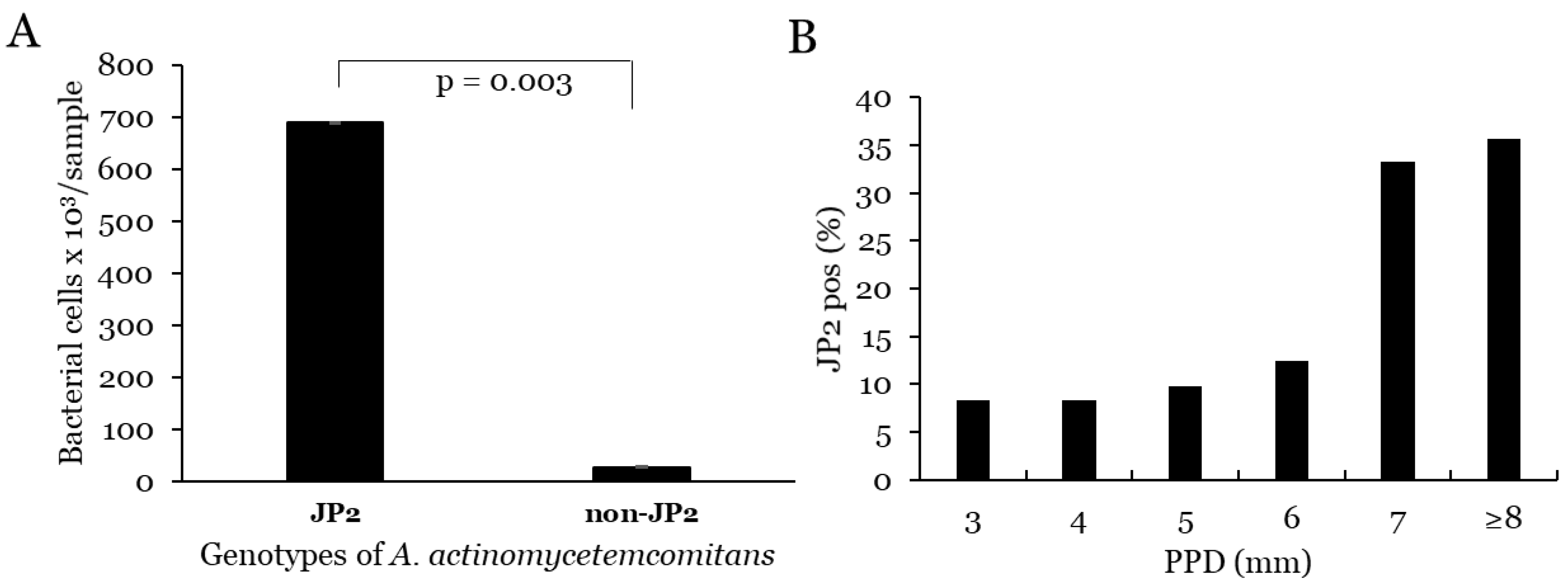
In comparison, between A. actinomycetemcomitans-positive samples with and without the JP2 genotype, the levels of cells of this species were significantly higher in the JP2-positive samples (p = 0.003) (Figure 3).

3.1.2. Correlation with PPD and Progression for A. actinomycetemcomitans

The levels of A. actinomycetemcomitans were not significantly correlated with increased PPD or with progression (Table 1). Samples with high levels of A. actinomycetemcomitans (>10,000 cells/sample) were significantly more prevalent from sites with increased PPD (p = 0.028) (Table 1) but were not associated with increased attachment loss progression (p = 0.076) (Table 1). The JP2-positive samples were significantly associated with sites with increased PPD (p = 0.024), as well as with progression (p = 0.003) (Table 1). The highest levels of PPD, as well as progression, were in sites positive for both the JP2 genotype and the ftxA gene of F. alocis (Table 1).

3.2. F. alocis

3.2.1. Prevalence and Levels of F. alocis

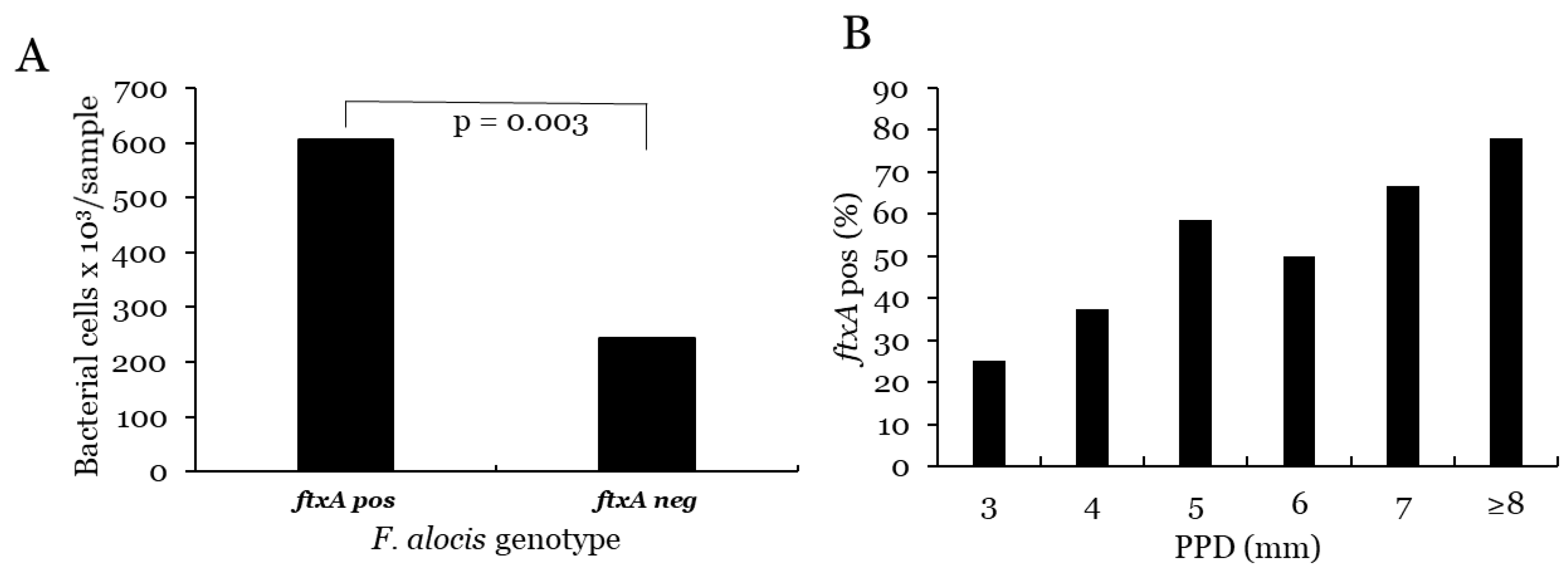
The prevalence of F. alocis was 92% (n = 157) of the sampled sites, and the ftxA gene of this bacterium was detected in 52% of samples (n = 90). The proportion of samples with higher levels of F. alocis (>10,000 cells/sample) was 72% (n = 123). The mean level of this bacterium in the 157 positive samples was 40.8 × 10⁴ cells/sample (median 10.5 × 10⁴). The distribution of samples with higher levels (>104 cells/sample) of F. alocis in relation to PPD is shown in Figure 2. In comparison, between F. alocis-positive samples with and without presence of the ftxA gene, the levels of F. alocis were significantly higher in the ftxA-positive samples (p = 0.003) (Figure 4).

3.2.2. Correlation with PPD and Progression for F. alocis

The prevalence of F. alocis was significantly associated with increased PPD (p = 0.015) but not with progression (p = 0.236). Samples with high levels of F. alocis (>10 000) were significantly more prevalent in samples from sites with increased PPD (p < 0.001) (Table 1), but not to increased progression (p < 0.001) (Table 1). The ftxA-positive samples were significantly associated with sites with increased PPD (p = 0.005), but not with progression (p = 0.123) (Table 1). In combination with JP2, the ftxA-positive sites showed the highest levels of PPD and progression (Table 1).

4. Discussion

Data from the present study confirmed the strong association between the presence of A. actinomycetemcomitans and progression of periodontal attachment loss [18,31]. In line with previous findings, the presence of the highly leukotoxic JP2 genotype of this bacterium further increased the correlation to onset and progression of periodontitis [12,13]. Earlier reported data are based of the prevalence on an individual basis, while the present study provided quantitative data from specific periodontal sites. It has previously been shown that the high levels of the JP2 genotype in saliva are associated to periodontal attachment loss [32]. A similar trend was found in the present study, where the sampled levels of A. actinomycetemcomitans in subgingival plaque were significantly increased between the two examinations in the sites positive for the JP2 genotype.
The RTX protein LtxA, which is highly expressed by the JP2 genotype, efficiently kills leukocytes during a rapid activation of a pro-inflammatory host response [33]. These properties of LtxA at least partly explain the strong correlation between the carriage of the JP2 genotype of A. actinomycetemcomitans and the onset and progression of periodontal attachment loss [34,35].
It has previously been reported that presence of F. alocis enhances the pathogenicity of A. actinomycetemcomitans [19]. Interestingly, it was recently demonstrated that F. alocis also encodes and expresses an RTX protein, FtxA [21,22]. The role of this potential virulence factor in periodontitis, as well as its mechanisms of action in host interaction, remain to be discovered. In the present study, we found a significant correlation between sample levels of F. alocis cell numbers and PPD or progression at the sampled site. Interestingly, the highest levels of PPD, as well as attachment loss progression, were documented in sites positive for both the JP2 genotype of A. actinomycetemcomitans and for ftxA-positive F. alocis. This is the first report that indicates a potential role of FtxA in the virulence of F. alocis. However, the lack of data for total bacterial loads in the samples of the present study made calculations of proportions in the microflora impossible.
It has recently been reported that the exposure of neutrophils to F. alocis expanded their lifespan by downregulating pro-apoptotic genes and upregulating anti-apoptotic genes [36]. Although F. alocis strains known to encode ftxA evidently express this protein, refs. [21,22] it is not known if this RTX protein is involved in the effects observed on neutrophils [36,37]. In contrast, LtxA is known to activate and efficiently kill all subsets of leukocytes [33]. In addition, LtxA activates bone resorption through promoting osteoclast differentiation by activating the NLRP3 inflammasome and, consequently, the release of bioactive IL-1β [38,39]. How the prolonged leukocyte survival induced by F. alocis interferes with the pro-inflammatory response by LtxA remains to be discovered [23,40]. Despite the gap in knowledge concerning mechanisms of action of FtxA and its possible interaction with LtxA, it may function as a potential risk marker, alone or in combination with virulent genotypes of A. actinomycetemcomitans. As the mode of action of FtxA, and its potential role in periodontal disease, is not yet understood, this aspect may be considered a limitation of the present study.

5. Conclusions

A. actinomycetemcomitans and F. alocis are common inhabitants in the biofilm associated with sites with periodontal attachment loss of Ghanaian adolescents. The levels of these two periodontitis-associated species were significantly associated with both enhanced PPD and tooth attachment loss progression. These associations were further pronounced, if there was a co-carriage of the highly leukotoxic JP2 genotype of A. actinomycetemcomitans and ftxA-positive F. alocis.

Supplementary Materials

The following supporting information can be downloaded at: https://www.mdpi.com/article/10.3390/microorganisms10122511/s1, Table S1: Primer sequences; Table S2: Cycle settings for PCR.

Author Contributions

Conceptualization, A.J., Z.R. and J.O.; methodology, R.C., Z.R. and J.O.; software, A.J. and Z.R.; validation, A.J., J.O. and R.C.; formal analysis, R.C., Z.R. and J.O.; investigation, C.H.Å., D.H., F.K. and A.J.; resources, A.J. and J.O.; data curation, A.J. and C.H.Å.; writing—original draft preparation, A.J. and Z.R.; writing—review and editing, C.H.Å., J.O., R.C., F.K. and D.H.; funding acquisition, A.J., J.O. and C.H.Å. All authors have read and agreed to the published version of the manuscript.

Funding

This research was funded by TUA grants from the County Council of Västerbotten, Sweden (A.J.; 7003193 and J.O.; 7003766), and by grants from the Medical Faculty of Umeå University (Insamlingsstiftelsen; to A.J. and J.O.; Research trainee program; to Z.R.).

Data Availability Statement

Data are available from the corresponding author (A.J.).

Acknowledgments

We are grateful to all the participants involved in the study as well as to the staff at the different Schools in Accra, Ghana, for helping with the clinical examinations. We also thank Björn Tavelin, statistician at the department of radiation sciences, Umeå University, for his help with the statistical calculations and Carina Öhman at the Department of Odontology, Umeå University, for valuable technical assistance.

Conflicts of Interest

The authors declare no conflict of interest.

References

  1. Könönen, E.; Gursoy, M.; Gursoy, U. Periodontitis: A Multifaceted Disease of Tooth-Supporting Tissues. J. Clin. Med. 2019, 8, 1135. [Google Scholar] [CrossRef] [PubMed] [Green Version]
  2. Yucel-Lindberg, T.; Båge, T. Inflammatory mediators in the pathogenesis of periodontitis. Expert Rev. Mol. Med. 2013, 15, e7. [Google Scholar] [CrossRef] [PubMed] [Green Version]
  3. Susin, C.; Haas, A.N.; Albandar, J.M. Epidemiology and demographics of aggressive periodontitis. Periodontology 2000 2014, 65, 27–45. [Google Scholar] [CrossRef] [PubMed]
  4. Norderyd, O.; Kochi, G.; Papias, A.; Köhler, A.A.; Helkimo, A.N.; Brahm, C.O.; Lindmark, U.; Lindfors, N.; Mattsson, A.; Rolander, B. Oral health of individuals aged 3-80 years in Jönköping, Sweden during 40 years (1973–2013). II. Review of clinical and radiographic findings. Swed. Dent. J. 2015, 39, 69–86. [Google Scholar] [PubMed]
  5. Zhang, X.; Wang, X.; Wu, J.; Wang, M.; Hu, B.; Qu, H.; Zhang, J.; Li, Q. The global burden of periodontal diseases in 204 countries and territories from 1990 to 2019. Oral Dis. 2022. [Google Scholar] [CrossRef]
  6. Kassebaum, N.J.; Smith, A.G.C.; Bernabé, E.; Fleming, T.D.; Reynolds, A.E.; Vos, T.; Murray, C.J.L.; Marcenes, W.; GBD 2015 Oral Health Collaborators. Global, Regional, and National Prevalence, Incidence, and Disability-Adjusted Life Years for Oral Conditions for 195 Countries, 1990–2015: A Systematic Analysis for the Global Burden of Diseases, Injuries, and Risk Factors. J. Dent. Res. 2017, 96, 380–387. [Google Scholar] [CrossRef]
  7. Tonetti, M.S.; Greenwell, H.; Kornman, K.S. Staging and grading of periodontitis: Framework and proposal of a new classification and case definition. J. Periodontol. 2018, 89 (Suppl. S1), S159–S172. [Google Scholar] [CrossRef] [Green Version]
  8. Graetz, C.; Mann, L.; Krois, J.; Sälzer, S.; Kahl, M.; Springer, C.; Schwendicke, F. Comparison of periodontitis patients’ classification in the 2018 versus 1999 classification. J. Clin. Periodontol. 2019, 46, 908–917. [Google Scholar] [CrossRef]
  9. Bouziane, A.; Hamdoun, R.; Abouqal, R.; Ennibi, O. Global prevalence of aggressive periodontitis: A systematic review and meta-analysis. J. Clin. Periodontol. 2020, 47, 406–428. [Google Scholar] [CrossRef]
  10. Yoshida, A.; Bouziane, A.; Erraji, S.; Lakhdar, L.; Rhissassi, M.; Miyazaki, H.; Ansai, T.; Iwasaki, M.; Ennibi, O. Etiology of aggressive periodontitis in individuals of African descent. Jpn. Dent. Sci. Rev. 2021, 57, 20–26. [Google Scholar] [CrossRef]
  11. Kissa, J.; El Houari, B.; Amine, K.; Chemlali, S.; Khlil, N.; Mikou, S.; Gharibi, A.; El Ouadnassi, I.; Rifki, C.; Albandar, J.M. Prevalence of periodontal disease in young Moroccans: A national survey. J. Periodontol. 2022. [Google Scholar] [CrossRef] [PubMed]
  12. Haubek, D.; Ennibi, O.-K.; Poulsen, K.; Væth, M.; Poulsen, S.; Kilian, M. Risk of aggressive periodontitis in adolescent carriers of the JP2 clone of Aggregatibacter (Actinobacillus) actinomycetemcomitans in Morocco: A prospective longitudinal cohort study. Lancet 2008, 371, 237–242. [Google Scholar] [CrossRef] [PubMed]
  13. Åberg, C.H.; Kwamin, F.; Claesson, R.; Dahlén, G.; Johansson, A.; Haubek, D. Progression of attachment loss is strongly associated with presence of the JP2 genotype of Aggregatibacter actinomycetemcomitans: A prospective cohort study of a young adolescent population. J. Clin. Periodontol. 2014, 41, 232–241. [Google Scholar] [CrossRef] [PubMed]
  14. Rylev, M.; Kilian, M. Prevalence and distribution of principal periodontal pathogens worldwide. J. Clin. Periodontol. 2008, 35, 346–361. [Google Scholar] [CrossRef]
  15. Dahlén, G.; Claesson, R.; Åberg, C.H.; Haubek, D.; Johansson, A.; Kwamin, F. Subgingival bacteria in Ghanaian adolescents with or without progression of attachment loss. J. Oral Microbiol. 2014, 6, 23977. [Google Scholar] [CrossRef] [PubMed]
  16. Åberg, C.H.; Kwamin, F.; Claesson, R.; Johansson, A.; Haubek, D. Presence of JP2 and Non-JP2 Genotypes of Aggregatibacter actinomycetemcomitans and attachment loss in adolescents in Ghana. J. Periodontol. 2012, 83, 1520–1528. [Google Scholar] [CrossRef] [PubMed]
  17. Brogan, J.M.; Lally, E.T.; Poulsen, K.; Kilian, M.; Demuth, D.R. Regulation of Actinobacillus actinomycetemcomitans leukotoxin expression: Analysis of the promoter regions of leukotoxic and minimally leukotoxic strains. Infect. Immun. 1994, 62, 501–518. [Google Scholar] [CrossRef] [PubMed] [Green Version]
  18. Fine, D.H.; Markowitz, K.; Furgang, D.; Fairlie, K.; Ferrandiz, J.; Nasri, C.; McKiernan, M.; Gunsolley, J. Aggregatibacter actinomycetemcomitans and Its Relationship to Initiation of Localized Aggressive Periodontitis: Longitudinal Cohort Study of Initially Healthy Adolescents. J. Clin. Microbiol. 2007, 45, 3859–3869. [Google Scholar] [CrossRef] [Green Version]
  19. Fine, D.H.; Markowitz, K.; Fairlie, K.; Tischio-Bereski, D.; Ferrendiz, J.; Furgang, D.; Paster, B.J.; Dewhirst, F.E. A Consortium of Aggregatibacter actinomycetemcomitans, Streptococcus parasanguinis, and Filifactor alocis Is Present in Sites Prior to Bone Loss in a Longitudinal Study of Localized Aggressive Periodontitis. J. Clin. Microbiol. 2013, 51, 2850–2861. [Google Scholar] [CrossRef] [Green Version]
  20. Arunraj, R.; Neelakandan, A.; Potluri, R.; Yadalam, P.K.; Chakraborty, P.; Saravanan, A. The varied proportion of Filifactor alocis in periodontal health and disease in the South Indian subpopulation. Contemp. Clin. Dent. 2021, 12, 433. [Google Scholar] [CrossRef]
  21. Oscarsson, J.; Claesson, R.; Bao, K.; Brundin, M.; Belibasakis, G. Phylogenetic Analysis of Filifactor alocis Strains Isolated from Several Oral Infections Identified a Novel RTX Toxin, FtxA. Toxins 2020, 12, 687. [Google Scholar] [CrossRef] [PubMed]
  22. Bao, K.; Claesson, R.; Gehrig, P.; Grossmann, J.; Oscarsson, J.; Belibasakis, G.N. Proteomic Characterization of the Oral Pathogen Filifactor alocis Reveals Key Inter-Protein Interactions of Its RTX Toxin: FtxA. Pathogens 2022, 11, 590. [Google Scholar] [CrossRef] [PubMed]
  23. Ozuna, H.; Snider, I.; Belibasakis, G.N.; Oscarsson, J.; Johansson, A.; Uriarte, S.M. Aggregatibacter actinomycetemcomitans and Filifactor alocis: Two exotoxin-producing oral pathogens. Front. Oral Health 2022, 3, 981343. [Google Scholar] [CrossRef] [PubMed]
  24. Möller, A.J. Microbiological examination of root canals and periapical tissues of human teeth. Methodological studies. Odontol. Tidskr. 1966, 74, 1–380. [Google Scholar]
  25. Rylev, M.; Abduljabar, A.B.; Reinholdt, J.; Ennibi, O.-K.; Haubek, D.; Birkelund, S.; Kilian, M. Proteomic and immunoproteomic analysis of Aggregatibacter actinomycetemcomitans JP2 clone strain HK1651. J. Proteom. 2011, 74, 2972–2985. [Google Scholar] [CrossRef]
  26. Aruni, A.W.; Roy, F.; Sandberg, L.; Fletcher, H.M. Proteome variation among Filifactor alocis strains. Proteomics 2012, 12, 3343–3364. [Google Scholar] [CrossRef] [Green Version]
  27. Jalava, J.; Eerola, E. Phylogenetic analysis of Fusobacterium alocis and Fusobacterium sulci based on 16S rRNA gene sequences: Proposal of Filifactor alocis (Cato, Moore and Moore) comb. nov. and Eubacterium sulci (Cato, Moore and Moore) comb. nov. Int. J. Evol. Microbiol. 1999, 49, 1375–1379. [Google Scholar] [CrossRef] [Green Version]
  28. Kirakodu, S.S.; Govindaswami, M.; Novak, M.J.; Ebersole, J.L.; Novak, K.F. Optimizing qPCR for the Quantification of Periodontal Pathogens in a Complex Plaque Biofilm. Open Dent. J. 2008, 2, 49–55. [Google Scholar] [CrossRef] [Green Version]
  29. Siqueira, J.F., Jr.; Rôças, I.N. Detection of Filifactor alocis in endodontic infections associated with different forms of periradicular diseases. Oral Microbiol. Immunol. 2003, 18, 263–265. [Google Scholar] [CrossRef]
  30. Poulsen, K.; Ennibi, O.-K.; Haubek, D. Improved PCR for Detection of the Highly Leukotoxic JP2 Clone of Actinobacillus actinomycetemcomitans in Subgingival Plaque Samples. J. Clin. Microbiol. 2003, 41, 4829–4832. [Google Scholar] [CrossRef] [Green Version]
  31. Van Der Velden, U.; Abbas, F.; Armand, S.; Loos, B.G.; Timmerman, M.F.; Van Der Weijden, G.A.; Van Winkelhoff, A.J.; Winkel, E.G. Java project on periodontal diseases. The natural development of periodontitis: Risk factors, risk predictors and risk determinants. J. Clin. Periodontol. 2006, 33, 540–548. [Google Scholar] [CrossRef] [PubMed]
  32. Ennibi, O.K.; Claesson, R.; Akkaoui, S.; Reddahi, S.; Kwamin, F.; Haubek, D.; Johansson, A. High salivary levels of JP2 genotype of Aggregatibacter actinomycetemcomitans is associated with clinical attachment loss in Moroccan adolescents. Clin. Exp. Dent. Res. 2019, 5, 44–51. [Google Scholar] [CrossRef] [PubMed]
  33. Johansson, A. Aggregatibacter actinomycetemcomitans Leukotoxin: A Powerful Tool with Capacity to Cause Imbalance in the Host Inflammatory Response. Toxins 2011, 3, 242–259. [Google Scholar] [CrossRef] [Green Version]
  34. Kelk, P.; Claesson, R.; Chen, C.; Sjöstedt, A.; Johansson, A. IL-1beta secretion induced by Aggregatibacter (Actinobacillus) actinomycetemcomitans is mainly caused by the leukotoxin. Int. J. Med. Microbiol. 2008, 298, 529–541. [Google Scholar] [CrossRef] [PubMed]
  35. Åberg, C.H.; Kelk, P.; Johansson, A. Aggregatibacter actinomycetemcomitans: Virulence of its leukotoxin and association with aggressive periodontitis. Virulence 2014, 6, 188–195. [Google Scholar] [CrossRef] [PubMed] [Green Version]
  36. Miralda, I.; Vashishta, A.; Rogers, M.N.; Lamont, R.J.; Uriarte, S.M. The emerging oral pathogen, Filifactor alocis, extends the functional lifespan of human neutrophils. Mol. Microbiol. 2022, 117, 1340–1351. [Google Scholar] [CrossRef]
  37. Armstrong, C.L.; Klaes, C.K.; Vashishta, A.; Lamont, R.J.; Uriarte, S.M. Filifactor alocis manipulates human neutrophils affecting their ability to release neutrophil extracellular traps induced by PMA. Innate Immun. 2018, 24, 210–220. [Google Scholar] [CrossRef] [PubMed] [Green Version]
  38. Kelk, P.; Claesson, R.; Hanstrom, L.; Lerner, U.H.; Kalfas, S.; Johansson, A. Abundant secretion of bioactive interleukin-1beta by human macrophages induced by Actinobacillus actinomycetemcomitans leukotoxin. Infect Immun. 2005, 73, 453–458. [Google Scholar] [CrossRef] [Green Version]
  39. Kelk, P.; Moghbel, N.S.; Hirschfeld, J.; Johansson, A. Aggregatibacter actinomycetemcomitans Leukotoxin Activates the NLRP3 Inflammasome and Cell-to-Cell Communication. Pathogens 2022, 11, 159. [Google Scholar] [CrossRef]
  40. Miralda, I.; Uriarte, S.M. Periodontal Pathogens’ strategies disarm neutrophils to promote dysregulated inflammation. Mol. Oral Microbiol. 2020, 36, 103–120. [Google Scholar] [CrossRef]
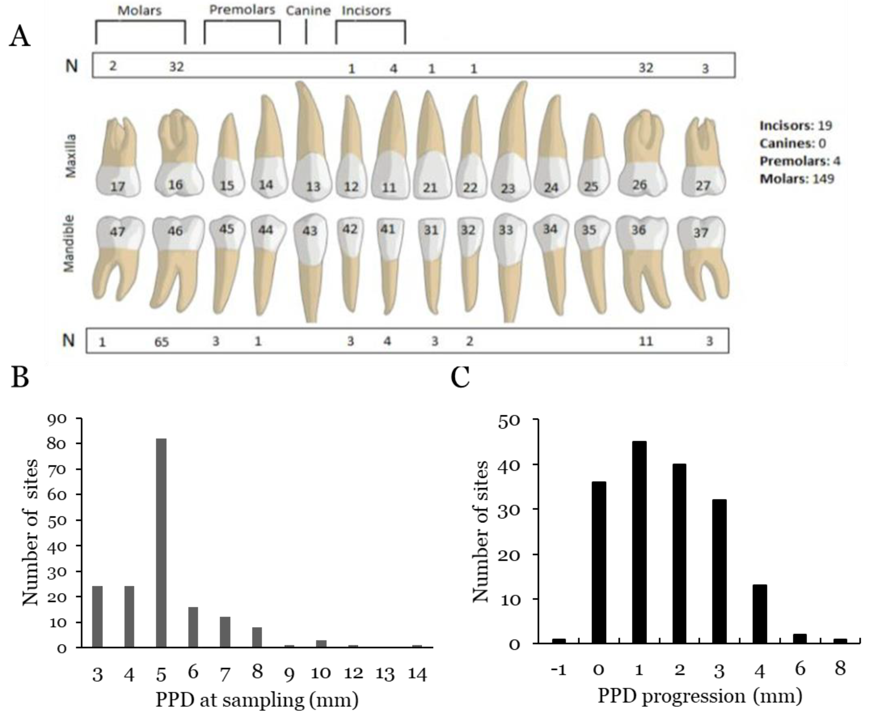
Figure 1. Illustration of number (N) and location of sampled sites (A), PPD at FU sampling (B), and progression of PPD (C) in the sampled sites from BL to FU examinations. Subgingival plaque samples are collected with paper points in the area between the teeth and the periodontal tissue.
Microorganisms 10 02511 g001
Figure 2. Proportion of samples with high levels (>104 cells/sample) of A. actinomycetemcomitans (Aa) or F. alocis (Fa), respectively, in relation to PPD.
Microorganisms 10 02511 g002
Figure 3. (A) Mean levels of A. actinomycetemcomitans cells as determined in sites with presence (JP2) and absence (non-JP2) of this genotype of the bacterium. (B) Proportion of JP2-positive sites associated with PPD.
Microorganisms 10 02511 g003
Figure 4. (A) Mean levels of F. alocis cells in sites positive for this bacterium with (ftxA pos) or without (ftxA neg) presence of the ftxA gene. (B) Proportion of ftxA-positive sites associated with PPD.
Microorganisms 10 02511 g004
Table 1. PPD and progression in relation to carriership of A. actinomycetemcomitans (Aa) and F. alocis (Fa) in the sampled sites. Significant differences in clinical parameters between sample groups were calculated with the Mann–Whitney U test.
PPD Progresssion
NMean (mm)SDp-ValueNMean (mm)SDp-Value
Aa pos955.24±1.430.027951.83±1.410.163
Aa neg775.05±1.91 751.57±1.41
Aa high335.67±1.800.027332.03±1.550.225
Aa low1395.04±1.61 1371.64±1.37
JP2245.79±1.580.013232.48±1.540.009
Non-JP21485.05±1.66 1471.60±1.36
Fa pos1575.24±1.680.0161551.74±1.410.384
Fa neg144.21±1.12 141.43±1.45
Fa high1235.49±1.690.0011211.93±1.370.001
Fa low484.31±1.27 481.17±1.39
ftxA pos905.52±1.650.001891.85±1.600.398
ftxA neg824.76±1.59 811.57±1.16
ftxA-JP2 156.20±1.320.001152.93±1.530.002
Other1575.06±1.66 1551.60±1.35
Publisher’s Note: MDPI stays neutral with regard to jurisdictional claims in published maps and institutional affiliations.