Article

Molecular Epidemiology of mcr-1-Positive Escherichia coli and Klebsiella pneumoniae Isolates: Results from Russian Sentinel Surveillance (2013–2018)

1
Federal State Budgetary Institution, Centre for Strategic Planning and Management of Biomedical Health Risks, Federal Medical Biological Agency, 119121 Moscow, Russia
2
Institute of Antimicrobial Chemotherapy, Smolensk State Medical University, 214019 Smolensk, Russia
3
Central Research Institute of Epidemiology, 111123 Moscow, Russia
*
Author to whom correspondence should be addressed.
Microorganisms 2022, 10(10), 2034; https://doi.org/10.3390/microorganisms10102034
Submission received: 27 September 2022 / Revised: 11 October 2022 / Accepted: 12 October 2022 / Published: 14 October 2022
(This article belongs to the Special Issue Antimicrobial Resistance Mechanisms in Bacteria)

Abstract

:
Background: The dissemination of mobile colistin resistance (mcr) genes is a serious healthcare threat because polymyxins represent “last-line” therapeutics for multi-drug-resistant Gram-negative pathogens. This study aimed to assess the prevalence of colistin resistance and mcr genes and characteristics of clinical Escherichia coli (Eco) and Klebsiella pneumoniae (Kpn) isolates and plasmids carrying these genes in Russia. Methods: A total of 4324 Eco and 4530 Kpn collected in the frame of sentinel surveillance in 2013–2018 were tested for susceptibility to colistin and other antibiotics using the broth microdilution method. mcr genes were screened by real-time PCR. Phylogeny, genomic features and plasmids of mcr-positive isolates were assessed using whole-genome sequencing and subsequent bioinformatic analysis. Results: Colistin resistance was detected in 2.24% Eco and 9.3% Kpn. Twenty-two (0.51%) Eco and two (0.04%) Kpn from distant sites carried mcr-1.1. Most mcr-positive isolates co-harbored ESBLs and other resistance determinants to various antibiotic classes. The mcr-positive Eco belonged to 16 MLST types, with ST359 being most common; Kpn belonged to ST307 and ST23. mcr-1.1 was carried mainly in IncI2 (n = 18) and IncX4 (n = 5) plasmids highly similar to those identified previously in human, animal and environmental isolates. Conclusion: This study demonstrated a dissemination of “typical” mcr-bearing plasmids among diverse Eco and Kpn genotypes and across a wide geographic area in Russia. Given the frequent association of mcr with other resistance determinants and potential clinical impact, the continual surveillance of this threat is warranted.

1. Introduction

Polymyxins (including colistin, or polymyxin E and polymyxin B), the cationic peptide antibiotics, were discovered more than 70 years ago and become clinically available in 1960s; however, their therapeutic use was long since abandoned because of nephrotoxicity and availability of alternative effective and better-tolerated antimicrobial agents [1]. In the last two decades, however, the emergence and global rise in resistance to all classes of antibiotics, including extended-spectrum cephalosporins and carbapenems, in Gram-negative bacteria have led to the reintroduction of polymyxins as “last-resort” drugs against extremely multi-resistant pathogens.
Polymyxins exert their antimicrobial action via initial interaction with negatively charged lipid A components of the lipopolysaccharide (LPS) in the outer membrane of Gram-negative bacteria. Until 2015, all known colistin resistance mechanisms were chromosomally mediated and usually related to mutations in several genes included in the regulation (pmrAB, phoPQ, and mgrB), biosynthesis (pmrHFIJKLM) and structural modification (pmrC and pmrE) of lipid A [2,3]. However, the discovery by Liu et al. [4] of the plasmid-borne mobile colistin resistance (mcr-1) gene for lipid A phosphoethanolamine transferase revealed a mechanism for horizontal spread of polymyxin resistance. Since then, mcr-1 was detected in various species of Enterobacterales from different sources (humans, animals and the environment) and different countries across Asia, Europe, Africa, the Americas and Oceania. Notably, a retrospective screening of the collection of poultry E. coli isolates by the same research group that discovered mcr-1 revealed its presence in isolates collected back in the 1980s when colistin first started to be used in food-producing animals in China [5]. In addition, several new variants of mcr genes sharing from 31% to 83% amino acid sequence identity to mcr-1 have been identified in different Gram-negative bacteria, with mcr-1 being the most frequently detected [6,7,8]. These genes have been associated with plasmids of diverse incompatibility types, among which, IncI2, IncHI2 and IncX4 were the most abundant [9].
Whole-genome sequencing (WGS) has become an essential tool for surveillance and molecular epidemiology of antimicrobial resistance (AMR). It provides precise spatial and temporal delineations of the spread of pathogens and mobile resistance genes. Coupled with epidemiological and phenotypic antimicrobial susceptibility investigations, it delivers ultimate resolution for tracing AMR [10]. Numerous studies have used WGS and bioinformatics analysis to characterize mcr-bearing plasmids and strains. Short-read sequencing (e.g., with Illumina platform) has been commonly applied to assess phylogenetic relationship of isolates based on analysis of core genome and to determine the presence of plasmids, virulence and AMR genes, while long-read sequencing (e.g., with the Oxford Nanopore platform) has been used to effectively resolve complex plasmid structures and genomic context of AMR genes. A hybrid assembly with short and long reads has been used to provide complete and accurate bacterial genomes [11].
Several research groups have previously reported on the sporadic occurrence of mcr-1 in Russia and described the corresponding mcr-bearing plasmids [12,13,14,15]. In this study, we aimed to systematically assess the prevalence of colistin resistance and of mcr genes in clinical Escherichia coli and Klebsiella pneumoniae isolates from Russian sentinel surveillance and to determine phenotypic and genomic characteristics of mcr-carrying isolates and plasmids.

2. Materials and Methods

2.1. Bacterial Isolates

All studied isolates and accompanying metadata were collected as part of the national sentinel surveillance program of antimicrobial resistance in bacterial pathogens isolated from hospitalized patients [16]. Between 1 January 2013 and 31 December 2018, participating laboratories from 36 cities across Russia contributed a total of 4324 E. coli and 4530 K. pneumoniae clinical isolates. The isolates were recovered from representative clinical specimens (blood, tissue biopsies, cerebrospinal fluid, bronchoalveolar lavage, sputum, urine, etc.) of patients with clinical symptoms of infections; isolates from surface swab specimens, screening, and environmental samples were spared. Only one isolate of each species per patient/case of infection was allowed.

2.2. Species Identification, Antimicrobial Susceptibility Testing and PCR Screening of mcr Genes

In the central surveillance laboratory, the species identity of the isolates was confirmed by MALDI-TOF mass spectrometry with the Microflex LT-MALDI Biotyper System (Bruker Daltonics, Bremen, Germany).
The susceptibility to a range of antibiotics, including ampicillin, amoxicillin-clavulanic acid, piperacillin-tazobactam, ceftazidime, ceftazidime-avibactam, cefepime, aztreonam, cefotaxime, ertapenem, imipenem, meropenem, gentamicin, amikacin, ciprofloxacin, doxycycline, tigecycline, chloramphenicol, trimethoprim-sulfamethoxazole, fosfomycin, and colistin, was determined by a reference broth microdilution method according to ISO 20776-2:2021 [17] and European committee on antimicrobial susceptibility testing (EUCAST) [18] methodology. Susceptibility testing results were interpreted according to EUCAST v.12.0 Clinical breakpoints [19].
All isolates with colistin minimum inhibitory concentration (MIC) >0.5 mg/L (1105 E. coli and 1483 K. pneumoniae) were screened for the presence of mcr genes by means of real-time PCR as described elsewhere [20].

2.3. Whole-Genome Sequencing

Twenty-four mcr-positive isolates were characterized by WGS. Genomic DNA was extracted using DNeasy Blood & Tissue Kit and QIAcube Connect Device (QIAGEN, Hilden, Germany). For short-read sequencing, library preparation was performed using Illumina DNA prep kit (Illumina, San Diego, CA, USA) according to manufacturer instructions. Capillary electrophoresis using the Agilent 2100 Bioanalyzer system and Agilent High Sensitivity DNA Kit (Agilent, Santa Clara, CA, USA) was used for estimation of library quality. Concentration of the libraries was measured using the Qubit 4.0 fluorimeter with Qubit dsDNA HS Assay Kit (Thermo Fisher Scientific, Waltham, MA, USA). Sequencing was performed on the Illumina NextSeq 550 System with the NextSeq 500/550 Mid Output Kit v2.5 (300 Cycles) and on the Illumina MiSeq System with MiSeq Reagent Kit v2 (500-cycles) (Illumina, USA). For long-read sequencing, library preparation was performed with NEBNext Companion Module (New England Biolabs, Ipswich, MA, USA) and Ligation Sequencing Kit (Oxford Nanopore Technologies (ONT), Oxford, UK) in accordance with manufacturer’s protocol. Samples were multiplexed using Native Barcoding Expansion 1–12 (ONT, UK). Library concentration was measured with Qubit dsDNA HS Assay Kit and Qubit 4 Fluorometer (Thermo Fisher Scientific, USA). Sequencing was performed on the MinION platform and R9.4.1 Flow Cells (ONT, UK).

2.4. Bioinformatics Analysis

Quality assessment of raw reads was performed using a FASTQC v.0.11.9 tool [21]. The bioinformatics pipeline TORMES v.1.2.1 [22] was used for the reads quality filtering with Trimmomatic v.0.39 [23] and Prinseq v.0.20.4 [24], de novo genome assembly with Spades v.3.14.1 [25], quality assessment of assemblies with Quast v.5.0.2 [26], and taxonomic identification by k-mer matching with Kraken2 v2.1.1 [27] and 16S rRNA gene analysis with RDP Classifier v.2.10.2 [28]. All steps were performed with default parameters, except for Spades assembly, which was run with the flag --cov-cutoff set to ‘auto’ and the additional flag --careful. The minimum contig size threshold was set to 500 bp in length. Contamination removal was performed based on determining the coverage cutoffs for each assembly after visual inspection of Coverage-vs.-Length (CVL) plots as described by Douglass et al. [29]. PlasmidSPAdes [30] was used for de novo reconstruction of plasmids.
Base-calling and demultiplexing were conducted using Guppy v5.0.16 with the dna_r9.4.1_450bps_sup model. Quality control of sequencing was assessed with MinIONQC v1.4.2 [31]. Hybrid assemblies were prepared by using the Trycycler pipeline v0.5.1 [32], which included quality-control filtering of short reads by fastp v0.20.1 [33] with default parameters and filtering of long reads by Filtlong v0.2.0 [34] with a minimum read length of 1 kbp and a kept-base percentage of 95%. Long reads were randomly subsampled into 12 read sets, which were assembled using three different assemblers: Flye v2.9 [35], Miniasm 0.3/Minipolish v0.1.3 [36], and Raven v1.6.1 [37]. The contigs from different assemblies were then clustered by Trycycler to produce a consensus contig for each cluster. The consensus contigs were polished with Medaka v.1.4.4 [38]. Short-read polishing was performed by Polypolish tool v0.5.0 [39] and POLCA script [40]. Quast tool [26] was used to obtain the metrics of the final hybrid assemblies. The genome completeness of each assembly was assessed by performing a benchmarking universal single-copy ortholog (BUSCO) analysis using BUSCO 4.0.6 [41]. The level of genome completeness was expressed as BUSCO scores, including complete, fragmented, and missing BUSCOs that indicate the fractions of high-identity full-length genes, partially present genes, and absent genes, respectively. Gene prediction was performed for complete genome sequences with the PROKKA v1.14.5 [42]. Phylogroup, sequence type, serotype, and FimH type of E. coli were identified using ClermonTyping [43], MLST v2.1 [44] with PubMLST typing database [45], SerotypeFinder v.2.0 [46], and FimTyper v.1.1 [47]. Kleborate v2.1.0 [48] was used for K and O antigen typing of K. pneumonia genomes. Acquired antimicrobial resistance genes and known resistance-associated point mutations were annotated using AMRFinderPlus tool v. 3.10.5 [49] with BLASTP, BLASTX and HMMER algorithms and ABRicate tool v1.0.1 [50] with Resfinder database [51]. Searches for virulence factor genes and plasmid replicons were performed using the ABRicate with EcOH database [52], VFDB [53], PlasmidFinder [54] and ISFinder databases [55]. The hybrid assemblies were additionally checked for the presence of multiple copies of AMR genes using BLASTN 2.4.0+ [56]. The core genomes were defined by Prokka and Roary [57] and the resulting core-genome alignment file from Roary was used as input into IQTREE v.2.2.0.3 [58] to construct a phylogenetic tree based on the best fit model (GTR+F+I+I+R7) identified by ModelFinder [59]. Bootstrapping was performed with 100 replicates and with parameter-b. The branches had bootstrap values above 98 for branch support. Core-genome single-nucleotide polymorphisms (SNPs) were extracted from the core-genome alignment using the SNP-sites v.2.5.1 [60]. iTol v5 [61] was used for visualizing the trees. Plasmids were reconstructed by Spades or plasmidSpades preferably from hybrid assemblies or from the longest contigs of two independent short-read assemblies. MOB-suite tool [62] was also used to assist in plasmid reconstruction in cases where mcr and plasmid replicon genes were on different contigs Alignment and circular comparisons of plasmids was performed with BRIG v0.95 [63]. The National Center for Biotechnology Information (NCBI) Basic Local Alignment Search Tool (BLAST) was used to identify published plasmid sequences most similar to those reported herein.

2.5. Data Availability

All Illumina and MinION sequences were deposited in the European Nucleotide Archive under the project accession number PRJEB53051.

3. Results

A total of 4324 E. coli and 4530 K. pneumoniae consecutive non-duplicate clinical isolates collected in 36 cities across Russia between 1 January 2013 and 31 December 2018 were assessed for susceptibility to colistin and for the presence of mcr genes. Phenotypic colistin resistance was detected in 97 (2.24%) E. coli and 420 (9.3%) K. pneumoniae isolates, while the presence of mcr genes was detected in 22 (0.51%) E. coli and 2 (0.04%) K. pneumoniae isolates. The geographic origins of mcr-positive isolates are shown in Figure 1. These isolates represented community-onset (n = 8) and hospital-onset (n = 16) infections of the urinary tract (n = 8), abdominal cavity (n = 7), lower respiratory tract (n = 5), skin and soft tissue (n = 3) and bloodstream (n = 1). Detailed clinical information on these isolates is presented in Supplementary Table S1.
The colistin MICs of mcr-positive isolates ranged between 4 and 16 mg/L. In addition to colistin, E. coli isolates were resistant to multiple antibiotics, including (in order of decreasing occurrence) ampicillin (n = 21), ciprofloxacin (n = 21), cefotaxime (n = 17), aztreonam (n = 16), amoxicillin-clavulanic acid (n = 16), cefepime (n = 14), trimethoprim-sulfamethoxazole (n = 14), chloramphenicol (n = 12), ceftazidime (n = 10), gentamicin (n = 10), tigecycline (n = 6), fosfomycin (n = 4), piperacillin-tazobactam (n = 3), and amikacin (n = 1), but were all susceptible to carbapenems and ceftazidime-avibactam. The two mcr-positive K. pneumoniae isolates were resistant to ampicillin, amoxicillin-clavulanic acid, piperacillin-tazobactam, cefotaxime, ceftazidime, cefepime, aztreonam, ciprofloxacin, gentamicin and chloramphenicol, but were susceptible to imipenem, meropenem, ceftazidime-avibactam, amikacin and fosfomycin at EUCAST clinical breakpoints. One K. pneumoniae isolate was additionally resistant to ertapenem and the other one to trimethoprim-sulfamethoxazole (Supplementary Table S1).
Illumina short-read whole-genome sequencing was performed on all 24 mcr-positive isolates. The summary of Illumina sequencing reads and assembly statistics are provided in Supplementary Table S2. The number of contigs in genome assemblies ranged from 70 to 269, while the N50 of contigs ranged from 43 kb to 349 kb. Five E. coli isolates (ec_75812, ec_76498, ec_92380, ec_102833, ec_103195) and one K. pneumoniae (kp_87441) were selected for complementary long-read sequencing using Oxford Nanopore Technologies (ONT) MinION platform to generate complete chromosome and plasmid assemblies (Supplementary Table S3). The total number of contigs in the resulting hybrid assemblies combining ONT and Illumina reads ranged from 5 to 6 per assembly. The largest contig of K. pneumoniae isolate was 5.376 Mb, and the largest contig of each E. coli isolate was >4.7 Mb, indicating that chromosomal DNA of each isolate was assembled into a single contig (Supplementary Tables S3 and S4). In addition, a quality check was performed using BUSCO software to ensure >99% completeness (Supplementary Table S3).
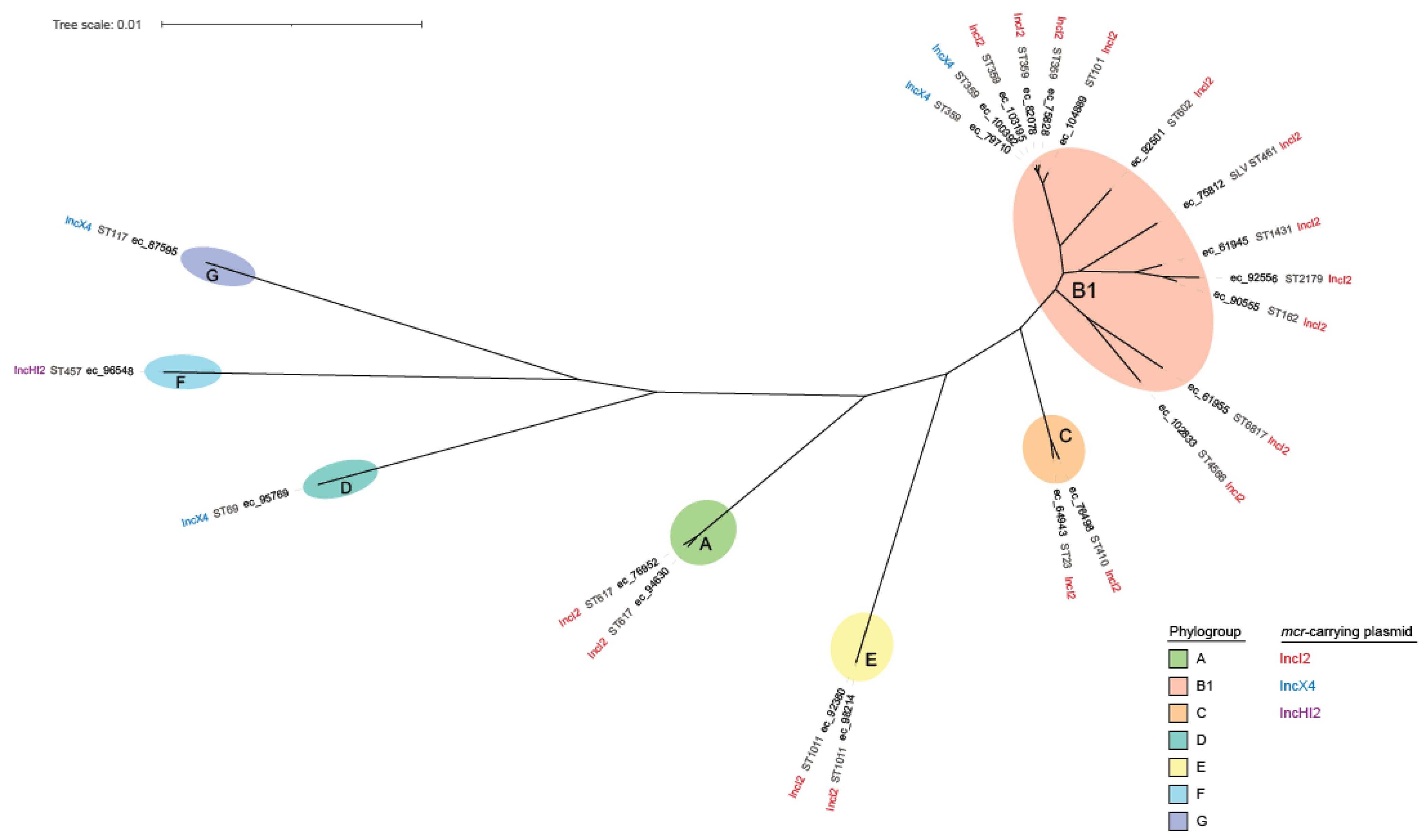
For all sequenced E. coli isolates, a core genome alignment of 2,976,673 bases was produced from 3066 (≥99%) core genes, yielding a final alignment of 150,635 single-nucleotide polymorphism (SNP) sites. In silico analysis assigned these isolates to seven different phylogroups (A, B1, C, D, F, G, and E) and 16 multilocus sequence types (STs) (Table 1, Figure 2). Notably, three STs included more than one epidemiologically unrelated isolates collected in geographically distinct regions: ST359 with five E. coli isolates (ec_79710, ec_100392, ec_103195, ec_75828, and ec_82078), the first three of which had the same serotype O36:H31 and carried fimH35 allele; ST1011 with two isolates (ec_92380 and ec_98214), which belonged to serotype H28:O188 and fimH31 type; and ST617 with two isolates (ec_76952 and ec_94630), which belonged to different serotypes and fimH types. One isolate (ec_75812) was found to belong to a new ST that differed from the most closely related ST461 at a single (adk) locus.
The two K. pneumoniae isolates (kp_87441 and kp_95329) belonged to ST307 and ST23 and carried the capsular biosynthesis loci (KL) KL102 and KL57, respectively (Table 2). Notably, the latter isolate carried typical for ST23 genotype hypervirulence marker genes, including aerobactin biosynthesis and receptor genes (iucABCD-iutA) and regulators of mucoid phenotype (rmpA and rmpA2).
Analysis of antibiotic resistance genes (ARGs) confirmed the presence of mcr-1.1 variant gene in all sequenced E. coli and K. pneumoniae genomes. Consistent with phenotypic antibiotic susceptibility patterns, multiple ARGs to various antibiotic classes were found. (Table 1 and Table 2).
All but one E. coli genomes contained acquired β-lactamase genes, including class A and D penicillinase genes: blaTEM-1 (n = 13) and blaOXA-1 (n = 1) and, most notably, the genes for various CTX-M-type extended-spectrum β-lactamases (ESBLs): blaCTX–M–15 (n = 5), blaCTX–M–1 (n = 4), blaCTX–M–55 (n = 3), blaCTX–M–65 (n = 2), blaCTX–M–27 (n = 1), blaCTX–M–169 (n = 1), and genes for acquired AmpC cephalosporinases: blaCMY-2 (n = 2). All E. coli isolates also carried one or several ARGs to multiple classes of non-β-lactam antibiotics, aminoglycosides (n = 19) (aadA1, aadA2, aadA5, aadA16, aac(3)-IId, aac(3)-IVa, aac(3)-VIa, aac(6′)-Ib-cr, aph(3′)-Ia, aph(3′)-Ib, aph(3′)-Id, aph(3′)-IIa, armA), quinolones (n = 7) (aac(6′)-Ib-cr, qnrB6, qnrB19, qnrS1), sulphonamides (n = 18) (sul1, sul2, sul3), trimethoprim (n = 14) (dfrA12, dfrA14, dfrA17, dfrA27), macrolides (n = 11) (mph(A), mph(E), msr(E)), phenicols (n = 11) (catA1, catB3, cmlA1, floR), tetracyclines (n = 16) (tetA, tetB, tetM), fosfomycin (n = 2) (fosA3, fosA7.5), and rifampicin (n = 3) (arr-3), as well as known mutations in the quinolone resistance-determining regions (QRDRs) of gyrA (n = 21) (S83L, D87N, D87Y), parC (n = 21) (S80I, S80R, E84G), and parE gene (n = 8) (S458A), and mutations in the fosfomycin resistance-determining region of uhpT (n = 3) (E350Q) and cyaA gene (n = 3) (S352T).
The genomes of two K. pneumoniae isolates contained, in addition to chromosomal species-specific penicillinase (blaSHV-1 or blaSHV-28) and glutathione_transferase (fosA) genes, the genes for acquired penicillinases (blaOXA-1 and blaLAP-2 or blaTEM-1), ESBLs (blaCTX-M-15 or blaCTX-M-55), as well as various aminoglycoside (aadA1, aadA2, aac(3)-IIe, aac(6′)-Ib-cr, aph(3″)-Ib, aph(6)-Id), tetracycline (tetA), sulfonamide (sul2, sul3), trimethoprim (dfrA14), phenicol (catB3, cmlA1), quinolone (aac(6′)-Ib-cr, oqxA, oqxB, qnrB1, qnrS1) ARGs and point mutations in gyrA (S83I) and parC (S80I) QRDRs.
Interestingly, using ONT long-read sequencing revealed the presence of multiple copies of certain ARGs in the individual sequenced genomes. Thus, a region of three tandem repeats containing aac(6′)-Ib-cr, blaOXA-1, catB3::IS26, tetR/tetA and ΔTnAs1 was found in IncFIB plasmid of K. pneumoniae isolate kp_87441, which was supported by 20 individual long sequencing reads spanning the entire repeat region (Supplementary Figure S1, Supplementary Table S5). Two copies of blaCTX–M–15 were found in the IncM2 plasmid of E. coli isolate ec_92380, with six individual long reads spanning the ARG region (Supplementary Figure S2, Supplementary Table S6); and two copies of blaTEM–1, one of which was carried on IncFIB(AP001918)/IncQ1/IncFIC(FII) plasmid and the other on the chromosome, were found in another E. coli isolate, ec_76498.
Analysis mcr-1.1 gene context revealed its association with plasmids of three replicon types: (i) IncI2 in 14 E. coli and 1 K. pneumoniae, (ii) IncX4 in 3 E. coli and 1 K. pneumoniae, and (iii) IncHI2 in a single E. coli isolate (Table 1 and Table 2). Mcr-1.1 gene was detected on the same contig with IncI2 plasmid marker genes in 17 isolates and with IncX4 plasmid marker genes in 5 isolates. The mcr-bearing plasmids of the remaining two isolates were partially reconstructed using MOB-suite and were assigned to replicon types IncI2 and IncHI2.
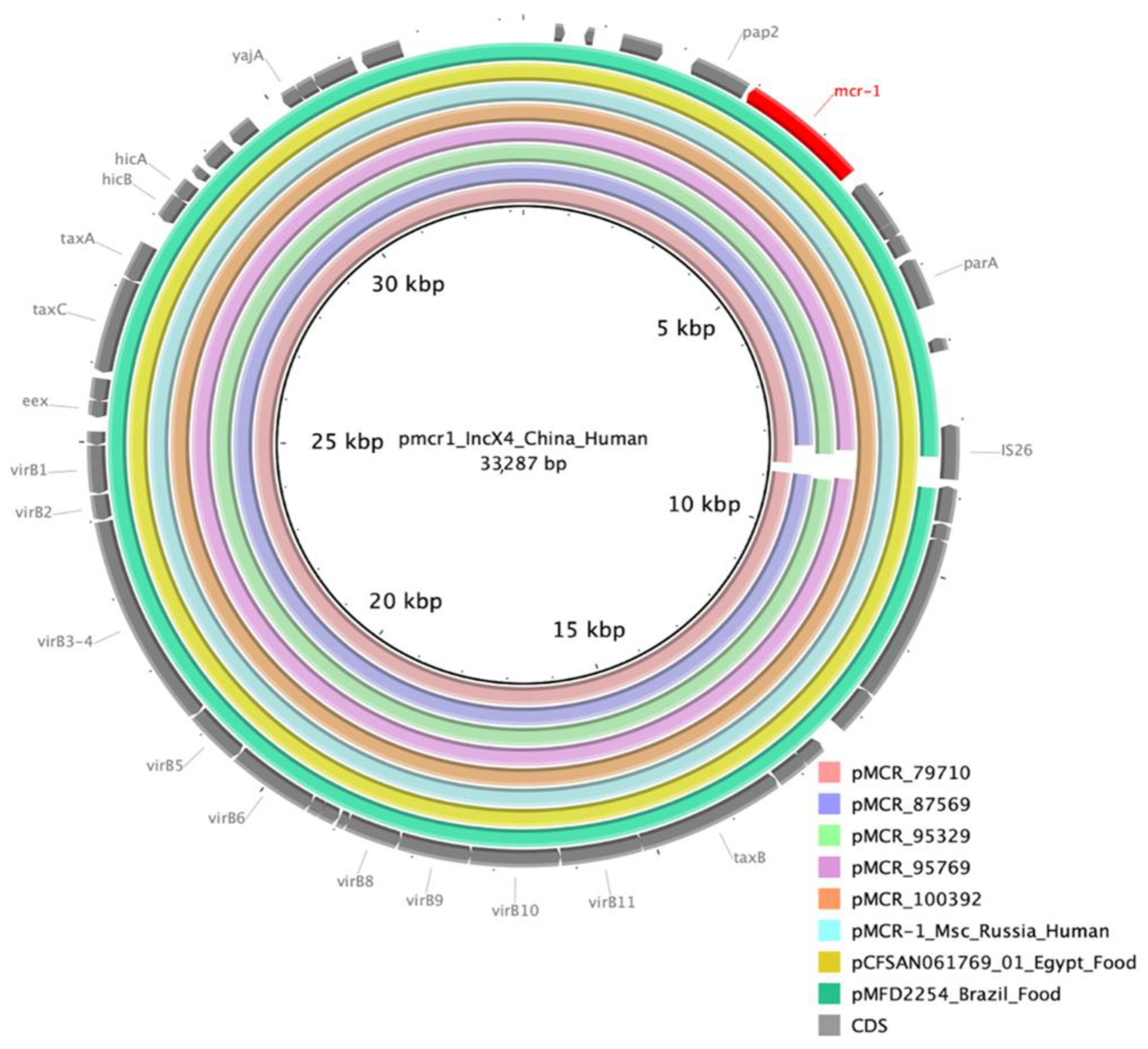
The size of the five IncX4 plasmids varied between 32,744 and 33,437 bp. A BLAST search against the NCBI nr/nt database showed high-nucleotide sequence identity (99% to 100%) and >99% coverage between these plasmids and the known mcr-bearing IncX4 plasmids reported from diverse epidemiological sources and geographic locations across the globe, such as pCFSAN061769_01 (GenBank acc. no. CP042970.1) of E. coli isolated from raw milk cheese in Egypt, pMCR-1_Msc (GenBank acc. no. NZ_MK172815.1) of E. coli isolated from human respiratory specimen in Moscow, Russia, pMFDS2254 (GenBank acc. no. MK869758.1) of E. coli isolated from chicken meat in Brazil, and pCFSA231 (GenBank acc. no. CP033349.2) of Salmonella enterica isolated from food sample in China. In addition to similar size and common backbone structure, all the above IncX4 plasmids showed the absence of ISApl1 element up- and downstream of mcr-1.1 and differed mainly in the presence or absence of IS26 element (Figure 3).
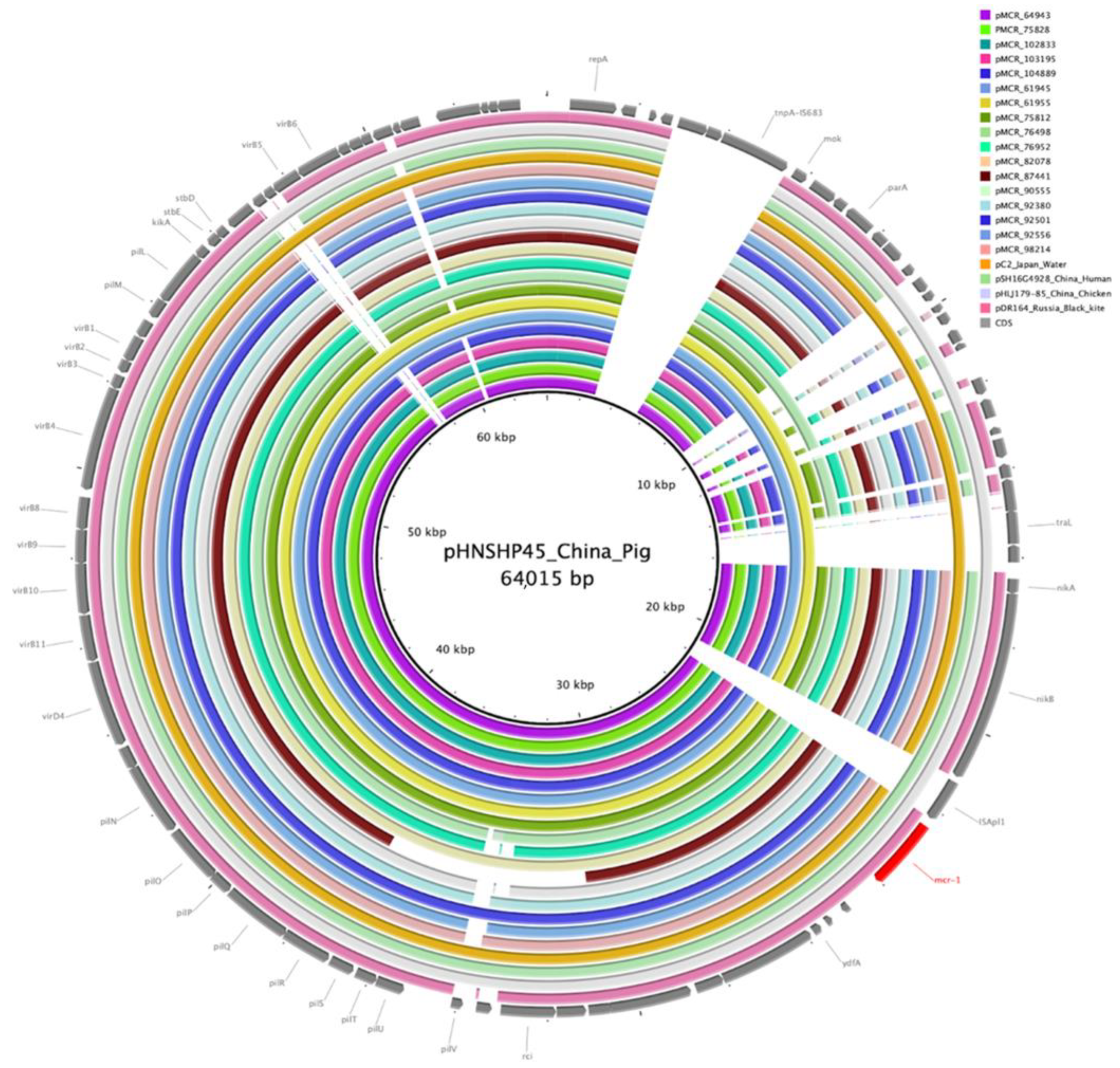
The length of the contigs that contained mcr-1.1 and IncI2 replicon genes ranged from 55,661 to 62,806 bp. These contigs contained typical IncI2 backbone elements, including the plasmid stability, replication, maintenance, and horizontal gene transfer regions. mcr-1.1 gene was located immediately downstream of the nikB gene encoding plasmid relaxase without an upstream ISApl1. The reported mcr-carrying IncI2 plasmids showed the highest (99% to 100%) sequence identity to the plasmids pC2 (GenBank acc. no. LC473131.1) of E. coli isolated from municipal wastewater in Japan, pDR164 (GenBank acc. no. MK542639.1) of E.coli isolated from wild bird in Russia, and several plasmids reported in China: pSh125-m2 (GenBank acc. no. KY363998.1) of Shigella sonnei and pSH16G4928 (GenBank acc. no. MH522426.1) of Salmonella enterica ser. Typhimurium isolated from human specimens, p5ZF15-2-1 (GenBank acc. no. CP079893.1) and pHLJ179-85 (GenBank acc. no. MN232214.1) of swine and chicken Escherichia fergusonii isolates (Figure 4).

4. Discussion

In this study, the prevalence of phenotypic colistin resistance and of the mobile colistin resistance (mcr-1) genes among clinical E. coli and K. pneumoniae isolates collected from hospitalized patients in Russia in 2013–2018 as part of sentinel surveillance was systematically assessed, and in-depth analysis of the genome sequences of mcr-bearing isolates and plasmids was performed. While there have been several reports of mcr-1 positive isolates recovered from human and animal sources in Russia [12,13,14,15], to our knowledge, this study is the first to report the prevalence and genomic context of mcr-1 in clinical isolates collected from geographically distant sites across Russia.
It should be noted that mcr has been observed even in isolates with colistin MICs below the clinical susceptibility breakpoint of ≤2 mg/L [63]. Therefore, in this study, PCR was used to screen for mcr in all isolates with colistin MIC of >0.5 mg/L to maximize the likelihood of detection. All mcr-positive isolates detected in this study, however, were resistant to colistin with MICs of 4 to16 mg/L. Phenotypic colistin resistance was detected in 2.24% of all E. coli and 9.3% of all K. pneumoniae isolates obtained in surveillance, but the prevalence of mcr-1 was relatively low: 0.51% and 0.04% (22.68% and 0.48% among colistin-resistant isolates), respectively. Similar prevalence of colistin resistance (2.1% in all Enterobacterales except Morganellaceae) and mcr gene presence (42.19% among colistin-resistant E. coli and 0.21% among colistin-resistant K. pneumoniae) have been observed in the INFORM global surveillance program [64]. These data likely suggest that, in human clinical isolates, mcr is still a less common mechanism of resistance to polymyxins than chromosomal mutations. Though important, the analysis of mutational resistance mechanisms to polymyxins in mcr-negative isolates was out of scope of this study, which was specifically focused on characterization of mcr-positive isolates. Worryingly, a frequent association of colistin resistance with resistances to other classes of antibiotics among mcr-positive isolates, especially to oxyimino-cephalosporins due to the presence of ESBL (blaCTX-M) and acquired cephalosporinase (blaCMY-2) genes, and to fluoroquinolones due to the presence of plasmid-mediated qnr and aac(6′)-Ib-cr genes and mutations in chromosomal genes, was observed in this study, as well as in others [65]. One E. coli isolate was found to co-harbor a ribosomal methyltransferase (armA) gene conferring pan-aminoglycoside resistance. None of the isolates carried carbapenemase genes; however, one K. pneumoniae isolate exhibited resistance to ertapenem presumably mediated by overproduction of CTX-M-55 ESBL and porin alteration [66].
Despite relatively low prevalence, wide geographical dissemination of mcr-1 was observed in Russia. All detected cases of infections due to mcr-positive isolates were epidemiologically unrelated and occurred in 17 hospitals in 16 geographically distant cities. The 22 E. coli isolates with mcr-1 were assigned to seven different phylogroups and 16 STs. The major clonal group was composed of ST359 and its double-locus variant, ST101, included six isolates. Both ST359 and ST101 have been previously reported among mcr-positive isolates of human and animal origin in Asia, Europe, Americas and Oceania [67]. The two K. pneumoniae isolates with mcr-1 were assigned to ST307 and ST23, which are both known as international ‘high-risk clones’ and have been commonly found in clinical settings in Russia [68,69,70,71,72,73]. To our knowledge, however, this is the first documented report of ST23 K. pneumoniae carrying both hypervirulence marker genes characteristic for this lineage and multiple acquired resistance genes, including mcr-1.1 and blaCTX-M-55.
The mcr-1 gene has been previously found in plasmids of more than 30 different replicon types, of which IncI2 and IncX4 have been the most common globally [74]. In this study, it was also detected mostly in IncI2 (n = 18) and IncX4 (n = 5) plasmids. Furthermore, significant structure and sequence similarity between the mcr-1-bearing IncI2 and IncX4 plasmids from this study and those reported earlier in isolates of different Enterobacterales species from different world regions and sources, including humans, livestock, wildlife animals, food and the environment, was observed. This once again confirms that the above plasmids are the main vehicle for mcr-1 transmission. Of notice, stable genetic environment of mcr-1 was found in all described plasmids and neither the presence of upstream ISApl1 element nor of the composite transposon Tn6330 (ISApl1-mcr-1-orf-ISApl1 structure), which has been proposed to be involved in initial mobilization and translocation of mcr-1 between various plasmid backbones [75], was detected.
In summary, this report provides a systematic and comprehensive analysis of the prevalence and molecular epidemiology of mcr-1-positive E. coli and K. pneumoniae in Russia. Further to previous reports, it shows the wider geographic spread of mcr-1, despite low prevalence in clinical settings. Given the frequent association of mcr-1 with other resistance determinants and its potential clinical impact, the continual surveillance of this threat is warranted.

Supplementary Materials

The following supporting information can be downloaded at: https://www.mdpi.com/article/10.3390/microorganisms10102034/s1, Table S1. Clinical information and antibiotic susceptibilities of mcr-positive isolates. Table S2. Illumina sequencing read and assembly statistics. Table S3. MinION sequencing read statistics and hybrid assembly summary. Table S4. Features of hybrid assembly contigs. Table S5. Summary of MinION reads spanning the entire tandem repeat region in IncFIB(K) plasmid of K. pneumoniae kp_87441. Table S6. Summary of MinION reads spanning the region with two copies of blaCTX-M-15 gene in IncM2 plasmid of E. coli ec_92380. Figure S1. Structure of tandem repeat region in 175,835-bp, IncFIB(K) plasmid carried by of K. pneumoniae kp_87441. Figure S2. Structure of repeat region in IncM2 plasmid of E. coli ec_92380.

Author Contributions

Conceptualization, V.S., I.A. and M.E.; methodology and experimental work: E.S. (Elvira Shaidullina), E.S. (Eugene Sheck), I.A., A.M. (Alexey Martinovich) A.M. (Alina Matsvay), M.D. and Y.S.; data validation and analysis, V.S., E.S. (Elvira Shaidullina) and M.E.; writing—original draft preparation, V.S.; writing—review and editing, M.E.; supervision, M.E. and K.K.; project administration, R.K. and G.S. All authors have read and agreed to the published version of the manuscript.

Funding

This research was funded by Russian Federal research grants No. 1022040800384-4-1.6.2 and No. 388-00154-22-00.

Data Availability Statement

All Illumina and MinION sequences and assemblies were deposited in the European Nucleotide Archive under the project accession number PRJEB53051.

Acknowledgments

We thank all the participants of the Russian sentinel antimicrobial resistance surveillance program for collection of clinical bacterial isolates used in this study. We also thank the entire IAC laboratory staff for maintaining the collection and susceptibility testing of isolates.

Conflicts of Interest

The authors declare no conflict of interest.

References

  1. Falagas, M.E.; Rafailidis, P.I. Nephrotoxicity of Colistin: New Insight into an Old Antibiotic. Clin. Infect. Dis. 2009, 48, 1729–1731. [Google Scholar] [CrossRef] [Green Version]
  2. Olaitan, A.; Morand, S.; Rolain, J.-M. Mechanisms of polymyxin resistance: Acquired and intrinsic resistance in bacteria. Front. Microbiol. 2014, 5, 643. [Google Scholar] [CrossRef] [PubMed] [Green Version]
  3. Gogry, F.A.; Siddiqui, M.T.; Sultan, I.; Haq, Q.M.R. Current Update on Intrinsic and Acquired Colistin Resistance Mechanisms in Bacteria. Front. Med. 2021, 8, 677720. [Google Scholar] [CrossRef] [PubMed]
  4. Liu, Y.-Y.; Wang, Y.; Walsh, T.R.; Yi, L.-X.; Zhang, R.; Spencer, J.; Doi, Y.; Tian, G.; Dong, B.; Huang, X.; et al. Emergence of Plasmid-Mediated Colistin Resistance Mechanism MCR-1 in Animals and Human Beings in China: A microbiological and Molecular Biological Study. Lancet Infect Dis. 2016, 16, 161–168. [Google Scholar] [CrossRef]
  5. Shen, Z.; Wang, Y.; Shen, Y.; Shen, J.; Wu, C. Early emergence of mcr-1 in Escherichia coli from food-producing animals. Lancet Infect. Dis. 2016, 16, 293. [Google Scholar] [CrossRef] [Green Version]
  6. Cherak, Z.; Loucif, L.; Moussi, A.; Rolain, J.-M. Epidemiology of Mobile Colistin Resistance (Mcr) Genes in Aquatic Environments. J Glob Antimicrob Resist 2021, 27, 51–62. [Google Scholar] [CrossRef]
  7. Li, X.-P.; Sun, R.-Y.; Song, J.-Q.; Fang, L.-X.; Zhang, R.-M.; Lian, X.-L.; Liao, X.-P.; Liu, Y.-H.; Lin, J.; Sun, J. Within-host heterogeneity and flexibility of mcr-1 transmission in chicken gut. Int. J. Antimicrob. Agents 2020, 55, 105806. [Google Scholar] [CrossRef]
  8. Martiny, H.-M.; Munk, P.; Brinch, C.; Szarvas, J.; Aarestrup, F.M.; Petersen, T.N. Global Distribution of mcr Gene Variants in 214K Metagenomic Samples. mSystems 2022, 7, 2. [Google Scholar] [CrossRef]
  9. Matamoros, S.; van Hattem, J.M.; Arcilla, M.S.; Willemse, N.; Melles, D.C.; Penders, J.; Vinh, T.N.; Hoa, N.T.; Bootsma, M.C.J.; van Genderen, P.J.; et al. Global phylogenetic analysis of Escherichia coli and plasmids carrying the mcr-1 gene indicates bacterial diversity but plasmid restriction. Sci. Rep. 2017, 7, 15364. [Google Scholar] [CrossRef] [Green Version]
  10. World Health Organization. GLASS Whole-Genome Sequencing for Surveillance of Antimicrobial Resistance, Electronic version; World Health Organization: Geneva, Switzerland, 2020; ISBN 9789240011007. [Google Scholar]
  11. Smelikova, E.; Tkadlec, J.; Krutova, M. How to: Screening for mcr-mediated resistance to colistin. Clin. Microbiol. Infect. 2022, 28, 43–50. [Google Scholar] [CrossRef] [PubMed]
  12. Tarabai, H.; Valcek, A.; Jamborova, I.; Vazhov, S.V.; Karyakin, I.V.; Raab, R.; Literak, I.; Dolejska, M. Plasmid-Mediated mcr-1 Colistin Resistance in Escherichia coli from a Black Kite in Russia. Antimicrob. Agents Chemother. 2019, 63, e01266-19. [Google Scholar] [CrossRef] [PubMed] [Green Version]
  13. Ageevets, V.; Lazareva, I.; Mrugova, T.; Gostev, V.; Lobzin, Y.; Sidorenko, S. IncX4 plasmids harbouring mcr-1 genes: Further dissemination. J. Glob. Antimicrob. Resist. 2019, 18, 166–167. [Google Scholar] [CrossRef]
  14. Sulian, O.; Ageevets, V.; Lazareva, I.; Gostev, V.; Popov, D.; Vostrikova, T.; Sukhinin, A.; Lobzin, Y.; Sidorenko, S. Co-production of MCR-1 and NDM-1 by Escherichia coli sequence type 31 isolated from a newborn in Moscow, Russia. Int. J. Infect. Dis. 2020, 101, 4–5. [Google Scholar] [CrossRef]
  15. Kuleshov, K.V.; Pavlova, A.S.; Shedko, E.D.; Mikhaylova, Y.V.; Margos, G.; Hepner, S.; Chebotar, I.V.; Korneenko, E.V.; Podkolzin, A.T.; Akimkin, V.G. Mobile Colistin Resistance Genetic Determinants of Non-Typhoid Salmonella enterica Isolates from Russia. Microorganisms 2021, 9, 2515. [Google Scholar] [CrossRef]
  16. Kuzmenkov, A.Y.; Trushin, I.V.; Vinogradova, A.G.; Avramenko, A.A.; Sukhorukova, M.V.; Malhotra-Kumar, S.; Dekhnich, A.V.; Edelstein, M.V.; Kozlov, R.S. AMRmap: An Interactive Web Platform for Analysis of Antimicrobial Resistance Surveillance Data in Russia. Front. Microbiol. 2021, 12, 620002. [Google Scholar] [CrossRef]
  17. ISO. Available online: https://www.iso.org/standard/70464.html (accessed on 20 September 2022).
  18. EUCAST. Available online: https://www.eucast.org/fileadmin/src/media/PDFs/EUCAST_files/Disk_test_documents/2022_manuals/ReadinR_guide_BMD_v_4.0_2022.pdf (accessed on 20 September 2022).
  19. EUCAST Clinical Breakpoints. Available online: https://www.eucast.org/clinical_breakpoints (accessed on 20 September 2022).
  20. Daniels, J.B.; Campbell, D.; Boyd, S.; Ansari, U.; Lutgring, J.; Rasheed, J.K.; Halpin, A.L.; Sjölund-Karlsson, M. Development and Validation of a Clinical Laboratory Improvement Amendments-Compliant Multiplex Real-Time PCR Assay for Detection of mcr Genes. Microb. Drug Resist. 2019, 25, 991–996. [Google Scholar] [CrossRef]
  21. Simon Andrews FastQC. Available online: https://www.bioinformatics.babraham.ac.uk/projects/fastqc/ (accessed on 14 June 2022).
  22. Quijada, N.M.; Rodríguez-Lázaro, D.; Eiros, J.M.; Hernández, M. TORMES: An automated pipeline for whole bacterial genome analysis. Bioinformatics 2019, 35, 4207–4212. [Google Scholar] [CrossRef]
  23. Bolger, A.M.; Lohse, M.; Usadel, B. Trimmomatic: A flexible trimmer for Illumina sequence data. Bioinformatics 2014, 30, 2114–2120. [Google Scholar] [CrossRef] [Green Version]
  24. Schmieder, R.; Edwards, R. Quality control and preprocessing of metagenomic datasets. Bioinformatics 2011, 27, 863–864. [Google Scholar] [CrossRef] [Green Version]
  25. Bankevich, A.; Nurk, S.; Antipov, D.; Gurevich, A.A.; Dvorkin, M.; Kulikov, A.S.; Lesin, V.M.; Nikolenko, S.I.; Pham, S.; Prjibelski, A.D.; et al. SPAdes: A new genome assembly algorithm and its applications to single-cell sequencing. J. Comput. Biol. 2012, 19, 455–477. [Google Scholar] [CrossRef]
  26. Gurevich, A.; Saveliev, V.; Vyahhi, N.; Tesler, G. QUAST: Quality assessment tool for genome assemblies. Bioinformatics 2013, 29, 1072–1075. [Google Scholar] [CrossRef] [Green Version]
  27. Wood, D.E.; Lu, J.; Langmead, B. Improved metagenomic analysis with Kraken 2. Genome Biol. 2019, 20, 257. [Google Scholar] [CrossRef] [PubMed] [Green Version]
  28. Wang, Q.; Garrity, G.M.; Tiedje, J.M.; Cole, J.R. Naïve Bayesian Classifier for Rapid Assignment of rRNA Sequences into the New Bacterial Taxonomy. Appl. Environ. Microbiol. 2007, 73, 5261–5267. [Google Scholar] [CrossRef] [PubMed] [Green Version]
  29. Douglass, A.P.; O’Brien, C.E.; Offei, B.; Coughlan, A.Y.; Ortiz-Merino, R.A.; Butler, G.; Byrne, K.P.; Wolfe, K.H. Coverage-Versus-Length Plots, a Simple Quality Control Step for de Novo Yeast Genome Sequence Assemblies. G3 Genes Genomes Genet. 2019, 9, 879–887. [Google Scholar] [CrossRef] [Green Version]
  30. Antipov, D.; Raiko, M.; Lapidus, A.; Pevzner, P.A. Plasmid detection and assembly in genomic and metagenomic data sets. Genome Res. 2019, 29, 961–968. [Google Scholar] [CrossRef] [PubMed] [Green Version]
  31. Lanfear, R.; Schalamun, M.; Kainer, D.; Wang, W.; Schwessinger, B. MinIONQC: Fast and simple quality control for MinION sequencing data. Bioinformatics 2019, 35, 523–525. [Google Scholar] [CrossRef] [Green Version]
  32. Wick, R.R.; Judd, L.M.; Cerdeira, L.T.; Hawkey, J.; Méric, G.; Vezina, B.; Wyres, K.L.; Holt, K.E. Trycycler: Consensus long-read assemblies for bacterial genomes. Genome Biol. 2021, 22, 266. [Google Scholar] [CrossRef]
  33. Chen, S.; Zhou, Y.; Chen, Y.; Gu, J. fastp: An ultra-fast all-in-one FASTQ preprocessor. Bioinformatics 2018, 34, i884–i890. [Google Scholar] [CrossRef]
  34. Ryan Wick Filtlong. Available online: https://github.com/rrwick/Filtlong (accessed on 12 June 2022).
  35. Kolmogorov, M.; Yuan, J.; Lin, Y.; Pevzner, P.A. Assembly of long, error-prone reads using repeat graphs. Nat. Biotechnol. 2019, 37, 540–546. [Google Scholar] [CrossRef]
  36. Wick, R.R.; Holt, K.E. Benchmarking of long-read assemblers for prokaryote whole genome sequencing. F1000Research 2019, 8, 2138. [Google Scholar] [CrossRef]
  37. Vaser, R.; Šikić, M. Time- and memory-efficient genome assembly with Raven. Nat. Comput. Sci. 2021, 1, 332–336. [Google Scholar] [CrossRef]
  38. Oxford Nanopore Technologies. Medaka. Available online: https://github.com/nanoporetech/medaka (accessed on 12 June 2022).
  39. Wick, R.R.; Holt, K.E. Polypolish: Short-read polishing of long-read bacterial genome assemblies. PLoS Comput. Biol. 2022, 18, e1009802. [Google Scholar] [CrossRef] [PubMed]
  40. Zimin, A.V.; Salzberg, S.L. The genome polishing tool POLCA makes fast and accurate corrections in genome assemblies. PLoS Comput. Biol. 2020, 16, e1007981. [Google Scholar] [CrossRef]
  41. Manni, M.; Berkeley, M.R.; Seppey, M.; Zdobnov, E.M. BUSCO: Assessing Genomic Data Quality and Beyond. Curr. Protoc. 2021, 1, e323. [Google Scholar] [CrossRef]
  42. Seemann, T. Prokka: Rapid Prokaryotic Genome Annotation. Bioinformatics 2014, 30, 2068–2069. [Google Scholar] [CrossRef] [Green Version]
  43. Beghain, J.; Bridier-Nahmias, A.; Le Nagard, H.; Denamur, E.; Clermont, O. ClermonTyping: An easy-to-use and accurate in silico method for Escherichia genus strain phylotyping. Microb. Genom. 2018, 4, e000192. [Google Scholar] [CrossRef] [PubMed]
  44. Torsten Seemann Mlst. Available online: https://github.com/tseemann/mlst (accessed on 17 June 2022).
  45. Jolley, K.A.; Maiden, M.C. BIGSdb: Scalable analysis of bacterial genome variation at the population level. BMC Bioinform. 2010, 11, 595. [Google Scholar] [CrossRef] [Green Version]
  46. Joensen, K.G.; Tetzschner, A.M.; Iguchi, A.; Aarestrup, F.M.; Scheutz, F. Rapid and Easy In Silico Serotyping of Escherichia coli Isolates by Use of Whole-Genome Sequencing Data. J. Clin. Microbiol. 2015, 53, 2410–2426. [Google Scholar] [CrossRef] [PubMed] [Green Version]
  47. Roer, L.; Tchesnokova, V.; Allesøe, R.; Muradova, M.; Chattopadhyay, S.; Ahrenfeldt, J.; Thomsen, M.C.F.; Lund, O.; Hansen, F.; Hammerum, A.M.; et al. Development of a Web Tool for Escherichia coli Subtyping Based on fimH Alleles. J. Clin. Microbiol. 2017, 55, 2538–2543. [Google Scholar] [CrossRef] [Green Version]
  48. Lam, M.M.C.; Wick, R.R.; Watts, S.C.; Cerdeira, L.T.; Wyres, K.L.; Holt, K.E. A genomic surveillance framework and genotyping tool for Klebsiella pneumoniae and its related species complex. Nat. Commun. 2021, 12, 4188. [Google Scholar] [CrossRef]
  49. Feldgarden, M.; Brover, V.; Gonzalez-Escalona, N.; Frye, J.G.; Haendiges, J.; Haft, D.H.; Hoffmann, M.; Pettengill, J.B.; Prasad, A.B.; Tillman, G.E.; et al. AMRFinderPlus and the Reference Gene Catalog facilitate examination of the genomic links among antimicrobial resistance, stress response, and virulence. Sci. Rep. 2021, 11, 12728. [Google Scholar] [CrossRef] [PubMed]
  50. Torsten Seemann Abricate. Available online: https://github.com/tseemann/abricate (accessed on 17 June 2022).
  51. Zankari, E.; Hasman, H.; Cosentino, S.; Vestergaard, M.; Rasmussen, S.; Lund, O.; Aarestrup, F.M.; Larsen, M.V. Identification of acquired antimicrobial resistance genes. J. Antimicrob. Chemother. 2012, 67, 2640–2644. [Google Scholar] [CrossRef] [PubMed]
  52. Ingle, D.J.; Valcanis, M.; Kuzevski, A.; Tauschek, M.; Inouye, M.; Stinear, T.; Levine, M.M.; Robins-Browne, R.M.; Holt, K.E. In silico serotyping of E. coli from short read data identifies limited novel O-loci but extensive diversity of O:H serotype combinations within and between pathogenic lineages. Microb. Genom. 2016, 2, e000064. [Google Scholar] [CrossRef] [PubMed]
  53. Chen, L.; Zheng, D.; Liu, B.; Yang, J.; Jin, Q. VFDB 2016: Hierarchical and refined dataset for big data analysis—10 years on. Nucleic Acids Res. 2016, 44, D694–D697. [Google Scholar] [CrossRef]
  54. Carattoli, A.; Zankari, E.; García-Fernández, A.; Voldby Larsen, M.; Lund, O.; Villa, L.; Møller Aarestrup, F.; Has-man, H. In Silico Detection and Typing of Plasmids Using PlasmidFinder and Plasmid Multilocus Sequence Typ-ing. Agents Chemother. 2014, 58, 3895–3903. [Google Scholar] [CrossRef] [Green Version]
  55. Siguier, P.; Perochon, J.; Lestrade, L.; Mahillon, J.; Chandler, M. ISfinder: The reference centre for bacterial insertion sequences. Nucleic Acids Res. 2006, 34, D32–D36. [Google Scholar] [CrossRef] [Green Version]
  56. Camacho, C.; Coulouris, G.; Avagyan, V.; Ma, N.; Papadopoulos, J.; Bealer, K.; Madden, T.L. BLAST+: Architecture and applications. BMC Bioinform. 2009, 10, 421. [Google Scholar] [CrossRef] [Green Version]
  57. Page, A.J.; Cummins, C.A.; Hunt, M.; Wong, V.K.; Reuter, S.; Holden, M.T.G.; Fookes, M.; Falush, D.; Keane, J.A.; Parkhill, J. Roary: Rapid large-scale prokaryote pan genome analysis. Bioinformatics 2015, 31, 3691–3693. [Google Scholar] [CrossRef] [Green Version]
  58. Minh, B.Q.; Schmidt, H.A.; Chernomor, O.; Schrempf, D.; Woodhams, M.D.; von Haeseler, A.; Lanfear, R. IQ-TREE 2: New Models and Efficient Methods for Phylogenetic Inference in the Genomic Era. Mol. Biol. Evol. 2020, 37, 1530–1534. [Google Scholar] [CrossRef] [Green Version]
  59. Kalyaanamoorthy, S.; Minh, B.Q.; Wong, T.K.F.; Von Haeseler, A.; Jermiin, L.S. ModelFinder: Fast model selection for accurate phylogenetic estimates. Nat. Methods 2017, 14, 587–589. [Google Scholar] [CrossRef]
  60. Page, A.J.; Taylor, B.; Delaney, A.J.; Soares, J.; Seemann, T.; Keane, J.A.; Harris, S.R. SNP-sites: Rapid efficient extraction of SNPs from multi-FASTA alignments. Microb. Genom. 2016, 2, e000056. [Google Scholar] [CrossRef] [PubMed] [Green Version]
  61. Letunic, I.; Bork, P. Interactive Tree Of Life (iTOL) v5: An online tool for phylogenetic tree display and annotation. Nucleic Acids Res. 2021, 49, W293–W296. [Google Scholar] [CrossRef] [PubMed]
  62. Robertson, J.; Nash, J.H.E. MOB-suite: Software tools for clustering, reconstruction and typing of plasmids from draft assemblies. Microb. Genom. 2018, 4, e000206. [Google Scholar] [CrossRef] [PubMed]
  63. Alikhan, N.-F.; Petty, N.K.; Ben Zakour, N.L.; Beatson, S.A. BLAST Ring Image Generator (BRIG): Simple prokaryote genome comparisons. BMC Genom. 2011, 12, 402. [Google Scholar] [CrossRef] [PubMed] [Green Version]
  64. Wise, M.G.; Estabrook, M.A.; Sahm, D.F.; Stone, G.G.; Kazmierczak, K.M. Prevalence of mcr-type genes among colistin-resistant Enterobacteriaceae collected in 2014-2016 as part of the INFORM global surveillance program. PLoS ONE 2018, 13, e0195281. [Google Scholar] [CrossRef] [Green Version]
  65. Luo, Q.; Wang, Y.; Xiao, Y. Prevalence and transmission of mobilized colistin resistance (mcr) gene in bacteria common to animals and humans. Biosaf. Health 2020, 2, 71–78. [Google Scholar] [CrossRef]
  66. Doumith, M.; Ellington, M.J.; Livermore, D.M.; Woodford, N. Molecular mechanisms disrupting porin expression in ertapenem-resistant Klebsiella and Enterobacter spp. clinical isolates from the UK. J. Antimicrob. Chemother. 2009, 63, 659–667. [Google Scholar] [CrossRef] [PubMed]
  67. Dadashi, M.; Sameni, F.; Bostanshirin, N.; Yaslianifard, S.; Khosravi-Dehaghi, N.; Nasiri, M.J.; Goudarzi, M.; Hashemi, A.; Hajikhani, B. Global prevalence and molecular epidemiology of mcr-mediated colistin resistance in Escherichia coli clinical isolates: A systematic review. J. Glob. Antimicrob. Resist. 2022, 29, 444–461. [Google Scholar] [CrossRef] [PubMed]
  68. Lev, A.I.; Astashkin, E.I.; Kislichkina, A.A.; Solovieva, E.V.; Kombarova, T.I.; Korobova, O.V.; Ershova, O.N.; Alexandrova, I.A.; Malikov, V.E.; Bogun, A.G.; et al. Comparative analysis of Klebsiella pneumoniae strains isolated in 2012–2016 that differ by antibiotic resistance genes and virulence genes profiles. Pathog. Glob. Health 2018, 112, 142–151. [Google Scholar] [CrossRef] [PubMed]
  69. Shamina, O.; Kryzhanovskaya, O.; Lazareva, A.; Alyabieva, N.; Polikarpova, S.; Karaseva, O.; Mayanskiy, N. Emergence of a ST307 clone carrying a novel insertion element MITEKpn1 in the mgrB gene among carbapenem-resistant Klebsiella pneumoniae from Moscow, Russia. Int. J. Antimicrob. Agents 2020, 55, 105850. [Google Scholar] [CrossRef]
  70. Shaidullina, E.; Shelenkov, A.; Yanushevich, Y.; Mikhaylova, Y.; Shagin, D.; Alexandrova, I.; Ershova, O.; Akimkin, V.; Kozlov, R.; Edelstein, M. Antimicrobial Resistance and Genomic Characterization of OXA-48- and CTX-M-15-co-Producing Hypervirulent Klebsiella pneumoniae ST23 Recovered from Nosocomial Outbreak. Antibiotics 2020, 9, 862. [Google Scholar] [CrossRef] [PubMed]
  71. Fursova, N.K.; Astashkin, E.I.; Gabrielyan, N.I.; Novikova, T.S.; Fedyukina, G.N.; Kubanova, M.K.; Esenova, N.M.; Sharapchenko, S.O.; Volozhantsev, N.V. Emergence of Five Genetic Lines ST395NDM-1, ST13OXA-48, ST3346OXA-48, ST39CTX-M-14, and Novel ST3551OXA-48 of Multidrug-Resistant Clinical Klebsiella pneumoniae in Russia. Microb. Drug Resist. 2020, 26, 924–933. [Google Scholar] [CrossRef] [PubMed]
  72. Fursova, N.K.; Astashkin, E.I.; Ershova, O.N.; Aleksandrova, I.A.; Savin, I.A.; Novikova, T.S.; Fedyukina, G.N.; Kislichkina, A.A.; Fursov, M.V.; Kuzina, E.S.; et al. Multidrug-Resistant Klebsiella pneumoniae Causing Severe Infections in the Neuro-ICU. Antibiotics 2021, 10, 979. [Google Scholar] [CrossRef] [PubMed]
  73. Khrulnova, S.; Fedorova, A.; Frolova, I.; Tandilova, K.; Likold, E.; Klyasova, G. Distribution of virulence genes and capsule types in Klebsiella pneumoniae among bloodstream isolates from patients with hematological malignancies. Diagn. Microbiol. Infect. Dis. 2022, 104, 115744. [Google Scholar] [CrossRef] [PubMed]
  74. Mmatli, M.; Marylucy Mbelle, N.; Sekyere, J.O. Global Epidemiology and Genetic Environment of Mcr Genes: A One Health Systematic Review of Current and Emerging Trends. medRxiv 2022. [Google Scholar] [CrossRef]
  75. Wang, R.; van Dorp, L.; Shaw, L.P.; Bradley, P.; Wang, Q.; Wang, X.; Jin, L.; Zhang, Q.; Liu, Y.; Rieux, A.; et al. The global distribution and spread of the mobilized colistin resistance gene mcr-1. Nat. Commun. 2018, 9, 117. [Google Scholar] [CrossRef] [PubMed]
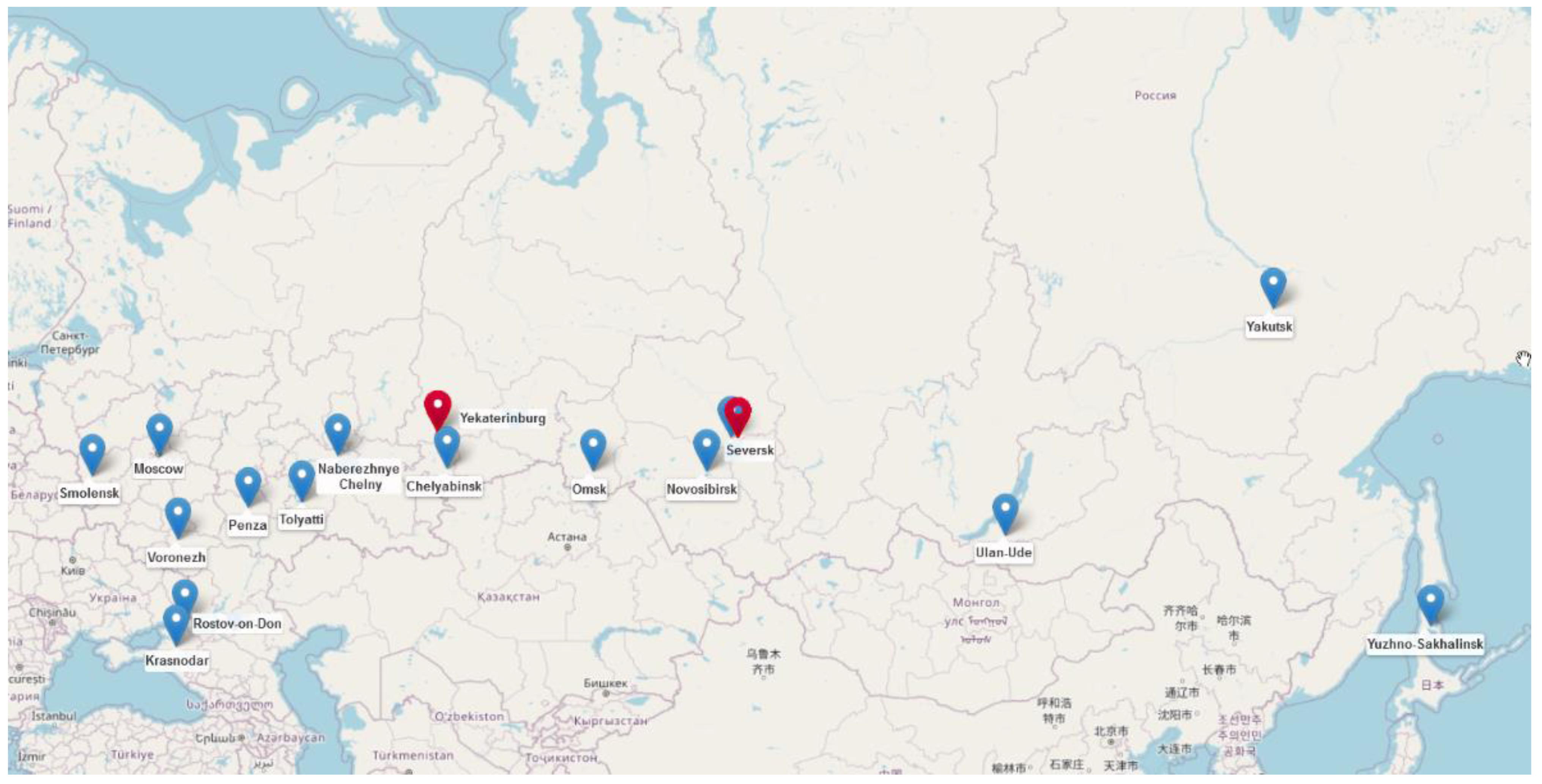
Figure 1. Map showing the geographic origins of mcr-positive E. coli (blue markers) and K. pneumoniae (red markers) isolates from this study.
Microorganisms 10 02034 g001
Figure 2. Core-genome SNP tree of 22 mcr-positive E. coli isolates. Sequence types (STs) and mcr-plasmid types are indicated next to the isolate names. Different E. coli phylogroups are shown by colored ellipses.
Microorganisms 10 02034 g002
Figure 3. Sequence alignment of five IncX4 mcr-bearing plasmids from this study and references from GenBank. The sequence of IncX4 plasmid pmcr1 (GenBank acc. no. KU761327) was used as a reference (inner circle). The concentric circles show similar plasmids from this study and GenBank references and are colored according to legend. Gaps in the circles correspond to plasmid regions which are missing compared to the reference. The key plasmid genes and mcr-1.1 are shown in the outer circle with gray and red arrows.
Microorganisms 10 02034 g003
Figure 4. Sequence alignment of seventeen IncI2 mcr-bearing plasmids from this study and references from GenBank. The sequence of IncI2 plasmid pHNSHP45 (GenBank acc no. KP347127) was used as a reference (inner circle). The concentric circles show similar plasmids from this study and GenBank references and are colored according to legend. Gaps in the circles correspond to plasmid regions which are missing compared to the reference. The key plasmid genes and mcr-1.1 are shown in the outer circle with gray and red arrows.
Microorganisms 10 02034 g004
Table 1. Characteristics of mcr-positive E. coli isolates.
IDCollection DatePhylo-GroupSTSerotypefimH TypePlasmid Type (Length of mcr-1.1 Contig)ARGs
ec_619451 January 2013B1ST1431O8:H19fimH32IncI2 (60859 bp)mcr-1.1, blaCTX-M-1, aac(6′)-Ib-cr, arr-3, dfrA27, aadA16, qacEdelta1, sul1, sul2, tet(A)
ec_619555 August 2013B1ST6817O85:H23fimH31IncI2 (60860 bp)mcr-1.1, blaTEM-1, blaCTX-M-15, dfrA12, aadA2, cmlA1, aadA1, qacL, tet(A), tet(M), sul3, aph(3′)-Ia
ec_6494330 May 2014CST23O78:H4fimH35IncI2 (61030 bp)mcr-1.1, catA1, dfrA17, aadA5, tet(B), blaTEM-1, sul2, aph(3″)-Ib, aph(6)-Id, aph(4)-Ia, aac(3)-IVa, qnrB19
ec_758121 June 2015B1new (SLV ST461)O178:H28fimH31IncI2 (60903 bp)mcr-1.1, sul1, qacEdelta1, aac(3)-VIa, aadA1, qnrS1, blaCTX-M-15
ec_758282 June 2015B1ST359O23:H21fimH35IncI2 (61036 bp)mcr-1.1, aph(3′)-Ia, aph(6)-Id, aph(3″)-Ib, tet(B), blaTEM-1, sul2
ec_7649816 June 2015CST410O66:H45fimH24IncI2 (62806 bp)mcr-1.1, blaCTX-M-27, floR, aac(6′)-Ib-cr, arr-3, dfrA27, aadA16, qacEdelta1, sul1, mph(A), blaTEM-1, aac(3)-IId, bleO, dfrA17, catA1, tet(B), blaTEM-1, sul2, aph(3″)-Ib, aph(6)-Id, aph(3′)-Ia, blaCMY-2
ec_7695222 October 2015AST617O101:H10fimH29IncI2 (59391 bp)mcr-1.1, mph(A), catB3, blaOXA-1, aac(6′)-Ib-cr, aph(4)-Ia, aac(3)-IVa, aph(6)-Id, aph(3″)-Ib, tet(B), blaCTX-M-15, sul1, qacEdelta1, aadA5, dfrA17
ec_7971016 March 2016B1ST359O36:H31fimH35IncX4 (33106 bp)mcr-1.1, blaCTX-M-1, tet(A), sul1, qacEdelta1, aac(3)-VIa, aadA1, aac(3)-IId, blaTEM-1, mph(A)
ec_820789 June 2016B1ST359O29:H31fimH35IncI2 (61024 bp)mcr-1.1, tet(A), dfrA12, mph(A), sul1, qacEdelta1, cmlA1, aadA1, qacL, sul3, aph(4)-Ia, aac(3)-IVa, aph(6)-Id, aph(3″)-Ib, aadA2
ec_8759515 March 2017GST117O45:H51fimH97IncX4 (32744 bp)mcr-1.1, blaCMY-2, mph(A), sul1, qacEdelta1, aadA2, dfrA12, tet(A), sul2, aph(3″)-Ib, aph(6)-Id
ec_905557 March 2017B1ST162O88:H21fimH32IncI2 (60006 bp)mcr-1.1, blaTEM-1, blaCTX-M-65, mph(A), sul1, qacEdelta1, aadA5, dfrA17, aac(3)-IId, floR, fosA3, aph(3″)-Ib, aph(6)-Id, tet(A), sul2
ec_9238017 July 2017EST1011O188:H28fimH31IncI2 (60934 bp)mcr-1.1, blaTEM-1, blaTEM-1, armA, msr(E), mph(E), blaCTX-M-15, blaCTX-M-15
ec_9250114 August 2017B1ST602O-*:H21fimH86IncI2 (61163 bp)mcr-1.1, floR, blaTEM-1, aph(6)-Id, aph(3″)-Ib, dfrA17, aac(3)-IId, fosA7.5, sul2, catA1, blaCTX-M-169
ec_9255623 September 2017B1ST2179O9/09a:H9fimH32IncI2 (59951 bp)mcr-1.1, blaTEM-1, blaCTX-M-65, tet(A), aph(6)-Id, aph(3″)-Ib, sul2, mph(A), floR
ec_9463010 October 2017AST617O101/O9:H9*IncI2 ** (23072 bp)mcr-1.1, blaCTX-M-55, blaTEM, aph(3′)-IIa, ble, dfrA14, aac(6′)-Ib-cr, arr-3, dfrA27, aadA16, qacEdelta1, sul1, qnrB6, qacEdelta1, sul1, aph(6)-Id, aph(3″)-Ib, sul2, floR
ec_9576928 November 2017DST69O15:H18fimH27IncX4 (32819 bp)mcr-1.1, blaCTX-M-15, qnrS1, blaTEM-1, aph(6)-Id, aph(3″)-Ib, sul2, tet(A), dfrA14
ec_965481 November 2017FST457O11:H25fimH145IncHI2 * (2325 bp)mcr-1.1, blaTEM-1, aadA2, tet(A), sul3, qacL, aadA1, cmlA1, floR, sul1, qacEdelta1, lnu(F), dfrA14, mph(A)
ec_9821422 March 2018EST1011O188:H28fimH31IncI2 (60456 bp)mcr-1.1, blaTEM-1
ec_1003923 April 2018B1ST359O36:H31fimH35IncX4 (33437 bp)mcr-1.1, blaCTX-M-55
ec_10283328 April 2018B1ST4566O163:H23fimH60IncI2 (61680 bp)mcr-1.1, tet(A), sul2, blaCTX-M-1
ec_10319527 September 2018B1ST359O36:H31fimH35IncI2 (60820 bp)mcr-1.1, sul2, blaCTX-M-1, tet(A), mph(A), dfrA17, aac(3)-IId, blaTEM-1
ec_10488931 October 2018B1ST101O115:H31fimH86IncI2 (61030 bp)mcr-1.1, aph(3″)-Ib, aph(6)-Id, blaCTX-M-55, catA1, dfrA14, mph(A), tet(B)
* Not determined; ** Partial sequence.
Table 2. Characteristics of mcr-positive K. pneumoniae isolates.
IDCollection DateSTK_LocusO_LocusPlasmid Type (Length of mcr-1.1 Contig)ARGs
kp_874419 February 2017307KL102O2v2IncI2 (55661 bp)mcr-1.1, oqxB19, oqxA, blaSHV-28, fosA, sul3, qacL, aadA1, cmlA1, aadA2, sul2, aph(3″)-Ib, aph(6)-Id, blaTEM-1, blaCTX-M-15, dfrA14, aac(3)-IIe, qnrB1, tet(A), catB3, blaOXA-1, aac(6′)-Ib-cr, tet(A), catB3, blaOXA-1, aac(6′)-Ib-cr, tet(A), catB3, blaOXA-1, aac(6′)-Ib-cr
kp_9532927 December 201723KL57O2v2IncX4 (32850 bp)mcr-1.1, aac(3)-IIe, catB3, blaOXA-1, aac(6′)-Ib-cr, oqxA, oqxB, blaSHV-1, fosA, qnrS1, blaLAP-2, tet(A), sul2, blaCTX-M-55
Publisher’s Note: MDPI stays neutral with regard to jurisdictional claims in published maps and institutional affiliations.