Article

Molecular Detection of Tick-Borne Bacteria from Amblyomma (Acari: Ixodidae) Ticks Collected from Reptiles in South Africa

by
Lehlohonolo S. Mofokeng
,
Nico J. Smit
and
Courtney A. Cook
*
Water Research Group, Unit for Environmental Sciences and Management, North-West University, Potchefstroom 2531, South Africa
*
Author to whom correspondence should be addressed.
Microorganisms 2022, 10(10), 1923; https://doi.org/10.3390/microorganisms10101923
Submission received: 2 September 2022 / Revised: 19 September 2022 / Accepted: 23 September 2022 / Published: 28 September 2022
(This article belongs to the Special Issue Molecular Detection and Genotypic Analysis of Tick-Borne Pathogens)

Abstract

:
Reptiles are hosts for various tick species and tick-associated organisms, many of which are zoonotic. However, little is known about the presence and diversity of tick-borne bacteria infecting reptiles and their ticks in South Africa. Amblyomma ticks (n = 253) collected from reptiles were screened for the presence of Coxiella, Anaplasma, Rickettsia, and Borrelia species by amplification, sequencing and phylogenetic analysis of the 16S rRNA, 23S rRNA, gltA, OmpA, and Flagellin genes, respectively. This study recorded the presence of reptile associated Borrelia species and Coxiella-like endosymbiont in South Africa for the first time. Furthermore, a spotted fever group Rickettsia species was observed in 7 Amblyomma marmoreum and 14 Amblyomma sylvaticum from tortoises of genera Kinixys and Chersina. Francisella-like endosymbiont was observed from 2 Amblyomma latum collected from the Mozambique spitting cobra, Naja mossambica. Coxiella burnetii and Anaplasma spp., were not detected from the current samples. Although the direct evidence that reptiles can act as reservoir hosts remains to be determined, observations from this study provide indications that reptilian ticks may play a role in the transmission of pathogenic bacteria to homothermic animals. Furthermore, the absence of Anaplasma spp., and C. burnetii does not mean that these pathogens should be completely neglected.

1. Introduction

Tick-borne pathogens (TBPs) are responsible for some of the most severe emerging infectious diseases across the world [1,2]. The wildlife-domestic-human interaction has the potential to accelerate the spread of zoonotic tick-borne pathogens that cause significant diseases and eventually death [2,3]. To date, roughly 61% of human diseases have been documented to have a zoonotic origin, and wildlife is associated with approximately 75% of emerging zoonotic infections globally [4]. In South Africa, four tick-borne spotted fever groups (SFG) Rickettsia spp. (Rickettsia africae, Rickettsia conorii conorii, Rickettsia aeschlimannii and Rickettsia sibirica mongolitimonae) have been associated with human diseases [5,6,7]. Additionally, members of the family Ixodidae are involved in the epidemiology of other pathogens such as Borrelia, Anaplasma and Coxiella spp. An increasing number of studies have implicated reptiles and their associated ticks as potential reservoirs involved in the transmission cycles of TBPs [2,8,9]. This fact is of great concern in tick-borne disease control strategies, as these ectotherms are usually asymptomatic carriers that may serve as reservoirs of the infection. Although diverse tick-borne pathogens are emerging across the world, with documented effects on the health of humans and livestock, studies into their diversity and specific interactions with ticks and their reptilian host species in South Africa are lacking. In particular, the role of these ectotherms in the epidemiology of TBPs remains unknown, despite the fact that exported ticks and reptiles (Amblyomma marmoreum, tortoises and lizards) have been implicated with TBPs of livestock [2,10]. This leads, therefore, to a lack of knowledge on the diversity of these pathogens in reptiles and whether or not reptiles can act as reservoir hosts for TBPs infecting domestic animals and humans.
In addition to pathogens, ticks also harbor various microbiomes which include symbiotic and commensal bacteria [11]. Tick endosymbionts have gained interest over the last decade and are now being investigated due to their physiological significance in ticks [12]. These endosymbionts are involved in the reproduction and growth of ticks by providing these arthropods with the necessary compounds that are absent in their haematophagous diet [13,14]. Furthermore, they are also involved in the maintenance of pathogens in arthropods [15]. To date, three genera of symbionts (Coxiella-like endosymbionts, Francisella-like endosymbionts, and Midichloria) have been documented to be exclusive to ticks, and some of these symbionts are mutualistic, whereas others may be facultative [11,16].
Despite the above, the presence and diversity of bacteria associated with ticks of reptiles, particularly those species that can also infest livestock and are close to human settlements, have not been studied in South Africa. This lack of information on tick-borne bacteria of reptilian ticks in South Africa warrants novel research in this area. Furthermore, since the literature related to tick-borne bacteria in reptiles and their associated ectoparasites is very recent, and most of these studies have been conducted in South America [9,17,18], the aim of this study was to investigate the presence of bacterial microorganisms from reptile associated ticks in South Africa.

2. Materials and Methods

2.1. Sample Collection

Ticks from 36 reptiles were collected in the Ndumo Game Reserve, KwaZulu-Natal (KZN) (32°18′49″ E; 26°54′33″ S) (2014–2022) and the region near the village of Paternoster, Western Cape (WC) (17°51′56″ E; 32°49′2″ S) (2011). Upon collection, the ticks were placed in falcon tubes with a damp piece of cottonwool, kept in a dark, cool environment, and allowed to digest their blood meals for approximately 10–20 days [19,20]. Thereafter, they were fixed in molecular grade 70% ethanol (Sigma-Aldrich, St. Louis, MI, USA). Reptiles were restrained by hand whilst ticks were collected in situ. Sampling was carried out under the relevant permits (KZN: OP 839/2014, OP 4374/2015, OP 4092/2016, OP 4264/2017; WC: 0035-AAA004-00383) and was approved by the North- West University Anim-Care Animal Research Ethics Committee, South Africa (NWU-00372-16-A5). Species identification of the collected ticks was carried out under a Nikon AZ100M stereo-microscope, and guidelines for tick identification were conducted as provided by Theiler [21] and Theiler and Salisbury [22]. The molecular methods as described in [20] were used to confirm morphological identification.

2.2. PCR and Phylogenetic Analysis

The DNA from whole tick specimens were extracted using the NucleoSpin®Tissue Genomic DNA Tissue Kit (Machery-Nagel, Duren, Germany). DNA was then frozen at −20 °C for further molecular studies.
Molecular characterization of bacterial microorganisms was performed via PCR amplification, amplifying different bacterial genes (Table 1). Conditions for both PCR and nested PCR of all bacteria were as follows: initial denaturation at 95 °C for 3 min, followed by 35 cycles, entailing a 95 °C denaturation for 30 s, annealing at different temperatures as indicated in Table 1, for 30 s with an end extension at 72 °C for 2 min, and following the cycles a final extension of 72 °C for 10 min. PCR reactions were conducted in a 25 μL reaction mixture containing 12.5 μL Thermo Scientific DreamTaq PCR master mix (2×) (2× DreamTaq buffer, 0.4 mM of each dNTP, and 4 mM MgCl2), 1.25 μL of each primer (10 μM), and at least 25 ng of DNA template. PCR grade nuclease free water (Thermo Scientific, Vilnius, Lithuania) was used to make up the final reaction volume. Reactions were undertaken in a Bio-Rad C1000 Touch™ Thermal Cycler PCR machine (Bio-Rad, Hemel Hempstead, UK). PCR and nested PCR amplicons were visualized on 1% GelRed® (Biotium Inc., Hayward, CA, USA) stained agarose gels. Visualization was performed under UV light.
The positive products of PCR and nested PCR were purified and sequenced by Inqaba Biotechnical Industries (Pty) Ltd. (Pretoria, South Africa). The obtained sequences were compared with GenBank references using the Basic Local Alignment Search Tool (BLAST), and deposited in the NCBI GenBank database.
In addition, phylogenetic reconstruction was performed. For phylogenetic analysis, comparative sequences of the Coxiella spp., Borrelia and Rickettsia species were downloaded from GenBank and aligned to the sequences generated within this study. Sequences were aligned using the Clustal W alignment tool under the default settings and implemented in MEGA Ver 11.1 [23]. To infer phylogenetic relationships, the maximum likelihood (ML) method was used. Prior to the analyses, a model test was performed to determine the most suitable nucleotide substitution model according to the Akaike information criterion (AIC) using jModelTest 2.1.7 [24,25]. For the ML analysis, nodal support was assessed using 1000 rapid bootstrap inferences.
Table 1. List of primer sequences used for the detection of bacterial species.
SpeciesAssayPrimer SequenceAnnealing (°C)Product SizeTarget GeneReferences
Universal bacterial primersPCRTCAAAKGAATTGACGGGGGC
GCCCGGGAACGTATTCAC
50478 bp16S rRNA[26]
Anaplasma spp.PCRCAGAGTTTGATCCTGGCTCAGAACG
GAGTTTGCCGGGACTTCTTCTGAA
50467 bp16S rRNA[27]
Borrelia spp.PCRAGAGCAACTTACAGACGAAATTAAT
CAAGTCTATTTTGGAAAGCACCTAA
54482 bpFlagellin gene[28]
Borrelia spp.PCRTGCGTCTTAAGCATGCAAGT
GTACAAAGGCCCGAGAACGTA
561344 bp16S rRNA[29]
nPCRCATGCAAGTCAAACGGAATG
ACTGTTTCGCTTCGCTTTGT
561205 bp16S rRNA[29]
Rickettsia spp.PCRGGGGGCCTGCTCACGGCGG
ATTGCAAAAAGTACAGTGAAC
48382 bpgltA gene[30]
Rickettsia spp.PCRTGGCGAATATT TCTCCAAAAA
GTTCCGTTA ATGGCAGCATCT
46631 bpompA gene[31]
Coxiella spp.PCRCGTAGGAATCTACCTTRTAGWGG
GCCTACCCGCTTCTGGTACAATT
52832–939 bp16S rRNA[32]
nPCRCGTAGGAATCTACCTTRTAGWGG
TGAGAACTAGCTGTTGGTTAGT
52624–627 bp16S rRNA[32]
Coxiella spp.PCRGCCTGCGAWAAGCTTCGGGGAG
CTCCTAKCCACASCTCATCCCC
56694–1188 bp23S rRNA[32]
nPCRGATCCGGAGATWTCYGAATGGGG
TCGYTCGGTTTCGGGTCKACTC
56583–867 bp23S rRNA[32]
Coxiella burnetiiPCRACT CAA CGC ACT GGA ACC GC
TAG CTG AAG CCA ATT CGC C
50257 bphtpAB[33]

2.3. Statistical Analysis

The 95% confidence interval was calculated using the formula: P ± Z × P   ( 1 P ) n .

3. Results

3.1. Overall Prevalence

Two hundred and fifty-three ticks (80 males, 21 females, and 12 larval stages of A. marmoreum; 40 males and three females of A. latum; 36 males and one female of A. exornatum; and 49 males and 11 females of A. sylvaticum) (Table S1) were collected from 36 reptiles and screened for tick-borne bacterial microorganisms (142 ticks in KZN; 111 ticks in WC). Based on the PCR results, all four tick species showed evidence of infection with at least one bacterial microorganism (see Table 2). A spotted fever group Rickettsia species was detected from 21 out of 253 samples (8.3%) (95%CI = ±3.39), reptile associated Borrelia species was detected in 16 out of 253 samples (6.3%) (95%CI = ±2.99), Francisella-like endosymbiont in two out of 253 samples (0.8%) (95%CI = ±1.10), and Coxiella-like endosymbiont (CLE) in 104 out of 253 samples (41.1%) (95%CI = ±6.06) (Table 2). Mixed infections of CLE and Borrelia spp. were observed in nine out of 253 samples (3.6%) (95%CI = ±2.21). The Anaplasma species and Coxiella burnetii were not found in the ticks examined.
In KZN, three genera of bacteria were observed in Amblyomma ticks. Borrelia spp. was observed at a prevalence of 5.6% (95%CI = ±3.78), Francisella-like endosymbiont at 1.4% (95%CI = ±1.93), and Coxiella-like endosymbiont at 21.1% (95%CI = ±6.71), while in WC, Borrelia sp. was observed at 7.2% (95%CI = ±4.81), Coxiella-like endosymbiont at 66.7% (95%CI = ±8.77), and Rickettsia sp., at 18.9% (95%CI = ±7.28).
Francisella-like endosymbiont was observed only in A. latum at a prevalence of 4.7% (95%CI = ±6.33), while A. sylvaticum harbored Rickettsia sp. at a prevalence of 23.3% (95%CI = ±10.61), Coxiella-like endosymbiont at a prevalence of 45.9% (95%CI = ±12.61), and Borrelia sp. at a prevalence of 6.7% (95%CI = ±6.33). Three bacterial genera were observed in A. marmoreum. Borrelia sp. was observed at a prevalence of 10.6% (95%CI = ±5.68), Coxiella-like endosymbiont at 35.4%, (95%CI = ±8.82) and Rickettsia spp. at 6.2% (95%CI = ±1.16). Coxiella-like endosymbiont was observed at a prevalence of 32.4% (95%CI = ±15.08) in A. exornatum. Mixed infections of Coxiella-like endosymbiont and Borrelia sp. were observed at a prevalence of 6.19% (95%CI = ±4.44) in A. marmoreum, and 3.33% (95%CI = ±4.52) in A. sylvaticum.

3.2. Phylogenetic Analysis of Bacterial Microorganisms in Amblyomma Ticks

According to the BLASTn analysis, the ~478 bp fragment of 16S rRNA for the universal bacterial microorganisms of A. latum yielded 98–99% with uncultured Francisella species (MN998636-MN998638) from France, and these sequences further clustered within the monophyletic clade of previously described FLEs rather than with pathogenic F. tularensis (Figure S1). The same gene from A. sylvaticum yielded (449/457, 98.25%) with uncultured Rickettsia spp. isolated from Amblyomma helvolum and Aponomma varanensis (KF318168.1 and JQ412124.1) in Thailand, and this sequence was submitted to GenBank as uncultured Rickettsia sp. under accession number (OP068356).
The 482 bp fragment of the flagellin gene (Fla) from A. marmoreum yielded an identity of 95% with Borrelia turcica (MK604332.1) from Poland, while the 1205 bp fragment of the 16S rRNA from both A. marmoreum and A. sylvaticum yielded an identity of 99.3% with Borrelia sp. tortoise (AB473532.1) and Borrelia turcica (CP028884.1). The sequences from this study fall within a monophyletic group with other sequences of the reptile-associated Borrelia group that is distinct but close with relapsing fever borreliae (Figure 1 and Figure 2).
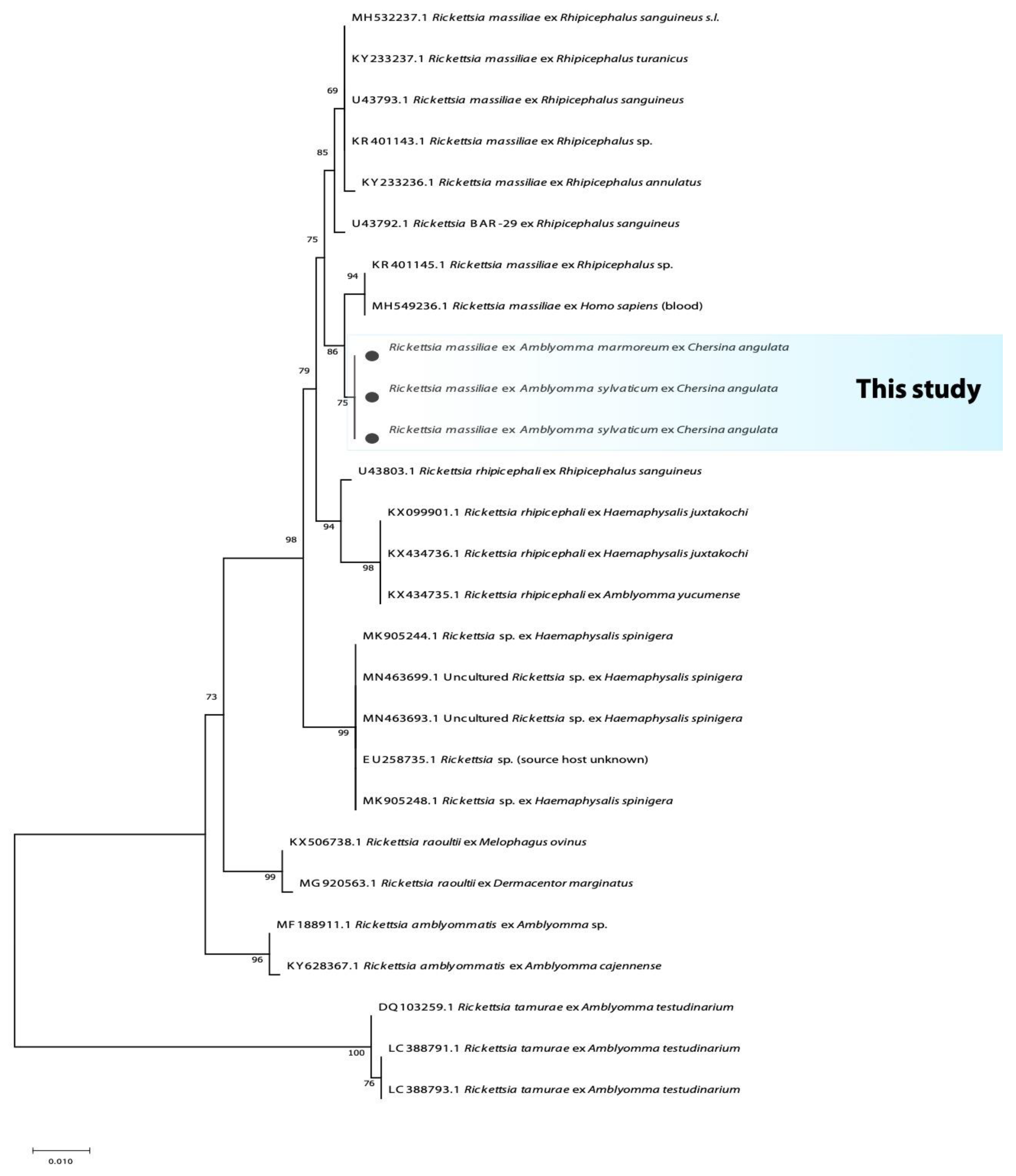
On the other hand, the 396 bp fragment of the citrate synthase gene (gltA) for Rickettsia amplified from A. marmoreum and A. sylvaticum yielded an identity of 97.9% with the spotted fever group Rickettsia massiliae (KY640405.1) and R. rhipicephali (KX018048.1). However, all the sequences of the 631 bp fragment of ompA gene for Rickettsia amplified from both A. marmoreum and A. sylvaticum yielded 99.1% with only R. massiliae (MH549236.1 and KR401145.1). The phylogenetic analysis of the fragment of the ompA clusters the current study’s Rickettsia sequences recovered from Amblyomma ticks in a monophyletic group with a support value of 75%. With high support (86%), these sequences form a sister clade to R. massiliae; this group in turn clusters within a larger monophyletic clade containing unnamed Rickettsia species and R. massiliae (Figure 3).
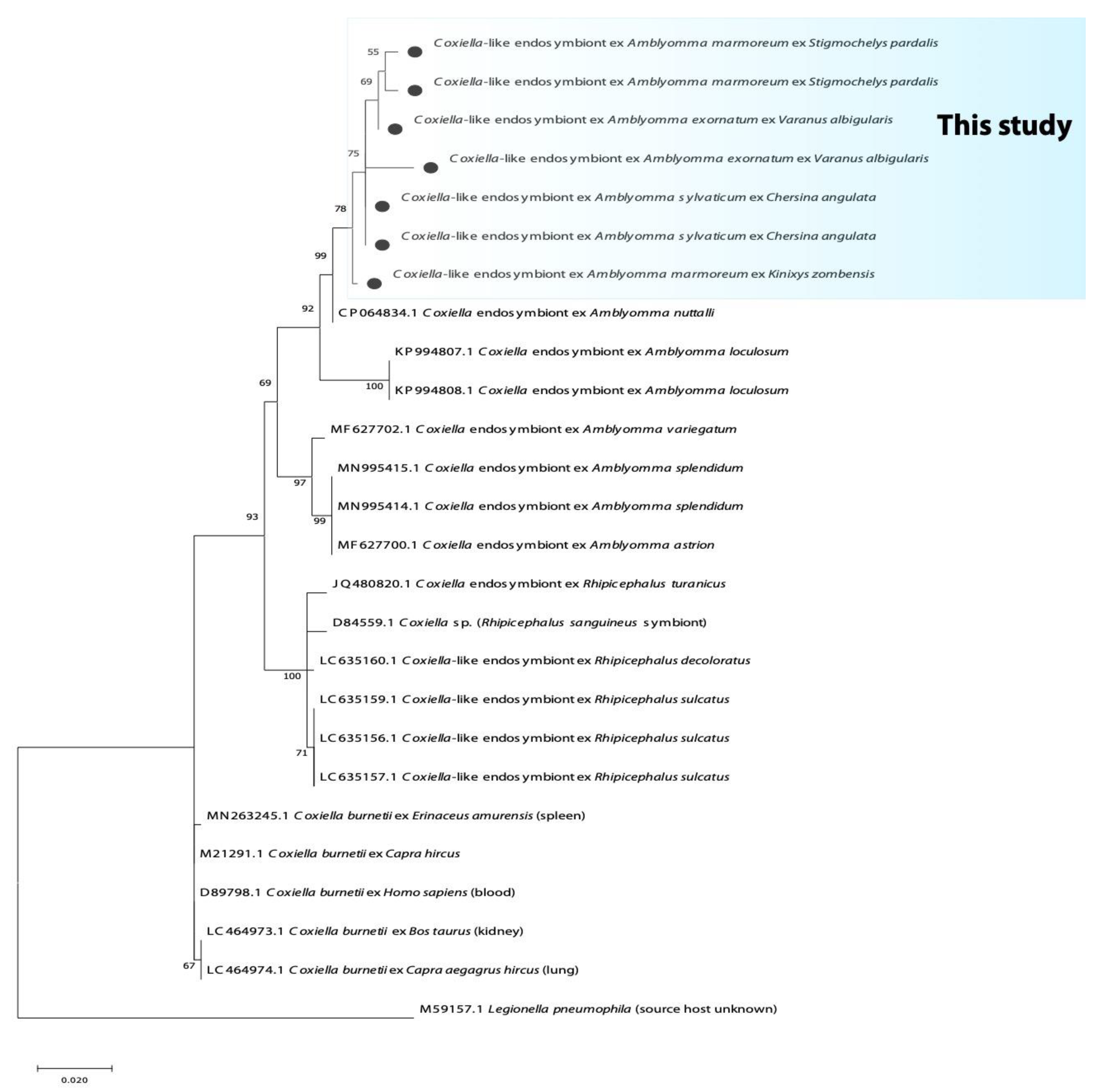
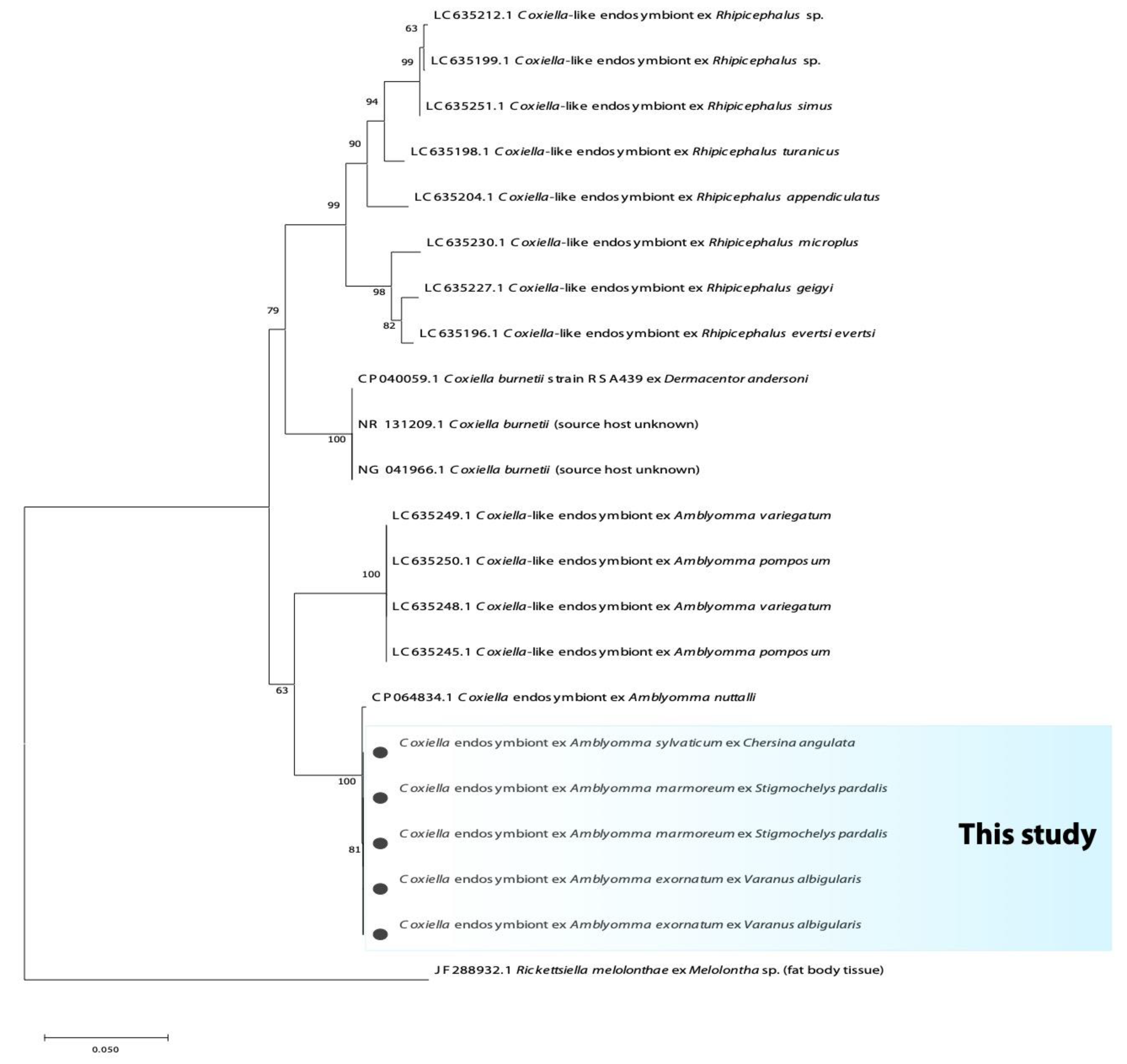
The fragment of the 16S rRNA and 23S rRNA yielded an identity of 99% with the complete genome of Coxiella-like endosymbiont (CP064834.1) from Amblyomma nutalli. Furthermore, these sequences also yielded an identity of 95.9% with the complete genome of the Coxiella burnetii strain RSA439 (CP040059.1) and C. burnetti isolate from Hyalomma asiaticum (MN880312.1). The phylogenetic analysis based on the 16S rRNA gene and 23S rRNA showed that Coxiella-like endosymbiont from reptilian Amblyomma ticks are grouped into an independent and distinct clade; it further clustered in a large monophyletic clade of Coxiella-like endosymbionts of Amblyomma tick species previously reported (Figure 4 and Figure 5).
The sequences derived during this study were submitted to the GenBank database under the accession numbers: Coxiella spp. (16S rRNA gene: OP070964-OP070972; 23S rRNA gene: OP071239-071243); Borrelia spp. (16S rRNA gene: OP081024-OP081026; flagellin gene: OP082442-OP082443); Francisella spp. (16S rRNA: OP068354 & OP068355); Rickettsia spp. (16S rRNA: OP068356; ompA gene: OP329173-OP329175).

4. Discussion

Notwithstanding the fact that tick-borne bacterial microorganisms represent serious emerging and re-emerging health problems to humans and other vertebrates, the surveillance of these pathogens in wildlife has focused mainly on mammals, birds and their ectoparasites, whereas reptiles and their associated ectoparasites remain unexplored [16,34]. Reptiles from Africa have been reported to carry Borrelia and Rickettsia infected ticks [8,17]. However, studies focusing on the detection of bacterial microorganisms in these ectothermic hosts, these hosts’ ability to act as reservoirs of infection or their ability to be refractory to infection [34], and their associated ticks, are only recent and scarce, making their inventory incomplete. This demonstrates the critical need to investigate their potential role in introducing and spreading new tick-borne bacterial diseases in South Africa in order to understand their transmission and zoonotic potential.
Beati et al. [35] isolated Rickettsia massiliae for the first time from Rhipicephalus sanguineus s.l. collected from canines in France, identifying it as a distinct species within the spotted fever group of rickettsiae. It has been mainly associated with ticks of the genus Rhipicephalus. It was then detected from various tick species associated with several vertebrate hosts in Europe, Africa and the Middle East [7,8,36,37]. Even though a human case of R. massiliae has not been reported in South Africa, it has been reported in Europe and Argentina, and as such should be considered in patients with tick bites [38,39,40]. Recently, this Rickettsia species was also detected in Rhipicephalus ticks infesting domestic small ruminants in Nigeria [41]. The finding of R. massiliae in reptilian ticks from South Africa suggests that human cases of this pathogen can occur. Therefore, ticks associated with reptiles should be considered in the epidemiology of R. massiliae, although more studies investigating the potential role of these ticks as vectors and reservoirs of this pathogen are needed. In the current study, R. massiliae was detected from A. sylvaticum and A. marmoreum collected from tortoises in South Africa, and these results are in accordance with a previous report on the presence of R. massiliae in Amblyomma ticks in this country [8]. However, it is unfortunate that the R. massiliae detected by Halajian et al. [8] could not be compared by phylogenetic analysis to R. massiliae in the present study, as different genes were amplified between these two studies.
Anaplasma species are tick-transmitted intracellular bacteria that infect a multitude of wild and domestic animals, as well as humans [42]. Although Anaplasma species have been documented from ticks and reptiles [43,44], they have not been recorded from African reptiles and their associated ticks, and we did not detect Anaplasma species in the current samples.
Coxiella burnetii is the causative agent of Q fever, a zoonotic disease with a global distribution, and it has been reported throughout the African continent in ticks, animal production and humans [45,46,47,48]. Ticks may become infected with C. burnetii during feeding but are not recognized as the actual vectors of the pathogen [49]. However, the current study could not detect C. burnetii. Nevertheless, for the first time in South Africa, the present study reports on the occurrence of Coxiella-like endosymbiont in reptilian ticks. These endosymbionts have been documented as vitamin provisioning endosymbionts, and it was reported that the biosynthetic pathways for various vitamins are found in all CLE [50,51]. Furthermore, Nardi et al. [14], in their genomic study, have suggested that these CLEs have independently evolved from C. burnetii. The prevalence of CLE in the current study was 41.1% among all the screened ticks, and it was detected in three of four Amblyomma species identified with different percentages. This was not surprising, as it has been documented that the prevalence of these bacteria varies widely in various species of ticks. This prevalence was in accordance with studies by Arthan et al. [52] and Rahal et al. [53], who reported prevalences of 42.2% and 51.7% in ticks from vegetation and cattle, respectively. Taking this into consideration, the potential of these CLEs to adapt to vertebrate hosts and evolve into pathogenic Coxiella cannot be ruled out. Recently, CLE was confirmed to be a new agent of human infections such as neck lymphadenopathy syndrome and scalp eschar through tick bites [54]. Furthermore, Shivaprasad et al. [55] and Vapniarsky et al. [56] reported avian infections of Coxiella-like bacteria even though there was no link to ticks. Therefore, the pathogenicity and zoonotic potential of these Coxiella-like bacteria merits further investigation. The negative results for Coxiella-like bacteria in A. latum may be due to the presence of other symbionts such as Francisella-like bacteria, as it was detected in some of the A. latum using bacteria specific primers, thus the presence of other endosymbionts in these ticks must not be ruled out.
Although Anaplasma spp., and C. burnetii were not detected, they cannot be completely disregarded, as their absence could be attributed to the limited number of ticks and localities analysed in the current study. More studies should be done in more localities to assess the impact of these bacterial microorganisms in reptiles and the risk of transmission to domestic animals and humans.
Takano et al. [57] identified reptile-associated-Borrelia spp. as a novel group of Borrelia spp. from imported reptiles and/or their ectoparasites, although this group have been found to be associated with species of the order Monotremata [29,58,59]. The transmission of these species is thought to be through hard-bodied ticks belonging to the genera Hyalomma or Amblyomma, while they appear to have evolutionarily diverged from an ancestor shared with RF Borrelia spp. [57]. In Africa, these species were only documented from Amblyomma ticks. Reptile-associated-Borrelia isolated from Amblyomma ticks clustered with the RF Borrelia based on a phylogenetic analysis study conducted by Takano et al. [60]. However, in 2014, based on the analysis of the 16S rRNA gene, Adelou and Gupta [61] grouped reptilian Borrelia species in a third distinct clade.
In this study, Borrelia spp. were detected in two species of Amblyomma ticks collected from Chersina angulata and Kinixys spp., and it was closely related to the REP borreliae group. This group has been previously detected in reptiles and their ticks imported from Africa into Japan [57]. These findings showed the presence of REP Borrelia in South Africa for the first time. Amblyomma marmoreum have been reported to feed on mammals and exported reptiles [62], which causes a serious concern on the spreading of pathogens by these Amblyomma ticks. Thus far, the impact of REP borreliae in humans and animals in South Africa is unknown. However, more detailed phylogenetic studies of the four groups of the genus Borrelia are needed to elucidate the origin of REP borreliae and that in turn may aid in clarifying the taxonomic status of species in this genus [63,64].
Moreover, co-infection of Coxiella-like endosymbiont and Borrelia sp. was found in A. marmoreum and A. sylvaticum collected from tortoises. This could be due to co-transmission of the pathogens during blood feeding on the host, or as a result of infection during different feedings [65,66].
Recently, many species of reptiles have been and continue to be traded and kept as pets in several countries across the world. As a result, tick-borne pathogens pose potential health threats to humans, associated pets and livestock; therefore, knowledge of the dynamics of the circulation of tick-borne bacteria in the environment is important to assess the risk of infection to both livestock and humans. Although the direct evidence remains to be determined, observations from this study provide clues for the possibility that reptile associated Amblyomma ticks can play a role in the maintenance and transmission of pathogenic bacteria to homothermic animals. The accumulation of this epidemiological information from multiple individuals and/or species of reptiles and different stages of ticks in future studies should be informative for this goal. More work will need to be done to determine the prevalence and pathogenic potential of R. massilae, REP Borrelia and Coxiella-like endosymbionts in South Africa. Recognition of the potential of Rickettsia species, Borrelia species and CLEs in reptiles and their Amblyomma ticks will help further our understanding of the emerging bacterial diseases in the country and the potential threat they pose to a global market in reptiles and their associated products.
This study also observed Francisella-like endosymbionts from Amblyomma latum using bacteria-specific primers. Though the role of most bacteria has not yet been clearly elucidated, the essential role of Francisella-like endosymbionts have been reported in several tick species in which it may provide essential nutrition for the tick [58,67,68].

5. Conclusions

The present study demonstrates for the first time the presence of REP Borrelia and Coxiella-like endosymbionts in Amblyomma ticks infesting South African reptiles, and corroborates the presence of R. massilae in South Africa. These results should increase the awareness on the emergence of these pathogens in this region, and additional studies should elucidate the role of these ectotherms in the transmission of different tick-borne bacterial microorganisms to domestic animals and humans. Findings from this study should stimulate further wide-scale investigation to determine the role of Francisella-like endosymbionts and Coxiella-like endosymbionts in tick physiology and vector competence.

Supplementary Materials

The following supporting information can be downloaded at: https://www.mdpi.com/article/10.3390/microorganisms10101923/s1, Table S1: Tick bacteria data; Figure S1: Phylogenetic tree based on the sequences of the 16S rRNA gene.

Author Contributions

Individual contributions include the conceptualization, L.S.M., N.J.S. and C.A.C.; formal analysis, L.S.M.; investigation, L.S.M., N.J.S. and C.A.C.; resources, N.J.S. and C.A.C.; writing—original draft preparation, L.S.M. and C.A.C.; writing—review and editing, L.S.M., N.J.S. and C.A.C.; visualization, L.S.M., N.J.S. and C.A.C.; supervision, N.J.S. and C.A.C.; project administration, L.S.M., N.J.S. and C.A.C.; funding acquisition, L.S.M. and C.A.C. All authors have read and agreed to the published version of the manuscript.

Funding

This research was funded by the National Research Foundation (NRF)—The Foundational Biodiversity Information Programme (FBIP) Scholarship (Grant number: 128335 & 138681). The NRF also provided funding to CAC (NRF project CSRP190414430265, grant 120395; NRF incentive RA161107208698, grant 120237).

Institutional Review Board Statement

Reptiles were restrained by hand whilst ticks were collected in situ. Sampling was carried out under the relevant permits (KZN: OP 839/2014, OP 4374/2015, OP 4092/2016, OP 4264/2017; WC: 0035-AAA004-00383) and was approved by the North- West University Anim-Care Animal Research Ethics Committee, South Africa (NWU-00372-16-A5).

Data Availability Statement

The data presented in this study are openly available in the NCBI Databases.

Acknowledgments

Ezemvelo KZN Wildlife, South Africa, is thanked for research permits OP 839/2014, OP 4374/2015, OP 4092/2016, OP 4264/2017, as well as Cape-Nature for research permit 0035-AAA004-00383. This is contribution No. 703 from the NWU-Water Research Group.

Conflicts of Interest

The authors declare that they have no conflict of interest. The funders had no role in the design of the study; in the collection, analyses, or interpretation of data; in the writing of the manuscript; or in the decision to publish the results.

References

  1. Parola, P.; Paddock, C.D.; Raoult, D. Tick-borne rickettsioses around the world: Emerging diseases challenging old concepts. Clin. Microbiol. 2005, 18, 719–756. [Google Scholar] [CrossRef]
  2. Omondi, D.; Masiga, D.K.; Fielding, B.C.; Kariuki, E.; Ajamma, Y.U.; Mwamuye, M.M.; Ouso, D.O.; Villinger, J. Molecular detection of tick-borne pathogen diversities in ticks from livestock and reptiles along the shores and adjacent islands of Lake Victoria and Lake Baringo, Kenya. Front. Vet. Sci. 2017, 4, 73. [Google Scholar] [CrossRef] [PubMed]
  3. Uilenberg, G. International collaborative research: Significance of tick-borne hemoparasitic diseases to world animal health. Vet. Parasitol. 1995, 57, 19–41. [Google Scholar] [CrossRef]
  4. Warwick, C.; Corning, S. Managing patients for zoonotic disease in hospitals. J. R. Soc. Med. Sh. Rep. 2013, 4, 1–9. [Google Scholar] [CrossRef] [PubMed]
  5. Pretorius, A.M.; Birtles, R.J. Rickettsia aeschlimannii: A new pathogenic spotted fever group rickettsia, South Africa. Emerg. Infect. Dis. 2002, 8, 874. [Google Scholar] [CrossRef]
  6. Pretorius, A.M.; Birtles, R.J. Rickettsia mongolotimonae infection in South Africa. Emerg. Infect. Dis. 2004, 10, 125. [Google Scholar] [CrossRef]
  7. Parola, P.; Paddock, C.D.; Socolovschi, C.; Labruna, M.B.; Mediannikov, O.; Kernif, T.; Abdad, M.Y.; Stenos, J.; Bitam, I.; Fournier, P.E.; et al. Update on tick-borne rickettsioses around the world: A geographic approach. Clin. Microbiol. 2013, 26, 657–702. [Google Scholar] [CrossRef]
  8. Halajian, A.; Palomar, A.M.; Portillo, A.; Heyne, H.; Luus-Powell, W.J.; Oteo, J.A. Investigation of Rickettsia, Coxiella burnetii and Bartonella in ticks from animals in South Africa. Ticks Tick Borne Dis. 2016, 7, 361–366. [Google Scholar] [CrossRef]
  9. Santodomingo, A.; Cotes-Perdomo, A.; Foley, J.; Castro, L.R. Rickettsial infection in ticks (Acari: Ixodidae) from reptiles in the Colombian Caribbean. Ticks Tick Borne Dis. 2018, 9, 623–628. [Google Scholar] [CrossRef]
  10. Peter, T.F.; Burridge, M.J.; Mahan, S.M. Competence of the African tortoise tick, Amblyomma marmoreum (acari: Ixodidae), as a vector of the agent of heartwater (Cowdria ruminantium). J. Parasitol. 2000, 86, 438–441. [Google Scholar]
  11. Daveu, R.; Laurence, C.; Bouju-Albert, A.; Sassera, D.; Plantard, O. Symbiont dynamics during the blood meal of Ixodes ricinus nymphs differ according to their sex. Ticks Tick Borne Dis. 2021, 12, 101707. [Google Scholar] [CrossRef] [PubMed]
  12. Kobayashi, T.; Chatanga, E.; Qiu, Y.; Simuunza, M.; Kajihara, M.; Hang’ombe, B.M.; Eto, Y.; Saasa, N.; Mori-Kajihara, A.; Simulundu, E.; et al. Molecular Detection and Genotyping of Coxiella-Like Endosymbionts in Ticks Collected from Animals and Vegetation in Zambia. Pathogens 2021, 10, 779. [Google Scholar] [CrossRef] [PubMed]
  13. Rio, R.V.; Attardo, G.M.; Weiss, B.L. Grandeur alliances: Symbiont metabolic integration and obligate arthropod hematophagy. Trends Parasitol. 2016, 32, 739–749. [Google Scholar] [CrossRef] [PubMed]
  14. Nardi, T.; Olivieri, E.; Kariuki, E.; Sassera, D.; Castelli, M. Sequence of a Coxiella endosymbiont of the tick Amblyomma nuttalli suggests a pattern of convergent genome reduction in the Coxiella genus. Genome Biol. Evol. 2021, 13, evaa253. [Google Scholar] [CrossRef]
  15. Moreira, L.A.; Iturbe-Ormaetxe, I.; Jeffery, J.A.; Lu, G.; Pyke, A.T.; Hedges, L.M.; Rocha, B.C.; Hall-Mendelin, S.; Day, A.; Riegler, M.; et al. A Wolbachia symbiont in Aedes aegypti limits infection with dengue, Chikungunya, and Plasmodium. Cell 2009, 139, 1268–1278. [Google Scholar] [CrossRef]
  16. Sánchez-Montes, S.; Isaak-Delgado, A.B.; Guzmán-Cornejo, C.; Rendón-Franco, E.; Muñoz-García, C.I.; Bermúdez, S.; Morales-Diaz, J.; Cruz-Romero, A.; Romero-Salas, D.; Dzul-Rosado, K.; et al. Rickettsia species in ticks that parasitize amphibians and reptiles: Novel report from Mexico and review of the worldwide record. Ticks Tick Borne Dis. 2019, 10, 987–994. [Google Scholar] [CrossRef]
  17. Andoh, M.; Sakata, A.; Takano, A.; Kawabata, H.; Fujita, H.; Une, Y.; Goka, K.; Kishimoto, T.; Ando, S. Detection of Rickettsia and Ehrlichia spp. in ticks associated with exotic reptiles and amphibians imported into Japan. PLoS ONE 2015, 10, e0133700. [Google Scholar] [CrossRef]
  18. Muñoz-Leal, S.; Tarragona, E.L.; Martins, T.F.; Martín, C.M.; Burgos-Gallardo, F.; Nava, S.; Labruna, M.B.; González-Acuña, D. Liolaemus lizards (Squamata: Liolaemidae) as hosts for the nymph of Amblyomma parvitarsum (Acari: Ixodidae), with notes on Rickettsia infection. Exp. Appl. Acarol. 2016, 70, 253–259. [Google Scholar] [CrossRef]
  19. Desser, S.S.; Hong, H.; Martin, D.S. The life history, ultrastructure, and experimental transmission of Hepatozoon catesbianae n. comb., an apicomplexan parasite of the bullfrog, Rana catesbeiana and the mosquito, Culex territans in Algonquin Park, Ontario. J. Parasitol. 1995, 81, 212–222. [Google Scholar] [CrossRef]
  20. Mofokeng, L.S.; Smit, N.J.; Cook, C.A. Molecular screening of ticks of the genus Amblyomma (Acari: Ixodidae) infesting South African reptiles with comments on their potential to act as vectors for Hepatozoon fitzsimonsi (Dias, 1953) (Adeleorina: Hepatozoidae). Int. J. Parasitol. Parasites Wildl. 2021, 16, 163–167. [Google Scholar] [CrossRef]
  21. Theiler, G. Ticks in the South African zoological survey collection. Part III. The ornate Aponommas. Onderstepoort J. Vet. Sci. Anim. Ind. 1945, 20, 165–178. Available online: http://hdl.handle.net/2263/59279 (accessed on 21 July 2020).
  22. Theiler, G.; Salisbury, L.E. Ticks in the South African Zoological Survey Collection-Part IX-The Amblyomma marmoreum Group. Onderstepoort J. Vet. Sci. Anim. Ind. 1959, 28, 54–98. Available online: http://hdl.handle.net/2263/57905 (accessed on 21 July 2021).
  23. Tamura, K.; Stecher, G.; Kumar, S. MEGA11: Molecular evolutionary genetics analysis version 11. Mol. Biol. Evol. 2021, 38, 3022–3027. [Google Scholar] [CrossRef]
  24. Guindon, S.; Gascuel, O. A simple, fast, and accurate algorithm to estimate large phylogenies by maximum likelihood. Syst. Biol. 2003, 52, 696–704. [Google Scholar] [CrossRef]
  25. Darriba, D.; Taboada, G.L.; Doallo, R.; Posada, D. jModelTest 2: More models, new heuristics and parallel computing. Nat. Methods 2012, 9, 772. [Google Scholar] [CrossRef]
  26. Relman, D.A.; Schmidt, T.M.; MacDermott, R.P.; Falkow, S. Identification of the uncultured Bacillus of Whipple’s disease. N. Engl. J. Med. 1992, 327, 293–301. [Google Scholar] [CrossRef]
  27. Stuen, S.; Nevland, S.; Moum, T. Fatal cases of tick-borne fever (TBF) in sheep caused by several 16S rRNA gene variants of Anaplasma phagocytophilum. Ann. N. Y. Acad. Sci. 2003, 990, 433–434. [Google Scholar] [CrossRef]
  28. Skotarczak, B.; Wodecka, B.; Cichocka, A. Coexistence DNA of Borrelia burgdorferi sensu lato and Babesia microti in Ixodes ricinus ticks from north-western Poland. Ann. Agric. Environ. Med. 2002, 9, 25–28. [Google Scholar]
  29. Loh, S.M.; Gillett, A.; Ryan, U.; Irwin, P.; Oskam, C. Molecular characterization of ‘Candidatus Borrelia tachyglossi’ (family Spirochaetaceae) in echidna ticks, Bothriocroton concolor. Int. J. Syst. Evol. 2017, 67, 1075. [Google Scholar] [CrossRef]
  30. Dupont, H.T.; Cornet, J.P.; Raoult, D. Identification of rickettsiae from ticks collected in the Central African Republic using the polymerase chain reaction. Am. J. Trop. Med. 1994, 50, 373–380. [Google Scholar] [CrossRef]
  31. Roux, V.; Fournier, P.E.; Raoult, D. Differentiation of spotted fever group rickettsiae by sequencing and analysis of restriction fragment length polymorphism of PCR-amplified DNA of the gene encoding the protein rOmpA. J. Clin. Microbiol. 1996, 34, 2058–2065. [Google Scholar] [CrossRef]
  32. Duron, O.; Noël, V.; McCoy, K.D.; Bonazzi, M.; Sidi-Boumedine, K.; Morel, O.; Vavre, F.; Zenner, L.; Jourdain, E.; Durand, P.; et al. The recent evolution of a maternally-inherited endosymbiont of ticks led to the emergence of the Q fever pathogen, Coxiella burnetii. PLoS Pathog. 2015, 11, e1004892. [Google Scholar] [CrossRef]
  33. Stein, A.; Raoult, D. Detection of Coxiella burnetti by DNA amplification using polymerase chain reaction. J. Clin. Microbiol. 1992, 30, 2462–2466. [Google Scholar] [CrossRef]
  34. Lane, R.S.; Mun, J.; Eisen, L.; Eisen, R.J. Refractoriness of the western fence lizard (Sceloporus occidentalis) to the Lyme disease group spirochete Borrelia bissettii. J. Parasitol. 2006, 92, 691–696. [Google Scholar] [CrossRef]
  35. Beati, L.; Finidori, J.P.; Gilot, B.; Raoult, D. Comparison of serologic typing, sodium dodecyl sulfate-polyacrylamide gel electrophoresis protein analysis, and genetic restriction fragment length polymorphism analysis for identification of rickettsiae: Characterization of two new rickettsial strains. J. Clin. Microbiol. 1992, 30, 1922–1930. [Google Scholar] [CrossRef]
  36. Blanda, V.; Torina, A.; La Russa, F.; D’Agostino, R.; Randazzo, K.; Scimeca, S.; Giudice, E.; Caracappa, S.; Cascio, A.; de la Fuente, J. A retrospective study of the characterization of Rickettsia species in ticks collected from humans. Ticks Tick Borne. Dis. 2017, 8, 610–614. [Google Scholar] [CrossRef]
  37. Demir, S.; Erkunt Alak, S.; Köseoğlu, A.E.; Ün, C.; Nalçacı, M.; Can, H. Molecular investigation of Rickettsia spp. and Francisella tularensis in ticks from three provinces of Turkey. Exp. Appl. Acarol. 2020, 81, 239–253. [Google Scholar] [CrossRef]
  38. Vitale, G.; Mansueto, S.; Rolain, J.M.; Raoult, D. Rickettsia massiliae human isolation. Emerg. Infect. Dis. 2006, 12, 174. [Google Scholar] [CrossRef]
  39. García-García, J.C.; Portillo, A.; Núñez, M.J.; Santibáñez, S.; Castro, B.; Oteo, J.A. Case report: A patient from Argentina infected with Rickettsia massiliae. Am. J. Trop. Med. Hyg. 2010, 82, 691. [Google Scholar] [CrossRef]
  40. Cascio, A.; Torina, A.; Valenzise, M.; Blanda, V.; Camarda, N.; Bombaci, S.; Iaria, C.; De Luca, F.; Wasniewska, M. Scalp eschar and neck lymphadenopathy caused by Rickettsia massiliae. Emerg. Infect. Dis. 2013, 19, 836. [Google Scholar] [CrossRef] [PubMed]
  41. Elelu, N.; Ola-Fadunsin, S.D.; Bankole, A.A.; Raji, M.A.; Ogo, N.I.; Cutler, S.J. Prevalence of tick infestation and molecular characterization of spotted fever Rickettsia massiliae in Rhipicephalus species parasitizing domestic small ruminants in north-central Nigeria. PLoS ONE 2022, 17, e0263843. [Google Scholar] [CrossRef] [PubMed]
  42. Han, R.; Yang, J.F.; Mukhtar, M.U.; Chen, Z.; Niu, Q.L.; Lin, Y.Q.; Liu, G.Y.; Luo, J.X.; Yin, H.; Liu, Z.J. Molecular detection of Anaplasma infections in ixodid ticks from the Qinghai-Tibet Plateau. Infect. Dis. Poverty 2019, 8, 12. [Google Scholar] [CrossRef] [PubMed]
  43. Nieto, N.C.; Foley, J.E.; Bettaso, J.; Lane, R.S. Reptile infection with Anaplasma phagocytophilum, the causative agent of granulocytic anaplasmosis. J. Parasitol. 2009, 95, 1165–1170. [Google Scholar] [CrossRef]
  44. Kho, K.L.; Koh, F.X.; Tay, S.T. Molecular evidence of potential novel spotted fever group rickettsiae, Anaplasma and Ehrlichia species in Amblyomma ticks parasitizing wild snakes. Parasites Vectors 2015, 8, 112. [Google Scholar] [CrossRef] [Green Version]
  45. Dupont, H.T.; Brouqui, P.; Faugere, B.; Raoult, D. Prevalence of antibodies to Coxiella burnetii, Rickettsia conorii, and Rickettsia typhi in seven African countries. Clin. Infect. Dis. 1995, 21, 1126–1133. [Google Scholar] [CrossRef]
  46. Vanderburg, S.; Rubach, M.P.; Halliday, J.E.; Cleaveland, S.; Reddy, E.A.; Crump, J.A. Epidemiology of Coxiella burnetii infection in Africa: A One-Health systematic review. PLoS Negl. Trop. Dis. 2014, 8, e2787. [Google Scholar] [CrossRef]
  47. Mtshali, K.; Nakao, R.; Sugimoto, C.; Thekisoe, O. Occurrence of Coxiella burnetii, Ehrlichia canis, Rickettsia species and Anaplasma phagocytophilum-like bacterium in ticks collected from dogs and cats in South Africa. J. S. Afr. Vet. Assoc. 2017, 88, 1390. [Google Scholar] [CrossRef]
  48. Halajian, A.; Palomar, A.M.; Portillo, A.; Heyne, H.; Romero, L.; Oteo, J.A. Detection of zoonotic agents and a new Rickettsia strain in ticks from donkeys from South Africa: Implications for travel medicine. Travel Med. Infect. Dis. 2018, 26, 43–50. [Google Scholar] [CrossRef]
  49. Maurin, M.; Raoult, D.F. Q fever. Clin. Microbiol. Rev. 1999, 12, 518–553. [Google Scholar] [CrossRef]
  50. Gottlieb, Y.; Lalzar, I.; Klasson, L. Distinctive genome reduction rates revealed by genomic analyses of two Coxiella-like endosymbionts in ticks. Genome Biol. Evol. 2015, 7, 1779–1796. [Google Scholar] [CrossRef]
  51. Guizzo, M.G.; Parizi, L.F.; Nunes, R.D.; Schama, R.; Albano, R.M.; Tirloni, L.; Oldiges, D.P.; Vieira, R.P.; Oliveira, W.H.C.; de Souza Leite, M.; et al. A Coxiella mutualist symbiont is essential to the development of Rhipicephalus microplus. Sci. Rep. 2017, 7, 17554. [Google Scholar] [CrossRef] [PubMed]
  52. Arthan, W.; Sumrandee, C.; Hirunkanokpun, S.; Kitthawee, S.; Baimai, V.; Trinachartvanit, W.; Ahantarig, A. Detection of Coxiella-like endosymbiont in Haemaphysalis tick in Thailand. Ticks Tick Borne Dis. 2015, 6, 63–68. [Google Scholar] [CrossRef] [PubMed]
  53. Rahal, M.; Medkour, H.; Diarra, A.Z.; Bitam, I.; Parola, P.; Mediannikov, O. Molecular identification and evaluation of Coxiella-like endosymbionts genetic diversity carried by cattle ticks in Algeria. Ticks Tick Borne Dis. 2020, 11, 101493. [Google Scholar] [CrossRef] [PubMed]
  54. Guimard, T.; Amrane, S.; Prudent, E.; El Karkouri, K.; Raoult, D.; Angelakis, E. Case report: Scalp eschar and neck lymphadenopathy associated with bacteremia due to Coxiella-like bacteria. Am. J. Trop. Med. 2017, 97, 1319. [Google Scholar] [CrossRef]
  55. Shivaprasad, H.L.; Cadenas, M.B.; Diab, S.S.; Nordhausen, R.; Bradway, D.; Crespo, R.; Breitschwerdt, E.B. Coxiella-like infection in psittacines and a toucan. Avian Dis. 2008, 52, 426–432. [Google Scholar] [CrossRef]
  56. Vapniarsky, N.; Barr, B.C.; Murphy, B. Systemic Coxiella-like infection with myocarditis and hepatitis in an eclectus parrot (Eclectus roratus). Vet. Pathol. 2012, 49, 717–722. [Google Scholar] [CrossRef]
  57. Takano, A.; Goka, K.; Une, Y.; Shimada, Y.; Fujita, H.; Shiino, T.; Watanabe, H.; Kawabata, H. Isolation and characterization of a novel Borrelia group of tick-borne borreliae from imported reptiles and their associated ticks. Environ. Microbiol. 2010, 12, 134–146. [Google Scholar] [CrossRef]
  58. Panetta, J.L.; Šíma, R.; Calvani, N.E.; Hajdušek, O.; Chandra, S.; Panuccio, J.; Šlapeta, J. Reptile-associated Borrelia species in the goanna tick (Bothriocroton undatum) from Sydney, Australia. Parasites Vectors 2017, 10, 616. [Google Scholar] [CrossRef]
  59. Gofton, A.W.; Margos, G.; Fingerle, V.; Hepner, S.; Loh, S.M.; Ryan, U.; Irwin, P.; Oskam, C.L. Genome-wide analysis of Borrelia turcica and ‘Candidatus Borrelia tachyglossi’ shows relapsing fever-like genomes with unique genomic links to Lyme disease Borrelia. Infect. Genet. Evol. 2018, 66, 72–81. [Google Scholar] [CrossRef]
  60. Takano, A.; Fujita, H.; Kadosaka, T.; Konnai, S.; Tajima, T.; Watanabe, H.; Ohnishi, M.; Kawabata, H. Characterization of reptile-associated Borrelia sp. in the vector tick, Amblyomma geoemydae, and its association with Lyme disease and relapsing fever Borrelia spp. Environ. Microbiol. Rep. 2011, 3, 632–637. [Google Scholar] [CrossRef]
  61. Adeolu, M.; Gupta, R.S. A phylogenomic and molecular marker based proposal for the division of the genus Borrelia into two genera: The emended genus Borrelia containing only the members of the relapsing fever Borrelia, and the genus Borreliella gen. nov. containing the members of the Lyme disease Borrelia (Borrelia burgdorferi sensu lato complex). Antonie Leeuwenhoek 2014, 105, 1049–1072. [Google Scholar] [CrossRef]
  62. Horak, I.G.; McKay, I.J.; Henen, B.T.; Heyne, H.; Hofmeyr, M.D.; De Villiers, A.L. Parasites of Domestic and Wild Animals in South Africa. XLVII. Ticks of Tortoises and Other Reptiles. Onderstepoort J. Vet. Res. 2006, 73, 215–227. Available online: https://hdl.handle.net/10520/EJC86256 (accessed on 28 May 2020). [CrossRef]
  63. Margos, G.; Fingerle, V.; Cutler, S.; Gofton, A.; Stevenson, B.; Estrada-Peña, A. Controversies in bacterial taxonomy: The example of the genus Borrelia. Ticks Tick Borne Dis. 2020, 11, 101335. [Google Scholar] [CrossRef]
  64. Morales-Diaz, J.; Colunga-Salas, P.; Romero-Salas, D.; Sánchez-Montes, S.; Estrada-Souza, I.M.; Ochoa-Ochoa, L.M.; Becker, I.; Flores-Primo, A.; Cruz-Romero, A. Molecular detection of reptile-associated Borrelia in Boa constrictor (Squamata: Boidae) from Veracruz, Mexico. Acta Trop. 2020, 205, 105422. [Google Scholar] [CrossRef]
  65. Randolph, S.E.; Gern, L.; Nuttall, P.A. Co-feeding ticks: Epidemiological significance for tick-borne pathogen transmission. Parasitol. Today 1996, 12, 472–479. [Google Scholar] [CrossRef]
  66. Del Cerro, A.; Oleaga, A.; Somoano, A.; Barandika, J.F.; García-Pérez, A.L.; Espí, A. Molecular identification of tick-borne pathogens (Rickettsia spp., Anaplasma phagocytophilum, Borrelia burgdorferi sensu lato, Coxiella burnetii and piroplasms) in questing and feeding hard ticks from North-Western Spain. Ticks Tick Borne Dis. 2022, 13, 101961. [Google Scholar] [CrossRef]
  67. Zhang, C.M.; Li, N.X.; Zhang, T.T.; Qiu, Z.X.; Li, Y.; Li, L.W.; Liu, J.Z. Endosymbiont CLS-HI plays a role in reproduction and development of Haemaphysalis longicornis. Exp. Appl. Acarol. 2017, 73, 429–438. [Google Scholar] [CrossRef]
  68. Duron, O.; Morel, O.; Noël, V.; Buysse, M.; Binetruy, F.; Lancelot, R.; Loire, E.; Ménard, C.; Bouchez, O.; Vavre, F.; et al. Tick-bacteria mutualism depends on B vitamin synthesis pathways. Curr. Biol. 2018, 28, 1896–1902. [Google Scholar] [CrossRef] [Green Version]
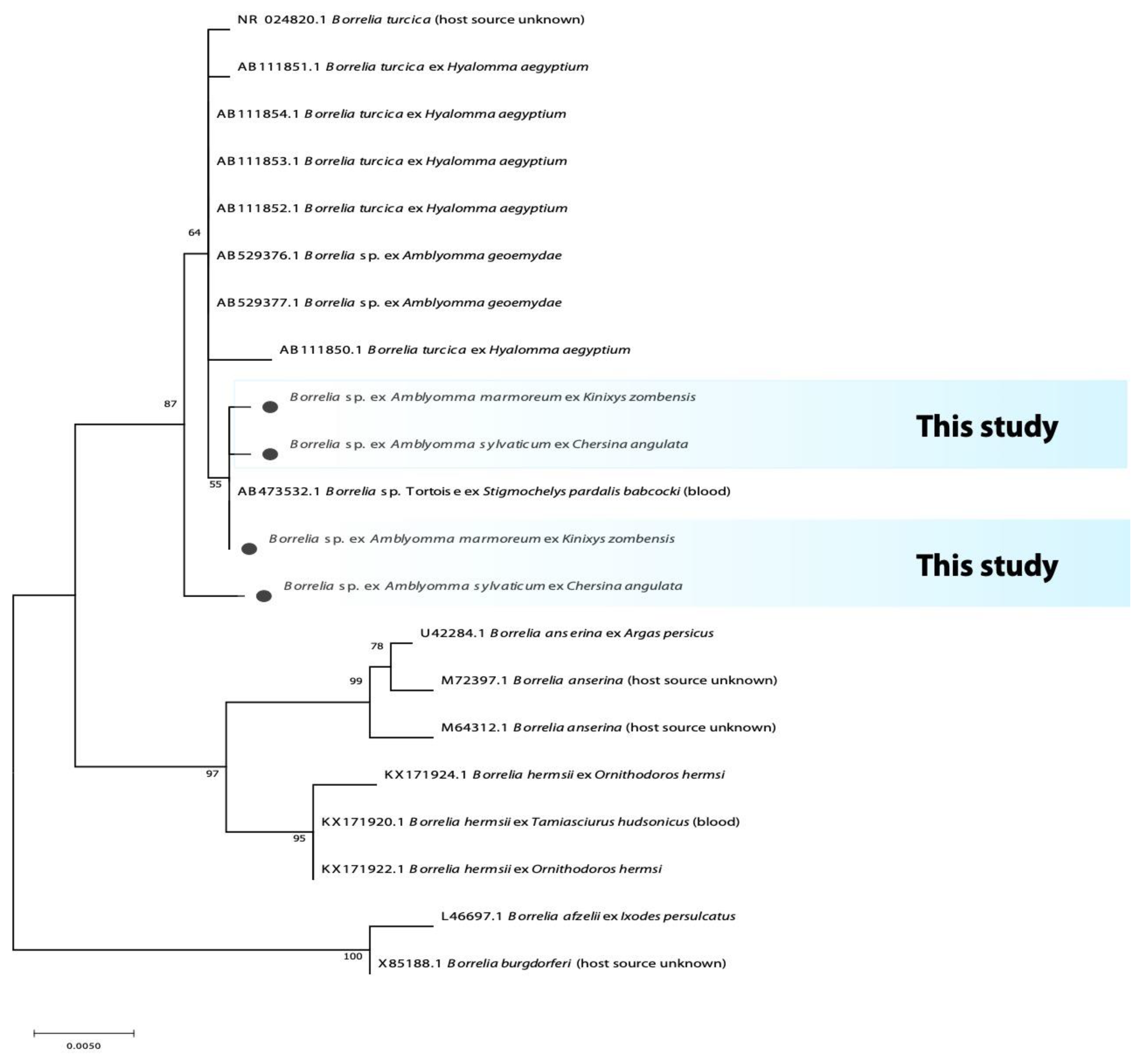
Figure 1. Phylogenetic tree based on the sequences of the Borrelia 16S rRNA gene. The tree was constructed using MEGA 11 based on the maximum likelihood, using the Hasegawa-Kishino-Yano Model. The sequences obtained in this study are indicated with bullets.
Microorganisms 10 01923 g001
Figure 2. Phylogenetic tree based on the sequences of the Borrelia Flagellin gene. The tree was constructed using MEGA 11 based on the maximum likelihood, using the Tamura 3-parameter Model. The sequences obtained in this study are indicated with bullets.
Microorganisms 10 01923 g002
Figure 3. Phylogenetic tree based on the sequences of the Rickettsia OmpA gene. The tree was constructed using MEGA 11 based on the maximum likelihood, using the Tamura 3-parameter model. The sequences obtained in this study are indicated with bullets.
Microorganisms 10 01923 g003
Figure 4. Phylogenetic tree based on the sequences of the Coxiella 16S rRNA gene. The tree was constructed using MEGA 11 based on the maximum likelihood, using the Kimura 2-parameter model. The sequences obtained in this study are indicated with bullets.
Microorganisms 10 01923 g004
Figure 5. Phylogenetic tree based on the sequences of the Coxiella 23S rRNA gene. The tree was constructed using MEGA 11 based on the maximum likelihood, using the Kimura 2-parameter model. The sequences obtained in this study are indicated with bullets.
Microorganisms 10 01923 g005
Table 2. Prevalence of bacterial microorganisms from reptilian Amblyomma ticks.
Province Tick SpeciesnHost (n)16S rRNA BacteriaRickettsia spp.Borrelia spp.Coxiella spp.
gltA GeneOmpA GeneFlagellin Gene16S rRNA16S rRNA23S rRNA
KZNA. marmoreum4Varanus albigularis (1)-----50% (2/4)-
A. marmoreum13Kinixys sp. (1)---46.2% (6/13)46.2 (6/13)46.2% (6/13)30.8% (4/13)
A. marmoreum11Kinixys zombensis (1)---18.2% (2/11)18.2% (2/11)45.5% (5/11)45.45% (5/11)
A. marmoreum12Stigmochelys pardalis (7)-----41.7% (5/12)16.7% (2/12)
A. latum43Naja mossambica (3), Dendroapis spp. (4)4.7% (2/43)
Francisella spp.
------
A. exornatum37Varanus albigularis (5)-----32.4% (12/37)24.3% (9/37)
WCA. marmoreum21Kinixys zombesis (3)---4.8% (1/21)4.8% (1/21)53.4% (11/21)38.1% (8/21)
A. marmoreum37Stigmochelys. pardalis (2)-----56.8% (21/37)40.5% (15/37)
A. marmoreum15Chersina angulata (9)-46.7% (7/15)33.3% (5/15)20% (3/15)20% (3/15)53.3% (8/15)53.3% (8/15)
A. sylvaticum60Chersina angulata (9)1.7% (1/60)
Rickettsia spp.
20.0% (12/60)23.3% (14/60)-6.7% (4/60)56.7% (34/60)23.3% (14/60)
(-) = negative for specific pathogen; KZN = KwaZulu-Natal; WC = Western Cape; n = number of ticks collected. Number of vertebrate hosts collected is indicted in parentheses next to the host’s name.
Publisher’s Note: MDPI stays neutral with regard to jurisdictional claims in published maps and institutional affiliations.