Article

Morphological and Molecular Identification of Fusarium ipomoeae as the Causative Agent of Leaf Spot Disease in Tobacco from China

1
Guizhou Provincial Academician Workstation of Microbiology and Health, Guizhou Academy of Tobacco Science, Guiyang 550081, China
2
College of Agriculture, Yangtze University, Jingzhou 434025, China
3
Guizhou Institute of Plant Protection, Guizhou Academy of Agricultural Sciences, Guiyang 550006, China
*
Authors to whom correspondence should be addressed.
Microorganisms 2022, 10(10), 1890; https://doi.org/10.3390/microorganisms10101890
Submission received: 26 August 2022 / Revised: 13 September 2022 / Accepted: 14 September 2022 / Published: 22 September 2022
(This article belongs to the Section Plant Microbe Interactions)

Abstract

:
Tobacco (Nicotiana tabacum L.), which creates jobs for 33 million people and contributes two trillion dollars’ tax annually, is one of the most important economic plants globally. However, tobacco is seriously threatened by numerous diseases during production. Previously, the field survey of tobacco diseases was conducted in the Guizhou and Guangxi provinces, the two main tobacco-producing areas in China. A serious leaf spot disease, with a 22% to 35% incidence, was observed in farming plants. In order to determine the causal agents, we collected the disease samples and isolated the pathogenic fungi. The pathogen was identified as Fusarium ipomoeae, based on the morphological characteristics and phylogenetic analysis. Pathogenicity tests showed that F. ipomoeae could induce tobacco leaf spot and blight. To our knowledge, this is the first report worldwide of F. ipomoeae causing leaf spots and stems on tobacco. Our study reveals the serious consequences of F. ipomoeae on tobacco filed production and provides information for future diagnosis and management of the Fusarium disease.

1. Introduction

Tobacco (Nicotiana tabacum L.) is one of the most crucial economic crops all over the world [1]. Globally, according to the estimates of tobacco industry, about 33 million people participate in tobacco planting, product manufacturing, distribution, and retailing [2]. Meanwhile, global revenues from the tobacco industry are estimated at 2 trillion dollars a year [3]. In China, tobacco was planted in more than 14,000 km2, and its yield of leafage reached 31.32 million tons in 2011 [4]. Its accounts for more than 39.6% of total global tobacco production [5], in which, farming is especially prevalent in Southwest China [6]. Among the main supplier provinces, Guizhou province produces nearly 30% of the total Chinese tobacco crop and ranks as the second-most tobacco-producing areas [7].
It is well-known that tobacco suffers from various fungal and oomycete pathogens during its whole growing season, such as Fusarium spp., Collectotrichum gloeosporioides, Alternaria alternata, Botrytis cinraea, etc. [6,7]. Among them, Fusarium spp., which can cause tobacco leaf, root, and stem diseases, have been a serious problem threatening to tobacco production in many countries [8]. The incidence of Fusarium spp. has increased considerably in recent years [6,7,8]. Furthermore, the mycotoxins and secondary metabolites produced by the Fusarium species in diseased leaves may be harmful to the health of humans [9]. Therefore, Fusarium identification was of particular importance for the effective management of tobacco diseases.
In August 2021, the field survey of tobacco disease was conducted in Zheng’an and Fenggang in Guizhou, Shanglin. The three main tobacco-producing areas in China are located in Guangxi. Tobacco plants with leaf spots and blights were frequently observed in the farming fields, with a 22 to 35% disease incidence. In order to determine the causal agents, provide information for future diagnosis, and help the management of this disease, the disease samples were collected, and the pathogenic fungi were isolated. Morphology characterization and multi-gene locus phylogenetic analysis were performed. Furthermore, the pathogenicity was also tested following the Koch postulates.

2. Materials and Methods

2.1. Isolation

In August 2021, diseased plants of tobacco were collected from three main producing counties (Zheng’an and Fenggang in Guizhou and Shanglin in Guangxi). For fungal isolation, the diseased tissues were cut into several small segments and placed on potato dextrose agar (PDA, Difcohai) at 25 °C in darkness for 3 to 5 days. To obtain pure cultures, hyphal tips from developed colonies were transferred to fresh PDA plates three times [10]. The isolates were inoculated to PDA test tube slants and stored at 4 °C [11]. The strain was deposited at Yangtze University, Jingzhou, Hubei, China.

2.2. Morphology

The edge of the colony was cut into 6 mm diameter plates with a sterile punch, and the mycelia plug was transferred to a 90 mm PDA plate and grown for 7 days at 25 °C in darkness. Then, the colony morphology was evaluated and visualized. For observing the morphological features of conidia, the marginal hyphae were transferred to carnation leaf-piece agar (CLA) medium (the sterilized carnation leaves were placed in water agar medium) and cultured at 22 °C under a light/dark period of 12/12 h [12]. After 7–10 days, conidia and chlamydospores were mounted in sterile water for microscopic observation using a Nikon ECLIPSE Ni–U microscope equipped with a Nikon DS–Ri2 digital camera (Tokyo, Japan) [12].

2.3. DNA Extraction and PCR Amplification

Genomic DNA was extracted from fresh mycelium grown on PDA using a method modified from Cenis [13]. Five loci, including the 5.8S nuclear ribosomal RNA gene with the two flanking internal transcribed spacers (ITS), translation elongation factor 1 alpha (EF–1α), calmodulin (CAM), RNA polymerase largest subunit (RPB1), and RNA polymerase second largest subunit (RPB2) gene regions, were amplified and sequenced. The detail information of corresponding primers were showed as follows: ITS (ITS4: TCCTCCGCTTATTGATATGC; ITS5: GGAAGTAAAAGTCGTAACAAGG) [12], EF–1α (EF1: ATGGGTAAGGARGACAAGAC; EF2: GGARGTACCAGTSATCATG) [14], CAM (CL1: GARTWCAAGGAGGCCTTCTC; CL2A: TTTTTGCATCATGAGTTGGAC) [15], RPB1 (Fa: CAYAARGARTCYATGATGGGWC; G2R: GTCATYTGDGTDGCDGGYTCDCC) [16], RPB2 (5f2: GAYGAYMGWGATCAYTTYGG; 7cr: CCCATRGCTTGYTTRCCCAT) [17]. The PCR programs were set as follows: initial denaturation at 95 °C for 90 s, followed by 35 cycles, at 95 °C for 30 s, annealing for 30 s, and extension at 72 °C for 1 min, as well as terminating with a final extension at 72 °C for 10 min. The annealing temperatures were 55 °C, 56 °C, 55 °C, 58 °C, and 58 °C for ITS, EF–1α, CAM, RPB1, and RPB2, respectively. The PCR products were sent to the company (TSINGKE, Beijing, China) for purification and sequencing.

2.4. Phylogenetic Analysis

The obtained sequences were analyzed by BLASTn (nucleotide blast) searches. The relevant strains (Table 1) were selected both according to BLAST searches and previous references [18]. The five gene sequences were concatenated and edited manually in MEGA v.7.0.26 [19], and the aligned dataset was deposited in TreeBASE. The maximum likelihood [20] and Bayesian inference (BI) methods were used to phylogenetic analysis the ITS, EF–1α, CAM, RPB1, and RPB2 combing sequences for Fusarium incarnatum–equiseti species complex (FIESC). ML analysis was performed using RAxML (Randomized Axelerated Maximum Likelihood) v.7.2.8 (A. Stamatakis, Heidelberg, Germany) [21]. The branch support was assessed with 1000 replicates. Bayesian inference (BI) analyses were conducted in MrBayes v.3.2.1 (Huelsenbeck J P and Ronquist F, Rochester, USA) by using the Markov chain Monte Carlo (MCMC) algorithm [22]. Mrmodel test v.2.3 (Posada D and Crandall K A, Oxford, England) [23] was used to determine the best fit evolutionary model (GTR + I + G) using the Akaike information criterion (AIC) parameter. Two MCMC chains were run in the random tree, with a total of 1 million generations, sampling once every 100 generations. When the average standard deviation of the separation frequency was less than 0.01, the first 25% of the samples were discarded, and the operation was stopped. The trees were viewed and edited with Figtree v.1.3.1 [19]. The ITS, EF–1, RPB2, RPB1, and CAM sequences of strain GZAX 312 were deposited under GenBank numbers ON961780, ON982724, ON982726, ON982728, and ON982722, respectively.

2.5. Pathogenicity Test

Pathogenicity was tested on living tobacco leaves and stems. Healthy tobacco were surface-sterilized in 2% sodium hypochlorite and washed 3 times with sterilized distilled water for 2 min before performing the test [12]. The experiment was repeated three times, with at least three plants for each time. For leaves and stems inoculation, the mycelium block (about 6 mm) cultured on the PDA medium was inoculated to the healthy leaves and base of the stalk and wrapped with absorbent cotton and plastic wrap to keep it moisturized. After inoculation, the plants were cultured in a greenhouse (22 °C, under a light/dark period of 12/12 h). Control plants were inoculated with PDA plugs. Lesion was observed daily, and photographic record for 7 days after inoculation. For root inoculation, fresh mycelium blocks were mixed with rye seeds and cultured at 28 °C for 7 days [1]. Then, 15 culture seeds were mixed into the 5 cm surface soil of each pot and cultured at 22 °C after adding sufficient water. Four days later, the healthy tobacco seedlings at 4-leaf stage were planted into the pot and cultured in greenhouse. Seven days later, the disease symptoms of roots were observed. Seedlings planted in sterile soil were used as controls. The pathogen was re-isolated from the inoculation site using PDA medium. The morphological characteristics and RPB2 sequence were compared with original strains.

3. Results

3.1. Serious Leaf Spot and Blight Disease Was Observed on Field Tobacco Plants

The field survey of tobacco disease was conducted in August 2021. During the surveillance, it was normally surrounded by a yellow halo, which appeared on the tobacco leaves, as shown in Figure 1. Surveys indicated a 22 to 35% disease incidence in three counties of Zheng’an and Fenggang in Guizhou province and Shanglin in Guangxi Zhuang Autonomous Region. With the development of the disease, spots were enlarged and concatenated. Severely infected leaves turned out to be blight, then defoliation. In order to determine the causal agents, 35 disease samples were collected from the three counties, and the pathogenic fungi were isolated in the laboratory.

3.2. Morphology Characterization Indicated a Fusarium spp.

In total, 77 fungal strains were isolated from those 35 samples. Among them, 68 strains showed similar cultural characters, such as the white colony, with cotton and flocculent aerial mycelium. Thus, they seem to be one species and could be the dominant pathogen for tobacco leaf spot and blight disease. Three represent strains (GZAX 307, GZAX 312, and GZAX 402) were randomly selected from all isolated strains for subsequent research. Observation indicated the three strains had same morphological characteristics. Therefore, the strain GZAX 307 was used for the following microscopy visualization and morphology description.
The fungal colonies reached 61–62 mm in diam. and were white in color, with cotton flocculent aerial mycelium after 7 days of incubation on PDA (Figure 2A,B). On the surface of the carnation leaves, the sporodochial macroconidia falcate, prominently curved, apical cell papillate to hooked, and basal cell had distinct foot shapes, 4–6 septa, most of which were 5 septa, macroconidia 44–118 × 4–11 μm (Figure 2C,D). Chlamydospores were not observed on PDA; few chlamydospores were produced singly, doubly, or in chains after 2 weeks of CLA under alternating 12 h darkness/12 h fluorescent light at 25 °C (Figure 2E,F). The conidiophores in the sporodochia were variable in length, verticillately branched, and densely packed, with most bearing the apical whorls of three monophialides, sporodochial phialides subulate to subcylindrical, thin-walled, hyaline (Figure 2G–I). No sexual structures were observed. These characteristics suggest the fungus was Fusarium sp. [24].

3.3. Phylogenetic Analyses Identified a Fusarium ipomoeae Agent

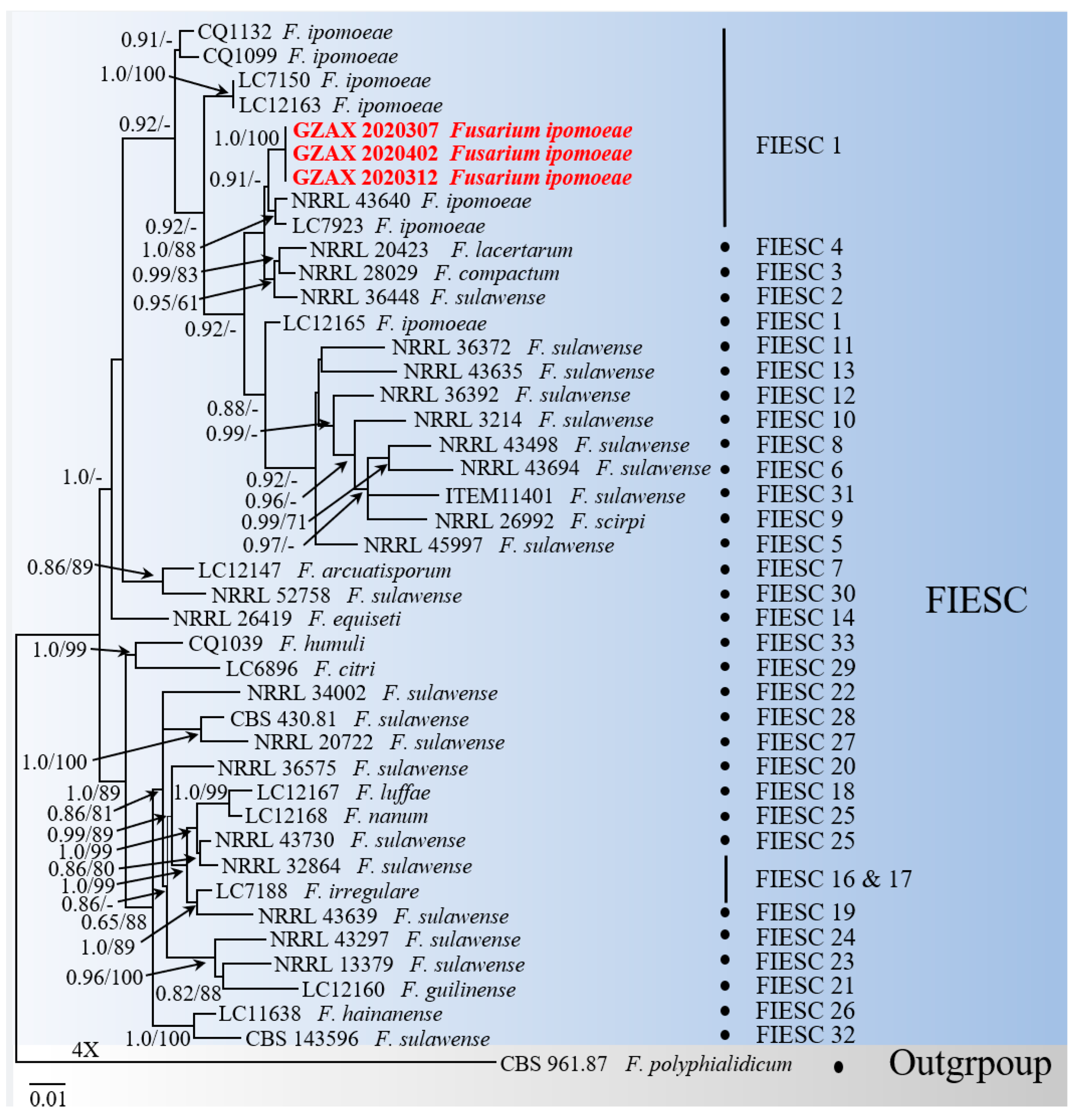
To further identify the causal agent, the phylogenetic analysis, based on the combination of five gene locus sequences, were performed (take GZAX 307 as an example). Firstly, the obtained sequences were analyzed on BLASTn, and the results showed that over 99% nucleotide sequence identity with members of the FIESC: the ITS sequence showed 100% identity (488/1132 bp) to the F. lacertarum strain NRRL 20423; the EF–1α sequence showed 99.56% identity (684/678 bp) to the F. ipomoeae strain NRRL 43640; the RPB2 sequence showed 100% identity (747/839 bp) to the F. ipomoeae strain CBS 140909; the RPB1 sequence showed 99.42% identity (1726/1578 bp) to the F. sulawense strain LC12173; and the CAM sequence showed 99.28% identity (715/698 bp) to the F. ipomoeae strain NRRL 34034. Then, phylogenetic analysis, using concatenated sequences of ITS, EF1–α, RPB2, RPB1, and CAM, showed that GZAX 307, GZAX 312, and GZAX 402 clustered monophyletically with strains of F. ipomoeae (the relevant strains are shown in Table 1). Therefore, based on the morphological and molecular characteristics, the isolates GZAX 307, GZAX 312, and GZAX 402 were identified as F. ipomoeae (Figure 3).

3.4. F. ipomoeae Showed Pathogenicity on Leaf and Stem

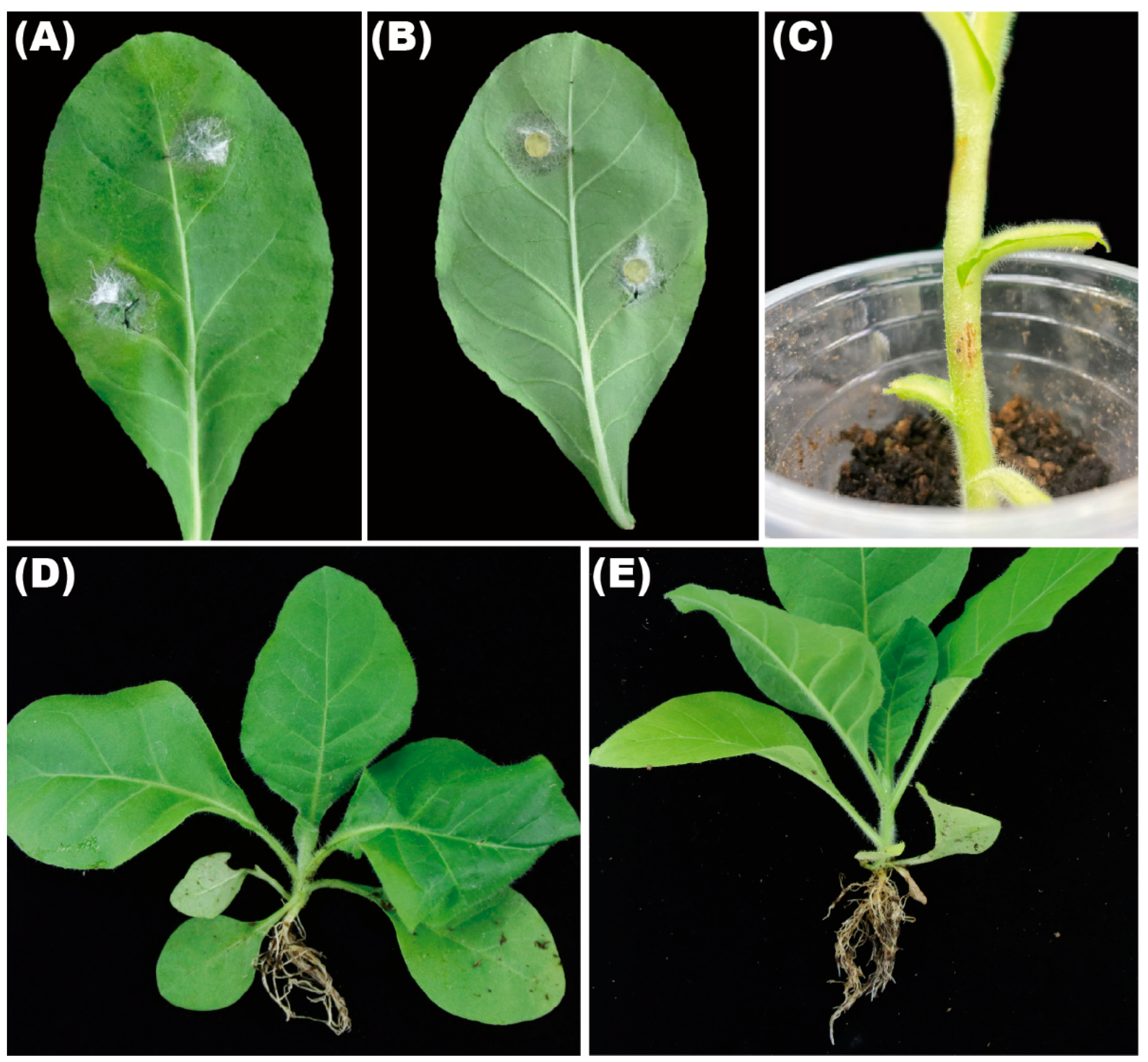
To complete the Koch postulates, the strains GZAX 307, GZAX 312, and GZAX 402 were inoculated on the leaves and stems, as well as the roots. The specific morphology and pathogenicity statistics are shown in Table 2. The pathogenicity results were consistent; therefore, the specific description took GZAX 307 as an example. The results of pathogenicity were the same, and strain 307 was taken as an example to show the symptoms. About 7 days post inoculation, white hyphae crawled on the leaves from the inoculation site; meanwhile, small brown spots appeared around the hyphae (Figure 4A,B). The lesion diameter can reach 42.6 ± 3.02 mm. The pathogen was re-isolated from the inoculated sites and further validated as the same fungus through morphological and phylogenetic analyses. Light brown spots appeared at the inoculation site of tobacco stem after 7 days (Figure 4C). After inoculation, no obvious pathogenicity symptoms occurred in the roots of the tobacco, compared with the control (Figure 4D,E).

4. Discussion

Dried and fermented tobacco leaves are raw resources for tobacco industry and product manufacturing [2]. Thus, owning to its important commercial values, this annual, leafy, solanaceous plant was planted globally; therefore, it creates jobs, increases incomes, maintains tax revenues, and sustains trade surpluses [2]. However, the field production of this cash crop is frequently seriously threatened by many fungal pathogens [7,25]. Thus, the safe and sustainable production of tobacco leaves required control of fungal diseases. Additionally, better understanding of the infection of pathogenic fungi is better for the control of the disease.
Previously, in order to know more about the incidence of fungal pathogens in the process of tobacco cultivation, we went to the tobacco-producing areas of Guizhou province and Guangxi Zhuang autonomous region to investigate. During the process of our investigation, we found that the symptoms were irregular brown spots, comprising whitish center, normally surrounded by yellow halo appearing on tobacco leaves. The disease was not deadly, but it seriously affected the quality and greatly reduced the economic value of tobacco. Further investigation revealed that a prevalence of the tobacco leaf spot was 22 to 35% in the field. In order to investigate the pathogen, we collected the samples of this disease from three random sites; through isolation and identification, we found that the dominant pathogen was Fusarium ipomoeae (Figure 2 and Figure 3).
Several studies have found that F. ipomoeae is associated with plant growth process, which serves as a causal pathogen that significantly affects the quality and quantity of products. Previously, F. ipomoeae have been reported to cause leaf spots on peanuts [26] and Bletilla striata [27] in China. However, to our knowledge, this is the first report worldwide of F. ipomoeae causing leaf spots on tobacco. Compared with the strain found in Bletilla striata, the colony and spore morphology in this study was consistent, but there were some differences in the spore sizes. In peanuts, the macroconidia were 4–7 septate, and 3.3–4.5 × 18.5–38.1 μm in size. In bletilla striata, the macroconidia were 3–5 septate, and 3.3–4.5 × 18.5–38.1 μm in size. In this study, macroconidia were 4–6 septa, most of which were 5 septa, with the macroconidia 44–118 × 4–11 μm in size. Czapek-Dox agar was used for determining the species strains from peanuts and PDA medium for strains from Bletilla striata, which were different from the CLA medium used in this study. However, CLA medium is commonly used to observe the spore morphology of Fusarium spp. [28]. The difference in spore size may also be related to host or cultural conditions, and it is necessary to study further.
Pathogenicity assays showed that leaf spot symptoms appeared on the inoculated leaves after 4 days post inoculation in Bletilla striata [27], and symptoms similar to those in the field were observed on leaves after 10 days inoculation in peanuts [26]. In the present study, leaf spot symptoms appeared on the inoculated tobacco leaves after one day. The pathogenicity of this pathogen to tobacco leaves was moderate and consistent with the field diseased symptom. A single inoculation site is not large, but when it spreads to the entirety of the tobacco leaves, tobacco production takes a pathogenic blow. Therefore, we should pay close attention to this pathogen in further disease control.

Author Contributions

Conceptualization, J.Y. and X.L.; methodology, H.W.; software, Y.L.; validation, X.L.; formal analysis, W.L. and X.L.; investigation, H.W.; resources, L.C. and J.M.; data curation, J.Y. and X.L.; writing—original draft preparation, J.Y. and G.X.; writing—review and editing, J.Y. and X.L.; visualization, H.W. and J.Y. All authors have read and agreed to the published version of the manuscript.

Funding

The Guizhou Tobacco Company (201914), China National Tobacco Corporation (110202001035 (LS–04), 110202101048 (LS–08)), Guizhou Science Technology Foundation (ZK (2021) Key036), National Natural Science Foundation of China (31960550, 32160522), Hundred Level Innovative Talent Foundation of Guizhou Province (GCC (2022) 028–1), Guizhou Provincial Academician Workstation of Microbiology and Health ((2020) 4004), and International Science and Technology Cooperation Base ((2020) 4102).

Institutional Review Board Statement

Not applicable.

Informed Consent Statement

Not applicable.

Data Availability Statement

The ITS, EF–1, RPB2, RPB1, and CAM sequences of strain GZAX 307 were deposited under GenBank numbers ON961779, ON982723, ON982725, ON982727, and ON982721, respectively. The ITS, EF–1, RPB2, RPB1, and CAM sequences of strain GZAX 312 were deposited under GenBank numbers ON961780, ON982724, OM982726, ON982728, and ON982722, respectively.

Acknowledgments

This work was supported by Guizhou Academy of Tobacco Science. We thank Changqing Zhang for kindly helping with sample collection and Jianxin Deng for providing technical support. The authors sincerely appreciate the two teachers’ valued help on the paper.

Conflicts of Interest

The authors declare no conflict of interest.

References

  1. Liu, X.; Li, Y.; Wang, M.; Cao, P.; Wang, H.; Yin, J. Stem-end Rot Caused by Lasiodiplodia brasiliense on Tobacco in China. Plant Dis. 2022. [Google Scholar] [CrossRef] [PubMed]
  2. Warner, K.E. The Economics of Tobacco: Myths and Realities. Tob. Control 2000, 1, 78–89. [Google Scholar] [CrossRef] [PubMed]
  3. Martins–da–Silva, A.S.; Torales, J.; Becker, R.F.V.; Moura, H.F. Tobacco growing and tobacco use. Int. Rev. Psychiatry 2022, 1, 51–58. [Google Scholar] [CrossRef] [PubMed]
  4. Guo, Z.; Xie, H.; Wang, H.; Huang, Y.; Chen, Q.; Xiang, L.; Yu, Z.; Yang, X. Leaf Spot Caused by Didymella segeticola on Tobacco in China. Plant Dis. 2019, 5, 1559. [Google Scholar] [CrossRef]
  5. Wang, H.C.; Wang, M.S.; Xia, H.Q.; Yang, S.J.; Shi, J.X. First Report of Fusarium Wilt of Tobacco Caused by Fusarium kyushuense in China. Plant Dis. 2013, 3, 424. [Google Scholar] [CrossRef] [PubMed]
  6. Jin, F.; Ding, Y.; Wei, D.; Reddy, M.S.; Du, B. Genetic Diversity and Phylogeny of Antagonistic Bacteria against Phytophthora nicotianae Isolated from Tobacco Rhizosphere. Int. J. Mol. Sci. 2011, 5, 3055–3071. [Google Scholar] [CrossRef] [PubMed]
  7. LaMondia, J.A. Fusarium wilt of tobacco. Crop Prot. 2015, 1, 73–77. [Google Scholar] [CrossRef]
  8. Wang, J.; Li, S.; Zhang, C.; Chao, F.; Kong, F. Occurrence and pathogen isolation of tobacco Fusarium root rot in main tobacco production regions of Shandong province. In Proceedings of the 2011 6th IEEE Conference on Industrial Electronics and Applications, Beijing, China, 21–23 June 2011; pp. 2637–2639. [Google Scholar]
  9. Gurdaswani, V.; Ghag, S.B. Toxins from Fusarium species and their role in animal and plant diseases. In New and Future Developments in Microbial Biotechnology and Bioengineering; Elsevier: Amsterdam, The Netherlands, 2020; pp. 7–27. [Google Scholar]
  10. Mincuzzi, A.; Sanzani, S.M.; Palou, L.; Ragni, M.; Ippolito, A. Postharvest Rot of Pomegranate Fruit in Southern Italy: Characterization of the Main Pathogens. J. Fungi 2022, 5, 475. [Google Scholar] [CrossRef]
  11. Cheng, H.; Zhao, L.; Wei, X.; Liu, X.; Gao, G.; Deng, J.; Li, M. Alternaria species causing leaf spot on hemp (Cannabis sativa) in Northern China. Eur. J. Plant Pathol. Vol. 2022, 4, 957–970. [Google Scholar] [CrossRef]
  12. Liu, H.; Zhou, J.; Liao, J.; Yi, J.; Ma, D.; Deng, J. Grafted twig rot on Citrus sinensis caused by a member of the Fusarium solani species complex. Can. J. Plant Pathol. Rev. Can. Phytopathol. 2020, 42, 133–139. [Google Scholar] [CrossRef]
  13. Cenis, J.L. Rapid extraction of fungal DNA for PCR amplification. Nucleic Acids Res. 1992, 9, 2380. [Google Scholar] [CrossRef] [PubMed]
  14. O’Donnell, K.; Cigelnik, E.; Nirenberg, H.I. Molecular systematics and phylogeography of the Gibberella fujikuroi species complex. Mycologia 1998, 3, 465–493. [Google Scholar] [CrossRef]
  15. O’Donnell, K.; Nirenberg, H.I.; Aoki, T.; Cigelnik, E. A Multigene phylogeny of the Gibberella fujikuroi species complex: Detection of additional phylogenetically distinct species. Mycoscience 2000, 1, 61–78. [Google Scholar] [CrossRef]
  16. O’Donnell, K. Phylogenetic analyses of RPB1 and RPB2 support a middle Cretaceous origin for a clade comprising all agriculturally and medically important Fusaria. Fungal Genet. Biol. 2013, 3, 20–31. [Google Scholar] [CrossRef]
  17. Mirghasempour, S.A.; Studholme, D.J.; Chen, W.; Zhu, W.; Mao, B. Molecular and Pathogenic Characterization of Fusarium Species Associated with Corm Rot Disease in Saffron from China. J. Fungi 2022, 5, 515. [Google Scholar] [CrossRef]
  18. Vignassa, M.; Meile, J.C.; Chiroleu, F.; Soria, C.; Leneveu–Jenvrin, C.; Schorr–Galindo, S.; Chillet, M. Pineapple Mycobiome Related to Fruitlet Core Rot Occurrence and the Influence of Fungal Species Dispersion Patterns. J. Fungi 2021, 3, 175. [Google Scholar] [CrossRef]
  19. He, L.; Cheng, H.; Htun, A.A.; Ge, H.; Xia, Z.Z. Phylogeny and taxonomy of two new Alternaria (Ascomycota: Pleosporaceae) species in section Gypsophilae from China. Mycol. Prog. 2021, 4, 355–363. [Google Scholar] [CrossRef]
  20. Sunpapao, A.; Suwannarach, N.; Kumla, J.; Dumhai, R.; Riangwong, K.; Sanguansub, S.; Wanchana, S.; Arikit, S. Morphological and Molecular Identification of Plant Pathogenic Fungi Associated with Dirty Panicle Disease in Coconuts (Cocos nucifera) in Thailand. J. Fungi 2022, 4, 335. [Google Scholar] [CrossRef]
  21. Stamatakis, A.; Ludwig, T.; Meier, H. RAxML-III: A fast program for maximum likelihood-based inference of large phylogenetic trees. Bioinformatics 2005, 21, 456–463. [Google Scholar] [CrossRef]
  22. Huelsenbeck, J.P.; Ronquist, F. MRBAYES: Bayesian inference of phylogenetic trees. Bioinformatics 2001, 17, 754–755. [Google Scholar] [CrossRef] [Green Version]
  23. Posada, D.; Crandall, K.A. MODELTEST: Testing the model of DNA substitution. Bioinformatics 1998, 14, 817–818. [Google Scholar] [CrossRef] [PubMed]
  24. Wang, M.; Chen, Q.; Diao, Y.; Duan, W.; Cai, L. Fusarium incarnatum-equiseti complex from China. Pers. Mol. Phylogeny Evol. Fungi 2019, 1, 70–89. [Google Scholar] [CrossRef] [PubMed]
  25. Chen, Q.L.; Cai, L.; Wang, H.C.; Cai, L.T. Fungal composition and diversity of the tobacco leaf phyllosphere during curing of leaves. Front. Microbiol. 2020, 11, 554051. [Google Scholar] [CrossRef] [PubMed]
  26. Xu, M.; Zhang, X.; Yu, J.; Guo, Z.; Li, Y.; Wu, J.; Chi, Y. First Report of Fusarium ipomoeae Causing Peanut leaf Spot in China. Plant Dis. 2021, 11, 3754. [Google Scholar] [CrossRef] [PubMed]
  27. Zhou, L.; Yang, S.; Wang, S.; Lv, J.; Zhou, H. Identification of Fusarium ipomoeae as the causative agent of leaf spot disease in Bletilla striata in China. Plant Dis. 2021, 4, 1204. [Google Scholar] [CrossRef]
  28. Leslie, J.F.; Summerell, B.A. The Fusarium Laboratory Manual—Species Concepts in Fusarium; Blackwell Publishing: Hoboken, NJ, USA, 2006. [Google Scholar]
Figure 1. (AC) Diseased tobacco leaf symptoms in the field.
Microorganisms 10 01890 g001
Figure 2. (A,B) Colony on PDA after incubation for 7 days. (C,D) Macroconidia. (E,F) Chlamydospores. (GI) Conidiogenous cells form on sporodochia. Bars: (C,D,G) = 50 μm; (E,F,H,I) = 10 μm.
Microorganisms 10 01890 g002
Figure 3. Phylogenetic tree was constructed using strains of Fusarium incarnatumequiseti complex (FIESC), based on ITS, EF–1α, CAM, RPB1, and RPB2 loci. MrBayes posterior probabilities (PP > 65) and ML bootstrap (BS > 60%) support values were showed at the nodes (PP/BS).
Microorganisms 10 01890 g003
Figure 4. Pathogenicity on tobacco (A,B) leaves, (C) stems, and (D) roots. (E) The roots of control plant.
Microorganisms 10 01890 g004
Table 1. Strains used in the phylogenetic analyses and their GenBank accession numbers.
SpeciesPhylogenetic SpeciesStrain HostLocationITSEF–1CAMRPB2RPB1
F. ipomoeaeFIESC 1CQ1099Rhododendron pulchrum leafJiangsu, China MK280853MK289573MK289715MK289727MK289861
LC7923Capsicum sp. Shandong, China MK280800MK289635MK289688MK289789MK289853
CQ1132Vinca major leaf Jiangsu, China MK280854MK289574MK289716MK289728MK289862
NRRL 43640 = UTHSC 04–123Dog noseTexas, America GQ505756GQ505667GQ505578GQ505845HM347191
LC12165 = CGMCC3.19496 (T) Ipomoea aquatica leafFujian, China MK280832MK289599MK289704MK289752MK289859
LC7150Bamboo Jiangxi, ChinaMK280818MK289627MK289678MK289781MK289852
LC12163Hibiscus syriacusFujian, ChinaMK280790MK289597MK289700MK289750MK289857
GZAX 402TobaccoGuizhou, ChinaOP454871OP432881OP432880OP432883OP432882
GZAX 307TobaccoGuizhou, ChinaON961779ON982723ON982721ON962725ON962727
GZAX 312TobaccoGuangxi, ChinaON961780ON982724ON982722ON982726ON982728
F. sulawenseFIESC 2NRRL 36448 = CBS 384.92 Phaseolus vulgaris seedSudan GQ505741GQ505652GQ505564GQ505830
F. compactumFIESC 3NRRL 28029 = CDC B–3335Human eyeCalifornia, America GQ505691GQ505602GQ505514GQ505780HM347150
F. lacertarumFIESC 4NRRL 20423 = IMI 300797 (T) Lizard skin IndiaGQ505682GQ505593GQ505505GQ505771JX171467
F. sulawenseFIESC 5NRRL 45997 = UTHSC 04–1902 Human sinusColorado, America GQ505761GQ505672GQ505583GQ505850
NRRL 34035 = UTHSC 91–569Human sinusColorado, America GQ505726GO505637GQ505549GQ505815
F. sulawenseFIESC 6NRRL 43694 = CDC 2006743607 Human eye Texas, AmericaGQ505757GQ505668GQ505579GQ505846HM347193
F. arcuatisporumFIESC 7LC12147 = CGMCC3.19493 (T) Brassica campestris pollen Hubei, ChinaMK280802MK289584MK289697MK289739MK289799
F. sulawenseFIESC 8NRRL 43498Human eye Pennsylvania, AmericaGQ505747GQ505658——GQ505836HM347181
F. sulawenseFIESC 30NRRL 52758 = ARSEF 4714 Prosapia nr. bicincta on CynodonCosta Rica JF740925JF740833——JF741159——
F. scirpiFIESC 9NRRL 26992 = CBS 610.95 SoilFranceGQ505681GQ505592GQ505504GQ505770——
F. sulawenseFIESC 31ITEM11401Avena sativaCanada ——LN901578LN901594LN901611——
F. sulawenseFIESC 10NRRL 3214 = FRC R–6054, 7.13 MRC Unknown UnknownGQ505676GQ5O5587GQ505499GQ505765——
F. sulawenseFIESC 13NRRL 43635 = UTHSC 06–638 Horse Nebraska GQ505751GQ505662GQ505573GQ505840HM347188
F. sulawenseFIESC 12NRRL 36392 = CBS 259.54 Unknown plant seedling GermanyGQ505739GQ505650GQ505562GQ505828——
F. sulawenseFIESC 11NRRL 36372 = CBS 235.79 AirAntilles, NetherlandsGQ505738GQ505649GQ505561GQ505827——
F. equisetiFIESC 14NRRL 26419 = CBS 307.94, BBA 68556 (NT) Soil Germany GQ505688GQ505599GQ505511GQ505777——
F. irregulareFIESC 15LC7188 = CGMCC3.19489 (T) Bamboo Guangdong, China MK280829MK289629MK289680MK289783MK289863
F. sulawenseFIESC 16 & 17NRRL 32864 = FRC R–7245Human Texas, America GQ505702GQ505613GQ505525GQ505791HM347160
NRRL 43730 = CDC 2006743605 Contact lens Mississippi, America EF453193GQ505669GQ505580GQ505847——
F. luffaeFIESC 18LC12167 = CGMCC3.19497 (T) Luffa aegyptiaca Fujian, China MK280852MK289569MK289711MK289723MK289870
F. sulawenseFIESC 19NRRL 43639 = UTHSC 04–135 Manatee Florida, AmericaGQ505755GQ505666GQ505577GQ505844HM347190
F. sulawenseFIESC 20NRRL 36575 = CBS 976.97 Juniperus chinensis leaf Hawaii, AmericaGQ505745GQ505656GQ505568GQ505834——
F. sulawenseFIESC 22NRRL 34002 = UTHSC 95–1545Human ethmoid sinusTexas, America GQ505715GQ505626GQ505538GQ505804HM347165
F. sulawenseFIESC 23NRRL 13379 = FRC R–5198, BBA 62200Oryza sativaIndiaGQ505680GQ505591GQ505503GQ505769——
F. sulawenseFIESC 24NRRL 43297 = W. Elmer 22 Spartina rhizomesConnecticut, America GQ505746GQ505657GQ505569GQ505835——
F. sulawenseFIESC 27NRRL 20722 = IMI 190455 Chrysanthemum sp. Kenya GQ505684GQ505595GQ505507GQ505773——
F. guilinenseFIESC 21LC12160 = CGMCC3.19495 (T)Musa nana leafGuangxi, ChinaMK280837MK289594MK289652MK289747MK289831
F. sulawenseFIESC 28CBS 430.81 = NRRL 28577Grave stoneRomania GQ505692GQ505603GQ505515GQ505781——
F. sulawenseFIESC 32CBS 143596Stereum irsutumIranLT970815LT970779LT970732LT970751——
F. nanumFIESC 25LC12168 = CGMCC3.19498 (T) Musa nana leaf Guangxi, China GQ505697GQ505608GQ505520GQ505786——
F. hainanenseFIESC 26LC11638 = CGMCC3.19478 (T) Oryza sp. stem Hainan, China MK280836MK289581MK289657MK289735MK289833
F. citriFIESC 29LC6896 = CGMCC3.19467 (T) Citrus reticulata leafHunan, ChinaMK280803MK289617MK289668MK289771MK289828
F. humuliFIESC 33CQ1039 = CGMCC3.19374 (T) Humulus scandens leaf Jiangsu, ChinaMK280845MK289570MK289712MK289724MK289840
F. polyphialidicum——CBS 961.87Plant debris South Africa GQ505763GQ505674GQ505585GQ505852——
Table 2. Morphology characterization and pathogenicity of F. ipomoeae.
Species (Strain)LocationColonies (mm)ConidiaPathogenicity on Tobacco Leaf (mm)On Stem (mm)On Root (mm)
Body (μm)SeptaWoundedUnwoundedWoundedUnwounded
F. ipomoeae (GZAX 307)Zheng’an in Guizhou province61.5 ± 0.544–118 × 4–114–637 ± 234 ± 318 ± 212 ± 6
F. ipomoeae (GZAX 312)Shanglin in Guangxi Zhuang autonomous region62 ± 147–123 × 2–94–635 ± 132 ± 220 ± 411 ± 5
F. ipomoeae (GZAX 402)Fenggang in Guizhou province62.5 ± 142–120 × 4–144–633 ± 434 ± 317 ± 210 ± 5
Publisher’s Note: MDPI stays neutral with regard to jurisdictional claims in published maps and institutional affiliations.