Article

Genomic Features and Construction of Streamlined Genome Chassis of Nisin Z Producer Lactococcus lactis N8

1
Key Laboratory of Molecular Microbiology and Technology, Ministry of Education, College of Life Sciences, Nankai University, Tianjin 300071, China
2
Department of Microbiology, Faculty of Agriculture and Forestry, University of Helsinki, 00014 Helsinki, Finland
3
Department of Bacteriology and Immunology, Human Microbiome Research Program, Faculty of Medicine, University of Helsinki, 00014 Helsinki, Finland
4
State Key Laboratory of Medicinal Chemical Biology and Tianjin Key Laboratory of Protein Sciences, College of Life Sciences, Nankai University, Tianjin 300071, China
5
School of Life Sciences, Shanxi University, Taiyuan 030006, China
*
Authors to whom correspondence should be addressed.
Microorganisms 2022, 10(1), 47; https://doi.org/10.3390/microorganisms10010047
Submission received: 4 November 2021 / Revised: 3 December 2021 / Accepted: 13 December 2021 / Published: 27 December 2021

Abstract

:
Lactococcus lactis is a commonly used fermenting bacteria in cheese, beverages and meat products. Due to the lack of simplified chassis strains, it has not been widely used in the fields of synthetic biology. Thus, the construction of lactic acid bacteria chassis strains becomes more and more important. In this study, we performed whole genome sequencing, annotation and analysis of L. lactis N8. Based on the genome analysis, we found that L. lactis N8 contains two large plasmids, and the function prediction of the plasmids shows that some regions are related to carbohydrate transport/metabolism, multi-stress resistance and amino acid uptake. L. lactis N8 contains a total of seven prophage-related fragments and twelve genomic islands. A gene cluster encoding a hybrid NRPS–PKS system that was found in L. lactis N8 reveals that the strain has the potential to synthesize novel secondary metabolites. Furthermore, we have constructed a simplified genome chassis of L. lactis N8 and achieved the largest amount of deletion of L. lactis so far. Taken together, the present study offers further insights into the function and potential role of L. lactis N8 as a model strain of lactic acid bacteria and lays the foundation for its application in the field of synthetic biology.

1. Introduction

Lactococcus lactis, a group of Gram-positive, catalase-negative, mesophilic fermentative bacteria producing lactic acid from sugar [1], has a long history of safe use in the fermented food industry and is granted a “GRAS” (generally regarded as safe) status. L. lactis has three subspecies, two of which are routinely employed in the dairy fermentation sector, i.e., subspecies (subsp.) lactis and subsp. lactis biovar diacetylactis, which distinguishes itself based on the citrate metabolism. The remaining is L. lactis subsp. hordniae isolated from the leafhopper Hordnia circellata [2]. Genetically, a typical L. lactis chromosome ranges in size from ~2.2 to 2.6 Mb and is often accompanied by plasmid complements [3] and multiple remnant prophages [4].
L. lactis N8 (N8) is a nisin Z producer isolated from milk in Finland, and the knowledge gained from fundamental research on this nisin production strain has been exploited for a wide variety of biotechnological applications. N8 is robust and genetically amenable, which has facilitated the analysis of introduced lactococcal and heterologous DNA. Due to the feature of considerable nisin yield, the research on this strain so far has mainly focused on its nisin synthetic gene cluster and increasing nisin yield [5,6]. The gene cluster for nisin synthetic (nisZBTCIPRKFEG) in strain N8 has been thoroughly studied in the past years. The NisB and NisC are responsible for the dehydration and cyclization of the precursor nisin [7]. The NisT is responsible for the transport of nisin and is also responsible for the dehydration and formation of lanthionine during the maturation of nisin, which occurs independently of the transport [5]. The NisI could have a supportive role in nisin immunity [8]. The NisP is essential to the maturation of nisin, and it has catalytic activity for nisin leader-peptide cleavage [9]. The NisRK, as a two-component regulatory system, has been characterized in other lactic acid bacteria [10]. The NisFEG, as a transporter complex, is important for the development of nisin immunity [8,11].
Researchers have tried to increase the nisin production of N8 through various methods, such as: (1) by improving the nisin immunity of the producer strain [12]; (2) by optimizing the culture conditions to achieve a high nisin yield of the producer strain [6]; (3) by using Mu transposition technology to build a library to screen genes that affect nisin production and nisin immunity [13,14].
Moreover, strain N8 has been used in food applications. For example, L. lactis N8 and Saccharomyces boulardii SAA655 were used in idli batter to increase the nutritional values of the food products by boosting the riboflavin and folate levels by 40 to 90% [15]. Recently, co-culturing L. lactis N8 with a bacterial cellulose-producing strain Enterobacter sp. FY-07 has successfully endowed bacterial cellulose with antibacterial properties and extended the application of N8 in food packaging [16]. Our group have heterologously secreted leucocin C in N8 and successfully achieved nisin and leucocin C co-expression. The recombinant bacteria exhibited highly efficient antimicrobial activity against L. monocytogenes [17].
In the past 30 years, a great deal of studies on N8 have been published, demonstrating its importance in the field of microbiology and biotechnology. However, the characteristics of its genome have not been deeply explored. Furthermore, the recent appraisal for the potential application of L. lactis strains in oral vaccine delivery may expand the importance of lactococcal investigations into the medical/pharmaceutical arena [18] as probiotic lactic acid bacteria are becoming more and more important in the field of synthetic biology [19].
In order to expand its application in the fields of the pharmaceutical arena, synthetic biology and metabolic engineering, in this study, we sequenced the whole genome of L. lactis N8. Through an in-depth analysis of the whole genome, we predicted the prophages and genomic islands of N8 that could be deleted to decrease the metabolic burden of the strain and increase the yield of useful substances. Through the analysis of the whole genome, we obtained the theoretical background, and, based on this, we carried out the construction of the N8 streamlined genome chassis using our established genome editing methods [20].

2. Materials and Methods

2.1. Strain and Media

All the strains used in this study were listed in Table 1. The original strain L. lactis N8 was isolated from milk in Finland [21]. M17 broth was purchased from Hope Bio-Technology (Qingdao, China). N8 and its derivatives were grown on GM17 agar plates (supplemented with 0.5% (w/v) glucose). After incubation overnight at 30 °C, a single colony was inoculated in GM17 and cultured overnight at 30 °C. Primers used in this study were purchased from GENEWIZ (Suzhou, China) and are listed in Supplementary Table S1.

2.2. DNA Extraction and Genome Sequencing

Genomic DNA (gDNA) was extracted from 2 mL overnight cultures of L. lactis N8 cells using MagAttract HMW DNA kit (Qiagen, Hilden, Germany) according to the manufacturer’s protocol. Isolated gDNA was subjected to whole genome sequencing performed by the DNA Sequencing and Genomics Laboratory (Helsinki Institute of Life Science, University of Helsinki, Helsinki, Finland). The high throughput sequencer Pacbio Sequel II System (Pacific Biosciences, San Francisco, CA, USA) was used for sequencing, and assembling was done by SMRT Link 9 Analysis software (Pacific Biosciences). Polished contigs were used for genome analysis. A more detailed explanation of the sequencing method can be found in the genome announcement of L. lactis subsp. lactis N8 [21].

2.3. Genome Analysis

2.3.1. Genome Annotation

The annotation of the genome was performed using the NCBI Prokaryotic Genome Annotation Pipeline (PGAP), and the resultant annotated proteome was further annotated with Cluster of Orthologous Groups (COG) categories and KEGG pathways using the Prokka version 1.14.6 and eggNOG mapper [26,27,28]. Circos software version 0.69-9 was used to make circle maps of the genome and large plasmids after genome assembly and annotation. Comparison of the homology of two large plasmids was performed and visualized using EasyFig [29]. All L. lactis plasmid data were extracted from NCBI https://www.ncbi.nlm.nih.gov/genome/browse/#!/plasmids/156/ (accessed on 5 July 2021). To differentiate plasmids and chromosomal accessory genes and highlight the plasmid contribution, COG annotation was conducted utilizing WebMGA server8 [30], using as input protein files predicted by Prokka. The flower plot was made using an online platform (https://www.genescloud.cn/; accessed on 5 July 2021).

2.3.2. Pan-Genome and Phylogenetic Analysis

To establish the accurate position of N8 within the population structure of L. lactis, we reconstructed the phylogeny using all available 202 L. lactis strains, including subsp. lactis, subsp. lactis bv. diacetylactis and subsp. hordniae (data retrieved by latest 5th July 2021, Supplementary Table S2). To ensure the assembly quality of dataset, only genomes with an N50 size over 20 kbp were selected. The pangenomes of L. lactis were inferred using Roary version 3.11.2 [31]. First, the nucleotide fasta files from all selected strains were downloaded from NCBI together with the GFF files and converted to GFF3 using a script (Supplementary Table S3). Regarding the strains for which GFF3 files were not provided by NCBI or could not pass the preliminary analysis by Roary, their nucleotide fasta files were downloaded and annotated using Prokka to obtain a compatible GFF3 file [26]. Second, the GFF3 annotations were provided to Roary to calculate the pangenome of the dataset and produce a multiple sequence alignment of the concatenated core genes (present in > 99% strains) using MAFFT. Third, the multiple sequence alignment of the core genome was used to generate a best-fit maximum likelihood phylogeny using IQTREE version 1.6.12 using ModelFinder optimization [32,33]. Finally, the trees were visualized in iTOL version 5.5 [34]. Pangenomes of closely related 16 strains to L. lactis N8 were also analyzed by roary_plots.py (https://github.com/sanger-pathogens/Roary/tree/master/contrib/roary_plots; accessed on 5 July 2021) to show the presence and absence of core and accessory genes.

2.3.3. Prophages Prediction

The nucleic acid of the lysogenic phage integrated into the host genome is referred to as prophage. In this study, we examined the prevalence of prophages in L. lactis N8 and its derivatives using online prediction tool PHASTER (https://phaster.ca/; accessed on 5 July 2021) [35].

2.3.4. Genomic Islands Prediction

Genomic islands can encode a variety of functions involving symbiosis and pathogenesis. IslandViewer 4 (http://www.pathogenomics.sfu.ca/islandviewer/; accessed on 5 July 2021) was used to predict genomic islands [36].

2.3.5. Genome Mining for Secondary Metabolite Biosynthetic Gene Clusters

Secondary metabolites by bacteria are an important source for identification of novel antimicrobial and bioactive compounds. To excavate this potential of L. lactis N8 on top of its excellence in antimicrobial nisin production, secondary metabolite biosynthetic gene clusters of N8 were predicted using the antiSMASH 5.0 (https://antismash.secondarymetabolites.org/; accessed on 5 July 2021) [37]. Swiss-model was used to predict the structure of antimicrobial peptides [38].

2.4. Methods of Genome Streamlining

The vectors for the deletion of nonessential fragments were constructed as described in our previous work [12]. Briefly, two fragments of the flanking region of nonessential fragment L1 were amplified by PCR with a proof-reading polymerase (Takara, Dalian, China), and the N8 chromosome was used as template. Then, the fragments were ligated into the XhoI–SwaI and SacI–BglII restriction sites of pNZ5319. The recombinant plasmid pNZ5319∆L1 obtained was transformed into E. coli DH5α cells strain by CaCl2 method [39]. Then, pNZ5319∆L1 was isolated and electroporated into L. lactis N8 competent cells to generate mutant L. lactis N8-1-cat. After the deletion of the L1 fragment, cat gene in the mutant was retrieved by introducing the plasmid pNZTS-Cre into L. lactis N8-1-cat strain. The final mutant was named as L. lactis N8-1. Seven nonessential DNA regions were deleted on schedule with the Cre-loxP system. The gene knockout vectors pNZ5319∆L2, pNZ5319∆L3 and pNZ5319∆L4, pNZ5319∆L5, pNZ5319∆L6, pNZ5319∆L7 and pNZ5319∆L8 were constructed subsequently. The second nonessential fragment was then deleted in L. lactis N8-1 with the plasmid pNZ5319∆L2. The derivative was named L. lactis N8-2. The deletion step was repeated with pNZ5319∆L3, pNZ5319∆L4, pNZ5319∆L5, pNZ5319∆L6, pNZ5319∆L7 and pNZ5319∆L8 plasmids. The resulting mutants were named L. lactis N8-3, L. lactis N8-4, L. lactis N8-5, L. lactis N8-6, L. lactis N8-7 and L. lactis N8-8, respectively. The correct deletions of the eight constructed strains were confirmed by resequencing.

2.5. Resequencing

Isolation of gDNA was carried out using SDS method. Total DNA obtained was subjected to quality control by agarose gel electrophoresis and quantified by Qubit (Thermo Fisher Scientific, Waltham, MA, USA). The genome of L. lactis N8-8 was sequenced with MPS (massively parallel sequencing) Illumina technology (Illumina, San Diego, CA, USA). The DNA library was constructed: a paired-end library with an insert size of 350 bp. The 350-bp library was sequenced using an Illumina PE150 strategy. Library construction and sequencing were performed at the Beijing Novogene Bioinformatics Technology Co., Ltd. Quality control of paired-end reads was performed using in-house program. Then, data processing, reads mapping and SV (structural variation) analysis were conducted as we described previously [12].

2.6. Phenotype Testing

2.6.1. Determination of Growth Profiles

To determine the growth profiles, L. lactis strains were cultured at 30 °C for 6 h in static (non-aerated) condition to log phase, harvested by centrifugation (5000× g, 3 min) and washed twice with PBS (phosphate buffered saline, pH 7.4). After washing, the cells were resuspended (adjusted to the same initial cell concentration) in GM17 medium as seed. Then, the seeds were inoculated into 100 mL GM17 flask culture medium with the ratio of 1% and cultured at 30 °C in static condition; samples were taken every hour for the determination of OD600, and the experiment was repeated three times independently. The maximum growth rate and generation time were calculated by using the data of growth profiles.

2.6.2. Determination of Nisin Titre

Nisin yield was determined by the agar well diffusion method [12] with minor modifications. Briefly, the broth of the tested strains after fermentation was boiled for 10 min and cells were removed by centrifugation at 8000 rpm for 3 min. Then, the supernatant was appropriately diluted with 0.02 M HCl. M. luteus (107 CFU/mL) was used as indicator and inoculated at a concentration of 1% (v/v) into 30 mL melted/cooled LB agar. To enhance nisin diffusion, 1.5% (v/v) Tween 80 (JiangTian, Tianjin, China) was added to the medium and mixed well. Then, the medium was quickly poured into sterile petri dish. After solidification and pre-cultivation, a 7 mm diameter sterile cork borer (MRS Scientific Ltd., Wickford, UK) was used to generate agar well for loading samples. Standard nisin solutions (concentrations of 20, 40, 80, 100, 200 and 400 IU/mL) were prepared using nisin powder (Sigma, St. Louis, MO, USA). Subsequently, standard nisin solutions and sample solutions were, respectively, loaded into the wells (80 μL per well), and the plates were incubated at 37 °C for 24 h. The diameter of inhibition zone was measured with a calliper. A regression equation was derived from the nisin standard data.

2.7. Statistical Analysis

The experiments to determine the growth profiles of L. lactis were performed in independent biological triplicates, and each sample was additionally collected in technical triplicates. For calculating the maximum growth rate and generation time, the experiments were independently repeated at least three times. Assays to determine the nisin yield of different L. lactis strains were independently repeated at least three times. Statistical analyses of data were performed using Origin 85 software version 8.5.0 SRI (OriginLab Corporation, Northampton, MA, USA) and GraphPad Prism 5 software version 5.01 (GraphPad software, Inc, San Diego, CA, USA).

3. Results and Discussion

3.1. General Features of the Genome

The assembly of the reads resulted in three contigs, implying that N8 contains one chromosome and two megaplasmids (Figure 1 and Table 2) [21]. N8 has fewer CDs than L. lactis KF147 (2446) and L. lactis KLDS 4.0325 (2596). The coding regions of N8 have a guanine plus cytosine (GC) content of 35.1 mol%, which is comparable to the other L. lactis strains.

3.2. Phylogeny and Core-Pan Genome

To visualize the position of L. lactis N8 within the species, we constructed an unrooted phylogenetic tree using all L. lactis currently published on NCBI (update on 5 July 2021) (Figure 2), and the rooted phylogenetic tree of all L. lactis revealed the distances among the strains, which is shown in Supplementary Figure S1. Fifteen strains are closely related to strain N8 and share the same evolutionary origin, as shown in Figure 2 marked with a green background.
The Roary matrix was generated from these 15 strains together with L. lactis N8, showing the genetic relatedness among them (Figure 3A). A pan-genome analysis shows that N8 shares the same immediate evolutionary origin with strains YF11, G423 and F44. The closest related L. lactis YF11 has a coding density of 95.52% similar to L. lactis N8. Meanwhile, the strain N8 has a coding density of 90.51% and 87.50% similar to L. lactis G423 and L. lactis F44, respectively. From the flower plot in Figure 3B, we can see that these 16 strains have 1682 genes in common, indicating that they are very closely related.
It is worth noting that, previously, L. lactis contained four subspecies: L. lactis subsp. lactis, L. lactis subsp. hordniae, L. lactis subsp. cremoris and L. lactis subsp. tructae. These four subspecies could be divided into two groups based on recA sequence analysis: L. lactis subsp. lactis and L. lactis subsp. hordniae; L. lactis subsp. cremoris and L. lactis subsp. tructae. The two groups had a relatively low DNA–DNA hybridization value (about 60%) [2]. In a recent study, L. lactis has been reclassified to three component subspecies, i.e., subsp. lactis, subsp. lactis biovar diacetylactis and subsp. hordniae. The former L. lactis subsp. cremoris is now an independent species L. cremoris sp. nov. and transfer L. lactis subsp. tructae to L. cremoris as L. cremoris subsp. tructae comb. nov. [40].

3.3. Megaplasmids of L. lactis N8

Plasmids, normally dispensable for bacterial growth, are autonomously replicating extrachromosomal DNA entities. They carry backbone genes important for the replication and maintenance in their host and might confer an advantage to their host in its ecological niche [41]. The genome of L. lactis N8 was revealed to possess two megaplasmids, designated pLLN8-1 (80,301 bp) and pLLN8-2 (71,261 bp) (Figure 4A). They are stably maintained in their natural host N8 and in total represent 5.89% (151,562 bp) of the N8 genome. An overview of the putative functions of plasmid-encoded genes is shown in Figure 4B. The two megaplasmids carry a number of genes known to be important for growth and survival under a specific environment. These two plasmids carry the genes encoding many functional proteins, for example proteins responsible for protein degradation, bacteriophage biogenesis, stress resistance, mobilization, partition systems, oligopeptide transporter, type I restriction-modification (R-M) system, metal transporters, enzymes and transcriptional regulators. The pLLN8-1 encodes XRE-family HTH domain transcriptional regulator, sugar transport proteins (lacRABCDFEGX), cadmium resistance proteins (cadAC), potassium transporter (trkAH), magnesium transporter (corA), putative Asp23/Gls24 family general stress response protein (ymgGIJ), oligopeptide transporter (oppAC), putative iron export ABC transporter (fetAB), chromosome partitioning proteins (parAB), ribonucleoside-diphosphate reductase (nrdFEI) and several IS3 and IS6 family transposases. Its replication initiator protein is the RepB family protein. The pLLN8-2 encodes functional copper resistance proteins (lcoRSABC), copper metabolism proteins (copYZ), cadmium resistance proteins (cadAC) and several IS3, IS6 and IS982 family transposases. Two highly homologous repB-containing replicons were found in the two plasmids, showing that both plasmids replicate via the theta-type mechanism [3]. Despite their enormous size, both plasmids appeared to be segregationally stable thanks to the possession of partition systems [41]. A detailed comparison of the homology of two large plasmids was shown in Figure 4C.
The plasmid-encoded and well-defined ability to rapidly ferment lactose amongst dairy-associated lactococci is a typical feature of LAB [42]. The lacABCDFEGX operon includes genes that function to encode lactose acquisition and utilization and is regulated by the repressor LacR encoded by the divergently oriented lacR gene. The Opp proteins belong to a superfamily of highly conserved ATP-binding cassette transporters that mediate the uptake of casein-derived peptides. The opp operon is important for the peptide uptake and the production of flavor compounds in food fermentation [3,43]. Together, the plasmid system is of great importance for N8 in terms of sugar acquisition and metabolism, flavor formation and tolerance to multiple environmental stresses.
In addition, we did a horizontal comparison of all L. lactis plasmids. At present, 38 strains of L. lactis have been announced, carrying 139 natural plasmids, and each strain contains 3.66 plasmids on average. The average size of all the L. lactis plasmids is 31,197 bp (Table 3). The average size of the plasmids of N8 is significantly larger than most of strains, indicating that the plasmids of N8 may contain more functional genes. The functional annotations of the two large plasmids can be viewed on the website (https://www.ncbi.nlm.nih.gov/nuccore/NZ_CP059051.1; accessed on 5 July 2021 and https://www.ncbi.nlm.nih.gov/nuccore/NZ_CP059050.1; accessed on 5 July 2021). Interestingly, we found that two large plasmids of N8 contain some unusual fragments of prophage. We postulated that natural plasmids, over a certain size, may have evolved from the prophage. Furthermore, the plasmids contain several IS family transposases that make the plasmid system more like a spare small genome looped from the genome, which can be used to store horizontally transferred fragments.

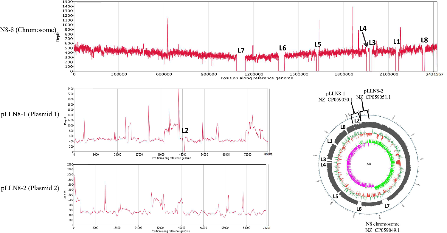
3.4. Prophage-Related Fragments of L. lactis N8

The N8 chromosome harbors seven regions that represent prophage-related fragments. Among these seven prophages, LLN8-1 (36,193 bp) and LLN8-2 (38,338 bp) appear to represent intact phages, LLN8-3 (34,367 bp), LLN8-4 (19,856 bp), LLN8-5 (44,895 bp), LLN8-6 (15,398 bp) and LLN8-7 (29,597 bp) appear to represent incomplete phages or questionable phages. The BLAST results of each prophage-related fragment are shown in Figure 5. Together, bacteriophage sequences encompass approximately 8.98%, representing a large portion of the N8 genome. The seven prophage-related fragments occupy various positions on the N8 genome. The G+C content of seven prophages ranges from 32.05 mol% to 36.09 mol%, similar to the value of 35.1 mol% calculated for the N8 chromosome (Table 4).
We have performed prophage predictions on all 42 L. lactis strains with complete genomes currently published by NCBI, and the results were listed in Supplementary Table S4. We found that all the published strains with complete genomes contain prophages to different degrees (intact, incomplete or questionable), indicating the symbiotic relationship between phages and L. lactis during the long-term evolution. Bacteriophages play a very important role in the evolution of lactic acid bacteria. There are 145 intact prophages identified from 42 L. lactis genomes (Supplementary Table S4). Among them, only five L. lactis strains carried no intact prophage; seventeen strains carried five or more intact prophages. On average, each strain has 6.5 prophage-related fragments.
Currently, 42 complete and 160 partially assembled L. lactis genomes are available in NCBI’s GenBank (update on 15 July 2021). The intact bacteriophages or prophage remnants have been identified in each one [44,45,46]. Possibly due to their long history in milk fermentation, L. lactis are regarded to contain the highest number of prophages among the LAB [47]. Prophage-related fragments encompass from 3 to 10% of the total genome of L. lactis strains [46]. The genome sequence analyses of L. lactis prophages indicated that they are affiliated with the P335 group of phages, and the two other species (c2- and 936-like) are composed of phages that are exclusively virulent [48]. In N8, LLN8-1, LLN8-2, LLN8-3, LLN8-4, LLN8-5, LLN8-6 and LLN8-7 have the highest homology with the staphylococcal phage phi3396, lactococcal phage TP901-1, Gordonia phage Hotorobo, lactococcal phage bIL310, staphylococcal bacteriophage SPbeta-like, staphylococcal bacteriophage SPbeta-like and clostridium phage phiCD6356, respectively.

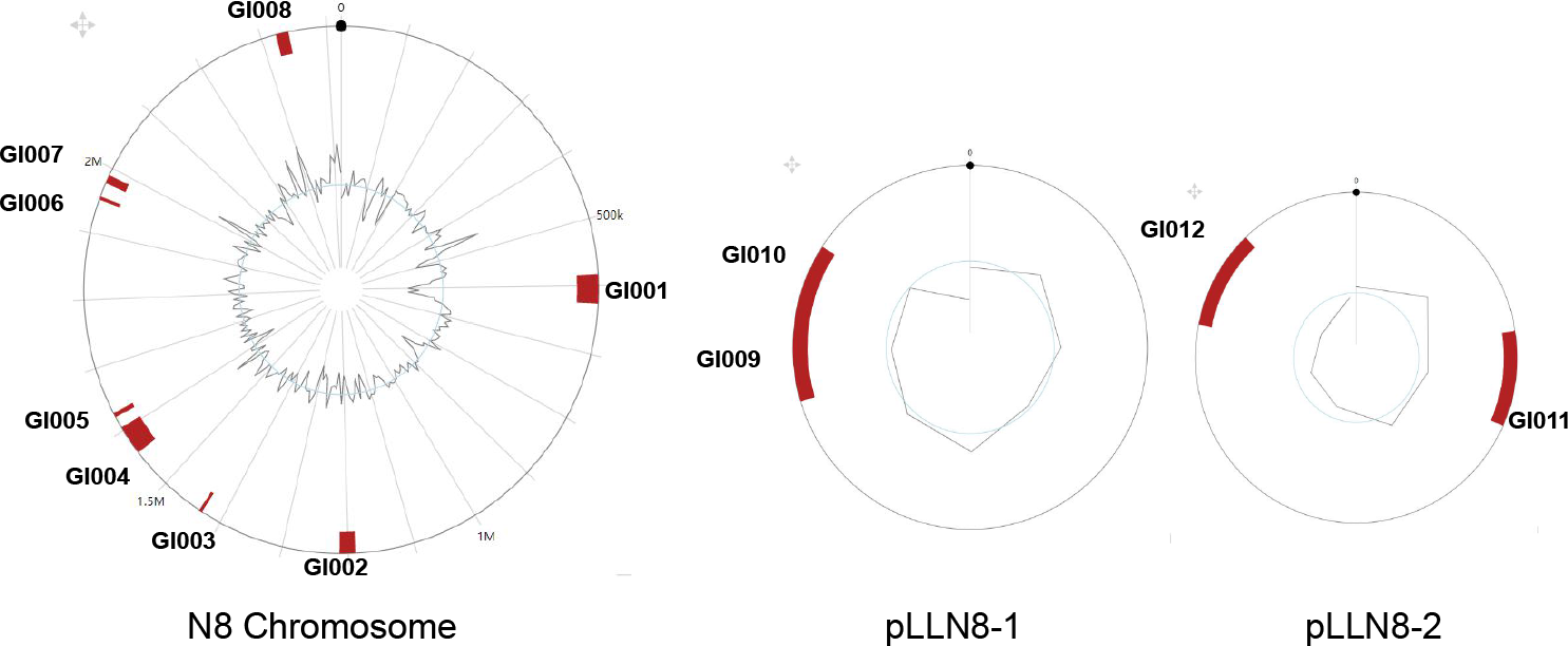
3.5. Genomic Islands of L. lactis N8

The genomic islands (GIs) are large DNA segments generally between 10 and 200 kb in length, with special structure and function. GIs have a variety of biological functions, such as antibiotic resistance, pathogenicity, xenobiotic degradation and heavy metal resistance [49,50]. The common features of GIs include: (1) integration hotspots for GIs are usually adjacent to RNA genes on chromosome; (2) GIs are flanked by direct repeats that are possibly related to the horizontal transfer of GIs; (3) horizontal transfer related genes, such as transposases, integrases and recombinases, are often found at the junction of GIs and core genome; (4) there are significant differences in G+C content between GIs and core genome [51].
In this study, twelve GIs in N8 accounting for 4.12% of the total genome were predicted, and their physical locations on the genome were marked in Figure 6. The detailed information of the GIs is shown in Table 5. Detailed gene content was listed in Supplementary Table S5. The prophage fragments and GIs overlap to a certain extent. This may be due to their exogenous DNA nature, obtained through horizontal transfer or other means of genomic evolution [52]. Owing to the diversity of the sources of GIs, their G+C content is different from the N8 genome.

3.6. Other Characteristics of L. lactis N8

3.6.1. Cell-Surface Protein
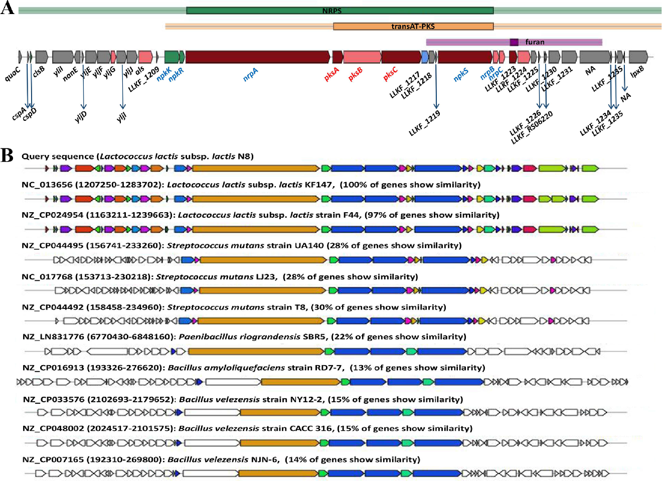
In L. lactis, csc genes can be found both on the chromosome and on the plasmids [53]. The chromosomally located csc loci are commonly flanked by IS elements and are, therefore, thought to be horizontally acquired and mobile. A csc gene cluster encoding exclusively cell-surface proteins was identified in N8. The gene cluster generally has one copy of four new gene families called cscA, cscB, cscC and cscD [53]. All the encoded proteins have a signal peptide for secretion by the secdependent pathway, while some have cell-surface anchors, novel WxL domains and putative domains for sugar binding and degradation. A proteomic analysis on N8 shows that the cscA-D genes are co-expressed [12], supporting their operon organization. Researchers propose that the CscA, CscB, CscC and CscD proteins form cell-surface protein complexes and play a role in carbon source acquisition [53]. Their presence in dairy lactococci is perhaps a relic of their plant ancestral heritage, although they may still provide some unknown benefits to their host in the dairy environment [3].

3.6.2. PKS/NRPS

L. lactis KF147 is the first reported L. lactis that has identified a gene cluster encoding a hybrid non-ribosomal peptide synthetase and polyketide synthase (NRPS–PKS) system [54]. Here, we reported that N8 also harbors the NRPS–PKS system (Figure 7A). Currently, PKS can be roughly divided into three types, namely PKS I (modular), PKS II (iterative) and PKS III (chalcone). The diverse activities of the PKS are completed by their protein modules, and the protein modules from different sources may be different. As a result, the chemical structure of polyketone compounds is also different. We compared the secondary metabolite systems of 42 L. lactis strains. We found that almost all the L. lactis contain the type Ⅲ PKS gene cluster. However, only eight contained NRPS gene clusters, namely L. lactis N8, L. lactis F44, L. lactis KF196, L. lactis KF147, L. lactis G423, L. lactis NCDO2118, L. lactis YF11, L. lactis KLDS 4.0325, all of which but L. lactis KLDS 4.0325 could produce lanthiopeptide (Supplementary Table S6).
The homology of the genes of the NRPS/PKS system in ten other strains was compared with N8, and the results are shown in Figure 7B. In the hybrid NRPS–PKS system of L. lactis N8, six NRPS modules and three PKS modules were identified (Figure 7A). Oxidative stress resistance and biofilm formation are the most probable functions of this hybrid system [55].

3.6.3. Bacteriocin Gene Cluster

There are two bacteriocin gene clusters in the genome of N8. The first one is the well-known nisin gene cluster encoding nisin synthesis-related protein. The second bacteriocin cluster in N8 consists of nine genes, including yujA, yujB, yujD, yujE1, yujE2, yujF, yujG and two genes of unknown function. Among them, yujG encodes a LytTR family transcriptional regulator YujG, and yujF encodes bacteriocin precursor peptide YujF, while yujD, yujE1 and yujE2 encode putative immunity proteins YujD, YujE1 and YujE2, respectively. Furthermore, yujB encodes uncharacterized integral membrane protein YujB, and yujA encodes class I SAM-dependent methyltransferase YujA, which catalyzes the methylation of one or more specific substrates using S-adenosyl-L-methionine (SAM or AdoMet) as the methyl donor. The remaining two genes encode membrane GTPase and hypothetical protein. The Swiss Model prediction results show that there are conserved regions between YujF and lactococcin 972. In addition, the prediction results indicate that YujF may have secretion signal peptide (Supplementary Figure S2).

3.7. Construction of Streamlined Genome Chassis of L. lactis N8

Through genome-wide analysis, we found prophages and GIs that are nonessential fragments for N8. These nonessential fragments were our preferred deletion targets for constructing the N8 streamlined genome chassis. In this study, we successfully deleted eight segments and realized the current maximum genome simplification of L. lactis (Figure 8). The cumulative deletion was 176.43 kb, which accounts for 6.86% of the whole N8 genome (Table 6). We verified the correctness of the knockout of the fragments through resequencing technology, and the physical location of each deleted fragment was also marked (Figure 8).
Then, we tested the growth phenotype of all the genome-streamlined strains. The growth status of the simplified genome strains is not much different from that of the wild-type strain N8 (Figure 9A). The final OD600 value is not significantly different, indicating that the deletion of these fragments did not cause a growth defect to the strains. However, it can be seen from the growth period that the deletion strains reach the stable period earlier than the wild type. Through the calculation of the generation time, we found that the generation times of all the deleted strains are shorter than that of the wild-type strain (Table 6). Especially, the generation time of strain N8-8 with the largest amount of deletion was shortened by 17.18% compared with the wild-type strain. Compared with the wild-type strain, the generation times of N8-3, N8-4 and N8-7 were shortened by 12.29%, 14.25% and 16.79%, respectively. Except for N8-5, the maximum growth rate of the deleted strains is also higher than that of the wild-type strains, which proves that deleting nonessential genome fragments is feasible for reducing the physiological burden of the strains (Table 6).
Due to the high nisin yield of N8, another phenotype we are more concerned about is how the deletion of nonessential fragments affects the yield of nisin. Therefore, we tracked the nisin production of all the strains (wild-type and genome-streamlined strains). As shown in Figure 9B, the nisin titre of N8-2 was slightly lower than that of the wild-type strain after 8 h of cultivation, but the difference in the nisin yield among all the strains disappeared after 12 h of cultivation. It shows that the deletion of nonessential fragments did not affect the overall nisin production, which is in line with our expectations. In the future, we will use the chassis strains to design a metabolic module for further increasing the production of nisin. In addition, the chassis strain N8-8 has the possibility of further genomic deletions, for example, at the area of remaining prophages and GIs. This strain possesses a great potential for the development of a model lactic acid bacterial strain. The construction of a simplified chassis strain of L. lactis is also important for understanding the minimal genome of its life-sustaining activities. In addition, the construction of simplified genome chassis strains also laid the foundation for the application of L. lactis in the field of synthetic biology.

4. Conclusions

In this study, we reported detailed genome features of L. lactis N8. Through the phylogenetic tree and core-pan genome analysis, we found that L. lactis N8 has the highest homology with L. lactis YF11. Through a genome-wide analysis, we found that N8 has two large plasmids harboring many functional genes. These two large plasmids play a very important role in N8 sugar transport and metabolism, flavor formation and stress tolerance under special environmental conditions. Then, we counted all the plasmids of L. lactis currently published and found that the plasmids possessed by N8 are significantly larger than most of other L. lactis. In addition, we predicted the prophage in N8 and counted all the prophages of L. lactis with the complete genome published so far. We also predicted the GIs in N8 and marked their precise locations. Based on these genome-wide findings, we set out to construct a streamlined chassis strain from L. lactis N8. Prophage and genomic island fragments were the preferred deletion targets, and we successfully deleted 6.86% of the N8 genome. As far as we know, we are the first to achieve this large an amount of deletion of an L. lactis genome, which is of great significance for the study of the simplest functional genome of lactic acid bacteria. The simplification of the L. lactis genome can also expand the application of lactic acid bacteria in the field of synthetic biology by, e.g., producing favorable metabolic modules in the L. lactis chassis strain.

Supplementary Materials

The following are available online at https://www.mdpi.com/article/10.3390/microorganisms10010047/s1, Supplementary Figure S1. The rooted phylogenetic tree of all 202 L. lactis. Supplementary Figure S2. Swiss Model prediction results of YujF. Supplementary Table S1. Primers used in this study. Supplementary Table S2. All 202 L. lactis data retrieved by latest 5th July 2021. Supplementary Table S3. Script for converting GFF files to GFF3 files. Supplementary Table S4. Prophage prediction results on 42 L. lactis strains with complete genomes currently published by NCBI. Supplementary Table S5. The detailed information of 12 GIs. Supplementary Table S6. Secondary metabolite biosynthetic gene cluster prediction results of 43 L. lactis strains (including L. lactis N8) with complete genomes currently published by NCBI.

Author Contributions

Conceptualization, W.Q. and M.Q.; Data curation, W.Q.; Formal analysis, W.Q., F.L., X.W., Y.Q. and R.L.; Funding acquisition, M.Q.; Investigation, W.Q., F.L., X.W., Y.Q. and R.L.; Methodology, W.Q. and X.W.; Supervision, Z.W., P.E.J.S., H.X. and M.Q.; Visualization, W.Q.; Writing—original draft, W.Q. and X.W.; Writing—review & editing, Z.W., P.E.J.S., H.X. and M.Q. All authors have read and agreed to the published version of the manuscript.

Funding

This research was funded by the National Natural Science Foundation of China (31770102), the Tianjin Key Research Program of Application Foundation and Advanced Technology (17JCZDJC32200) and the National Key Research and Development Program (2018YFA0900100). Xing Wan received a personal grant from the Jane and Aatos Erkko Foundation, Finland (grant No. 200050).

Institutional Review Board Statement

Not applicable.

Informed Consent Statement

Not applicable.

Data Availability Statement

Data are contained within the article or Supplementary Materials.

Conflicts of Interest

The authors declare no conflict of interest.

References

  1. Schleifer, K.H.; Kilpper-Bälz, R. Molecular and Chemotaxonomic Approaches to the Classification of Streptococci, Enterococci and Lactococci: A Review. Syst. Appl. Microbiol. 1987, 10, 1–19. [Google Scholar] [CrossRef]
  2. Pérez, T.; Balcázar, J.L.; Peix, A.; Valverde, A.; Velázquez, E.; De Blas, I.; Ruiz-Zarzuela, I. Lactococcus lactis subsp. tructae subsp. nov. isolated from the intestinal mucus of brown trout (Salmo trutta) and rainbow trout (Oncorhynchus mykiss). Int. J. Syst. Evol. Microbiol. 2011, 61, 1894–1898. [Google Scholar] [CrossRef] [Green Version]
  3. Ainsworth, S.; Stockdale, S.; Bottacini, F.; Mahony, J.; Van Sinderen, D. The Lactococcus lactis plasmidome: Much learnt, yet still lots to discover. FEMS Microbiol. Rev. 2014, 38, 1066–1088. [Google Scholar] [CrossRef] [Green Version]
  4. Kelleher, P.; Bottacini, F.; Mahony, J.; Kilcawley, K.N.; Van Sinderen, D. Comparative and functional genomics of the Lactococcus lactis taxon; insights into evolution and niche adaptation. BMC Genom. 2017, 18, 267. [Google Scholar] [CrossRef] [Green Version]
  5. Qiao, M.; Saris, P.E.J. Evidence for a role of NisT in transport of the lantibiotic nisin produced by Lactococcus lactis N8. FEMS Microbiol. Lett. 1996, 144, 89–93. [Google Scholar] [CrossRef]
  6. Simsek, Ö.; Saris, P.E.J. Cycle changing the medium results in increased nisin productivity per cell in Lactococcus lactis. Biotechnol. Lett. 2009, 31, 415–421. [Google Scholar] [CrossRef]
  7. Koponen, O.; Tolonen, M.; Qiao, M.; Wahlström, G.; Helin, J.; Saris, P.E.J. NisB is required for the dehydration and NisC for the lanthionine formation in the post-translational modification of nisin. Microbiology 2002, 148, 3561–3568. [Google Scholar] [CrossRef] [Green Version]
  8. Takala, T.M.; Koponen, O.; Qiao, M.; Saris, P.E.J. Lipid-free NisI: Interaction with nisin and contribution to nisin immunity via secretion. FEMS Microbiol. Lett. 2004, 237, 171–177. [Google Scholar] [CrossRef]
  9. Xu, Y.; Li, X.; Li, R.; Li, S.; Ni, H.; Wang, H.; Xu, H.; Zhou, W.; Saris, P.E.J.; Yang, W.; et al. Structure of the nisin leader peptidase NisP revealing a C-terminal autocleavage activity. Acta Crystallogr. Sect. D 2014, 70, 1499–1505. [Google Scholar] [CrossRef]
  10. Kuipers, O.P.; De Ruyter, P.G.G.A.; Kleerebezem, M.; De Vos, W.M. Quorum sensing-controlled gene expression in lactic acid bacteria. J. Biotechnol. 1998, 64, 15–21. [Google Scholar] [CrossRef] [Green Version]
  11. Immonen, T.; Saris, P.E.J. Characterization of the nisFEG Operon of the Nisin Z Producing Lactococcus lactis subsp. lactis N8 Strain. DNA Seq. 1998, 9, 263–274. [Google Scholar] [CrossRef] [PubMed]
  12. Qiao, W.; Qiao, Y.; Liu, F.; Zhang, Y.; Li, R.; Wu, Z.; Xu, H.; Saris, P.E.J.; Qiao, M. Engineering Lactococcus lactis as a multi-stress tolerant biosynthetic chassis by deleting the prophage-related fragment. Microb. Cell Fact. 2020, 19, 225. [Google Scholar] [CrossRef] [PubMed]
  13. Wu, Z.; Xuanyuan, Z.; Li, R.; Jiang, D.; Li, C.; Xu, H.; Bai, Y.; Zhang, X.; Turakainen, H.; Saris, P.E.J.; et al. Mu transposition complex mutagenesis in Lactococcus lactis–identification of genes affecting nisin production. J. Appl. Microbiol. 2009, 106, 41–48. [Google Scholar] [CrossRef] [PubMed]
  14. Zhu, D.; Li, R.; Liu, F.; Xu, H.; Li, B.; Yuan, Y.; Saris, P.E.J.; Qiao, M. Mu insertion in feuD triggers the increase in nisin immunity in Lactococcus lactis subsp. lactis N8. J. Appl. Microbiol. 2016, 120, 402–412. [Google Scholar] [CrossRef]
  15. Chandrasekar Rajendran, S.C.; Chamlagain, B.; Kariluoto, S.; Piironen, V.; Saris, P.E.J. Biofortification of riboflavin and folate in idli batter, based on fermented cereal and pulse, by Lactococcus lactis N8 and Saccharomyces boulardii SAA655. J. Appl. Microbiol. 2017, 122, 1663–1671. [Google Scholar] [CrossRef] [PubMed] [Green Version]
  16. Gao, G.; Fan, H.; Zhang, Y.; Cao, Y.; Li, T.; Qiao, W.; Wu, M.; Ma, T.; Li, G. Production of nisin-containing bacterial cellulose nanomaterials with antimicrobial properties through co-culturing Enterobacter sp. FY-07 and Lactococcus lactis N8. Carbohydr. Polym. 2021, 251, 117131. [Google Scholar] [CrossRef]
  17. Fu, Y.; Mu, D.; Qiao, W.; Zhu, D.; Wang, X.; Liu, F.; Xu, H.; Saris, P.; Kuipers, O.P.; Qiao, M. Co-expression of Nisin Z and Leucocin C as a Basis for Effective Protection Against Listeria monocytogenes in Pasteurized Milk. Front. Microbiol. 2018, 9, 547. [Google Scholar] [CrossRef] [PubMed]
  18. Bermúdez-Humarán, L.G.; Aubry, C.; Motta, J.-P.; Deraison, C.; Steidler, L.; Vergnolle, N.; Chatel, J.-M.; Langella, P. Engineering lactococci and lactobacilli for human health. Curr. Opin. Microbiol. 2013, 16, 278–283. [Google Scholar] [CrossRef]
  19. Zhu, D.; Fu, Y.; Liu, F.; Xu, H.; Saris, P.E.J.; Qiao, M. Enhanced heterologous protein productivity by genome reduction in Lactococcus lactis NZ9000. Microb. Cell Fact. 2017, 16, 1. [Google Scholar] [CrossRef] [Green Version]
  20. Zhu, D.; Zhao, K.; Xu, H.; Zhang, X.; Bai, Y.; Saris, P.E.J.; Qiao, M. Construction of thyA deficient Lactococcus lactis using the Cre-loxP recombination system. Ann. Microbiol. 2015, 65, 1659–1665. [Google Scholar] [CrossRef]
  21. Wan, X.; Takala Timo, M.; Qiao, M.; Saris Per, E.J. Complete Genome Sequence of Nisin-Producing Lactococcus lactis subsp. lactis N8. Microbiol. Resour. Announc. 2021, 10, e01120–e01147. [Google Scholar] [CrossRef] [PubMed]
  22. Woodcock, D.M.; Crowther, P.J.; Doherty, J.; Jefferson, S.; Decruz, E.; Noyer-Weidner, M.; Smith, S.S.; Michael, M.Z.; Graham, M.W. Quantitative evaluation of Escherichia coli host strains for tolerance to cytosine methylation in plasmid and phage recombinants. Nucleic Acids Res. 1989, 17, 3469–3478. [Google Scholar] [CrossRef] [PubMed] [Green Version]
  23. Qiao, M.; Ye, S.; Koponen, O.; Ra, R.; Usabiaga, M.; Immonen, T.; Saris, P.E.J. Regulation of the nisin operons in Lactococcus lactis N8. J. Appl. Bacteriol. 1996, 80, 626–634. [Google Scholar] [CrossRef]
  24. Qiao, M.; Immonen, T.; Koponen, O.; Saris, P.E.J. The cellular location and effect on nisin immunity of the NisI protein from Lactococcus lactis N8 expressed in Escherichia coli and L. lactis. FEMS Microbiol. Lett. 1995, 131, 75–80. [Google Scholar] [CrossRef] [PubMed]
  25. Lambert Jolanda, M.; Bongers Roger, S.; Kleerebezem, M. Cre-lox-Based System for Multiple Gene Deletions and Selectable-Marker Removal in Lactobacillus plantarum. Appl. Environ. Microbiol. 2007, 73, 1126–1135. [Google Scholar] [CrossRef] [PubMed] [Green Version]
  26. Seemann, T. Prokka: Rapid prokaryotic genome annotation. Bioinformatics 2014, 30, 2068–2069. [Google Scholar] [CrossRef]
  27. Tatusova, T.; Dicuccio, M.; Badretdin, A.; Chetvernin, V.; Nawrocki, E.P.; Zaslavsky, L.; Lomsadze, A.; Pruitt, K.D.; Borodovsky, M.; Ostell, J. NCBI prokaryotic genome annotation pipeline. Nucleic Acids Res. 2016, 44, 6614–6624. [Google Scholar] [CrossRef] [PubMed]
  28. Huerta-Cepas, J.; Forslund, K.; Coelho, L.P.; Szklarczyk, D.; Jensen, L.J.; Von Mering, C.; Bork, P. Fast Genome-Wide Functional Annotation through Orthology Assignment by eggNOG-Mapper. Mol. Biol. Evol. 2017, 34, 2115–2122. [Google Scholar] [CrossRef] [Green Version]
  29. Sullivan, M.J.; Petty, N.K.; Beatson, S.A. Easyfig: A genome comparison visualizer. Bioinformatics 2011, 27, 1009–1010. [Google Scholar] [CrossRef]
  30. Wu, S.; Zhu, Z.; Fu, L.; Niu, B.; Li, W. WebMGA: A customizable web server for fast metagenomic sequence analysis. BMC Genom. 2011, 12, 444. [Google Scholar] [CrossRef] [Green Version]
  31. Page, A.J.; Cummins, C.A.; Hunt, M.; Wong, V.K.; Reuter, S.; Holden, M.T.G.; Fookes, M.; Falush, D.; Keane, J.A.; Parkhill, J. Roary: Rapid large-scale prokaryote pan genome analysis. Bioinformatics 2015, 31, 3691–3693. [Google Scholar] [CrossRef]
  32. Nguyen, L.-T.; Schmidt, H.A.; Von Haeseler, A.; Minh, B.Q. IQ-TREE: A Fast and Effective Stochastic Algorithm for Estimating Maximum-Likelihood Phylogenies. Mol. Biol. Evol. 2015, 32, 268–274. [Google Scholar] [CrossRef]
  33. Kalyaanamoorthy, S.; Minh, B.Q.; Wong, T.K.F.; Von Haeseler, A.; Jermiin, L.S. ModelFinder: Fast model selection for accurate phylogenetic estimates. Nat. Methods 2017, 14, 587–589. [Google Scholar] [CrossRef] [PubMed] [Green Version]
  34. Letunic, I.; Bork, P. Interactive tree of life (iTOL) v3: An online tool for the display and annotation of phylogenetic and other trees. Nucleic Acids Res. 2016, 44, W242–W245. [Google Scholar] [CrossRef] [PubMed]
  35. Arndt, D.; Grant, J.R.; Marcu, A.; Sajed, T.; Pon, A.; Liang, Y.; Wishart, D.S. PHASTER: A better, faster version of the PHAST phage search tool. Nucleic Acids Res. 2016, 44, W16–W21. [Google Scholar] [CrossRef] [Green Version]
  36. Bertelli, C.; Laird, M.R.; Williams, K.P.; Simon Fraser University Research Computing Group; Lau, B.Y.; Hoad, G.; Winsor, G.L.; Brinkman, F.S.L. IslandViewer 4: Expanded prediction of genomic islands for larger-scale datasets. Nucleic Acids Res. 2017, 45, W30–W35. [Google Scholar] [CrossRef]
  37. Blin, K.; Shaw, S.; Steinke, K.; Villebro, R.; Ziemert, N.; Lee, S.Y.; Medema, M.H.; Weber, T. antiSMASH 5.0: Updates to the secondary metabolite genome mining pipeline. Nucleic Acids Res. 2019, 47, W81–W87. [Google Scholar] [CrossRef] [PubMed] [Green Version]
  38. Waterhouse, A.; Bertoni, M.; Bienert, S.; Studer, G.; Tauriello, G.; Gumienny, R.; Heer, F.T.; De Beer, T.A.p.; Rempfer, C.; Bordoli, L.; et al. SWISS-MODEL: Homology modelling of protein structures and complexes. Nucleic Acids Res. 2018, 46, W296–W303. [Google Scholar] [CrossRef] [Green Version]
  39. Tang, X.; Nakata, Y.; Li, H.O.; Zhang, M.; Gao, H.; Fujita, A.; Sakatsume, O.; Ohta, T.; Yokoyama, K. The optimization of preparations of competent cells for transformation of E. coli. Nucleic Acids Res. 1994, 22, 2857–2858. [Google Scholar] [CrossRef] [Green Version]
  40. Li, T.T.; Tian, W.L.; Gu, C.T. Elevation of Lactococcus lactis subsp. cremoris to the species level as Lactococcus cremoris sp. nov. and transfer of Lactococcus lactis subsp. tructae to Lactococcus cremoris as Lactococcus cremoris subsp. tructae comb. nov. Int. J. Syst. Evol. Microbiol. 2021, 71, 4727. [Google Scholar] [CrossRef]
  41. Malesevic, M.; Stanisavljevic, N.; Miljkovic, M.; Jovcic, B.; Filipic, B.; Studholme, D.J.; Kojic, M. The large plasmidome of Lactococcus lactis subsp. lactis bv. diacetylactis S50 confers its biotechnological properties. Int. J. Food Microbiol. 2021, 337, 108935. [Google Scholar] [CrossRef]
  42. Cords, B.R.; Mckay, L.L.; Guerry, P. Extrachromosomal Elements in Group N Streptococci1. J. Bacteriol. 1974, 117, 1149–1152. [Google Scholar] [CrossRef] [Green Version]
  43. Savijoki, K.; Ingmer, H.; Varmanen, P. Proteolytic systems of lactic acid bacteria. Appl. Microbiol. Biotechnol. 2006, 71, 394–406. [Google Scholar] [CrossRef] [PubMed]
  44. Chopin, A.; Bolotin, A.; Sorokin, A.; Ehrlich, S.D.; Chopin, M.-C. Analysis of six prophages in Lactococcus lactis IL1403: Different genetic structure of temperate and virulent phage populations. Nucleic Acids Res. 2001, 29, 644–651. [Google Scholar] [CrossRef]
  45. Ventura, M.; Zomer, A.; Canchaya, C.; O’Connell-Motherway, M.; Kuipers, O.; Turroni, F.; Ribbera, A.; Foroni, E.; Buist, G.; Wegmann, U.; et al. Comparative Analyses of Prophage-Like Elements Present in Two Lactococcus lactis Strains. Appl. Environ. Microbiol. 2007, 73, 7771–7780. [Google Scholar] [CrossRef] [PubMed] [Green Version]
  46. Kelleher, P.; Mahony, J.; Schweinlin, K.; Neve, H.; Franz, C.M.; Van Sinderen, D. Assessing the functionality and genetic diversity of lactococcal prophages. Int. J. Food Microbiol. 2018, 272, 29–40. [Google Scholar] [CrossRef]
  47. O’Sullivan, D.J.; Lee, J.H.; Dominguez, W. Lactic Acid Bacteria: | Genomics, Genetic Engineering. In Encyclopedia of Dairy Sciences; Fuquay, J.W., McSweeney, P.L., Fox, P.F., Eds.; Academic Press: Cambridge, MA, USA, 2011; pp. 67–77. [Google Scholar]
  48. Deveau, H.; Labrie Simon, J.; Chopin, M.-C.; Moineau, S. Biodiversity and Classification of Lactococcal Phages. Appl. Environ. Microbiol. 2006, 72, 4338–4346. [Google Scholar] [CrossRef] [PubMed] [Green Version]
  49. Hacker, J.; Kaper, J.B. Pathogenicity Islands and the Evolution of Microbes. Annu. Rev. Microbiol. 2000, 54, 641–679. [Google Scholar] [CrossRef] [Green Version]
  50. Langille, M.G.I.; Hsiao, W.W.L.; Brinkman, F.S.L. Detecting genomic islands using bioinformatics approaches. Nat. Rev. Microbiol. 2010, 8, 373–382. [Google Scholar] [CrossRef] [PubMed]
  51. Vernikos, G.S.; Parkhill, J. Resolving the structural features of genomic islands: A machine learning approach. Genome Res. 2008, 18, 331–342. [Google Scholar] [CrossRef] [Green Version]
  52. Umenhoffer, K.; Fehér, T.; Balikó, G.; Ayaydin, F.; Pósfai, J.; Blattner, F.R.; Pósfai, G. Reduced evolvability of Escherichia coli MDS42, an IS-less cellular chassis for molecular and synthetic biology applications. Microb. Cell Fact. 2010, 9, 38. [Google Scholar] [CrossRef] [PubMed] [Green Version]
  53. Siezen, R.; Boekhorst, J.; Muscariello, L.; Molenaar, D.; Renckens, B.; Kleerebezem, M. Lactobacillus plantarum gene clusters encoding putative cell-surface protein complexes for carbohydrate utilization are conserved in specific gram-positive bacteria. BMC Genom. 2006, 7, 126. [Google Scholar] [CrossRef] [PubMed] [Green Version]
  54. Golomb, B.L.; Yu, A.O.; Coates, L.C.; Marco, M.L. The Lactococcus lactis KF147 nonribosomal peptide synthetase/polyketide synthase system confers resistance to oxidative stress during growth on plant leaf tissue lysate. Microbiol. Open 2018, 7, e00531. [Google Scholar] [CrossRef]
  55. Khayatt, B.I.; Van Noort, V.; Siezen, R.J. The Genome of the Plant-Associated Lactic Acid Bacterium Lactococcus lactis KF147 Harbors a Hybrid NRPS-PKS System Conserved in Strains of the Dental Cariogenic Streptococcus mutans. Curr. Microbiol. 2020, 77, 136–145. [Google Scholar] [CrossRef] [PubMed]
Figure 1. Circular genome plot of L. lactis N8. From outer to inner circle: gene function annotation results, genome GC content, genome GC skew.
Microorganisms 10 00047 g001
Figure 2. Overall position of L. lactis N8 in the L. lactis phylogeny. An unrooted tree, representing the phylogenetic position of N8 (red) among 202 L. lactis.
Microorganisms 10 00047 g002
Figure 3. (A) Pan-genome matrix of 17 L. lactis. Blue, gene presence; white, gene absence. (B) Flower plot of 17 L. lactis.
Microorganisms 10 00047 g003
Figure 4. Plasmid genetic maps of L. lactis N8. (A) Arrows indicate positions, size and direction of predicted genes. Putative functions of genes are presented by colors. From outer to inner circle: gene function annotation results, genome GC content, genome GC skew. (B) Overview of the functions of plasmid-encoded genes in L. lactis N8. The sum of the sizes of the genes within a category relative to the total size of all plasmid genes was used. (C) Comparison of the homology of two large plasmids.
Microorganisms 10 00047 g004
Figure 5. Prediction of the functional protein of each prophage.
Microorganisms 10 00047 g005
Figure 6. Circular map of L. lactis N8 chromosome and two large plasmids showing the physical location of the 12 GIs.
Microorganisms 10 00047 g006
Figure 7. (A) The hybrid NRPS/PKS from L. lactis N8. Genes on the hybrid NRPS/PKS system are colored as follows: two-component transcriptional regulator (green), secondary metabolite core biosynthetic genes (dark red), additional biosynthetic genes (light red), transport-related genes (blue) and other genes (grey). (B) The homology of genes in ten other strains of L. lactis, S. mutans, P. riograndensis, B. amyloliquefaciens and B. velezensis (NCBI Reference Sequence numbers are written next to the strain names).
Microorganisms 10 00047 g007
Figure 8. Alignment of L. lactis N8 and L. lactis N8-8 genome sequences verified correct deletion.
Microorganisms 10 00047 g008
Figure 9. Phenotypes of wild-type strain and streamlined genome chassis strains. (A) Growth profiles s of the strains. (B) Nisin titer of different strains at different time points (8 h, 10 h and 12 h).
Microorganisms 10 00047 g009
Table 1. Bacterial strains and plasmids utilized in this study.
Strains or PlasmidsRelevant DescriptionsReference
Strains
E. coli DH5αCloning host; F-φ80lacZ∆M15endA1 recA1 endA1 hsdR17 (rK-mK+) supE44 thi-1 gyrA 96 relA1 ∆(lacZYA-argF)U169 deoR λ-[22]
E. coli DH5α-up-down (L1)Cmr, Emr, E. coli DH5α derivative containing the whole plasmid pNZ5319∆L1[12]
E. coli DH5α-up-down (L2)Cmr, Emr, E. coli DH5α derivative containing the whole plasmid pNZ5319∆L2This study
E. coli DH5α-up-down (L3)Cmr, Emr, E. coli DH5α derivative containing the whole plasmid pNZ5319∆L3This study
E. coli DH5α-up-down (L4)Cmr, Emr, E. coli DH5α derivative containing the whole plasmid pNZ5319∆L4This study
E. coli DH5α-up-down (L5)Cmr, Emr, E. coli DH5α derivative containing the whole plasmid pNZ5319∆L5This study
E. coli DH5α-up-down (L6)Cmr, Emr, E. coli DH5α derivative containing the whole plasmid pNZ5319∆L6This study
E. coli DH5α-up-down (L7)Cmr, Emr, E. coli DH5α derivative containing the whole plasmid pNZ5319∆L7This study
E. coli DH5α-up-down (L8)Cmr, Emr, E. coli DH5α derivative containing the whole plasmid pNZ5319∆L8This study
Micrococcus luteus NCIB 8166Indicator strains for Nisin agar gel diffusion assay[23]
L. lactis N8Wild-type (WT) Nisin Z producer[24]
L. lactis N8-1The first DNA region L1 deletion in L. lactis N8[12]
L. lactis N8-2The L2 deletion in L. lactis N8-1This study
L. lactis N8-3The L3 deletion in L. lactis N8-2This study
L. lactis N8-4The L4 deletion in L. lactis N8-3This study
L. lactis N8-5The L5 deletion in L. lactis N8-4This study
L. lactis N8-6The L6 deletion in L. lactis N8-5This study
L. lactis N8-7The L7 deletion in L. lactis N8-6This study
L. lactis N8-8The L8 deletion in L. lactis N8-7This study
Plasmids
pNZ5319Cmr, Emr, used as knock-out vector[25]
pNZTS-CreEmr, cre gene cloned at the EcoRI and HindIII sites (cat gene deletion vector) [20]
pNZ5319∆L1Cmr, Emr, upstream and downstream homology arm of L1 amplified from L. lactis N8 genome cloned into pNZ5319[12]
pNZ5319∆L2Cmr, Emr, upstream and downstream homology arm of L2 amplified from L. lactis N8 genome cloned into pNZ5319This study
pNZ5319∆L3Cmr, Emr, upstream and downstream homology arm of L3 amplified from L. lactis N8 genome cloned into pNZ5319This study
pNZ5319∆L4Cmr, Emr, upstream and downstream homology arm of L4 amplified from L. lactis N8 genome cloned into pNZ5319This study
pNZ5319∆L5Cmr, Emr, upstream and downstream homology arm of L5 amplified from L. lactis N8 genome cloned into pNZ5319This study
pNZ5319∆L6Cmr, Emr, upstream and downstream homology arm of L6 amplified from L. lactis N8 genome cloned into pNZ5319This study
pNZ5319∆L7Cmr, Emr, upstream and downstream homology arm of L7 amplified from L. lactis N8 genome cloned into pNZ5319This study
pNZ5319∆L8Cmr, Emr, upstream and downstream homology arm of L8 amplified from L. lactis N8 genome cloned into pNZ5319This study
Table 2. Basic genomic characteristics of L. lactis N8.
FeatureSize/Percentage/Number
Genome size (bp)2,421,567
G + C content35.1%
rRNAs19
tRNAs64
ncRNAs4
Total genes2521
Coding sequences (CDs)2434
Prophages7
Genomic islands (GIs)12
Plasmids2
Table 3. Horizontal comparison of all L. lactis plasmids.
Organism NameStrainBioSampleNumber of PlasmidsTotal Size of PlasmidsRelease Date
Lactococcus lactis subsp. lactisN8SAMN1550061820.15156222 October 2020
Lactococcus lactis subsp. lactisNo attributesSAMN1422393110.06023210 September 1998
Lactococcus lactis subsp. lactisCBA3619SAMN1184366310.10758616 August 2019
Lactococcus lactis subsp. lactisFDAARGOS_865SAMN1345039510.05833515 December 2020
Lactococcus lactis subsp. lactisFDAARGOS_887SAMN1345041740.18036215 December 2020
Lactococcus lactis subsp. lactisFDAARGOS_866SAMN1345039640.24110815 December 2020
Lactococcus lactis subsp. lactisFDAARGOS_1064SAMN1635723340.11297921 December 2020
Lactococcus lactis subsp. lactisWiKim0098SAMN1678872810.07698718 January 2021
Lactococcus lactis subsp. lactisA12SAMEA400523640.12672723 July 2016
Lactococcus lactis subsp. lactis229SAMN0495524950.1656855 April 2017
Lactococcus lactis subsp. lactis275SAMN0495525240.258015 April 2017
Lactococcus lactis subsp. lactisUC063SAMN0495630250.1490785 April 2017
Lactococcus lactis subsp. lactisUL8SAMN0495640230.03706710 April 2017
Lactococcus lactis subsp. lactis14B4SAMN0879243010.05971 June 2018
Lactococcus lactis subsp. lactis184SAMN0495524760.04241611 January 2019
Lactococcus lactis subsp. lactisC10SAMN0495626730.05686511 January 2019
Lactococcus lactis subsp. lactisUC06SAMN0495629260.15568713 January 2019
Lactococcus lactis subsp. lactisUC08SAMN0495629350.16676616 January 2019
Lactococcus lactis subsp. lactisUC77SAMN0495630350.1781843 January 2019
Lactococcus lactis subsp. lactisUC11SAMN0495629460.15638211 September 2019
Lactococcus lactis subsp. lactisG121SAMN1494368730.1233077 September 2020
Lactococcus lactis subsp. lactis223SAMN0984786960.0558725 February 2021
Lactococcus lactis subsp. lactisWM1SAMN0984764950.18067223 February 2021
Lactococcus lactis subsp. lactis267SAMN0984787050.16432727 February 2021
Lactococcus lactis subsp. lactisDRC3SAMN1660456770.23282210 April 2021
Lactococcus lactis subsp. lactisLac3SAMN1874031410.00736713 May 2021
Lactococcus lactis subsp. lactis bv. diacetylactisFM03SAMN0606193970.080924 May 2017
Lactococcus lactis subsp. lactis bv. diacetylactisSD96SAMN12502795100.2199317 October 2019
Lactococcus lactis subsp. lactis bv. diacetylactisBGBU1-4SAMN1262723110.0063317 November 2019
Lactococcus lactis subsp. lactis bv. diacetylactisS50SAMN1016714460.24041210 September 2020
Lactococcus lactis subsp. lactis bv. diacetylactisNo attributesSAMN1422608910.0189771 August 2011
Lactococcus lactis subsp. lactis bv. diacetylactisNo attributesSAMN1422608810.0217281 August 2011
Lactococcus lactis subsp. lactis bv. diacetylactisNo attributesSAMN1422608710.0221661 August 2011
Lactococcus lactis subsp. lactis bv. diacetylactisNo attributesSAMN1422608010.0538761 August 2011
Lactococcus lactis subsp. lactisCV56SAMN0260339850.1192791 May 2012
Lactococcus lactis subsp. lactisKF147SAMN0260308710.0375122 December 2009
Lactococcus lactis subsp. lactisKLDS 4.0325SAMN0260346860.17162210 May 2018
Lactococcus lactis subsp. lactisNCDO 2118SAMN0247137610.03757121 January 2015
Average3.660.031197
Table 4. Information of prophage-related fragments.
ProphageStartEndSize (bp)Att Core SequenceG+C ContentStatus
LLN8-1 (chromosome)1,369,3741,405,56636,193TTTAATTTAGAAA35.27%intact
LLN8-2 (chromosome)1,949,9301,988,31738,388AACGTAACTAAAAACGTAACTAA35.18%intact
LLN8-3 (chromosome)2,128,0372,162,40334,367AACTTATTTTTAT34.31%incomplete
LLN8-4 (chromosome)2,323,0152,342,87019,856ACGCTTTTTACTACGTTCG34.56%incomplete
LLN8-5 (plasmid1)40,33369,92929,597AAAATAAAAAGT32.05%incomplete
LLN8-6 (plasmid2)865853,55244,895TTTCGAACATTT36.09%questionable
LLN8-7 (plasmid2)53,72869,12515,398AGGTTCTGTTGCAAAGTT35.32%questionable
Table 5. Characterization of genomic islands in L. lactis N8.
Genomic IslandStartEndSize (bp)G+C Content %
GI001 (chromosome)583,454626,48643,03230.63
GI002 (chromosome)1,188,6971,213,41024,71332.54
GI003 (chromosome)1,431,0361,435,913487739.57
GI004 (chromosome)1,560,5791,604,86544,28635.98
GI005 (chromosome)1,622,3091,629,361705230.91
GI006 (chromosome)1,950,0281,955,065503730.06
GI007 (chromosome)1,976,1591,989,28113,12233.38
GI008 (chromosome)2,322,3082,341,77219,46434.55
GI009 (plasmid 1)56,38460,867448337.20
GI010 (plasmid 1)58,99667,736874033.58
GI011 (plasmid 2)15,92922,625669636.81
GI012 (plasmid 2)55,77462,660688632.64
Table 6. Growth parameters in shaken-flask cultures of L. lactis strains.
L. lactisRemoved (bp)Cumulative (bp)Deletion (%)μmax (h−1)Generation Time (min)
N8000%0.44 ± 0.0445.7 ± 2.7
N8-119,73919,7390.77%0.48 ± 0.0541.5 ± 5.0
N8-2163821,3770.83%0.49 ± 0.0642.7 ± 4.1
N8-318,62840,0051.55%0.50 ± 0.0340.7 ± 5.0
N8-410,65950,6641.97%0.47 ± 0.0340.0 ± 5.1
N8-513,50264,1662.49%0.44 ± 0.0241.3 ± 4.9
N8-639,203103,3694.02%0.53 ± 0.0941.1 ± 3.2
N8-758,594161,9636.29%0.52 ± 0.0339.3 ± 5.2
N8-814,465176,4286.86%0.50 ± 0.0539.0 ± 4.2
Publisher’s Note: MDPI stays neutral with regard to jurisdictional claims in published maps and institutional affiliations.

