Article

Fabric-Based, Pneumatic Exosuit for Lower-Back Support in Manual-Handling Tasks

by
Amit Nirmal Cuttilan
1,
Rainier F. Natividad
2 and
Raye Chen-Hua Yeow
2,*
1
Division of Graduate Medical Studies, National University of Singapore, Singapore 119077, Singapore
2
Department of Biomedical Engineering, National University of Singapore, Singapore 119077, Singapore
*
Author to whom correspondence should be addressed.
Actuators 2023, 12(7), 273; https://doi.org/10.3390/act12070273
Submission received: 6 April 2023 / Revised: 15 June 2023 / Accepted: 27 June 2023 / Published: 3 July 2023
(This article belongs to the Section Actuators for Robotics)

Abstract

:
Lower-back pain (LBP) is a major cause of occupational disability and morbidity. This study investigates the effectiveness of a fabric-based pneumatic exosuit in reducing discomfort and lumbar muscle activation in healthy individuals who are performing manual-handling tasks. The suit combines the comfort of soft exosuits and the support of rigid exoskeletons. Ten healthy subjects performed a circuit of lifting tasks, simulating manual-handling work, with and without AireLevate support. We assessed the comfort levels and ease of task completion via a questionnaire after each manual-handling task. There was no difference in spinal range of motion, local discomfort, or general discomfort of activities with or without the AireLevate. There was a statistically significant reduction in muscle activation of the erector spinae at the L-5 level with AireLevate support (p < 0.02). This study demonstrates the exosuit’s ability in reducing lower-back muscle activation during manual-handling tasks, while maintaining comfort and mobility. Practitioner summary: We developed a soft exosuit which was shown to significantly reduce the muscle action of the erector muscles of the lumbar spine. In addition, participants perceived that the suit was easy to use and did not limit manual-handling tasks.

1. Introduction

Lower-back pain (LBP) is a major cause of disability globally. It is also well-recognized as an important factor for disability and morbidity in various occupations [1,2,3,4]. In fact, 37% of LBP has been shown to be related to occupation, with work-related LBP estimated to contribute to 818,000 disability-adjusted life years (DALYs) lost annually [5]. LBP has also been a significant contributor to absenteeism rates. Previous studies indicate that 10.9 to 54.4% of healthcare workers with LBP took sick leave because of LBP [6,7]. The duration of sick leave for 71% of these workers ranged from 2 to 30 days [8]. Some individuals may even experience recurrent episodes of LBP. Approximately half of individuals with LBP continue to experience either symptoms or functional limitations even after 1 year, thus making it a chronic medical condition [9,10].
Spinal loading is a result of muscle forces that are required to counteract against both gravitational forces on the upper body and the additional carried load carried by the individual. This has been shown to be a significant risk factor for occupation-related LBP in multiple studies. Risk factors for LBP at work include carrying heavy loads, work position, repetitive actions, and the duration of work [11]. Despite technological improvements, approximately 30% of workers continue to carry heavy loads at least 25% of the time at work [12]. As the management of LBP may be challenging, and at times unpredictable, it is vital to prevent the underlying etiologies of pain where possible [4,13,14]. Decreasing the frequency and weight of loads reduces spinal loading and is an effective way to minimize musculoskeletal injuries and hence LBP in the workplace.
Back-support exoskeletons provide direct support to the back and have been gaining interest as a viable intervention to minimize LBP in the workplace [15,16,17]. These exoskeletons assist the wearer by providing back support during lifting, lowering, and static bending, activities which can predispose workers to LBP [15]. Broadly, exoskeletons can be classified as active (i.e., powered) or passive (i.e., not powered) [18,19,20].
Active exoskeletons are wearable devices that convert stored energy into kinetic energy to aid the wearer in manual-handling tasks. Powered exoskeletons typically employ a rigid frame around the wearer’s torso and thighs which is powered by rotary electric motors placed concentrically on the hip joint [21,22,23]. A limitation of this configuration is the necessity to position the motors precisely on the hip, increasing the width of the exoskeleton and thus hindering its use in cramped workspaces. Active exoskeletons can feature high torque outputs; however, these require complex control and power systems, which increases their cost and weight significantly [23,24,25]. In addition, complex suits might be limited to use by trained individuals.
Conversely, passive exoskeletons provide mechanical assistance using levers, gears, and other mechanical components, without the use of a power source. Through this, paravertebral muscles generate a smaller extension force during lifting and carrying, hence reducing the mechanical load on the spine. However, as passive exoskeletons rely on the energy provided by the wearer’s own movements to provide support, their support can be limited by an individual wearer’s physical capabilities.
Gas or coil linear springs are widely utilized in several exoskeletons, both in the academic literature and in the commercial space [25,26]. The use of springs allows exoskeletons to operate without control and power systems, but this requires the use of rigid structures, which adds considerable inertia to the user and may even introduce discomfort. There is thus an increased popularity of elastic band-powered exoskeletons that use a fabric structure intertwined with elastic bands that function similarly to rigid springs [26,27]. Aptly renamed exosuits owing to a lack of a rigid skeleton, these fabric suits have minimal mass and comfort penalties. However, the use of elastic bands limits their efficacy in providing extension moments and limits their ability to support the back.
With all these limitations in previous iterations of exoskeletons, there has been increased attention on hybrid active–passive pneumatic-driven exoskeletons [28,29,30]. They are like powered exoskeletons in that they require the application of externally stored energy to activate their pneumatic components, in this case compressed air. These activated components then behave as passive members by leveraging on the fluid mechanics of air. The compressibility of air facilitates human–robot interaction, and improves both comfort and safety [31]. When compared to other active exoskeletons, pneumatic exoskeletons also have a lower risk of malfunction. However, several pneumatic exoskeletons still feature predominantly rigid components which make it difficult for them to synergize with the body’s natural kinematics [20].
To address the current limitations of pneumatic exoskeletons, we developed the AireLevate (Figure 1). The AireLevate is a 1.5-kilogram, fabric-based exosuit with an apron-like form factor. It is primarily worn on the anterior side of the body and is composed of a singular fabric plate on the torso which transforms into two pieces on the thighs. It is secured to the body through straps located along multiple points on the torso and thighs. The AireLevate is a pneumatically powered exosuit configured to operate as a hybrid active–passive wearable exosuit. An experiment on healthy subjects was performed to ascertain the AireLevate’s ability to provide support to the lower back. Subjects were instructed to perform lifting motions, such as those performed in manual-handling tasks. The AireLevate supports the range of motion (ROM) of the lumbar spine from approximately 90 degrees of forward flexion to full extension of the spine. While it has a curved profile, its soft robotic properties allow it to conform geometrically to whatever it is attached to. The AireLevate was also designed so that the actuators are sufficiently restrained to the body, and thus conform to the user’s body once worn securely.
Surface Electromyography sensors (sEMG) were attached to measure muscle activation, while motion capture cameras were utilized to quantify subject movement to ensure that the manual-handling tasks were performed consistently regardless of the presence of the AireLevate.
The rest of this paper comprises four sections: (2) the Section 2, (3) the Section 3, (4) Section 4, and (5) Section 5. The methodology includes Section 2.1 describing the operation of AireLevate, Section 2.2 describing our experiment design, Section 2.3 our data processing, and Section 2.4 our data analysis. Section 3 documents the results from this trial. Section 3.1 contains the quantitative measures we picked up, Section 3.2 the range-of-motion results, Section 3.3 reduction in muscle activation, and Section 3.4 differences in the effect on the left and right sides of the body.

2. Methodology

2.1. AireLevate Operation

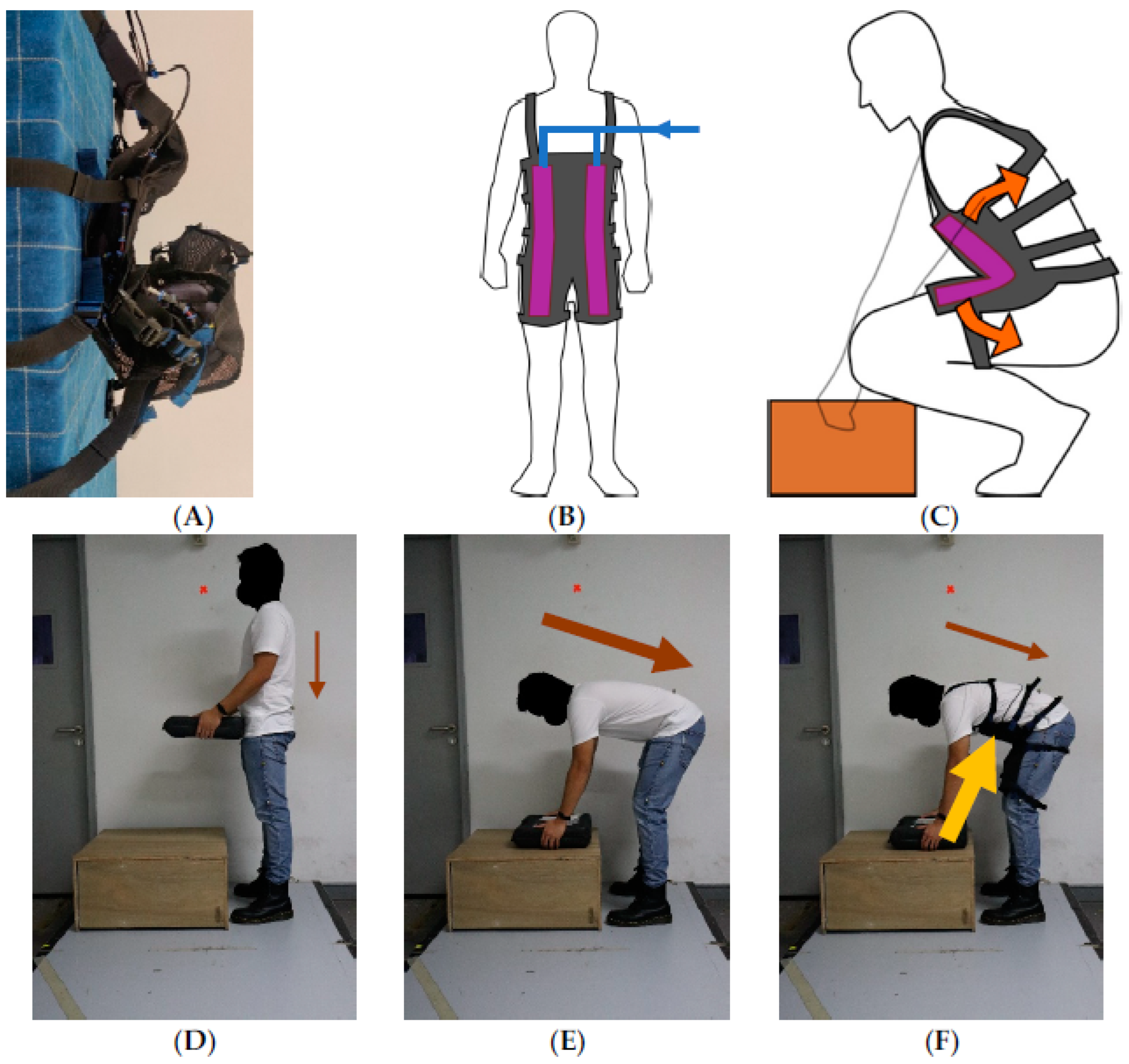
The AireLevate’s primary purpose is to provide support to the spine’s erectors. The exosuit is equipped with pneumatic fabric bending actuators first presented in [32]. The actuators follow a continuum trajectory when activated, curling into a circular profile after injection of compressed air (Figure 2A). A pair of actuators is attached to the anterior side of the AireLevate, with one actuator attached to each thigh and each side of the torso. Activation of the actuator is performed by injecting compressed air from an external source. (Figure 2B). When attached to the human body, the actuator’s curling motion is prevented by the user’s body, and instead the curling motion is converted into a distributed pushing force applied to a wide area of the abdomen and the thigh (Figure 2C). While the compressed air bladders are on the anterior abdominal wall, this stored energy in the air bladders pushes the torso into an upright position. This extends the flexed spine, and reduces the effort of the spinal erector muscles, especially in the lumbar region of the spine.
The AireLevate’s actuators are configured to operate in a passive manner. They are first activated through the application of compressed air. After this, the actuators are operated passively by leveraging on the actuators’ natural mechanics. An inflated actuator’s force naturally increases as it is folded onto itself [32]. The AireLevate is designed to leverage this mechanism, forcing the actuator to fold onto itself as the user bends down. In this manner, the suit’s force output is mechanically programmed to coincide with the increased load on the spine and its erectors as the torso is flexed (Figure 2F). The suit’s force output similarly decreases as the torso is extended.
There were no significant changes to the participant’s posture when not wearing the suit compared to wearing it (Figure 2E vs. Figure 2F).

2.2. Experiment Procedure

2.2.1. Participant Characteristics

Ten healthy subjects were recruited to participate in the experiment. Recruitment criteria were restricted to subjects who had no pre-existing musculoskeletal injuries to the upper body and had not undergone surgery in the prior 6 months. Upon arrival of the subject at the testing site, they were briefed on the various aspects of the experimental protocol. Written, informed consent was then acquired by a team member. Subjects had an average height of 1.7 m ± 0.08 m, an average weight of 66.4 kg ± 8.5 kg, and average age of 26.5 years ± 2.3 years. All subjects wore the same exosuit for this trial to ensure reproducibility of results.

2.2.2. Application of Suit and Sensors

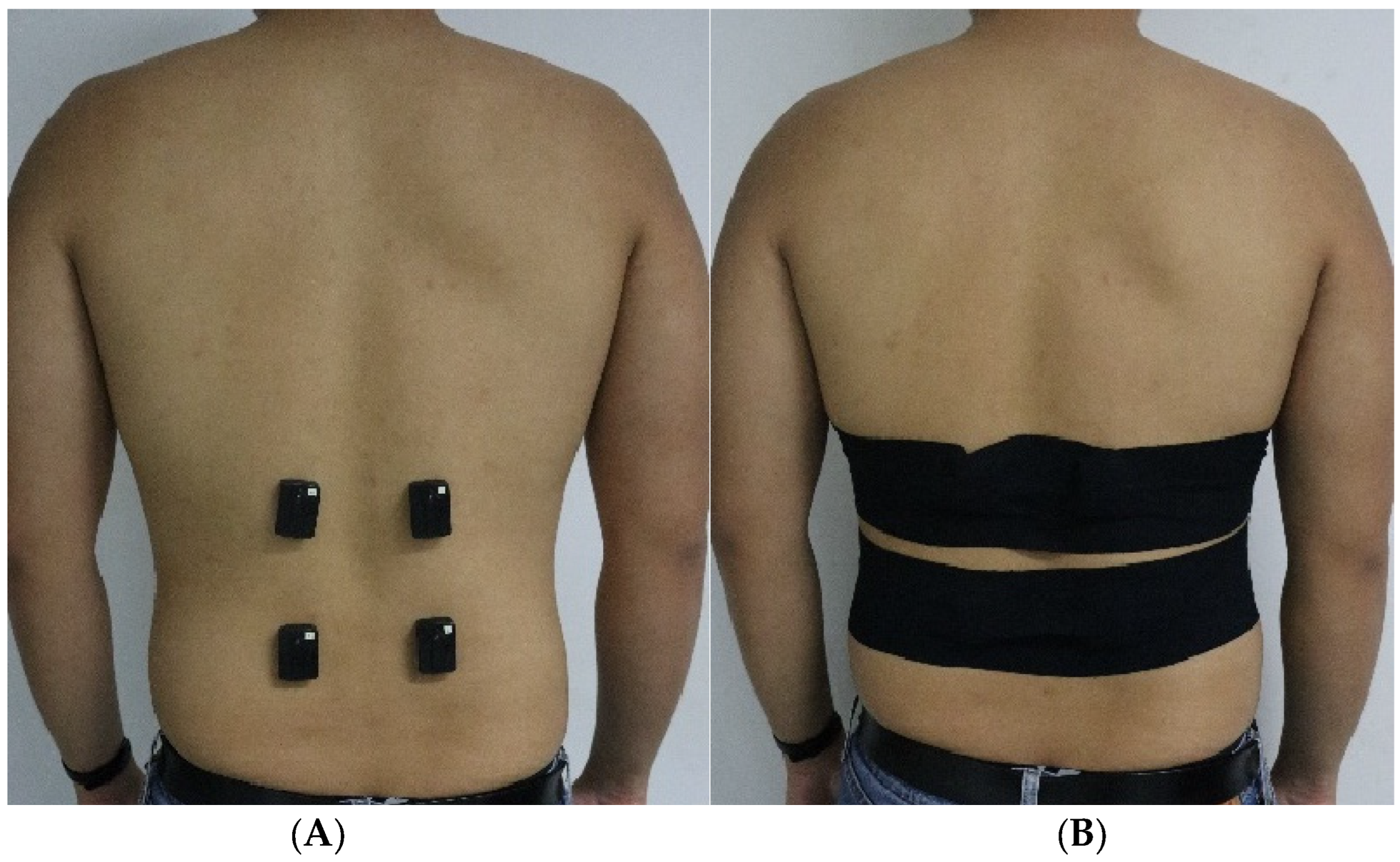
Fitting of the suit on subjects was performed by the researchers to ensure consistency of suit-fitting across subjects and prevention of interference of the suit with the sensors. sEMG sensors (Delsys Trigno, Delsys Inc., Natick, MA, USA) were attached bilaterally at the L1 and L5 spinal levels, corresponding with the muscle bellies of the iliocostalis lumborum and longissimus thoracis muscles in the erector spinae muscle group. The sensors were polled at a rate of 2000 Hz. Sensors were attached using SENIAM protocols [33]. The suit was removed and the risk of movement artifacts in sEMG readings was further reduced by applying kinesiology tape over the sensors and wrapped around the torso (Figure 3). Passive optical markers were then attached on the back and the thighs. Markers were placed on the C7 vertebra, T10 vertebra, on the left and right posterior superior iliac, and on the lateral point along the midplane of the left and right thighs. Optical markers were tracked by motion capture cameras (Vantage, Vicon) at a sampling rate of 100 Hz. Maximum voluntary isometric contractions (MVIC) were then acquired using techniques established by [34].

2.2.3. Circuit of Lifting Tasks

The experiment was designed to simulate work typically performed by manual-handling professionals [35] (Figure 4). Subjects were instructed to lift containers of varying weights (5 kg, 10 kg, 15 kg, and 20 kg). The container did not have any handles or other ergonomic features that aid lifting. At the start of each experiment, the subjects were instructed to stand in front of a 40-centimeter-high wooden platform with the container positioned on the platform. Subjects were instructed to grasp the sides of the container with outstretched arms in preparation to perform a lifting motion; this was designated as the flexed position. Subjects were then instructed to extend their lower back fully while carrying the container; this was designated as the standing position. After a brief pause, the subject was then instructed to lower the container back to its original position. One set consisted of 3 repetitions of lifting the box up from the platform and lowering the box back to the platform. Subjects started with a 5 kg container for the 1st set; subsequent sets increased container weight by 5 kg until the subject performed a 20 kg set.
Each set was performed twice, once with AireLevate support (powered sets) and once without AireLevate support (unpowered sets). Half the subjects performed the experiment starting with powered sets while the other half began with unpowered sets. Subjects were given a 20-minute period to rest between powered and unpowered tests. The AireLevate was pressurized to 150 kPa for supported sets.
At the end of each set, the participant was verbally asked to give a rating about their discomfort while performing the set. The purpose was to ascertain if use of the AireLevate introduced any perceived penalties to the comfort of the user. A rating of 10 corresponded to minimal discomfort whilst 1 corresponded to the worst discomfort they had faced prior to this experiment. Two discomfort measures were obtained: general discomfort and local discomfort. General discomfort pertained to any discomfort perceived throughout the entire body except the lumbar spine while local discomfort focused on the lumbar area only.
In addition to discomfort levels, participants were assessed on task difficulty (10 corresponding to an easy to do and 1 being very difficult, the overall suit comfort (10 corresponding to minimal discomfort), the overall ease of donning and doffing the suit (10 corresponding to minimal difficulty in donning and doffing the suit, and 1 corresponding to a very difficult application), and the design of the suit (10 corresponding to a very well-designed exosuit, and 1 corresponding to an extremely poor design).

2.3. Data Processing

sEMG data were processed based on recommendations outlined in [36]. EMG waveforms were passed through a 20 Hz high-pass, 80 dB impulse response filter. Power line interference was removed by a 48 Hz–52 Hz band stop filter with a 60 dB impulse response. The signal was then rectified and outliers with values greater than 150% of the top 75th percentile were removed. The trajectory of the participants was used to generate RMS measures of the EMG. Envelopes were extracted through a 500-wide sample, moving RMS window. EMG waveforms were then normalized using the measured MVIC of each subject.

2.4. Data Analysis

The torso’s trajectory was determined by creating a line segment between the midpoint of the left and right posterior superior iliac makers, and the marker on the T10 vertebra. The ROM of the thoracic spine, from fully erect to the lowest carrying position, was determined by comparing the angle between the flexed position and the standing position. Trajectory data were used to segment processed EMG waveforms into lifting and lowering segments. The RMS value of each lifting segment and each lowering segment was used as the primary measure for muscle activation. The results from left and right EMG sensors were combined by calculating the mean of the RMS values of the left and right L-1 and L-5 sEMG readings. Separated left and right sEMG sensor readings were also analyzed.
The RMS values for each segment and for each weight were then averaged, and the relative differences between supported and unsupported sets were determined. Metrics were tested for normality, and then statistically compared through Wilcoxon Signed Rank Tests (alpha = 0.05). Results for qualitative measures and measures for muscle activation were depicted in the form of box plots. Statistical significance was set at p < 0.05.

3. Results

3.1. Qualitative Measures

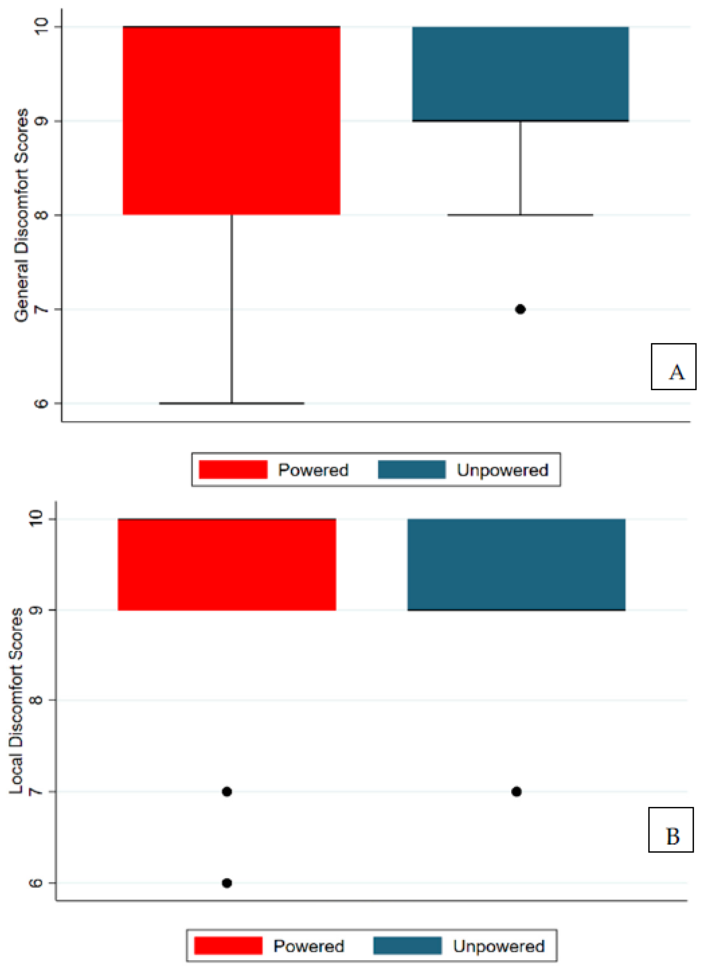
There was no statistically significant difference in the general discomfort and local discomfort of participants while performing the test with and without AireLevate support (Figure 5). Likewise, participants did not have difficulty with wearing the suit.

3.2. Range of Motion

Time series results for a representative subject lifting the 20 kg weight are shown in Figure 6. Results showed that use of the AireLevate did not result in any significant change in the user’s range of motion (ROM) when performing lifting tasks, with an average change in ROM of 4%. Participants who donned the AireLevate were able to perform all the lifting activities while maintaining their spinal curvature, as depicted in Figure 2, and there were no observable changes in posture during the lifting tasks.

3.3. Reduction in Muscle Activation

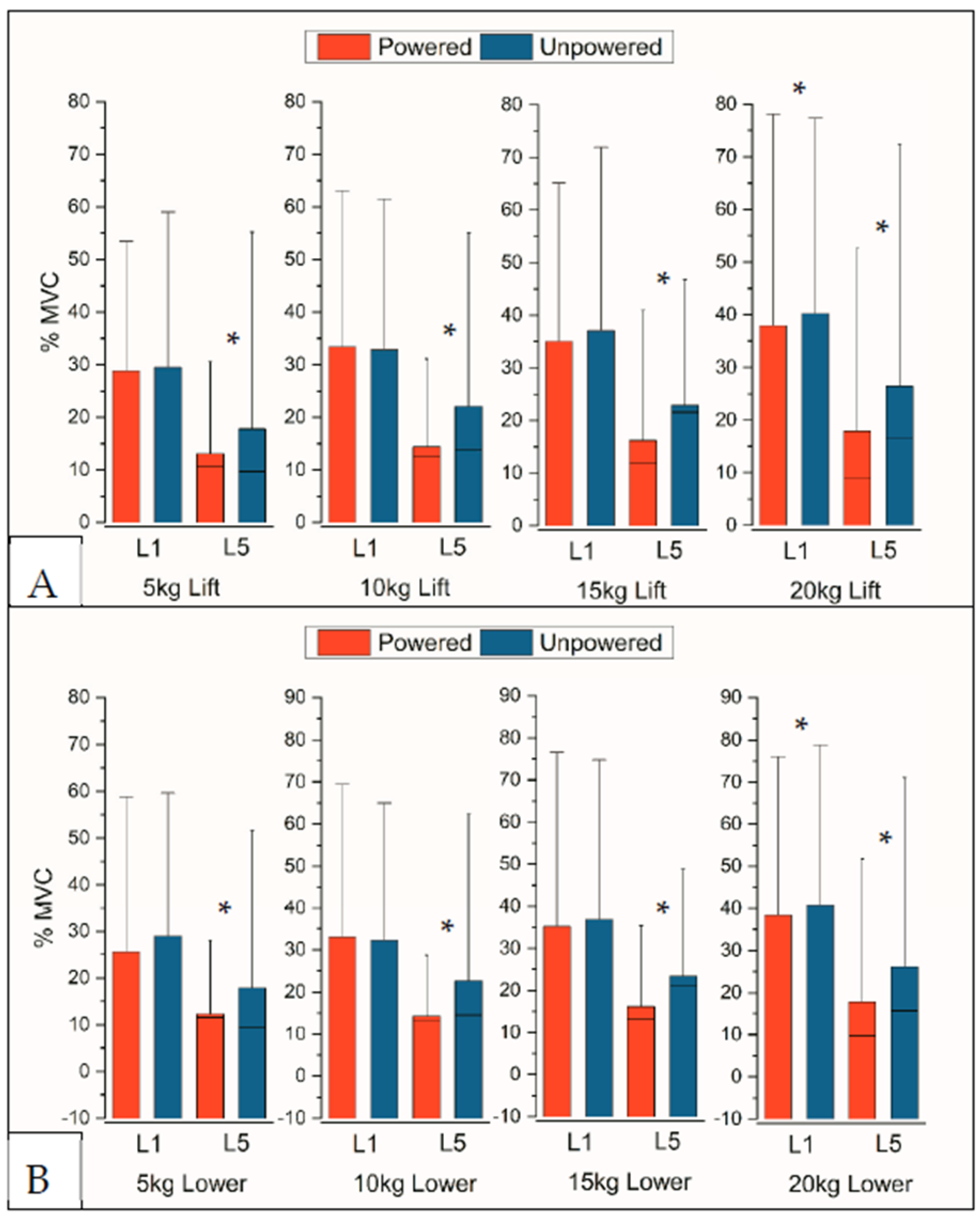
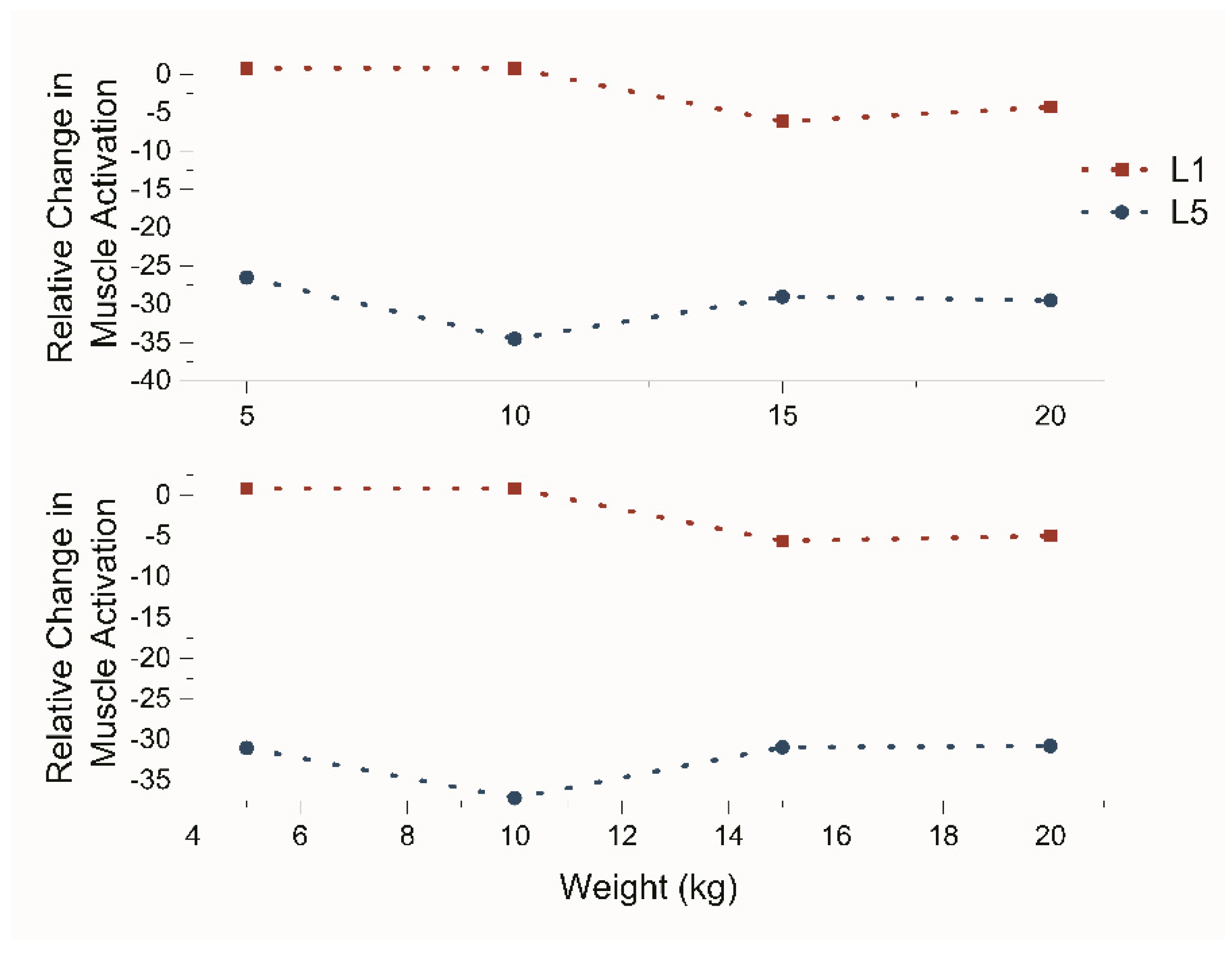
Figure 7 shows the summarized results of the experiment, separated into lifting and lowering motions. The results revealed that the AireLevate significantly reduced the muscle activation of the erector spinae on the L-5 region; all L-5 results were statistically significant (p-value < 0.02). Lifting motions showed average reduction of 27%, 35%, 29%, and 29% among all subjects for 5 kg, 10 kg, 15 kg, and 20 kg lifts, respectively; lowering motions showed average reduction of 31%, 37%, 31%, and 31% for 5 kg, 10 kg, 15 kg, and 20 kg lifts, respectively. There was no statistical difference in muscle activation in the L-1 region with or without the suit. While 20 kg lifting and lowering motions exhibited statistically significant results for the L-1 region, the reduction was relatively lower, with lifting and lowering motions exhibiting 4% and 5% reduction, respectively. The consistent reduction in muscle activation is due to the passive nature of the air in the device. The suit’s force output corresponds with the increased load on the spine and its erectors as the torso is flexed, and there is a proportionate increase in output based on the load borne by the erector spinae muscles.
The results also highlighted the consistency at which the AireLevate is able to provide assistance throughout a wide range of loads. Figure 8 presents the EMG results against the carried weight. While 10 kg lifting showed maximum reduction for both lifting and lowering motions, the range of the assistance provided did not vary significantly across various load levels. The suit exhibited average assistance levels between 27% and 35% for lifting motions and between 31% and 37% for lowering motions. The results appear to indicate a relatively better performance of the AireLevate in supporting lowering motions.

3.4. Effect on Left Side and Right Side

Comparisons between left and right side sEMG signals show a significant difference in the level of assistance between the left and right side of the body (Table 1). Left L-5 readings showed that the AireLevate was able to generate 63% of assistance for both the lifting and lowering of a 10 kg load; however, this support was reduced to 9% and 14%, respectively, on the Right L-5 during lowering motions. There is also no trend between level of support and weight lifted.

4. Discussion

This study provides insight on the impact of a pneumatic powered exoskeleton on manual-lifting tasks in a controlled setting. Our study revealed that the AireLevate significantly reduced muscle activation at the L-5 region of the thoracolumbar spine, especially on the left side of the body. The AireLevate also did not impede the user’s range of motion, nor was there significant discomfort when using the suit.
While pneumatic exoskeletons typically use rigid frames to provide support, the AireLevate exosuit utilizes pressurized air within its flexible fabric actuators. Due to its soft robotic nature, the suit’s actuators fit closely to the contour of the body in both its deflated and inflated state. They can perform effectively without the need for precise alignment between the joints and the actuators. These features reduce the mechanical impedance of the exosuit and preserves the user’s range of motion. Traditional exoskeleton power transmission systems, which predominantly employ rigid components, typically require precise alignment between the suit’s and body’s kinematic chains. A suit which does not impede the range of motion of a wearer would be easier to implement on the ground as workers would not have to change their current working postures when assisted by the suit [37]. User comfort is similarly paramount; exoskeletons which impose significant comfort penalties to the user will reduce adoption, regardless of their ability to provide support to the body. The AireLevate’s fabric design minimizes any user discomfort, allowing the user to perform their tasks in a more natural manner and with minimal impediment; these attributes will help boost its adoption rate. We acknowledge that there appears to be a ceiling effect for our comfort scores in this pilot study as participants gave high comfort scores without wearing the suit. However, similarly high comfort scores when wearing the suits led us to conclude that the suit in its current iteration does not affect the comfort of users when performing manual-handling tasks.
Simultaneously, the suit was able to reduce muscle activation both for lifting as well as lowering motions, especially at the L-5 region of the thoracolumbar spine. Moreover, the results showed the suit’s ability to deliver the same level of efficacy regardless of the load lifted by the user. This effect was more pronounced in the lower lumbar compared to the upper lumbar regions. The AireLevate was able to achieve a maximum reduction in L-5 muscle activation of 63%. However, the level of support measured was dissimilar between the left and right sides, and the difference in left–right muscle activation was more pronounced with the suit on compared to without the suit. Most individuals have asymmetrically developed paraspinal muscles, with the left side usually being the weaker non-dominant muscle group [38]. Thus, we hypothesize that the difference in level of assistance for the left lumbar muscles compared to the right can be explained by an asymmetry in muscle bulk for most individuals.
Despite this dissimilarity between left and right erector spinae muscles, the averaged values still produced a 35% reduction in muscle activation. This indicates a robust net effect on combined muscle activation of the left and right erector spinae. The reduction in EMG muscle readings is coincident with the literature. Among active exoskeletons, Glinski et al. [39] reported a decrease of 4.5% in muscle activation in the lumbar region. The pulley-driven exoskeleton from [23] reduced erector spinae activation by 23.5%. Madini et al. [26] performed a similar study on commercially available exoskeletons driven by gas springs and found that the Laevo™ exoskeletons and BackX™ exoskeletons produced trunk extensor muscle activation reductions of 10.4% and 17.3%, respectively. Lamers et al. [27] performed a test on an exosuit driven by elastic bands and found that it was able to reduce erector spinae activation by 16% when lifting a 24 kg weight. Ide et al. [29] performed a test on the Muscle Suit exoskeleton powered by McKibben pneumatic artificial muscles; however, the authors did not normalize EMG readings, making external analysis difficult. Nevertheless, these comparisons highlight the AireLevate’s ability in providing increased support and assistance to the user.
By assisting the user’s erector spinae, the decreases in muscle activation at the L-5 region for both lifting and lowering motions indicate that the AireLevate can potentially protect the wearer from developing LBP. Poor muscle tone and body posture predisposes workers to LBP [40]. It is also known that loading of the erector spinae muscles leads to compression of the spine, which can eventually lead to permanent LBP [41]. The reduction in erector spinae muscle activation also reduces the spinal load; this effect will eventually lead to a lower incidence of LBP. Therefore, the AireLevate has the potential to reduce the prevalence of occupational LBP through the minimization of both acute and chronic LBP. If the suit is worn on a longer term, this can result in two forms of cost savings for employers. First, there will be an increase in productivity if workers take fewer days of sick leave for LBP [42]. While acute LBP might resolve in a few days, chronic LBP tends to have a protracted course with longer days of absence [43]. Limiting the incidence of chronic LBP would not just represent increased productivity from having fewer days of sick leave, but it would also represent decreased healthcare costs. Workers who report sick less often would have lower healthcare costs and hence represent financial savings for both employers and insurers [44].
Moreover, reduced muscle activation correlates with decreased energy consumption with all other factors kept constant [44,45,46]. The EMG findings suggest that the AireLevate can potentially reduce energy expenditure during manual-handling tasks. This could also represent an increase in productivity for users of our suit who are performing manual-handling tasks. The suit would not only improve efficiency in terms of reduced sick days, but reduced energy consumption would minimize productivity dips due to fatigue.
While our suit showed promising results in assisting manual-lifting tasks, there were some limitations with our study. First, our sample size was small, hence making it challenging to extrapolate the findings to larger populations. Secondly, our participants were young healthy males. While this demographic might represent a high proportion of manual-handling workers, the results might not be reproducible to other groups such as females and older individuals. Furthermore, this study did not include participants who had existing or previous musculoskeletal injuries or surgical intervention within the past 6 months.
There were some limitations to the AireLevate as well. Firstly, the exoskeleton only supported the core muscles of the participant and did not cover the upper limbs. Previous studies [47] have shown that supporting the upper-limb and shoulder muscles helps to compressive loads on the lower back. However, other studies have shown the contrary, that upper limb exoskeletons increase the load on the lumbar spine [48]. This increase can be attributed to the added mass introduced by the upper-limb exoskeleton; a shoulder exosuit designed similarly to the AireLevate will have minimal mass, minimizing its effect on the spine.
The study also highlighted limitations in the current design. The AireLevate is primarily secured to the body through a series of adjustable straps buckled at the back. Such a design could impose a number of challenges in a practical setting. Not only does it extend the donning time of the exosuit, but it also hinders the rapid removal of the suit in emergency situations. The current version also requires a permanent air tether during operation, thus limiting the practical use of the suit in workspaces without readily available air and power sources. Future versions place importance on the retention of compressed air during extended operating periods.

5. Conclusions

This study identified that the AireLevate reduced muscle activation, mainly at the L-5 region of the thoracolumbar spine both for lifting and lowering activities, without worsened general or local discomfort. This suggests the potential of the exosuit in mitigating LBP in the workplace, where the carrying of heavy loads predisposes workers to high LBP risk. Future studies should consider including more participants and an evaluation of the efficacy of the device within actual workplaces; such a study may also expand EMG coverage to the entire posterior shoulder girdle and posterior thigh leg muscles, include VO2 tests to further assess energy expenditure, and add longitudinal tests to assess pain outcomes.

Author Contributions

Conceptualization, A.N.C. and R.F.N.; methodology, A.N.C. and R.F.N.; validation, A.N.C.; writing—original draft, A.N.C. and R.F.N.; writing—review and editing, A.N.C., R.F.N. and R.C.-H.Y. All authors have read and agreed to the published version of the manuscript.

Funding

This work was funded by the National Robotics Programme—Robotics Enabling Capabilities and Technologies (W2025d0243).

Institutional Review Board Statement

Subjects further consented to the use of their photographs, videos, and data in academic publications. The study’s protocol and methods were formulated following the principles stated in the Declaration of Helsinki and were reviewed and approved by the National University of Singapore’s Institutional Review Board (NUS-IRB-2021-25). Furthermore, all mandatory health and safety protocols by the university’s Department of Biomedical engineering were adhered to.

Informed Consent Statement

Subjects provided written consent before participating in the studies.

Data Availability Statement

Due to commercial factors related to the ARMAS device, the supporting data for this study is not available.

Conflicts of Interest

Rainier Natividad and Yeow Chen Hua are co-inventors on intellectual property related to the AireLevate. They are co-founders and have a financial interest in ArmasTec Pte. Ltd., a spinoff to commercialize the AireLevate. Amit Cuttilan is a clinician who conducted the data analysis and write up of this paper. He has no financial interests in ArmasTec Pte. Ltd.

References

  1. Alnefaie, M.; Alamri, A.; Hariri, A.; Alsaad, M.; Alsulami, A.; Abbas, A.; Alfozan, A.; Aljiffry, M.; Abbadi, H. Musculoskeletal Symptoms Among Surgeons at a Tertiary Care Center: A Survey Based Study. Med. Arch. 2019, 73, 49–54. [Google Scholar] [CrossRef] [PubMed]
  2. Abdul Monem, A.; Nizar, A.; Abdullaha, A. Prevalence and Associated Factors of Low Back Pain among Clinicians of A Major Referral Hospital. Med. J. Malays. 2015, 70, 12–17. [Google Scholar]
  3. Meisha, D.E.; Alsharqawi, N.S.; Samarah, A.A.; Al-Ghamdi, M.Y. Al-Ghamdi Prevalence of work-related musculoskeletal disorders and ergonomic practice among dentists in Jeddah, Saudi Arabia. Clin. Cosmet. Investig. Dent. 2019, 11, 171–179. [Google Scholar] [CrossRef] [PubMed] [Green Version]
  4. Waddell, G.; Burton, A.K.; Main, C.J. Screening to Identify People at Risk of Long-Term Incapacity for Work: A Conceptual and Scientific Review; Royal Society of Medicine Press: London, UK, 2003; Available online: www.rsmpress.co.uk (accessed on 20 August 2022).
  5. Punnett, L.; Prüss-Ütün, A.; Nelson, D.I.; Fingerhut, M.A.; Leigh, J.; Tak, S.; Phillips, S. Estimating the global burden of low back pain attributable to combined occupational exposures. Am. J. Ind. Med. 2005, 48, 459–469. [Google Scholar] [CrossRef]
  6. Kofi, M.; Abbas, M.A.F.; Lamiaa, Z.; Abu Zaid, A.; Fiala; Alhamdan, N.A. Prevalence and Risk Factors of Low Back Pain among Nurses in Four Tertiary Care Hospitals at King Fahad Medical City, Riyadh, KSA. 2010. Available online: www.medicaljournalofcairouniversity.comwww.medicaljournalofcairouniversity.com (accessed on 20 August 2022).
  7. Al Dajah, S.; Abdalhamed, H.; Daghdi, A. Prevalence and risk factors of low back pain among nurses in sudayr region. Eur. Sci. J. 2013, 9, 1857–7881. [Google Scholar]
  8. Al-Eisa, E.; Al-Abbad, H. Occupational Back Pain among Rehabilitation Nurses in Saudi Arabia. Workplace Health Saf. 2013, 61, 401–407. [Google Scholar] [CrossRef]
  9. Croft, P.R.; Macfarlane, G.J.; Papageorgiou, A.C.; Thomas, E.; Silman, A.J. General practice Outcome of low back pain in general practice: A prospective study. BMJ 1998, 316, 1356–1359. Available online: www.bmj.com (accessed on 20 August 2022). [CrossRef] [Green Version]
  10. Hestbaek, L.; Leboeuf-Yde, C.; Manniche, C. Low back pain: What is the long-term course? A review of studies of general patient populations. Eur. Spine J. 2003, 12, 149–165. [Google Scholar] [CrossRef]
  11. Andini, F. Risk Factors of Low Back Pain in Workers. J. Major. 2015, 4. [Google Scholar]
  12. Publications Office of the European Union. Fifth European Working Conditions Survey—Overview Report; Publications Office of the European Union: Luxembourg, 2012. [Google Scholar]
  13. Iles, R.A.; Davidson, M.; Taylor, N.F. Psychosocial predictors of failure to return to work in non-chronic non-specific low back pain: A systematic review. Occup. Environ. Med. 2008, 65, 507–517. [Google Scholar] [CrossRef] [Green Version]
  14. Steenstra, I.A.; Verbeek, J.H.; Heymans, M.W.; Bongers, P.M. Prognostic factors for duration of sick leave in patients sick listed with acute low back pain: A systematic review of the literature. Occup. Environ. Med. 2005, 62, 851–860. [Google Scholar] [CrossRef] [PubMed] [Green Version]
  15. De Looze, M.P.; Bosch, T.; Krause, F.; Stadler, K.S.; O’Sullivan, L.W. Exoskeletons for industrial application and their potential effects on physical work load. Ergonomics 2016, 59, 671–681. [Google Scholar] [CrossRef] [PubMed] [Green Version]
  16. Howard, J.; Murashov, V.; Lowe, B.D.; Lu, M.L. Industrial exoskeletons: Need for intervention effectiveness research. Am. J. Ind. Med. 2020, 63, 201–208. [Google Scholar] [CrossRef] [PubMed]
  17. Kermavnar, T.; de Vries, A.W.; de Looze, M.P.; O’Sullivan, L.W. Effects of industrial back-support exoskeletons on body loading and user experience: An updated systematic review. Ergonomics 2021, 64, 685–711. [Google Scholar] [CrossRef]
  18. Bosch, T.; van Eck, J.; Knitel, K.; de Looze, M. The effects of a passive exoskeleton on muscle activity, discomfort and endurance time in forward bending work. Appl. Ergon. 2016, 54, 212–217. [Google Scholar] [CrossRef]
  19. Gopura, R.A.R.C.; Bandara, D.S.V.; Kiguchi, K.; Mann, G.K.I. Developments in hardware systems of active upper-limb exoskeleton robots: A review. Rob. Auton. Syst. 2016, 75, 203–220. [Google Scholar] [CrossRef]
  20. Ali, A.; Fontanari, V.; Schmoelz, W.; Agrawal, S.K. Systematic Review of Back-Support Exoskeletons and Soft Robotic Suits. Front. Bioeng. Biotechnol. 2021, 9, 1027. [Google Scholar] [CrossRef]
  21. Shirazi-Adl, A.; El-Rich, M.; Pop, D.G.; Parnianpour, M. Spinal muscle forces, internal loads and stability in standing under various postures and loads—Application of kinematics-based algorithm. Eur. Spine J. 2004, 14, 381–392. Available online: https://www.academia.edu/12685444/Spinal_muscle_forces_internal_loads_and_stability_in_standing_under_various_postures_and_loads_application_of_kinematics_based_algorithm (accessed on 19 September 2022). [CrossRef] [Green Version]
  22. Hara, H.; Sankai, Y. Development of HAL for Lumbar Support. In Proceedings of the Joint 5th International Conference on Soft Computing and Intelligent Systems and 11th International Symposium on Advanced Intelligent Systems (SCIS & ISIS 2010), Okayama, Japan, 8–10 December 2010; pp. 416–421. [Google Scholar]
  23. Yong, X.; Yan, Z.; Wang, C.; Wang, C.; Li, N.; Wu, X. Ergonomic mechanical design and assessment of a waist assist exoskeleton for reducing lumbar loads during lifting task. Micromachines 2019, 10, 463. [Google Scholar] [CrossRef] [Green Version]
  24. Ko, H.K.; Lee, S.W.; Koo, D.H.; Lee, I.; Hyun, D.J. Waist-assistive exoskeleton powered by a singular actuation mechanism for prevention of back-injury. Rob. Auton. Syst. 2018, 107, 1–9. [Google Scholar] [CrossRef]
  25. Technology—Verve Motion. Available online: https://vervemotion.com/technology/ (accessed on 31 August 2022).
  26. Madinei, S.; Alemi, M.M.; Kim, S.; Srinivasan, D.; Nussbaum, M.A. Biomechanical assessment of two back-support exoskeletons in symmetric and asymmetric repetitive lifting with moderate postural demands. Appl. Ergon. 2020, 88, 103156. [Google Scholar] [CrossRef]
  27. Lamers, E.P.; Yang, A.J.; Zelik, K.E. Feasibility of a biomechanically-assistive garment to reduce low back loading during leaning and lifting. In IEEE Transactions on Biomedical Engineering; IEEE Computer Society United States of America: Washington, DC, USA, 2018; Volume 65, pp. 1674–1680. [Google Scholar] [CrossRef]
  28. LiftSuit|Auxivo. Available online: https://www.auxivo.com/liftsuit (accessed on 31 August 2022).
  29. Ide, M.; Hashimoto, T.; Matsumoto, K.; Kobayashi, H. Evaluation of the Power Assist Effect of Muscle Suit for Lower Back Support. IEEE Access 2021, 9, 3249–3260. [Google Scholar] [CrossRef]
  30. Inose, H.; Mohri, S.; Arakawa, H.; Okui, M.; Koide, K.; Yamada, Y.; Kikutani, I.; Nakamura, T. Semi-endoskeleton-type waist assist AB-wear suit equipped with compressive force reduction mechanism. In Proceedings of the 2017 IEEE International Conference on Robotics and Automation (ICRA), Singapore, 29 May–3 June 2017; pp. 6014–6019. [Google Scholar] [CrossRef]
  31. Veale, A.J.; Xie, S.Q. Towards compliant and wearable robotic orthoses: A review of current and emerging actuator technologies. Med. Eng. Phys. 2016, 38, 317–325. [Google Scholar] [CrossRef]
  32. Natividad, R.F.; Miller-Jackson, T.; Chen-Hua, R.Y. A 2-DOF Shoulder Exosuit Driven by Modular, Pneumatic, Fabric Actuators. IEEE Trans. Med. Robot. Bionics 2020, 3, 166–178. [Google Scholar] [CrossRef]
  33. Hermens, H.J.; Freriks, B.; Disselhorst-Klug, C.; Rau, G. Development of recommendations for SEMG sensors and sensor placement procedures. J. Electromyogr. Kinesiol. 2000, 10, 361–374. [Google Scholar] [CrossRef] [PubMed]
  34. Vera-Garcia, F.J.; Moreside, J.M.; McGill, S.M. MVC techniques to normalize trunk muscle EMG in healthy women. J. Electromyogr. Kinesiol. 2010, 20, 10–16. [Google Scholar] [CrossRef]
  35. Health and Safety Executive Manual Handling at Work: A Brief Guide. 2020. Available online: https://books.hse.gov.uk/ (accessed on 20 August 2022).
  36. Merletti, R.; Di Torino, P.J.J.E.K. Standards for Reporting EMG Data. J. Electromyogr. Kinesiol. 1999, 9, 3–4. [Google Scholar]
  37. Sposito, M.; di Natali, C.; Toxiri, S.; Caldwell, D.G.; de Momi, E.; Ortiz, J. Exoskeleton kinematic design robustness: An assessment method to account for human variability. Wearable Technol. 2020, 1, e7. [Google Scholar] [CrossRef]
  38. Xiao, J.; Gao, J.; Wang, H.; Liu, K.; Yang, X. The Surface EMG Characteristics between Erector Spinae and Vastus Lateralis during Bending Forward and Squatting Down Tasks. Physiol. J. 2015, 2015, 537379. [Google Scholar] [CrossRef] [Green Version]
  39. Von Glinski, A.; Yilmaz, E.; Mrotzek, S.; Marek, E.; Jettkant, B.; Brinkemper, A.; Fisahn, C.; Schildhauer, T.A.; Geßmann, J. Effectiveness of an on-body lifting aid (HAL® for care support) to reduce lower back muscle activity during repetitive lifting tasks. J. Clin. Neurosci. 2019, 63, 249–255. [Google Scholar] [CrossRef]
  40. National Safety Council. Know the Facts about Backs [Fact Sheet]. National Safety Council, United States of America. 2019. Available online: https://injuryfacts.nsc.org/ (accessed on 20 August 2022).
  41. Mazis, N. Does a History of Non Specific Low Back Pain Influence Electromyographic Activity of the Erector Spinae Muscle Group during Functional Movements? J. Nov. Physiother. 2014, 4, 226. [Google Scholar] [CrossRef]
  42. Kordt, M.; Marschall, J.; Hildebrandt-Heene, S.; Zich, K.; Sörensen, J.; Nolting, H.-D. Deutsche Angestellten-Krankenkasse-Gesundheitsreport 2018, Medhochzwei. 2018. Available online: https://www.dak.de/dak/download/gesundheitsreport-2018-pdf-2073702.pdf (accessed on 5 April 2023).
  43. Mehra, M.; Hill, K.; Nicholl, D.; Schadrack, J. The burden of chronic low back pain with and without a neuropathic component: A healthcare resource use and cost analysis. J. Med. Econ. 2012, 15, 245–252. [Google Scholar] [CrossRef] [PubMed]
  44. Kim, L.H.; Vail, D.; Azad, T.D.; Bentley, J.P.; Zhang, Y.; Ho, A.L.; Fatemi, P.; Feng, A.; Varshneya, K.; Desai, M.; et al. Expenditures and health care utilization among adults with newly diagnosed lowback and lower extremity pain. JAMA Netw. Open 2019, 2, e193676. [Google Scholar] [CrossRef] [Green Version]
  45. Hargreaves, M.; Spriet, L.L. Author Correction: Skeletal muscle energy metabolism during exercise. Nat. Metab. 2020, 2, 817–828, Erratum in Nat. Metab. 2020, 2, 990. [Google Scholar] [CrossRef]
  46. Tsianos, G.A.; MacFadden, L.N. Validated Predictions of Metabolic Energy Consumption for Submaximal Effort Movement. PLoS Comput. Biol. 2016, 12, e1004911. [Google Scholar] [CrossRef] [PubMed] [Green Version]
  47. Kim, S.; Nussbaum, M.A.; Esfahani, M.I.M.; Alemi, M.M.; Jia, B.; Rashedi, E. Assessing the influence of a passive, upper extremity exoskeletal vest for tasks requiring arm elevation: Part II—‘Unexpected’ effects on shoulder motion, balance, and spine loading. Appl. Ergon. 2018, 70, 323–330. [Google Scholar] [CrossRef]
  48. Weston, E.B.; Alizadeh, M.; Knapik, G.G.; Wang, X.; Marras, W.S. Biomechanical evaluation of exoskeleton use on loading of the lumbar spine. Appl. Ergon. 2018, 68, 101–108. [Google Scholar] [CrossRef]
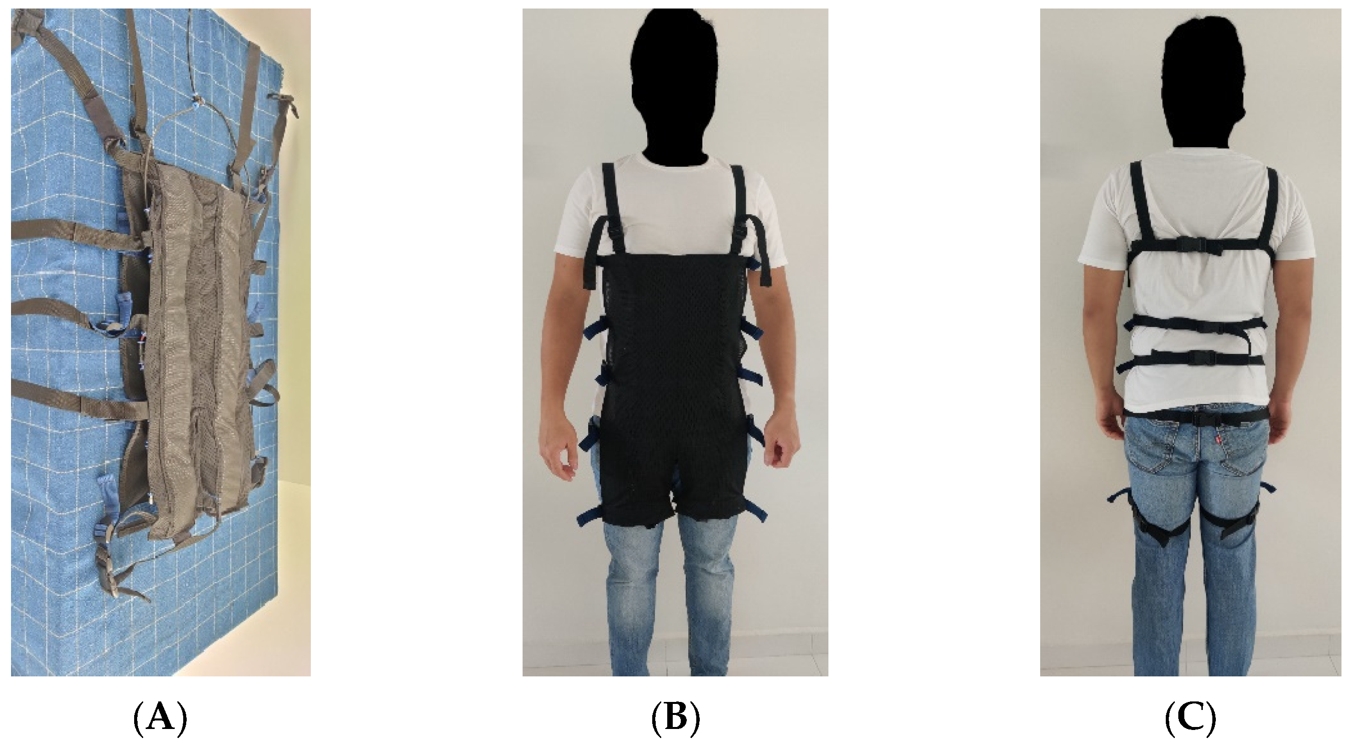
Figure 1. (A) The AireLevate. (B) Front view of the AireLevate as worn by the user. (C) Back view of the AireLevate as worn by a user.
Actuators 12 00273 g001
Figure 2. The actuators installed in the AireLevate generate a pushing force that causes the AireLevate to curl. (A) An inflated AireLevate creates a bending motion when unworn. (B) A pair of bending actuators are placed on the anterior section of the torso (location highlighted in purple). Compressed air is supplied externally and distributed to the actuators. Pneumatic tubes are externally located and are shown in blue. (C) When worn, this curling motion instead translates to a pushing force to the abdomen and thighs. The orange lines show the direction by which the actuators would curl if the user’s body was not present. (D) A user is carrying a container without AireLevate assistance. The brown arrow shows the force generated by the erectors. (E) A person bends down to pick up an object without wearing the AireLevate. As the user bends down, the demand on the erector increases. (F) With the assistance of the AireLevate, the demand on the erectors is reduced. The yellow arrow shows the direction of the force generated by the AireLevate as a result of the body preventing the actuators from curling.
Actuators 12 00273 g002
Figure 3. (A) the positioning of sEMG sensors. (B) Kinesiology tape was applied to secure the sEMG sensors.
Actuators 12 00273 g003
Figure 4. The supported lifting motion is shown. (A) Sets start in the flexed position. (B) The container is raised to the standing position (C) and is returned to the flexed position. The user is instructed to release the container.
Actuators 12 00273 g004
Figure 5. Box plots of qualitative results. (A) Results for general discomfort. (B) Results for local lumbar spine discomfort. On the y-axes, 10 corresponds to minimal discomfort and 1 to maximum discomfort during the lifting and lowering activities.
Actuators 12 00273 g005
Figure 6. Time series data showing a subject’s torso angle and Raw EMG readings on the Lumbar-5 region for one 20 kg set. At a 90° torso angle, the torso is fully flexed. (A) Unpowered. (B) Powered.
Actuators 12 00273 g006
Figure 7. Box plots showing the RMS of muscle activation among all subjects. (A) Results for lifting motions. (B) Results for lowering motions. * signifies statistically significant comparisons.
Actuators 12 00273 g007
Figure 8. Line graph summarizing the change in RMS muscle activation when carrying different weights. (Top) lifting motion; (Bottom) lowering motion.
Actuators 12 00273 g008
Table 1. Relative Change in the Separated Left and Right sEMG readings from the L-5 region.
Lifting MotionsLowering Motions
WeightLeft L-5Right L-5Left L-5Right L-5
5 kg−54%
(0.01)
−9%
(0.01)
−50%
(0.02)
−19%
(0.01)
10 kg−63%
(<0.005)
−9%
(0.03)
−63%
(<0.005)
−14%
(0.09)
15 kg−61%
(<0.005)
0%
(0.07)
−61%
(<0.005)
−4%
(0.07)
20 kg−55%
(<0.005)
−7%
(0.14)
−56%
(<0.005)
−8%
(0.15)
Values in bold exhibit p-values < 0.05. p-values are in parenthesis.
Disclaimer/Publisher’s Note: The statements, opinions and data contained in all publications are solely those of the individual author(s) and contributor(s) and not of MDPI and/or the editor(s). MDPI and/or the editor(s) disclaim responsibility for any injury to people or property resulting from any ideas, methods, instructions or products referred to in the content.