Review

Gelatin Soft Actuators: Benefits and Opportunities

1
Department of Mechanical Engineering, University of Illinois at Urbana-Champaign, Urbana, IL 61801, USA
2
Department of Bioengineering, University of Illinois at Urbana-Champaign, Urbana, IL 61801, USA
*
Author to whom correspondence should be addressed.
Actuators 2023, 12(2), 63; https://doi.org/10.3390/act12020063
Submission received: 1 November 2022 / Revised: 22 January 2023 / Accepted: 26 January 2023 / Published: 31 January 2023
(This article belongs to the Special Issue 10th Anniversary of Actuators)

Abstract

:
Soft robots are being developed as implantable devices and surgical tools with increasing frequency. As this happens, new attention needs to be directed at the materials used to engineer these devices that interface with biological tissues. Biocompatibility will increase if traditional materials are replaced with biopolymers or proteins. Gelatin-based actuators are biocompatible, biodegradable, versatile, and tunable, making them ideal for biomedical and biomechanical applications. While building devices from protein-based materials will improve biocompatibility, these new materials also bring unique challenges. The properties of gelatin can be tuned with the addition of several additives, crosslinkers, and plasticizers to improve mechanical properties while altering the characteristic fluid absorption and cell proliferation. Here, we discuss a variety of different gelatin actuators that allow for a range of actuation motions including swelling, bending, folding, and twisting, with various actuation stimulants such as solvent, temperature, pneumatic pressure, electric field, magnetic field, or light. In this review, we examine the fabrication methods and applications of such materials for building soft robots. We also highlight some ways to further extend the use of gelatin for biomedical actuators including using fiber-reinforced gelatin, gelatin cellular solids, and gelatin coatings. The understanding of the current state-of-the-art of gelatin actuators and the methods to expand their usage may expand the scope and opportunities for implantable devices using soft hydrogel robotics.

1. Introduction

Gelatin is a heterogeneous mixture of peptides and proteins derived from collagen through partial hydrolysis that destroys cross-linkages between polypeptide chains and breaks polypeptide bonds [1,2]. In this hydrolyzed state, gelatin is versatile and has many benefits. Gelatins are readily derived without the need for synthesis, are harmless to the environment due to rapid degradation rates, and allow for the incorporation of water-soluble additives [3]. Gelatin is used extensively in the food [2,4,5], pharmaceutical [6,7], cosmetic [8], and photography [9,10] industries. In food industries, gelatin is used for emulsification, texturization, and stabilization [11]. In pharmaceutical industries, gelatin is used for drug encapsulation [12], drug stabilization [13], wound dressing [14,15], plasma expansion [16], ointment filling [17], and emulsification [18]. After the long historical use of gelatin in these industries, applications of gelatin are now also used in diverse biomechanical fields from drug delivery [19] to bone tissue engineering [20,21].
Gelatin has more recently been studied for use in non-conventional active roles such as actuators in soft robotic devices [22,23], and wearables [3,24,25]. Soft actuators are sensitive to environmental fluctuations that convert chemical and physical energy into mechanical work [22]. However, hydrogels such as gelatin are considered to be mechanically weak materials [26], they are soft and brittle and cannot withstand large deformations from environmental fluctuations due to their polymer networks containing inhomogeneities such as dangling chains and loops [26]. Gelatin gels are also easily broken into fragments under a modest compressive load [27]. Gelatin-based gels have a moderate performance when strained and rapidly dry when operated in air, causing stiffening and limiting the stability and durability of the gelatin elements [3]. The mechanical properties of conventional hydrogels such as gelatin are also evaluated by shearing or compression rather than by stretching because of poor deformability [26].
Although alone they are characterized as mechanically weak materials, the physical properties of gelatin gels can be easily tuned with additives, nanocomposites, or plasticizers [28]. Gelatin hydrogels can also serve as a platform for building actuators for biomedical applications because of their biocompatibility [29,30], ability to promote cell adhesion and proliferation [31], tissue-like stiffness [32,33] and other attractive properties such as fluid absorbance [34]. With the use of various material additives and fabrication techniques that enhance gelatin’s mechanical properties, the benefits of gelatin hydrogel materials can be exploited and they can be applied as actuators. Many soft fluidic actuators have been modeled and controlled through various methods, including differential simulations, topology optimizations, data-driven modeling and control methods, hardware control boards, nonlinear estimations, and analytical and numerical modeling methods, such as dynamic modeling and finite element modeling [35,36]. Finite element modeling methods were found to be especially effective in capturing the strong nonlinearities and complex geometries of soft actuators [37]. Since gelatin can be both hyperelastic and viscoelastic [38,39], finite element modeling methods are useful to predict gelatin actuation performance and optimize designs.
In this review, we classify gelatin actuators based on their kinetic motion, i.e., linear type, bending type, and twisting type (Figure 1). We identify for each of these different motions the different ways in which gelatin can be engineered to be stimulated by environmental fluctuations in pH [40,41], solvent [22], temperature [42], humidity [43], electric field [44], magnetic field [45], or light [46]. The actuation motion and stimuli diversity of gelatin actuators is dictated by how the gelatin is modified with additives, such as crosslinkers, reinforcements, and plasticizers, and by actuator structural design [47]. The structural designs of these gelatin actuators can be fabricated through several techniques, such as mold casting [23,48], spin-coating [43], and 3D printing [49,50]. In classifying the gelatin actuators according to kinetic motions, it can be seen how currently widely used non-biocompatible biomedical actuators of a particular kinetic motion can be replaced with gelatin actuators that carry out the same motions, through different fabrication methods, depending on the stimulations, the additives, and the structural designs.
While gelatin has been demonstrated to be useful and functional as an actuator in the biomechanical field, there are many ways that evolving gelatin applications can be extended within soft robotics. This can be achieved by improving the material properties of gelatin with the use of reinforced fibers or with the use of gelatin cellular solids or foams. Gelatin coatings may also serve to enhance the functional biocompatibility and efficiency of synthetic actuators. With these methods, there is an opportunity to widen the scope for innovation with gelatin actuators and medical device design.

2. Versatile Actuation Using Gelatin

2.1. Linear Actuators

The majority of small gelatin linear actuators are stimulated by swelling. The swelling of hydrogels such as gelatin can be used for actuating and sensing applications [51]. The swelling of gelatin actuators is stimulated through solvents creating environmental fluctuations in terms of pH, compositions, and concentrations [52]. The swelling also depends on the substrate and solvent material synthesis as well as on the time spent in the solvent. Swelling-stimulated hydrogel actuators work as drug delivery systems by initially trapping molecules in their crosslinked polymer networks. When the hydrogel is in contact with a solution that causes it to swell, the distance between the polymer chains in the gel increase, and the drug is released into the bloodstream [53]. Gelatin is a hydrophilic material with a water uptake of above 100% when immersed for periods as short as 5 min, and above 500% when immersed for 15 min [54]. In general, a high temperature, low gelatin concentration, and deviation from the isoelectric point can promote the swelling or disintegration of the gelatin polymer networks [46,55].
Some linear actuation due to solvent swelling depends on the solvent’s pH. Ooi et al., studied the effects of cellulose nanocrystals (CNCs) on the swelling ratio of a CNC-reinforced gelatin hydrogel crosslinked with glutaraldehyde [53]. They found that the hydrogels were highly sensitive to changes in pH and had a maximum swelling ratio at pH 3. This ability to vary swelling ratios depending on the stimulant pH allows for the control of swelling with a viable pH in the body and makes it a good candidate for drug carriers. Hajikarimi and Sadeghi studied the effect of pH on the swelling of nanocomposite hydrogels of N-vinyl pyrrolidone via acrylic acid (gelatin-g-NVP-AA) with and without the addition of nanoclay crosslink sodium montmorillonite (gelatin-g-NVP-AA/MMT) [56]. The maximum drug absorption occurred at pH 8 and the rate of swelling increased with decreasing particle size. The hydrogel with the nanoclay significantly increased the absorption capacity of the nanocomposite.
Some linear actuation due to solvent swelling depends on the solvent’s and gelatin’s concentrations and compositions. Microbial transglutaminase (m-TG) is a common non-toxic crosslinking agent for gelatin [57,58]. A study by Besser et al., used a bi-layer gelatin-laminin hydrogel crosslinked with microbial transglutaminase that swells in a high glucose content solution [59]. The substrate with both laminin and gelatin more authentically recapitulated the composition and mechanical properties of the extracellular matrix. Using a gelatin concentration between 4% and 10% for the hydrogels allows for an elastic modulus of about 1–25 kPa which corresponds to the stiffnesses appropriate for multiple cell types. The crosslinked hydrogel acts as a substrate for human stem cells, Schwann cells, and skeletal muscle cells to adhere to and proliferate and the degree of swelling is inversely proportional to gelatin concentration. Li et al., developed a PHEMA-gelatin actuator with a ductile gelatin micro-disc embedded in a 3D rigid PHEMA polymeric network that can contract (swell) in high-concentration salt solutions and relax (de-swell) in dilute solutions [60]. This behavior is due to the synergy of phase separation and the Hofmeister effect of the hydrogel. Tattanon et al., studied the swelling of a gelatin hydrogel with hydroxyapatite crosslinked with genipin, a low toxicity, water-soluble bi-functional crosslinking reagent, to increase material strength [52,61]. The material has superior swelling behavior and degradation rate in deionized water compared to that in a phosphate-buffered saline solution; this is due to the hydroxyl groups of deionized water that react with gelatin’s hydrophilic functional groups.
Gelatin is also used to create actuators that disintegrate or dissolve when stimulated photothermally. Wu et al., developed a self-propelled biodegradable multilayer rocket for anticancer drug delivery to cancer cells [46]. The layers of the rocket are bovine serum albumin (BSA), poly-l-lysine (PLL), and heat-sensitive gelatin hydrogel with gold nanoparticles, doxorubicin, or catalase. Near-infrared irradiation is absorbed as heat into the gelatin which melts the actuator down and rapidly releases the drugs into cancer cells.

2.2. Bending Actuators

Bending actuators convert actuation energy into a bending motion. Although gelatin has a reputation for having relatively poor mechanical properties, it can produce a wide range of bending motions [3,44,62]. Gelatin is also extremely versatile and can be actuated by several stimuli, including pH [41], temperature [63], solvent [22,43], mechanically [64], pneumatically [3,23,28,48,49,65,66], or electrically [44,67,68,69,70,71]. The following section describes a variety of efforts to build bending actuators from gelatin-based materials.
A mechanically actuated gelatin finger (Figure 2A) was proposed by Harris et al. [64]. Its components were fabricated by crosslinking gelatin with microbial transglutaminase which allows for tuning the stiffness of the material and prevents the material from turning brittle by increasing the Young’s Modulus [72]. The resulting gelatin actuator compliance matches in vivo tissues and may be a favorable chemical interface for host tissues.
A temperature-responsive reversible bi-layer film actuator was proposed by Stroganov et al., with one of the layers being gelatin and the other being polycaprolactone (PCL) [63]. The actuator can be both folded and unfolded. The gelatin layer swells in water applying compressive stress on the hydrophobic PCL layer and the PCL layer bends and folds at high temperatures and unfolds at room temperature. This actuator has applications in cell encapsulation and release, and scaffold design [63].
Solvent-stimulated bending actuators based on solvent or gelatin concentrations have different forms. Self-folding robots were developed to undergo geometric transformations due to the varying thickness and stiffness of the gelatin layer crosslinked with microbial transglutaminase and chemically coupled to an aluminum-nylon tri-layer [62]. The constructs fold when dehydrated in a polyethylene glycol solution, where water diffuses out, and unfolds when rehydrated in a NaCl solution, where water diffuses into the system. The folding actuation is affected by the changes in hydrogel mechanics, thickness, and cross-linking density. The bilayer actuators produce a simple bending motion when stimulated to swell [22]. Hanzly et al., proposed a solvent-stimulated gelatin bi-layer actuator formed with two different gelatin layers of different stiffnesses [22]. Crosslinking gelatin with glutaraldehyde (GTA) increases cross-link density, increasing the stiffness of the gelatin layer. The higher modulus layer acts as a strain-limiting layer when the actuator swells in response to a soluble starch, causing bending. The actuator can then return to a relaxed state when the starch is hydrolyzed with the enzyme, α-amylase. The actuator can be used as a shape-shifting hydrogel in soft machines or to deliver nutraceuticals, flavors, or drugs upon ingestion. The actuators can also create folding motions.
A swelling-stimulated bending actuator based on pH sensitivity to the solvent was proposed by Riedel et al., in the form of a bilayer gelatin actuator [41]. It was appropriately modified with energetic electron beams to control the switching behavior with environmental conditions and programmable bio-absorbability (Figure 2B). They found that there is a strong dependence of the swelling on the pH, which is correlated to the isoelectric point, and that the strongest swelling occurs at a pH of 2–3. Varying material properties such as gel concentration, irradiation dose, pH value, and salt concentration can tune the stimuli’ responsiveness. This actuator has a wide range of applications in bio-sensors and self-expanding bio-actuators.
A magnetic field-stimulated bending actuator was designed by Helminger et al. [45]. They developed a gelatin-based ferrogel by embedding iron ions into the gelatin hydrogel. They do this by bonding gelatin molecules to ferrous metal cations which act as a template for the co-precipitation of the magnetic nanoparticles. These magnetic nanoparticles are stimulated by an external magnetic field to create a bending actuator. The study also found that the adsorption of these magnetic nanoparticles onto the polymer matrix limits the swelling of the hydrogel and makes them lose their thermoreversible properties.
The pneumatically-stimulated bending gelatin actuators have more variety in design and form. Some benefits of soft pneumatic actuation are that they are more lightweight, flexible, and versatile with actuation modes [73], and offer less pollutive and hazardous complications than actuating with non-sustainable chemicals or electric fields [74]. One of the trepidations of using soft natural biomaterials as a pneumatic actuator is that they may fail under the high, pressurized forces required of a pneumatic actuator to carry out the desired actuation; hydrogel actuators can typically handle around 15 psi actuation pressure [75]. However, by leveraging gelatin composites, a strong elastomeric material can be made that is comparable to silicone [48] or India rubber [76]. These new materials have then been used to fabricate effective pneumatic actuators from gelatin analogs.
When gelatin was mixed with glycerol and water in a ratio of 1:1:8, the resulting elastomer can be cast to create a new type of edible, biodegradable pneumatic actuator [23]. The same material was also used to fabricate a tube-like actuator that bends due to a strain-limiting layer on one side (Figure 3A) [66]. An “elephant trunk” actuator that can actuate into an s-shape or a u-shape guided by a crocheted cotton yarn exoskeleton was created using a modified mixture of these three ingredients that included citric acid [3]. This created an acidic environment and increased the safety of the material by preventing bacterial growth. They found that using a shellac resin as a biocompatible coating on the actuator delays dissolution even in acidic, stomach fluid-like environments. They also added other sugars and food additives to enhance the extensibility and structural properties of the actuator without sustainability and edibility being compromised. PneuNets (pneumatic network actuators) are a class of pneumatic soft actuators known for their ability to make sophisticated motions with simple controls [77]. It was found that modifying the gelatin, glycerol, and water mixture with citric acid and sodium chloride additives can mold room-temperature self-healing pneumatic actuators such as the pneuNets (Figure 3B) [28].
The combination of materials was also used to create pneumatic pouch-based actuators that were edible and biodegradable while also being untethered and self-actuating due to the food-safe cyclic chemical reactions between citric acid and sodium bicarbonate filling the pouch actuator with gases that release through a valve when pressurized [48]. The ratios of gelatin, glycerol, and water were altered to increase the glycerol content in a ratio of 1:6:5; in addition, the actuator was biodegradable in warm water. These characteristics allow the actuator to be used as deployable agricultural robots that roam freely, acting as sources of nutrients to various flora and fauna before degrading.
Fiber-reinforced elastic enclosure (FREE) gelatin actuators were also built to perform an omnidirectional movement at rapid response times of less than 1 s [49]. The FREE gelatin actuator is a cylindrical body with three chambers arranged in 120° rotation and was 3D printed based on the fused deposition modeling method. The 3D-printed stretchable waveguides were integrated with this soft robotic actuator to combine optical sensor networks to enable integrated curvature, direction, and force sensing with high precision. It was then reinforced with cotton fiber obliquely wound around the actuator body to improve bending performance. The robots were capable of real-time control and could be used to detect and remove obstacles.
Pneumatic gelatin actuators can also be made to be consumed. Since edible gummy candies use gelatin as a main ingredient, it was found that they could be melted and recast into edible pneuNet molds [65]. Although the resulting actuator was edible, it takes about three days to cure recast gummy bears, and the actuators had reduced elasticity. Thus, the authors cast an alternate mixture, made of gelatin, water, and corn syrup, that was more elastic and appealing to consume. A single-pour mold was used for more robust designs of the gelatin candy actuators [78].
Gelatin-based bending actuators can also be electromechanically stimulated. Electroactive polymers (EAPs) are electrically simulated materials with many unique appealing properties, including low weight, flexibility, and high energy density [44]. Gelatin alone is not an EAP because it has a low water resistance [67], limited mobility of polarized groups due to hydrogen bonds [44], and reduced mechanical properties when treated with an acid or base at high temperatures [67]. However, gelatin can be used for EAP applications by reinforcing the gelatin with filler materials or through chemical cross-linking [67].
Elhi et al., developed tri-layer electroactive polymer actuators out of polypyrrole and a gelatin hydrogel with choline acetate and choline isobutyrate ionic liquids [79]. The actuator was robust due to the high elasticity of the membrane and the ionic conductivity of the choline electrolytes. Another study used a gelatin-water hydrogel film to create a simple ionic actuator by immersing it in a 0.1 M NaOH solution with two contactless steel electrodes [69]. They assessed the electro-mechanical performance of the film and demonstrated the bending behavior of the swollen material in response to the electric field.
Graphene as a filler material in gelatin was studied for its electrical, thermal, and electromechanical properties under electric fields through copper electrodes [67]. It was found that the increased surface area and concentration of graphene led to increased storage modulus responses. A 0.1% volume graphene/gelatin hydrogel delivered the greatest deflection distance and di-electrophoresis force, making it a good candidate for actuation. Graphene additives were also found to exhibit high tensile strength [68]. Graphene oxide (GO) is easier to disperse in composite solvents than graphene [68,70]. Thus, GO is found to be a useful additive for electroactive gelatin polymers as it increases the tensile strength, Young’s modulus, and energy at the break of gelatin [80]. The electro-responsive properties of the GO gelatin composite were investigated under external electric fields through copper electrodes. It was found to have larger magnitude responses as compared to gelatin hydrogels and graphite-gelatin composites (Figure 4A) [44]. GO has also been incorporated in gelatin methacrylate (GelMA) due to the excellent photo-patternable properties of GelMA in the fabrication of bio-compatible microscale structures [70]. The resulting hydrogel had tunable mechanical strength and enhanced electrical properties. However, GO does not have as strong electrical properties as graphene does, so chemically reducing GO to graphene to form reduced graphene oxide (RGO) restores its properties, but requires reductants and suspension agents that are toxic, making it unsuitable for biomedical applications [68].
One study used multi-walled carbon nanotubes (MWCNTs) in gelatin composites because carbon nanotubes have excellent electrical properties [71]. The composite was dispersed with ionic surfactant and then placed in an aqueous medium of NaCl solution between two platinum electrodes generating a DC electric field. The resulting actuator bent due to the osmotic pressure difference at the solution gel interface and showed good reversible behavior compared to gelatin alone, which eroded significantly on continuous exposure to DC currents.
Alternatively, gelatin could also be used to build sensors, and electrodes, or even generate electric signals. Hardman et al., used a mixture of gelatin, glycerol, water, and citric acid in the ratio of 1:1.5:2.5:0.2, along with an additive of NaCl, that they extruded to 3D print ionic strain sensors that have highly linear responses to strain with impressive electrical properties (Figure 4B) [28]. NaCl reduces the baseline resistance of the material for strain sensing. They printed the sensors on a glove to read strain data while flexing their fingers. Choe et al., used electrohydrodynamic printing to fabricate gelatin-based electrodes with self-healing capabilities that could almost fully recover performance even if the electrodes were damaged due to tannic acid in the mixture forming hydrogen bonds [81]. These electrodes could be attached with elastomers to create dielectric elastomer actuators with good actuator operation and could also be used as strain sensors. Liu et al., created an actuator that generates a piezoelectric signal while being actuated by changes in humidity [43]. Gelatin was doped with poly(3,4-ethylenedioxythiophene)-poly(styrenesulfonate) (PEDOT:PSS) to improve mechanical integrity. The gelatin mixture was then spin-coated and combined with piezoelectric poly(vinylidene fluoride) (PVDF) film that can work as an alternating current generator when actuated. Energy from water vapor is absorbed into the water-responsive material and is converted to mechanical motion which then generates an electric signal. The actuator had good mechanical and humidity-responsive properties and may have applications in self-powering biomedicine robotic systems and sensors.

2.3. Twisting and Winding Actuators

While soft actuators have previously been widely studied for bending, expanding, and shrinking movements, there is now also more recent literature on twisting or torsional soft actuators [36,82]. However, there is a need to further explore twisting actuators in soft robotics, especially those made from biocompatible materials such as gelatin. These actuation motions are necessary to simulate the bio-morphology of the left ventricle of the heart, for example [83]. Twisting and winding gelatin actuators were inspired by self-winding and shape-changing mechanisms in nature such as seed dispersal units, climbing plants, carnivorous plants, and plant tendrils (Figure 5A,B) [84,85,86]. Helical shapes in nature are usually formed by competition between bending and in-plane stretching energy due to internal or external forces [47].
Many of the plant-inspired twisting gelatin actuators are stimulated by swelling in water. One study took inspiration from the fact that many of these shape-changing mechanisms in nature are due to cellulose microfibrils (CMFs) arranged in specific orientations that limit externally actuated swelling or shrinking in certain directions [84]. They simulate the shape-changing effect of the CMFs with anisotropic reinforcement microparticles, such as aluminum oxide platelets coated with superparamagnetic iron oxide nanoparticles electrostatically in a gelatin matrix. A weak external magnetic field is used to orient the particles in a similar way to natural systems. Hydrating and swelling of the gels in water create bending and twisting motions that can be brought back to their original flat geometry by drying. They simulated the twisting mechanisms of orchid tree chiral seed pods, the bending mechanisms of the opening and closing of a pinecone, and the opening and closing of a wheat awn [84]. Another study took inspiration from self-winding plant tendrils [85]. They created water-responsive gelatin actuators loaded with rigid left-handed amyloid fibrils which were then roll-dry spun into wires. The concentration and distribution of the amyloid fibrils in the matrix cause wire bending, while the orientation of the amyloid fibrils causes wire twisting.
Another study designed fluid-driven hydrogel actuators in the form of origami structures called cuboid actuator units (CAUs) to achieve diverse actuation movements such as twisting, bending, and linear contraction [88]. The origami structures have a predesigned crease pattern that guides the directional movements of the fluid-driven hydrogel actuators when actuated. Combining multiple types of CAUs achieved various actuation modes, including decoupling, superposition, and reprogramming. These actuation modes were useful for different simple applications such as a two-finger gripper and a three-finger gripper for grasping tasks, and a multi-way circuit switch.
Another winding actuator stimulated by temperature was made out of shape memory hydrogel synthesized by a reaction of glycidylmethacrylated gelatin with oligo(ethylene glycol) α,ω-dithiols, a bifunctional crosslinker, to establish a polymer network (Figure 5C) [87]. When the hydrogel was heated and subsequently cooled, the temporary winding shape of the material was activated due to the formation of triple helices acting as temporary netpoints. When the hydrogel was then heated, the temporary netpoints were dissociated and the permanent shape was re-established.

3. Expanding the Use of Gelatin for Actuation

The scope of research on gelatin in actuators is not widespread because gelatin is still considered a weak material with limitations in its mechanical properties. Despite this, gelatin is seen as a good candidate material for biomechanical actuators because of its attractive properties in biocompatibility, biodegradation, biomimicry, and cell proliferation. Thus, there have been several studies aiming to enhance and expand the mechanical properties of gelatin for actuation.
Gelatin can form various types of actuators with various stimuli methods. To expand their applications beyond what is currently available, the material properties of gelatin can be enhanced or developed with the use of reinforcing fibers or by foaming the gelatin material. Another way of expanding the use of gelatin in actuation is to use gelatin as coatings to enhance or support the functioning and efficiency of other working actuators and active substrates.

3.1. Reinforcing Gelatin with Fibers to Improve Material Characteristics

Gelatin can be reinforced with a variety of materials to improve material characteristics, improve mechanical properties, and constrain movement and actuation while maintaining biocompatibility and biodegradability. Gelatin has been reinforced with powders such as bio-ceramic powders that enable it to be used as bone implants or repair cartilage tissue [89,90,91]. Gelatin has also been reinforced by crystalline materials such as crystalline cellulose and cellulose nanocrystals [53,92,93,94], as they are found to have excellent biocompatibility and mechanical properties, along with water-resistive properties. Gelatin has been reinforced with nanosheet materials such as GO sheets that increase Young’s modulus by over 50% and increase fracture stress by over 60% [95]. These additives work well for reinforcement and expand the functionality and applications of gelatin. Gelatin has also been reinforced with a variety of fibers and nanofibers that are covered in the following section (Figure 6).
Natural fibers commonly used in fabrics such as silk, jute, and cotton have been used as reinforcement in gelatin composites. Shubhra et al., fabricated silk fiber-gelatin composites using compression molding [96,97]. They found that the composite had improved mechanical properties with the tensile strength, tensile modulus, bending strength, bending modulus, and impact strength of the composite having increased by 250 to 450%. Studies conducted on jute-gelatin composites [54] found that the tensile strength and bending strength of the jute composites increased by 212% and 241% when the fiber weight percent was increased from 0% to 50% due to a higher load transfer capacity. However, both tensile strength and bending strength decreased when the fiber content was >50% because of the presence of fiber ends that may initiate cracks. These fibers can also be used to guide the deformation shape on actuation like the use of crocheted cotton to constrain the pneumatic gelatin actuator when inflated to a particular shape [3].
Other natural fibers have also been used to reinforce gelatin. Liu et al., used vegetable-tanned collagen fibers (VCF) for gelatin reinforcement [98]. They found that the composite films had significantly improved mechanical properties compared to pure gelatin films in a wet state and had higher water and thermal resistance. Wei et al., used a combination of collagen fibers and mesoporous bioactive glass particles to reinforce gelatin so that they could cure the material in situ with UV light (Figure 6A) [99]. They found that the composite’s shear strength increased by 62% compared to pure gelatin. In addition, the composite’s stability was enhanced in an artificial saliva solution.
Figure 6. Fiber-reinforced gelatin enhancements. (A) Various sealant gelatin-collagen fibril compositions containing 20% mesoporous bioactive glass exposed to an LED light for different durations [99]. (B) Fabricating gelatin-nylon 66 nanofibers through solution blow spinning with improved mechanical properties [100]. (C) Cross- and sagittal-sectioned SEM images of (i,ii) gelatin; (iii,iv) CNFs; (v,vi) (Gel:CNFs)-75:25 [101]. (D) Delignified wood fiber reinforcement, gelatin, and delignified wood/gelatin hydrogel. SEM image of freeze-dried delignified wood with a well-preserved cellular structure, and optical microscopy image of a delignified wood/gelatin composite hydrogel [102]. (E) The SEM images of a gelatin biomaterial ink without suture fibers and with suture fibers [103]. (Images (AE) are licensed under CC BY 4.0 (http://creativecommons.org/licenses/by/4.0/ (accessed on 30 October 2022))).
Actuators 12 00063 g006
Synthetic fabric fibers that are biocompatible but not biodegradable, such as nylon and polyester, have been used as reinforcement in gelatin composites. Yang et al., used a mixture of nylon 66 and gelatin to fabricate nanofibers through a solution blow spinning technique to create composite films with improved mechanical properties (Figure 6B) [100]. The elongation at the break of the composited film increased from 7.98% to 30.36%, and the tensile strength increased from 0.03 MPa to 1.42 MPa. The composite produced an improved water barrier. Dacron fibers are woven or knitted polyester fibers currently used in biomedical practices as grafts that have also been gelatin reinforced to improve hydrophilicity and cell adhesion [104,105,106]. Dacron-gelatin composites that have been used in prosthetic heart valves can release lysozyme anti-bacterial proteins to reduce infections since impregnating the Dacron with gelatin increased the lysozyme loading capacity with a sustained release 30 h after implantation [106].
In recent years, nanocelluloses such as cellulose nanofibrils (CNFs) and cellulose nanocrystals (CNCs) have been evaluated for biomedical applications due to low toxicity, biocompatibility, and excellent mechanical properties [101,107]. Due to the synergistic interaction between CNF and gelatin, their bio-composites had higher cytocompatibility compared to pure gelatin and CNFs (Figure 6C) [101]. Reinforcing the gelatin with CNFs improved the maximum compressive breaking strength 5.75-fold more than that of pure gelatin, and significantly increased the strain [108]. The Young’s Modulus also increased linearly with increasing amounts of CNFs added to the gelatin [101]. Wang et al., developed a nanofiber cellulose (NFC) reinforced gelatin structure and found that it increased material hardness while reducing elasticity [109]. Wang et al., extracted lignin from wood to form delignified wood-gelatin composites crosslinked with genipin (Figure 6D) [102]. This approach takes advantage of the highly aligned cellulose nanofibril bundles in the cell walls of the delignified wood to increase mechanical support, mechanical strength, and stiffness, and also to improve liquid conduction through the naturally aligned micro and nanochannels.
Gelatin has also been reinforced with carbon nanofibers due to their excellent biocompatibility and successful use in several clinical applications [110]. It was found that for long carbon fibers in a gelatin composite, the increase in carbon fiber volume fraction increased the tensile strength, modulus, and shear strength. For short carbon fibers in a gelatin composite, the increase in carbon fiber volume fraction initially increased tensile strength and modulus, which then later decreased, while shear strength improved and then reached a constant value. Long carbon fiber composites consistently demonstrated higher mechanical properties than short fiber composites.
Since gelatin itself can be spun into fibers, Ravishankar et al., jet-spun gelatin into fibers when blended with polycaprolactone to mimic diameters in the range found in a heart valve extracellular matrix [111]. These fibers were embedded in a methacrylated hydrogel mixture of gelatin, sodium hyaluronate, and chondroitin sulfate to create reinforced composites. The reinforced composites were able to swell higher than the hydrogel alone and were able to mimic heart valve mechanical behavior and have a high cell viability. Gelatin fibers were also hand-spun and electrospun [112]. They found that gelatin fiber acts as a foaming agent when placed in Linear Low-density Polyethylene (LLDPE) and that mechanical properties such as tensile strength, bending strength, elongation at break, tensile modulus, bending modulus, and hardness decreased with the increase in gelatin fiber in the LLDPE composite.
Gelatin actuators and structures can also be used as biomaterial ink and 3D-printed fibers could be used to reinforce the ink to enhance mechanical properties [103,108]. Jiang et al., added cellulose microfibrils to their 3D printing gelatin inks and found that the resulting parts had improved mechanical strength [108]. Choi et al., added biodegradable suture fibers to the gelatin biomaterial ink which improved their printing accuracy to 97%, their mechanical strength 6-fold, and their dimensional stability (Figure 6E) [103].

3.2. Gelatin Cellular Solids to Improve Material Properties

Cellular structure solids are porous materials that offer increased benefits due to their low density large surface-to-volume ratio, energy absorption properties, high thermal and acoustic insulation, and low power loss factor. Gelatin cellular solids are a type of bio-foam that have increased benefits due to them being non-toxic, biocompatible, and biodegradable while maintaining the desirable properties of synthetic cellular solids, including improved thermal and acoustic insulation properties, increased biochemical activities, and a high specific surface area [113]. Gelatin foams have typically been used in the biomechanical field for tissue regeneration, the release of bioactive substances, and as biodegradable packaging materials.
There are a variety of material properties that affect foam behavior such as density, pore connectivity, pore size distribution, or mechanical properties. [114,115]. Pore connectivity defines whether the solid foam is a closed-cell foam (neighboring gas pores are separated by thin films) or an open-cell foam, also known as sponges (the gas phase is continuous) [115]. Closed-cell foams are useful for structural, protective, and thermal insulation applications as mechanical stiffness is ensured and air motion is avoided [115]. Open-cell foams are useful for the propagation and growth of 3D-cell networks, and acoustic insulation applications. These benefits of gelatin foams can be exploited and expanded for actuation by using them as coatings or alongside other actuators.
Whether a foam is open-cell or closed-cell is heavily influenced by its fabrication process [113]. Open-cell gelatin foams can be fabricated through conventional foaming methods such as mechanical foaming (whipping, stirring, shaking, static mixing, and ultrasonic cavitation) and foaming through physical or chemical blowing agents [113]. Mechanical foaming is non-toxic and safe compared to using blowing agents but is difficult to control and quantify [113]. Closed-cell gelatin foams can be fabricated through Thermally Induced Phase Separation (TIPS) with freeze-drying, or other lesser-known techniques, such as microwave foaming (Figure 7A) [116]. Of all methods, freeze drying and TIPS are the most widely reported for fabricating gelatin-based cellular solids because of their multiple advantages [113]. Freeze drying results in superior cellular structure stability and batch-to-batch consistency and allows for the creation of tailored morphologies (e.g., open/closed, fibrous/porous/membrane-like) by adjusting the processing parameters [113]. There are several other techniques to fabricate gelatin foams including 3D printing, electrospinning, gas foaming, and particle leaching methods. However, most techniques including freeze-drying, gas foaming, or salt leaching result in a combination of open- and closed-cell pores [116].
One of the common uses of fabricating gelatin foams is to fabricate scaffolds for tissue engineering [119,120], wound healing [14,15], artificial skin, sealants, bone repairing matrices [21,121], and blood plasma expanders [113]. In most of these applications, the gelatin foam is more of a passive foam rather than an actuated one.
However, gelatin foams can play more active roles as well. Fu et al., developed a muscle-like magnetorheological actuator with a magneto-restrictive component made of an alginate-gelatin sponge embedded with micron carbonyl iron particles and Fe3O4 nanoparticles wrapped with MWCNTs [122]. A polymer sponge matrix was used compared to a general elastomer because of its better flexibility and deformability, useful for soft actuation, and better ability to support magnetic particles than fluids and gels under a free boundary without encapsulation. The actuation performance was significantly influenced by the mass ratio of AL-GE and the mass fraction of the uniformly dispersed magnetic particles. The sponge exhibited superior flexibility and enhanced magneto-induced performance compared with other magnetic AL-GE sponge matrices. Chen et al., developed a wood-biomimetic macro-porous metal carbide/carbonitride (MXene) based hydrogel that can be enhanced by electric fields and sunlight irradiation to adsorb uranium from seawater (Figure 7B) [117]. The hydrogel has oriented micropores that allow for a high specific surface area for adsorption and has a good conductivity enhanced by the electric field/sunlight which accelerates the immigration of uranyl ions and the high efficiency of photothermal conversion. The uranium adsorption of the porous hydrogel increases by 79.95% when under both a 0.4 V electric field and sun irradiation.
Another active role played by gelatin foams as actuated devices are when the bubbles can be manipulated for drug delivery [123]. Ultrasound contrast agents (UCAs) are a special bubble type that were moved to tumor sites where an ultrasound or shock wave would excite it for drug delivery [124,125,126]. Acoustics could also be used to vaporize droplets to deliver drugs to cancer cells [127]. Similarly, gelatin was seen to liquefy when hit with a shock wave, and the shockwaves induced the collapse of gelatin bubbles with accompanying high-speed jets [128,129,130,131]. Another study used a shock tube to apply a planar shock front which provides an instantaneous pressure jump to a constant high pressure to induce the collapse of gas bubbles in a gelatinous mixture and could be used for targeted drug delivery and cancer research. [132]. Similar studies and results were found by Murakami et al., using ultrasonic irradiation to collapse bubbles in a gelatin gel where the bubbles were generated by focusing a laser pulse into a 6 wt% gelatin gel supersaturated with dissolved air and examined the role of viscoelasticity since human tissues are viscoelastic (Figure 7C) [118]. A better understanding of the role of viscoelasticity in acoustic bubble dynamics is desirable since collapsing bubbles and the cavitation of bubble activities do cause damage or heating to nearby tissues [125,131,133] and studies must be conducted on gelatin porous structures to estimate and control this damage.
Gelatin foams are also capable of enhancing actuator feedback and control by behaving as biodegradable sensors, such as capacitors [3,134]. Baumgartner et al., created a deformable gelatin foam capacitor that added tactile sense to their gelatin actuator [3]. They used food additives, mono- and di-glycerides of fatty acids (E471), to create the stable air bubbles of the foam sandwiched between two zinc electrodes such that an applied load could compress the soft foam, leading to impedance change. They were also able to use this foam to create a pressure-sensitive e-skin that was able to detect objects with complex shapes. Fan and Shen also developed a supercapacitor that is made of a microporous gelatin structure [135] since micropores can enhance electric double-layer capacitance. Since it is difficult for electrolyte ions to penetrate micropores, they improved ion transferring at high current densities with graphene oxide dispersed in the gelatin mixture. The resulting porous carbon nanosheet had excellent electrical conductivity and supercapacitor performance. Wang et al., created a gelatin-based microporous thermoelectric generator that was regulated and optimized by silica nanoparticles that improved ionic conductivity [136]. They created wearable devices such as wristbands and sleeves that could light up an LED or power a calculator by harvesting and storing low-grade body heat energy. Wearable energy harvesters such as these aid the problem of power supply for wearable devices.

3.3. Using Gelatin as Coatings on Other Actuators

Hydrogel coatings are commonly used for a variety of applications due to favorable traits such as biocompatibility, lubricity, and flexibility (Figure 8A) [137]. Gelatin coatings are widely used due to their ability to form uniform, strong, clear, and moderately flexible coatings that can readily swell and absorb water, making them ideal for the manufacture of capsules and photographic films [54]. Gelatin is currently widely used as a coating in the food industry due to its water-binding ability, gel-formation ability, water vapor barrier, film-forming ability, foam-forming ability, emulsification tendency, color-maintaining ability, and antioxidant activities [4,5]. Gelatin coatings and packaging films are also used to preserve foods and extend their shelf life [2]. In addition, gelatin coatings are used in the pharmaceutical industry because they are nonirritating, relatively low antigenic, inert, toxicity-reducing, and inexpensive, and they enhance the physical stability, targeting ability, and biocompatibility of nanoparticles [6,7]. Gelatin coatings were also used to encapsulate a probiotic yeast on chemically crosslinked gelatin hydrogels to protect the bioactive agents in different environments (Figure 8B) [138] and increase the efficiency of drug delivery into cancer cells by coating drug-encapsulating liposomes (Figure 8C) [7]. Gelatin can also be used on nanomaterials such as carbon nanotubes for superb affinity and uniformity wrapping on the surface of the carbon fiber while improving even distribution in a resin matrix to produce an advanced CNT-reinforced CFRP composite (Figure 8D) [139]. Since gelatin coatings are so versatile, they can be extended to support or expand actuating function on other actuators.
Wei et al., used gelatin methacryloyl (GelMA) to develop a biodegradable coating with mechanically tunable, anti-freezing, and long-term storage properties through facilely soaking strategies [137]. The coating is tunable due to the hydrophobic aggregation and hydrogen bonding of the hydrogel. They found that the coating was stable without disintegration on a variety of substrates of different geometrical shapes such as glass, ceramic, iron, wood, PTFE, and glass. They also found the coating to have superior interfacial adhesion and flexibility. These properties make the coating a good option for biomedical devices and actuators as this material coating can reduce friction between soft tissues and medical tools/rigid implantables.
Gelatin coatings also have thermal protective properties. Nickel-titanium shape memory alloys (NiTi SMAs) have been used for biomedical applications such as stents, orthodontic arc wires, and orthopedic staples due to their favorable features such as stable shape memory and small cross sections [140,141,142]. However, since they are thermally actuated, their surface temperatures limit biomedical applications. NiTi implants are also prone to nickel ion dissolution which could lead to medical complications. Simsek et al., developed a NiTi SMA with a coating made of a blend of gelatin and polyvinyl alcohol which provides a hydrophilic surface that forms a cushion at soft tissue-implant interfaces [140].
Gelatin coatings can also protect against humidity and swelling. Tan et al., used gelatin as a coating for long-period grating sensors used to measure relative humidity since gelatin is highly sensitive and can vary its index of refraction with relative humidity while protecting the sensor from water droplet condensation, short-circuiting, and corrosion [143]. Gelatin by itself exhibits poor barrier properties against water vapor due to its hydrophilic characteristics and being a hygroscopic material [94,144]. Thus, gelatin is a disadvantage in high moisture environments because the films may disintegrate in contact with water. A uniform dispersion of cellulose nanocrystals in nanocomposite films could block the permeating path of small molecules and lead to a good barrier performance [145]. The CNC-gelatin matrix was used in seed coatings and was found to reduce moisture absorption by a maximum of 39% due to CNC-gelatin hydrogen bonding interactions which reduced the chance of water molecules bonding to sorption sites and thus reduced the water uptake [94].
Baumgartner et al., developed an autonomous e-skin sensor patch made of a mixture of gelatin, glycerol, water, and citric acid and enhanced it with sensors to provide the multimodal sensing of temperature, humidity, and the state of deformation [3]. The temperature sensor was made of graphite powder and carnauba wax and could monitor temperature variations in proximity to a hot object. The humidity sensor was a designed interdigital electrode that could detect humidity changes induced by aspiration. Equipping soft actuators with such skins could bring them closer to autonomy.

4. Discussion

Soft robot actuators have several advantages over conventional robots such as safer human-machine interactions, adaptability to wearable devices, adept at navigating uneven terrains, resilience to perturbations, high conformity, and adaptability [146,147]. They have a wide range of functions, including wearables, grippers, and movement and locomotion, as well as biomechanical applications, such as rehabilitation devices, soft tools for surgery, drug delivery, artificial organs, and active simulators for training [148], and can be made in small scale [149,150]. In addition, the foreign body response to synthetic materials can dictate the long-term applicability of devices. Thus, biocompatibility and biomimicry are key considerations for soft robotics in biomedical engineering [148].
Gelatin is both biocompatible and biodegradable. The mechanical properties of gelatin are also tunable and can mimic human tissue due to its versatility. Therefore, it is a good candidate material to fabricate soft actuators for biomechanical applications. Gelatin also has many unique properties that promote cell proliferation and fluid absorbance that are beneficial for biomedical applications. The scope of research on gelatin actuators is increasing largely because their properties can be improved, and their applications in actuation can be vastly expanded with the use of plasticizers, crosslinkers, nanoparticles, fiber reinforcements, and foam structures. These additives leverage gelatin as a foundation and mold its properties to enhance and even transform them. Efforts to tune the mechanical properties of gelatin have yielded composites of a variety of stiffnesses to match a variety of biological tissues (Figure 9). The stiffness and porosity of gelatin-based hydrogels, such as GelMA hydrogel, can be controlled by tuning the hydrogel concentration, degree of functionalization, UV intensity, temperature, and additive supplementation [151,152,153].
As such, gelatin has been explored for actuation with different modes, i.e., linear, bending, folding, and twisting. It also has different methods of stimulation or actuation mediums, such as pneumatic, electric, magnetic, pH, light, and temperature, due to different additives and crosslinkers. We can also find the maximum actuation deflection to size percent of the different types of actuators. The swelling-stimulated linear actuators are typically small but can swell up to 500% of their initial size when solvent-stimulated [54]. Bending gelatin actuators have a deflection to size percent of 30 to 50% when stimulated thermally or with solvent [22,41,63], and larger ratios of 100 to 130% when stimulated pneumatically, electrically, or mechanically [3,23,44,64]. The linear deflection to size percent for twisting gelatin actuators is typically around 75% for any stimulant, including solvent, pneumatic, or thermal [84,85].
In addition to actuators, gelatin is also extendable as sensors, capacitors, generators, and coatings for other non-gelatin actuators and active materials that we have discussed throughout this paper. Furthermore, using gelatin as a coating may provide enhanced biocompatibility for implantable devices. The soft robotics field is rapidly growing with innovations in biomechanical actuators. As there are many benefits to gelatin’s properties and there are relevant ways that gelatin can be enhanced, there is the potential for typical actuators to be built and enhanced with gelatin. Thus, gelatin can increase the opportunities to more safely interface soft robotics in implantable systems and broaden the scope for innovation in biomechanics.

Author Contributions

Conceptualization, S.E. and H.M.G.; writing—original draft preparation, S.E.; writing—review and editing, H.M.G.; visualization, S.E. and H.M.G.; supervision, H.M.G.; All authors have read and agreed to the published version of the manuscript.

Funding

This research was funded by the Department of Bioengineering at the University of Illinois at Urbana Champaign.

Data Availability Statement

Not applicable.

Acknowledgments

The authors thank Lucy Brizzolara for her help with schematics.

Conflicts of Interest

The authors declare no conflict of interest.

References

  1. Rahman, S.; Islam, M.; Islam, S.; Zaman, A.; Ahmed, T.; Biswas, S.; Sharmeen, S.; Rashid, T.U.; Rahman, M.M. Morphological Characterization of Hydrogels. In Cellulose-Based Superabsorbent Hydrogels; Mondal, M.I.H., Ed.; Springer: Cham, Switzerland, 2019; pp. 819–863. [Google Scholar]
  2. Liu, D.; Nikoo, M.; Boran, G.; Zhou, P.; Regenstein, J.M. Collagen and Gelatin. Annu. Rev. Food Sci. Technol. 2015, 6, 527–557. [Google Scholar] [CrossRef] [PubMed]
  3. Baumgartner, M.; Hartmann, F.; Drack, M.; Preninger, D.; Wirthl, D.; Gerstmayr, R.; Lehner, L.; Mao, G.; Pruckner, R.; Demchyshyn, S.; et al. Resilient yet entirely degradable gelatin-based biogels for soft robots and electronics. Nat. Mater. 2020, 19, 1102–1109. [Google Scholar] [CrossRef] [PubMed]
  4. Abdallah, M.R.; Mohamed, M.A.; Mohamed, H.; Emara, M.T. Application of alginate and gelatin-based edible coating materials as alternatives to traditional coating for improving the quality of pastirma. Food Sci. Biotechnol. 2018, 27, 1589–1597. [Google Scholar] [CrossRef] [PubMed]
  5. Lu, Y.; Luo, Q.; Chu, Y.; Tao, N.; Deng, S.; Wang, L.; Li, L. Application of Gelatin in Food Packaging: A Review. Polymers 2022, 14, 436. [Google Scholar] [CrossRef]
  6. Babu, R.J.; Annaji, M.; Alsaqr, A.; Arnold, R.D. Animal-Based Materials in the Formulation of Nanocarriers for Anticancer Therapeutics. In Polymeric Nanoparticles as a Promising Tool for Anti-Cancer Therapeutics; Elsevier: Amsterdam, The Netherlands, 2019; pp. 319–341. [Google Scholar]
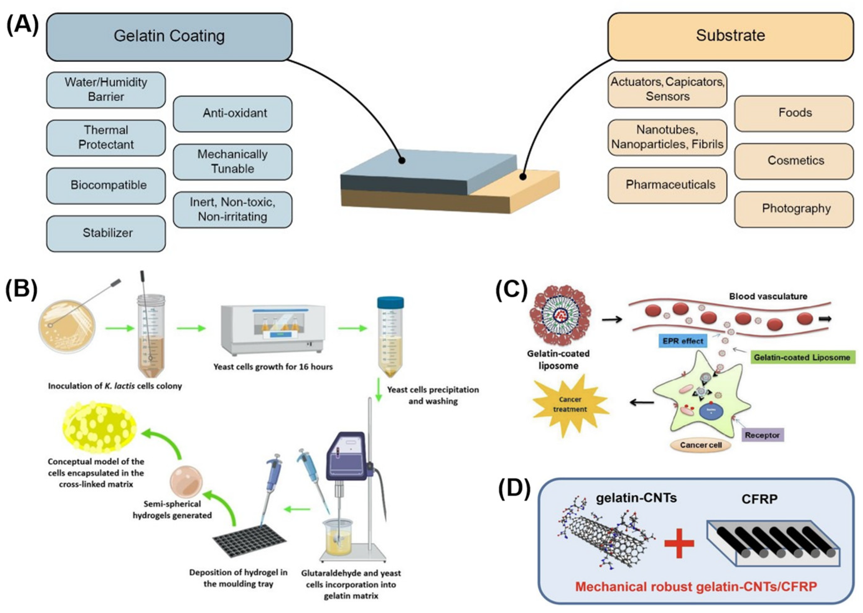
  7. Battogtokh, G.; Joo, Y.; Abuzar, S.M.; Park, H.; Hwang, S.-J. Gelatin Coating for the Improvement of Stability and Cell Uptake of Hydrophobic Drug-Containing Liposomes. Molecules 2022, 27, 1041. [Google Scholar] [CrossRef]
  8. Al-Nimry, S.; Dayah, A.; Hasan, I.; Daghmash, R. Cosmetic, Biomedical and Pharmaceutical Applications of Fish Gelatin/Hydrolysates. Mar. Drugs 2021, 19, 145. [Google Scholar] [CrossRef]
  9. Calixto, S.; Ganzherli, N.; Gulyaev, S.; Figueroa-Gerstenmaier, S. Gelatin as a Photosensitive Material. Molecules 2018, 23, 2064. [Google Scholar] [CrossRef] [Green Version]
  10. Liu, J.; Li, Y.; Hu, D.; Chao, X.; Zhou, Y.; Wang, J. An Essential Role of Gelatin in the Formation Process of Curling in Long Historical Photos. Polymers 2021, 13, 3894. [Google Scholar] [CrossRef]
  11. Chen, Z.; Shi, X.; Xu, J.; Du, Y.; Yao, M.; Guo, S. Gel properties of SPI modified by enzymatic cross-linking during frozen storage. Food Hydrocoll. 2016, 56, 445–452. [Google Scholar] [CrossRef]
  12. Saxena, A.; Sachin, K.; Bohidar, H.; Verma, A.K. Effect of molecular weight heterogeneity on drug encapsulation efficiency of gelatin nano-particles. Colloids Surf. B Biointerfaces 2005, 45, 42–48. [Google Scholar] [CrossRef]
  13. Santoro, M.; Tatara, A.M.; Mikos, A.G. Gelatin Carriers for Drug and Cell Delivery in Tissue Engineering. J. Control. Release 2014, 190, 210–218. [Google Scholar] [CrossRef] [Green Version]
  14. Ndlovu, S.P.; Ngece, K.; Alven, S.; Aderibigbe, B.A. Gelatin-Based Hybrid Scaffolds: Promising Wound Dressings. Polymers 2021, 13, 2959. [Google Scholar] [CrossRef]
  15. Sandrasegaran, K.; Lall, C.; Rajesh, A.; Maglinte, D.T. Distinguishing Gelatin Bioabsorbable Sponge and Postoperative Abdominal Abscess on CT. Am. J. Roentgenol. 2005, 184, 475–480. [Google Scholar] [CrossRef]
  16. Saw, M.M.; Chandler, B.; Ho, K.M. Benefits and Risks of Using Gelatin Solution as a Plasma Expander for Perioperative and Critically Ill Patients: A Meta-Analysis. Anaesth. Intensive Care 2012, 40, 17–32. [Google Scholar] [CrossRef] [Green Version]
  17. Ichwan, A.M.; Karimi, M.; Dash, A.K. Use of gelatin–acacia coacervate containing benzocaine in topical formulations. J. Pharm. Sci. 1999, 88, 763–766. [Google Scholar] [CrossRef]
  18. Wang, T.; Zhu, X.-K.; Xue, X.-T.; Wu, D.-Y. Hydrogel sheets of chitosan, honey and gelatin as burn wound dressings. Carbohydr. Polym. 2012, 88, 75–83. [Google Scholar] [CrossRef]
  19. Luo, Z.; Sun, W.; Fang, J.; Lee, K.; Li, S.; Gu, Z.; Dokmeci, M.R.; Khademhosseini, A. Biodegradable Gelatin Methacryloyl Microneedles for Transdermal Drug Delivery. Adv. Health Mater. 2018, 8, e1801054. [Google Scholar] [CrossRef]
  20. Echave, M.C.; Pimenta-Lopes, C.; Pedraz, J.L.; Mehrali, M.; Dolatshahi-Pirouz, A.; Ventura, F.; Orive, G. Enzymatic crosslinked gelatin 3D scaffolds for bone tissue engineering. Int. J. Pharm. 2019, 562, 151–161. [Google Scholar] [CrossRef] [Green Version]
  21. Singh, D.; Tripathi, A.; Zo, S.; Singh, D.; Han, S.S. Synthesis of composite gelatin-hyaluronic acid-alginate porous scaffold and evaluation for in vitro stem cell growth and in vivo tissue integration. Colloids Surf. B Biointerfaces 2014, 116, 502–509. [Google Scholar] [CrossRef]
  22. Hanzly, L.E.; Kristofferson, K.A.; Chauhan, N.; Barone, J.R. Biologically Controlled Gelatin Actuators. Green Mater. 2021, 9, 157–166. [Google Scholar] [CrossRef]
  23. Shintake, J.; Sonar, H.; Piskarev, E.; Paik, J.; Floreano, D. Soft Pneumatic Gelatin Actuator for Edible Robotics. In Proceedings of the 2017 IEEE/RSJ International Conference on Intelligent Robots and Systems (IROS), Vancouver, BC, Canada, 24–28 September 2017; pp. 6221–6226. [Google Scholar]
  24. Wang, H.; Xiang, J.; Wen, X.; Du, X.; Wang, Y.; Du, Z.; Cheng, X.; Wang, S. Multifunctional skin-inspired resilient MXene-embedded nanocomposite hydrogels for wireless wearable electronics. Compos. Part A Appl. Sci. Manuf. 2022, 155, 106835. [Google Scholar] [CrossRef]
  25. Wang, X.; Bai, Z.; Zheng, M.; Yue, O.; Hou, M.; Cui, B.; Su, R.; Wei, C.; Liu, X. Engineered gelatin-based conductive hydrogels for flexible wearable electronic devices: Fundamentals and recent advances. J. Sci. Adv. Mater. Devices 2022, 7, 100451. [Google Scholar] [CrossRef]
  26. Ebara, M.; Kotsuchibashi, Y.; Uto, K.; Aoyagi, T.; Kim, Y.-J.; Narain, R.; Idota, N.; Hoffman, J.M. Smart Hydrogels. In Smart Biomaterials; Springer: Tokyo, Japan, 2014; pp. 9–65. [Google Scholar]
  27. Nakayama, A.; Kakugo, A.; Gong, J.P.; Osada, Y.; Takai, M.; Erata, T.; Kawano, S. High Mechanical Strength Double-Network Hydrogel with Bacterial Cellulose. Adv. Funct. Mater. 2004, 14, 1124–1128. [Google Scholar] [CrossRef]
  28. Hardman, D.; Thuruthel, T.G.; Iida, F. Self-healing ionic gelatin/glycerol hydrogels for strain sensing applications. NPG Asia Mater. 2022, 14, 11. [Google Scholar] [CrossRef]
  29. Lai, J.-Y. Biocompatibility of chemically cross-linked gelatin hydrogels for ophthalmic use. J. Mater. Sci. Mater. Med. 2010, 21, 1899–1911. [Google Scholar] [CrossRef]
  30. Stevens, K.R.; Einerson, N.J.; Burmania, J.A.; Kao, W.J. In vivo biocompatibility of gelatin-based hydrogels and interpenetrating networks. J. Biomater. Sci. Polym. Ed. 2002, 13, 1353–1366. [Google Scholar] [CrossRef]
  31. Wu, S.-C.; Chang, W.-H.; Dong, G.-C.; Chen, K.-Y.; Chen, Y.-S.; Yao, C.-H. Cell Adhesion and Pro-liferation Enhancement by Gelatin Nanofiber Scaffolds. J. Bioact. Compat. Polym. 2011, 26, 565–577. [Google Scholar] [CrossRef]
  32. Gattazzo, F.; De Maria, C.; Rimessi, A.; Donà, S.; Braghetta, P.; Pinton, P.; Vozzi, G.; Bonaldo, P. Gelatin-Genipin-Based Biomaterials for Skeletal Muscle Tissue Engineering: Gelatin-Genipin-Based Biomaterials. J. Biomed. Mater. Res. 2018, 106, 2763–2777. [Google Scholar] [CrossRef]
  33. Sadeghi, A.H.; Shin, S.R.; Deddens, J.C.; Fratta, G.; Mandla, S.; Yazdi, I.K.; Prakash, G.; Antona, S.; Demarchi, D.; Buijsrogge, M.P.; et al. Engineered 3D Cardiac Fibrotic Tissue to Study Fibrotic Remodeling. Adv. Health Mater. 2017, 6, 1601434. [Google Scholar] [CrossRef]
  34. Sundaram, C.P.; Keenan, A.C. Evolution of hemostatic agents in surgical practice. Indian J. Urol. 2010, 26, 374–378. [Google Scholar] [CrossRef]
  35. Zolfagharian, A.; Mahmud, M.A.P.; Gharaie, S.; Bodaghi, M.; Kouzani, A.Z.; Kaynak, A. 3D/4D-Printed Bending-Type Soft Pneumatic Actuators: Fabrication, Modelling, and Control. Virtual Phys. Prototyp. 2020, 15, 373–402. [Google Scholar] [CrossRef]
  36. Xavier, M.S.; Tawk, C.D.; Zolfagharian, A.; Pinskier, J.; Howard, D.; Young, T.; Lai, J.; Harrison, S.M.; Yong, Y.K.; Bodaghi, M.; et al. Soft Pneumatic Actuators: A Review of Design, Fabrication, Modeling, Sensing, Control and Applications. IEEE Access 2022, 10, 59442–59485. [Google Scholar] [CrossRef]
  37. Xavier, M.S.; Fleming, A.J.; Yong, Y.K. Finite Element Modeling of Soft Fluidic Actuators: Overview and Recent Developments. Adv. Intell. Syst. 2021, 3, 2000187. [Google Scholar] [CrossRef]
  38. Czerner, M.; Fellay, L.S.; Suárez, M.P.; Frontini, P.M.; Fasce, L.A. Determination of Elastic Modulus of Gelatin Gels by Indentation Experiments. Procedia Mater. Sci. 2015, 8, 287–296. [Google Scholar] [CrossRef] [Green Version]
  39. Bracq, A.; Haugou, G.; Bourel, B.; Maréchal, C.; Lauro, F.; Roth, S.; Mauzac, O. On the modeling of a visco-hyperelastic polymer gel under blunt ballistic impacts. Int. J. Impact Eng. 2018, 118, 78–90. [Google Scholar] [CrossRef]
  40. Bello, J.; Bello, H.R.; Vinograd, J.R. The mechanism of gelation of gelatin the influence of pH, concentration, time and dilute electrolyte on the gelation of gelatin and modified gelatins. Biochim. Biophys. Acta 1962, 57, 214–221. [Google Scholar] [CrossRef]
  41. Riedel, S.; Heyart, B.; Apel, K.S.; Mayr, S.G. Programing stimuli-responsiveness of gelatin with electron beams: Basic effects and development of a hydration-controlled biocompatible demonstrator. Sci. Rep. 2017, 7, 17436. [Google Scholar] [CrossRef] [Green Version]
  42. Sha, X.-M.; Hu, Z.-Z.; Ye, Y.-H.; Xu, H.; Tu, Z.-C. Effect of extraction temperature on the gelling properties and identification of porcine gelatin. Food Hydrocoll. 2019, 92, 163–172. [Google Scholar] [CrossRef]
  43. Liu, Y.; Sun, X.-C.; Lv, C.; Xia, H. Green nanoarchitectonics with PEDOT:PSS–gelatin composite for moisture-responsive actuator and generator. Smart Mater. Struct. 2021, 30, 125014. [Google Scholar] [CrossRef]
  44. Chungyampin, S.; Niamlang, S. The Soft and High Actuation Response of Graphene Oxide/Gelatin Soft Gel. Materials 2021, 14, 7553. [Google Scholar] [CrossRef]
  45. Helminger, M.; Wu, B.; Kollmann, T.; Benke, D.; Schwahn, D.; Pipich, V.; Faivre, D.; Zahn, D.; Cölfen, H. Synthesis and Characterization of Gelatin-Based Magnetic Hydrogels. Adv. Funct. Mater. 2014, 24, 3187–3196. [Google Scholar] [CrossRef] [PubMed] [Green Version]
  46. Wu, Z.; Lin, X.; Zou, X.; Sun, J.; He, Q. Biodegradable Protein-Based Rockets for Drug Transportation and Light-Triggered Release. ACS Appl. Mater. Interfaces 2015, 7, 250–255. [Google Scholar] [CrossRef] [PubMed]
  47. Chen, Z.; Huang, G.; Trase, I.; Han, X.; Mei, Y. Mechanical Self-Assembly of a Strain-Engineered Flexible Layer: Wrinkling, Rolling, and Twisting. Phys. Rev. Appl. 2016, 5, 017001. [Google Scholar] [CrossRef] [Green Version]
  48. Hughes, J.; Rus, D. Mechanically Programmable, Degradable & Ingestible Soft Actuators. In Proceedings of the 3rd IEEE International Conference on Soft Robotics (RoboSoft), New Haven, CT, USA, 15 May–15 July 2020; pp. 836–843. [Google Scholar]
  49. Heiden, A.; Preninger, D.; Lehner, L.; Baumgartner, M.; Drack, M.; Woritzka, E.; Schiller, D.; Gerstmayr, R.; Hartmann, F.; Kaltenbrunner, M. 3D Printing of Resilient Biogels for Omnidirectional and Exteroceptive Soft Actuators. Sci. Robot. 2022, 7, eabk2119. [Google Scholar] [CrossRef] [PubMed]
  50. Tawk, C.; Alici, G. A Review of 3D-Printable Soft Pneumatic Actuators and Sensors: Research Challenges and Opportunities. Adv. Intell. Syst. 2021, 3, 2000223. [Google Scholar] [CrossRef]
  51. Ehrenhofer, A.; Elstner, M.; Wallmersperger, T. Normalization of hydrogel swelling behavior for sensoric and actuatoric applications. Sens. Actuators B Chem. 2018, 255, 1343–1353. [Google Scholar] [CrossRef]
  52. Tattanon, T.; Pongprayoon, T.; Arpornmaeklong, P.; Ummartyotin, S. Development of Hydroxy-apatite from Cuttlebone and Gelatin-Based Hydrogel Composite for Medical Materials. J. Polym. Res. 2022, 29, 364. [Google Scholar] [CrossRef]
  53. Ooi, S.Y.; Ahmad, I.; Amin, M.C.I.M. Cellulose nanocrystals extracted from rice husks as a reinforcing material in gelatin hydrogels for use in controlled drug delivery systems. Ind. Crop. Prod. 2016, 93, 227–234. [Google Scholar] [CrossRef]
  54. Shubhra, Q.T.H. Gelatin Film and Fiber Reinforced Gelatin Composites. Gelatin: Production, Applications and Health Implications; Boran, G., Ed.; Nova Publishers: New York, NY, USA, 2013. [Google Scholar]
  55. Qiao, C.; Cao, X.; Wang, F. Swelling Behavior Study of Physically Crosslinked Gelatin Hydrogels. Polym. Polym. Compos. 2012, 20, 53–58. [Google Scholar] [CrossRef]
  56. Hajikarimi, A.; Sadeghi, M. Free radical synthesis of cross-linking gelatin base poly NVP/acrylic acid hydrogel and nanoclay hydrogel as cephalexin drug deliver. J. Polym. Res. 2020, 27, 57. [Google Scholar] [CrossRef]
  57. Barthélémy, F.; Santoso, J.W.; Rabichow, L.; Jin, R.; Little, I.; Nelson, S.F.; McCain, M.L.; Miceli, M.C. Modeling Patient-Specific Muscular Dystrophy Phenotypes and Therapeutic Responses in Reprogrammed Myotubes Engineered on Micromolded Gelatin Hydrogels. Front. Cell Dev. Biol. 2022, 10, 830415. [Google Scholar] [CrossRef]
  58. Gupta, D.; Santoso, J.; McCain, M. Characterization of Gelatin Hydrogels Cross-Linked with Microbial Transglutaminase as Engineered Skeletal Muscle Substrates. Bioengineering 2021, 8, 6. [Google Scholar] [CrossRef]
  59. Besser, R.R.; Bowles, A.C.; Alassaf, A.; Carbonero, D.; Claure, I.; Jones, E.; Reda, J.; Wubker, L.; Batchelor, W.; Ziebarth, N.; et al. Enzymatically Cross-linked Gelatin–Laminin Hydrogels for Applications in Neuromuscular Tissue Engineering. Biomater. Sci. 2020, 8, 591–606. [Google Scholar] [CrossRef]
  60. Li, J.; Chee, H.L.; Chong, Y.T.; Chan, B.Q.Y.; Xue, K.; Lim, P.C.; Loh, X.J.; Wang, F. Hofmeister Effect Mediated Strong PHEMA-Gelatin Hydrogel Actuator. ACS Appl. Mater. Interfaces 2022, 14, 23826–23838. [Google Scholar] [CrossRef]
  61. Wang, Y.; Bamdad, F.; Song, Y.; Chen, L. Hydrogel Particles and Other Novel Protein-Based Methods for Food Ingredient and Nutraceutical Delivery Systems: Encapsulation Technologies and Delivery Systems for Food Ingredients and Nutraceuticals; Elsevier: Amsterdam, The Netherlands, 2012; pp. 412–450. [Google Scholar]
  62. Gomes, C.M.; Liu, C.; Paten, J.A.; Felton, S.M.; Deravi, L.F. Protein-Based Hydrogels That Actu-ate Self-Folding Systems. Adv. Funct. Mater. 2019, 29, 1805777. [Google Scholar] [CrossRef]
  63. Stroganov, V.; Al-Hussein, M.; Sommer, J.-U.; Janke, A.; Zakharchenko, S.; Ionov, L. Reversible Thermosensitive Biodegradable Polymeric Actuators Based on Confined Crystallization. Nano Lett. 2015, 15, 1786–1790. [Google Scholar] [CrossRef]
  64. Harris, H.; Radecka, A.; Malik, R.; Pineda Guzman, R.A.; Santoso, J.; Bradshaw, A.; McCain, M.; Kersh, M.; Golecki, H. Development and Characterization of Biostable Hydrogel Robotic Actuators for Implantable Devices: Tendon Actuated Gelatin. In Proceedings of the 2022 Design of Medical Devices Conference, American Society of Mechanical Engineers, Minneapolis, MN, USA, 11–14 April 2022; p. V001T05A004. [Google Scholar]
  65. Sardesai, A.N.; Segel, X.M.; Baumholtz, M.N.; Chen, Y.; Sun, R.; Schork, B.W.; Buonocore, R.; Wagner, K.O.; Golecki, H.M. Design and Characterization of Edible Soft Robotic Candy Actuators. MRS Adv. 2018, 3, 3003–3009. [Google Scholar] [CrossRef]
  66. Nagai, T.; Kurita, A.; Shintake, J. Characterization of Sustainable Robotic Materials and Finite Element Analysis of Soft Actuators Under Biodegradation. Front. Robot. AI 2021, 8, 760485. [Google Scholar] [CrossRef]
  67. Tungkavet, T.; Seetapan, N.; Pattavarakorn, D.; Sirivat, A. Graphene/Gelatin Hydrogel Compo-sites with High Storage Modulus Sensitivity for Using as Electroactive Actuator: Effects of Surface Area and Electric Field Strength. Polymer 2015, 70, 242–251. [Google Scholar] [CrossRef]
  68. Wang, W.; Wang, Z.; Liu, Y.; Li, N.; Wang, W.; Gao, J. Preparation of Reduced Graphene Ox-ide/Gelatin Composite Films with Reinforced Mechanical Strength. Mater. Res. Bull. 2012, 47, 2245–2251. [Google Scholar] [CrossRef]
  69. Chambers, L.D.; Winfield, J.; Ieropoulos, I.; Rossiter, J. Biodegradable and Edible Gelatine Actuators for Use as Artificial Muscles. In Electroactive Polymer Actuators and Devices (EAPAD); Bar-Cohen, Y., Ed.; SPIE: San Diego, CA, USA, 2014; p. 90560B. [Google Scholar]
  70. Shin, S.R.; Bolagh, B.A.G.; Dang, T.; Topkaya, S.N.; Gao, X.; Yang, S.Y.; Jung, S.M.; Oh, J.H.; Dokmeci, M.R.; Tang, X.; et al. Cell-laden Microengineered and Mechanically Tunable Hybrid Hydrogels of Gelatin and Graphene Oxide. Adv. Mater. 2013, 25, 6385–6391. [Google Scholar] [CrossRef] [PubMed]
  71. Haider, S.; Park, S.-Y.; Saeed, K.; Farmer, B. Swelling and electroresponsive characteristics of gelatin immobilized onto multi-walled carbon nanotubes. Sens. Actuators B Chem. 2007, 124, 517–528. [Google Scholar] [CrossRef]
  72. Bigi, A. Relationship between Triple-Helix Content and Mechanical Properties of Gelatin Films. Biomaterials 2004, 25, 5675–5680. [Google Scholar] [CrossRef] [PubMed]
  73. Firouzeh, A.; Salerno, M.; Paik, J. Soft Pneumatic Actuator with Adjustable Stiffness Layers for Multi-DoF Actuation. In Proceedings of the IEEE/RSJ International Conference on Intelligent Robots and Systems (IROS), Hamburg, Germany, 28 September 2015; pp. 1117–1124. [Google Scholar]
  74. Su, H.; Hou, X.; Zhang, X.; Qi, W.; Cai, S.; Xiong, X.; Guo, J. Pneumatic Soft Robots: Challenges and Benefits. Actuators 2022, 11, 92. [Google Scholar] [CrossRef]
  75. Na, H.; Kang, Y.-W.; Park, C.S.; Jung, S.; Kim, H.-Y.; Sun, J.-Y. Hydrogel-based strong and fast actuators by electroosmotic turgor pressure. Science 2022, 376, 301–307. [Google Scholar] [CrossRef]
  76. Dick, W.B. Cements and Uniting Bodies, Part 3. In Encyclopedia of Practical Receipts and Processes; Dick and Fitzgerald Publishers: New York, NY, USA, 1872. [Google Scholar]
  77. Mosadegh, B.; Polygerinos, P.; Keplinger, C.; Wennstedt, S.; Shepherd, R.; Gupta, U.; Shim, J.; Bertoldi, K.; Walsh, C.J.; Whitesides, G.M. Pneumatic Networks for Soft Robotics that Actuate Rapidly. Adv. Funct. Mater. 2014, 24, 2163–2170. [Google Scholar] [CrossRef] [Green Version]
  78. Greer, A.H.; King, E.; Lee, E.H.; Sardesai, A.N.; Chen, Y.; Obuz, S.E.; Graf, Y.; Ma, T.; Chow, D.Y.; Fu, T.; et al. Soluble Polymer Pneumatic Networks and a Single-Pour System for Improved Accessibility and Durability of Soft Robotic Actuators. Soft Robot. 2021, 8, 144–151. [Google Scholar] [CrossRef]
  79. Elhi, F.; Karu, K.; Rinne, P.; Nadel, K.-A.; Järvekülg, M.; Aabloo, A.; Tamm, T.; Ivaništšev, V.; Põhako-Esko, K. Understanding the Behavior of Fully Non-Toxic Polypyrrole-Gelatin and Polypyrrole-PVdF Soft Actuators with Choline Ionic Liquids. Actuators 2020, 9, 40. [Google Scholar] [CrossRef]
  80. Wan, C.; Frydrych, M.; Chen, B. Strong and Bioactive Gelatin–Graphene Oxide Nanocomposites. Soft Matter 2011, 7, 6159. [Google Scholar] [CrossRef]
  81. Choe, G.; Tang, X.; Wang, R.; Wu, K.; Jin Jeong, Y.; Kyu An, T.; Hyun Kim, S.; Mi, L. Printing of Self-Healable Gelatin Conductors Engineered for Improving Physical and Electrical Functions: Exploring Potential Application in Soft Actuators and Sensors. J. Ind. Eng. Chem. 2022, 116, 171–179. [Google Scholar] [CrossRef]
  82. Li, D.; Fan, D.; Zhu, R.; Lei, Q.; Liao, Y.; Yang, X.; Pan, Y.; Wang, Z.; Wu, Y.; Liu, S.; et al. Origami-Inspired Soft Twisting Actuator. Soft Robot. 2021. [Google Scholar] [CrossRef]
  83. Roche, E.T.; Wohlfarth, R.; Overvelde, J.T.B.; Vasilyev, N.V.; Pigula, F.A.; Mooney, D.J.; Bertoldi, K.; Walsh, C.J. A Bioinspired Soft Actuated Material. Adv. Mater. 2014, 26, 1200–1206. [Google Scholar] [CrossRef]
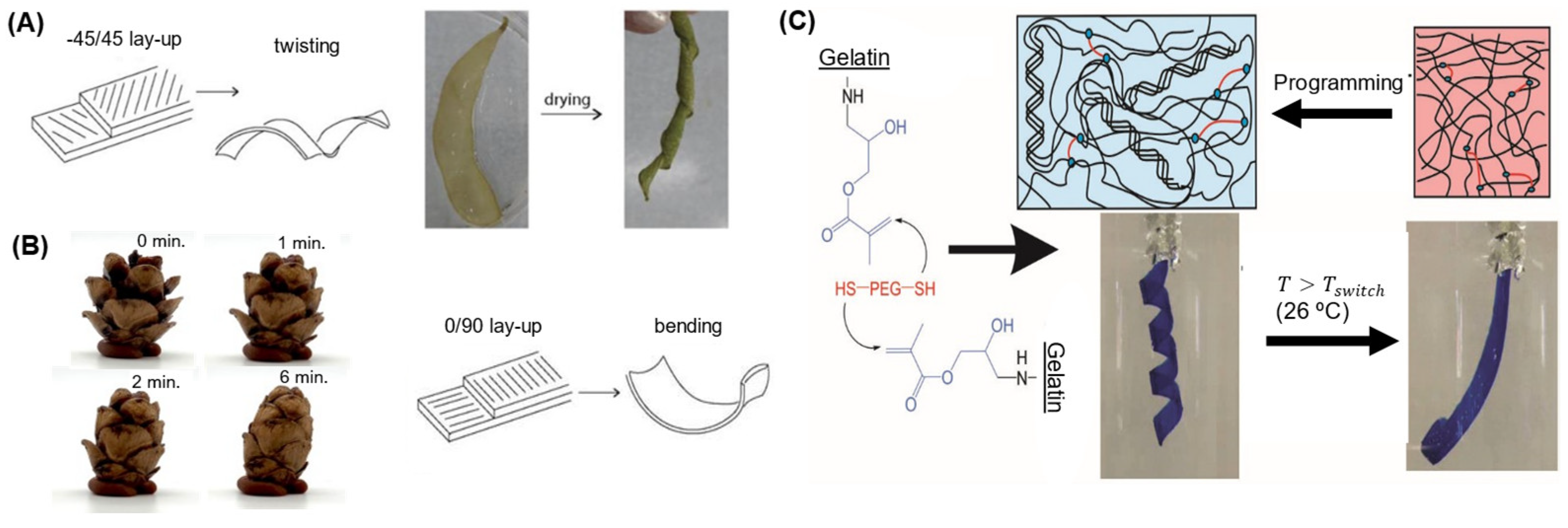
  84. Erb, R.M.; Sander, J.S.; Grisch, R.; Studart, A.R. Self-shaping composites with programmable bioinspired microstructures. Nat. Commun. 2013, 4, 1712. [Google Scholar] [CrossRef] [Green Version]
  85. Lutz-Bueno, V.; Bolisetty, S.; Azzari, P.; Handschin, S.; Mezzenga, R. Self-Winding Gelatin–Amyloid Wires for Soft Actuators and Sensors. Adv. Mater. 2020, 32, 2004941. [Google Scholar] [CrossRef]
  86. Zhang, Y.; Le Ferrand, H. Bioinspired Self-Shaping Clay Composites for Sustainable Development. Biomimetics 2022, 7, 13. [Google Scholar] [CrossRef]
  87. Neffe, A.; Löwenberg, C.; Julich-Gruner, K.; Behl, M.; Lendlein, A. Thermally-Induced Shape-Memory Behavior of Degradable Gelatin-Based Networks. Int. J. Mol. Sci. 2021, 22, 15892. [Google Scholar] [CrossRef]
  88. Huang, Z.; Wei, C.; Dong, L.; Wang, A.; Yao, H.; Guo, Z.; Mi, S. Fluid-driven hydrogel actuators with an origami structure. Iscience 2022, 25, 104674. [Google Scholar] [CrossRef]
  89. Sasaki, N.; Umeda, H.; Okada, S.; Kojima, R.; Fukuda, A. Mechanical Properties of Hydroxyapatite-Reinforced Gelatin as a Model System of Bone. Biomaterials 1989, 10, 129–132. [Google Scholar] [CrossRef]
  90. Lin, F.-H.; Yao, C.-H.; Sun, J.-S.; Liu, H.-C.; Huang, C.-W. Biological effects and cytotoxicity of the composite composed by tricalcium phosphate and glutaraldehyde cross-linked gelatin. Biomaterials 1998, 19, 905–917. [Google Scholar] [CrossRef]
  91. Avila-Ramirez, A.; Catzim-Ríos, K.; Guerrero-Beltrán, C.E.; Ramírez-Cedillo, E.; Ortega-Lara, W. Reinforcement of Alginate-Gelatin Hydrogels with Bioceramics for Biomedical Applications: A Comparative Study. Gels 2021, 7, 184. [Google Scholar] [CrossRef]
  92. Bhowmik, S.; Islam, J.; Debnath, T.; Miah, M.; Bhattacharjee, S.; Khan, M. Reinforcement of Gelatin-Based Nanofilled Polymer Biocomposite by Crystalline Cellulose from Cotton for Advanced Wound Dressing Applications. Polymers 2017, 9, 222. [Google Scholar] [CrossRef] [PubMed] [Green Version]
  93. Alves, J.S.; dos Reis, K.C.; Menezes EG, T.; Pereira, F.V.; Pereira, J. Effect of Cellulose Nano-crystals and Gelatin in Corn Starch Plasticized Films. Carbohydr. Polym. 2015, 115, 215–222. [Google Scholar] [CrossRef] [PubMed] [Green Version]
  94. Dogaru, B.-I.; Stoleru, V.; Mihalache, G.; Yonsel, S.; Popescu, M.-C. Gelatin Reinforced with CNCs as Nanocomposite Matrix for Trichoderma harzianum KUEN 1585 Spores in Seed Coatings. Molecules 2021, 26, 5755. [Google Scholar] [CrossRef] [PubMed]
  95. Panzavolta, S.; Bracci, B.; Gualandi, C.; Focarete, M.L.; Treossi, E.; Kouroupis-Agalou, K.; Rubini, K.; Bosia, F.; Brely, L.; Pugno, N.M.; et al. Structural reinforcement and failure analysis in composite nanofibers of graphene oxide and gelatin. Carbon 2014, 78, 566–577. [Google Scholar] [CrossRef] [Green Version]
  96. Shubhra, Q.T.H.; Alam, A.M.; Khan, M.A.; Saha, M.; Saha, D.; Khan, J.A.; Quaiyyum, M.A. The Preparation and Characterization of Silk/Gelatin Biocomposites. Polym. Technol. Eng. 2010, 49, 983–990. [Google Scholar] [CrossRef]
  97. Shubhra, Q.T.; Alam, A.; Beg, M.D.H. Mechanical and degradation characteristics of natural silk fiber reinforced gelatin composites. Mater. Lett. 2011, 65, 333–336. [Google Scholar] [CrossRef]
  98. Liu, J.; Liu, C.-K.; Brown, E. Development and Characterization of Genipin Cross-Linked Gelatin Based Composites Incorporated with Vegetable-Tanned Collagen Fiber (VCF). J. Am. Leather Chem. Assoc. 2017, 112, 410–419. [Google Scholar]
  99. Wei, S.-M.; Pei, M.-Y.; Pan, W.-L.; Thissen, H.; Tsai, S.-W. Gelatin Hydrogels Reinforced by Absorbable Nanoparticles and Fibrils Cured In Situ by Visible Light for Tissue Adhesive Applications. Polymers 2020, 12, 1113. [Google Scholar] [CrossRef]
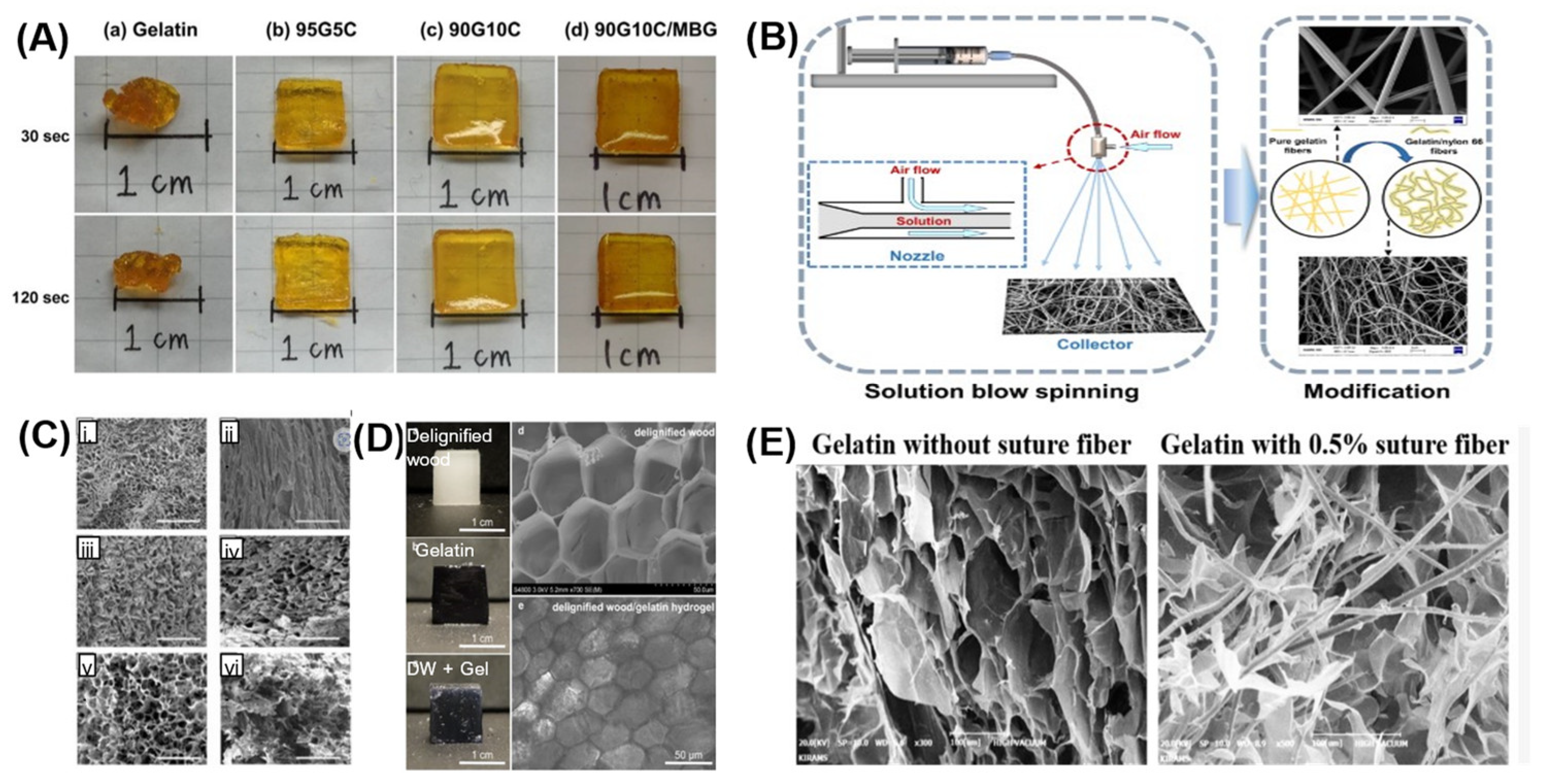
  100. Yang, Z.; Shen, C.; Zou, Y.; Wu, D.; Zhang, H.; Chen, K. Application of Solution Blow Spinning for Rapid Fabrication of Gelatin/Nylon 66 Nanofibrous Film. Foods 2021, 10, 2339. [Google Scholar] [CrossRef]
  101. Campodoni, E.; Montanari, M.; Dozio, S.M.; Heggset, E.B.; Panseri, S.; Montesi, M.; Tampieri, A.; Syverud, K.; Sandri, M. Blending Gelatin and Cellulose Nanofibrils: Biocomposites with Tunable Degradabil-ity and Mechanical Behavior. Nanomaterials 2020, 10, 1219. [Google Scholar] [CrossRef]
  102. Wang, S.; Li, K.; Zhou, Q. High strength and low swelling composite hydrogels from gelatin and delignified wood. Sci. Rep. 2020, 10, 17842. [Google Scholar] [CrossRef]
  103. Choi, D.J.; Choi, K.; Park, S.J.; Kim, Y.-J.; Chung, S.; Kim, C.-H. Suture Fiber Reinforcement of a 3D Printed Gelatin Scaffold for Its Potential Application in Soft Tissue Engineering. Int. J. Mol. Sci. 2021, 22, 11600. [Google Scholar] [CrossRef]
  104. Singh, C.; Wong, C.S.; Wang, X. Medical Textiles as Vascular Implants and Their Success to Mimic Natural Arteries. J. Funct. Biomater. 2015, 6, 500–525. [Google Scholar] [CrossRef] [Green Version]
  105. Hiob, M.A.; She, S.; Muiznieks, L.D.; Weiss, A.S. Biomaterials and Modifications in the Development of Small-Diameter Vascular Grafts. ACS Biomater. Sci. Eng. 2017, 3, 712–723. [Google Scholar] [CrossRef]
  106. Kuijpers, A.; van Wachem, P.; van Luyn, M.; Engbers, G.; Krijgsveld, J.; Zaat, S.; Dankert, J.; Feijen, J. In vivo and in vitro release of lysozyme from cross-linked gelatin hydrogels: A model system for the delivery of antibacterial proteins from prosthetic heart valves. J. Control. Release 2000, 67, 323–336. [Google Scholar] [CrossRef]
  107. Hassan, M.L.; Fadel, S.M.; El-Wakil, N.A.; Oksman, K. Chitosan/Rice Straw Nanofibers Nano-composites: Preparation, Mechanical, and Dynamic Thermomechanical Properties. J. Appl. Polym. Sci. 2012, 125, E216–E222. [Google Scholar] [CrossRef]
  108. Jiang, Y.; Xv, X.; Liu, D.; Yang, Z.; Zhang, Q.; Shi, H.; Zhao, G.; Zhou, J. Preparation of cellulose nanofiber-reinforced gelatin hydrogel and optimization for 3D printing applications. Bioresources 2018, 13, 5909–5924. [Google Scholar] [CrossRef]
  109. Wang, W.; Zhang, X.; Teng, A.; Liu, A. Mechanical Reinforcement of Gelatin Hydrogel with Nanofiber Cellulose as a Function of Percolation Concentration. Int. J. Biol. Macromol. 2017, 103, 226–233. [Google Scholar] [CrossRef]
  110. Wan, Y.Z.; Wang, Y.L.; Luo, H.L.; Cheng, G.X.; Yao, K.D. Carbon Fiber-Reinforced Gelatin Composites. I. Preparation and Mechanical Properties. J. Appl. Polym. Sci. 2000, 75, 987–993. [Google Scholar] [CrossRef]
  111. Ravishankar, P.; Ozkizilcik, A.; Husain, A.; Balachandran, K. Anisotropic Fiber-Reinforced Gly-cosaminoglycan Hydrogels for Heart Valve Tissue Engineering. Tissue Eng. Part A 2021, 27, 513–525. [Google Scholar] [CrossRef]
  112. Khan, R.A.; Khan, M.A.; Sarker, B.; Saha, S.; Das, A.K.; Noor, N.; Huq, T.; Khan, A.; Dey, K.; Saha, M. Fabrication and Characterization of Gelatin Fiber-based Linear Low-density Polyethylene Foamed Composite. J. Reinf. Plast. Compos. 2010, 29, 2438–2449. [Google Scholar] [CrossRef]
  113. Torrejon, V.M.; Song, J.; Yu, Z.; Hang, S. Gelatin-Based Cellular Solids: Fabrication, Structure and Properties. J. Cell. Plast. 2022, 58, 797–858. [Google Scholar] [CrossRef]
  114. Gibson, L.J.; Ashby, M.F. Cellular Solids: Structure and Properties; Cambridge University Press: Cambridge, UK, 1997. [Google Scholar]
  115. Andrieux, S.; Quell, A.; Stubenrauch, C.; Drenckhan, W. Liquid foam templating—A route to tailor-made polymer foams. Adv. Colloid Interface Sci. 2018, 256, 276–290. [Google Scholar] [CrossRef]
  116. Frazier, S.D.; Aday, A.N.; Srubar, W. On-Demand Microwave-Assisted Fabrication of Gelatin Foams. Molecules 2018, 23, 1121. [Google Scholar] [CrossRef] [PubMed] [Green Version]
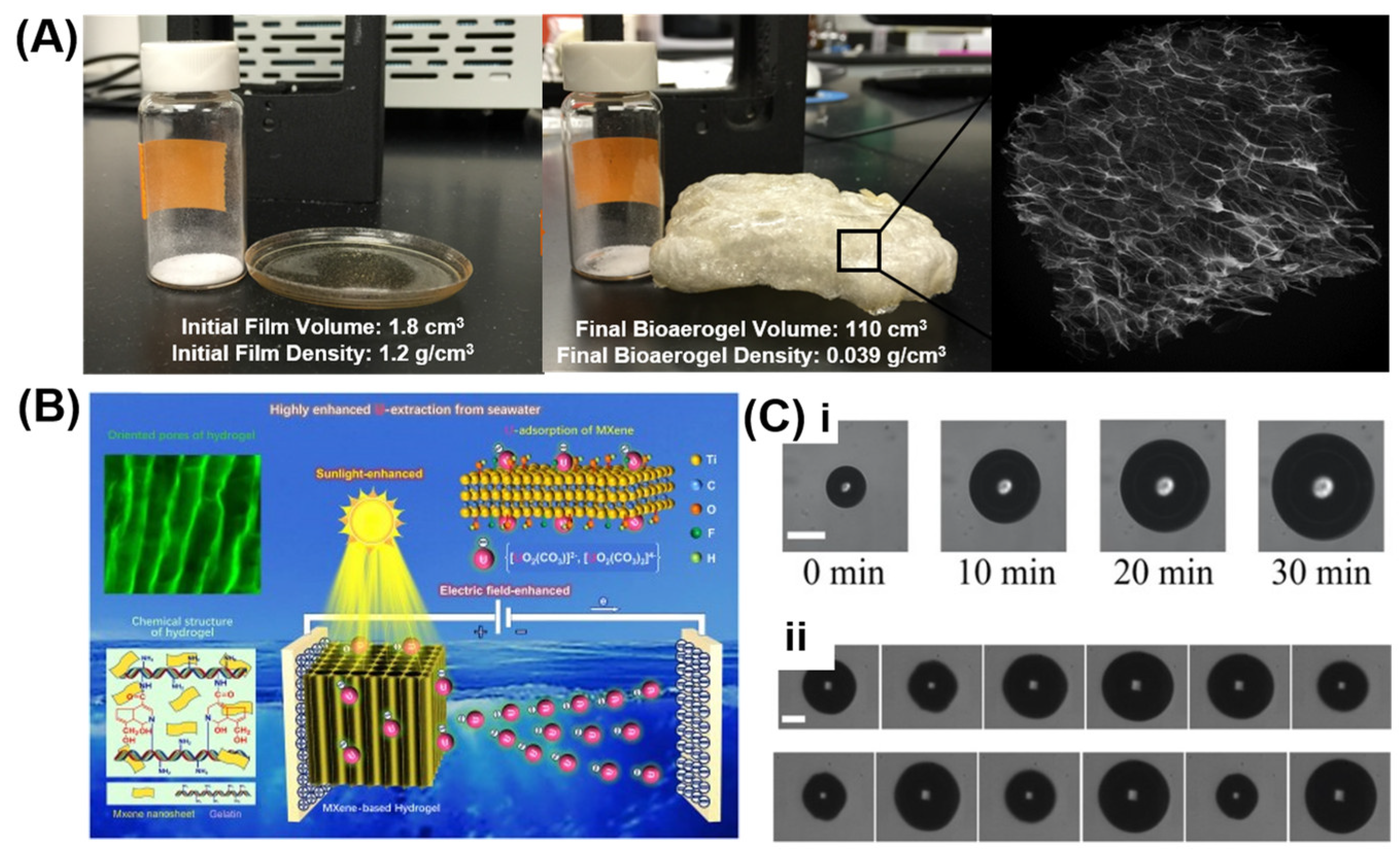
  117. Chen, L.; Sun, Y.; Wang, J.; Ma, C.; Peng, S.; Cao, X.; Yang, L.; Ma, C.; Duan, G.; Liu, Z.; et al. A wood-mimetic porous MXene/gelatin hydrogel for electric field/sunlight bi-enhanced uranium adsorption. E-Polymers 2022, 22, 468–477. [Google Scholar] [CrossRef]
  118. Murakami, K.; Yamakawa, Y.; Zhao, J.; Johnsen, E.; Ando, K. Ultrasound-Induced Nonlinear Oscillations of a Spherical Bubble in a Gelatin Gel. J. Fluid Mech. 2021, 924, A38. [Google Scholar] [CrossRef]
  119. Pezeshki Modaress, M.; Mirzadeh, H.; Zandi, M. Fabrication of a Porous Wall and Higher Inter-connectivity Scaffold Comprising Gelatin/Chitosan via Combination of Salt-Leaching and Lyophilization Methods. Iran. Polym. J. 2012, 21, 191–200. [Google Scholar] [CrossRef]
  120. Lu, B.; Wang, T.; Li, Z.; Dai, F.; Lv, L.; Tang, F.; Yu, K.; Liu, J.; Lan, G. Healing of skin wounds with a chitosan–gelatin sponge loaded with tannins and platelet-rich plasma. Int. J. Biol. Macromol. 2016, 82, 884–891. [Google Scholar] [CrossRef]
  121. La Gatta, A.; Tirino, V.; Cammarota, M.; La Noce, M.; Stellavato, A.; Pirozzi, A.V.A.; Portaccio, M.; Diano, N.; Laino, L.; Papaccio, G.; et al. Gelatin-Biofermentative Unsulfated Glycosaminoglycans Semi-Interpenetrating Hydrogels via Microbial-Transglutaminase Crosslinking Enhance Osteogenic Poten-tial of Dental Pulp Stem Cells. Regen. Biomater. 2021, 8, rbaa052. [Google Scholar] [CrossRef]
  122. Fu, Y.; Yao, J.; Zhao, H.; Zhao, G.; Wan, Z.; Guo, R. A muscle-like magnetorheological actuator based on bidisperse magnetic particles enhanced flexible alginate-gelatin sponges. Smart Mater. Struct. 2020, 29, 015019. [Google Scholar] [CrossRef]
  123. Ohl, S.-W.; Klaseboer, E.; Khoo, B.C. Bubbles with shock waves and ultrasound: A review. Interface Focus 2015, 5, 20150019. [Google Scholar] [CrossRef]
  124. Ohl, C.D.; Ikink, R. Shock-Wave-Induced Jetting of Micron-Size Bubbles. Phys. Rev. Lett. 2003, 90, 214502. [Google Scholar] [CrossRef] [Green Version]
  125. Ter Haar, G. Ultrasonic imaging: Safety considerations. Interface Focus 2011, 1, 686–697. [Google Scholar] [CrossRef]
  126. Liang, H.-D.; Noble, J.A.; Wells, P.N.T. Recent advances in biomedical ultrasonic imaging techniques. Interface Focus 2011, 1, 475–476. [Google Scholar] [CrossRef]
  127. Wong, Z.Z.; Kripfgans, O.D.; Qamar, A.; Fowlkes, J.B.; Bull, J.L. Bubble evolution in acoustic droplet vaporization at physiological temperature via ultra-high speed imaging. Soft Matter 2011, 7, 4009–4016. [Google Scholar] [CrossRef]
  128. Dear, J.P.; Field, J.E. A study of the collapse of arrays of cavities. J. Fluid Mech. 1988, 190, 409–425. [Google Scholar] [CrossRef]
  129. Dear, J.P.; Field, J.E.; Walton, A.J. Gas compression and jet formation in cavities collapsed by a shock wave. Nature 1988, 332, 505–508. [Google Scholar] [CrossRef]
  130. Bourne, N.K.; Field, J.E. Shock-induced collapse of single cavities in liquids. J. Fluid Mech. 1992, 244, 225–240. [Google Scholar] [CrossRef]
  131. Kodama, T.; Takayama, K. Dynamic behavior of bubbles during extracorporeal shock-wave lithotripsy. Ultrasound Med. Biol. 1998, 24, 723–738. [Google Scholar] [CrossRef]
  132. Hopfes, T.; Wang, Z.; Giglmaier, M.; Adams, N.A. Collapse Dynamics of Bubble Pairs in Gelati-nous Fluids. Exp. Therm. Fluid Sci. 2019, 108, 104–114. [Google Scholar] [CrossRef]
  133. Gillitzer, R.; Neisius, A.; Wöllner, J.; Hampel, C.; Brenner, W.; Bonilla, A.A.; Thüroff, J. Low-Frequency Extracorporeal Shock Wave Lithotripsy Improves Renal Pelvic Stone Disintegration in a Pig Model. BJU Int. 2009, 103, 1284–1288. [Google Scholar] [CrossRef] [PubMed]
  134. Saliu, O.D.; Mamo, M.; Ndungu, P.; Ramontja, J. The Making of a High Performance Supercapac-itor Active at Negative Potential Using Sulphonic Acid Activated Starch-Gelatin-TiO2 Nano-Hybrids. Arab. J. Chem. 2021, 14, 103242. [Google Scholar] [CrossRef]
  135. Fan, H.; Shen, W. Gelatin-Based Microporous Carbon Nanosheets as High Performance Superca-pacitor Electrodes. ACS Sustain. Chem. Eng. 2016, 4, 1328–1337. [Google Scholar] [CrossRef]
  136. Wang, S.; Han, L.; Liu, H.; Dong, Y.; Wang, X. Ionic Gelatin-Based Flexible Thermoelectric Gener-ator with Scalability for Human Body Heat Harvesting. Energies 2022, 15, 3441. [Google Scholar] [CrossRef]
  137. Wei, L.; Huang, J.; Yan, Y.; Cui, J.; Zhao, Y.; Bai, F.; Liu, J.; Wu, X.; Zhang, X.; Du, M. Substrate-Independent, Mechanically Tunable, and Scalable Gelatin Methacryloyl Hydrogel Coating with Drag-Reducing and Anti-Freezing Properties. ACS Appl. Polym. Mater. 2022, 4, 4876–4885. [Google Scholar] [CrossRef]
  138. Patarroyo, J.L.; Florez-Rojas, J.S.; Pradilla, D.; Valderrama-Rincón, J.D.; Cruz, J.C.; Reyes, L.H. Formulation and Characterization of Gelatin-Based Hydrogels for the Encapsulation of Kluyveromyces lactis—Applications in Packed-Bed Reactors and Probiotics Delivery in Humans. Polymers 2020, 12, 1287. [Google Scholar] [CrossRef]
  139. Zeng, L.; Tao, W.; Zhao, J.; Li, Y.; Li, R. Mechanical performance of a CFRP composite reinforced via gelatin-CNTs: A study on fiber interfacial enhancement and matrix enhancement. Nanotechnol. Rev. 2022, 11, 625–636. [Google Scholar] [CrossRef]
  140. Simsek, G.M.; Barthes, J.; Muller, C.; McGuinness, G.B.; Vrana, N.E.; Yapici, G.G. PVA/gelatin-based hydrogel coating of nickel-titanium alloy for improved tissue-implant interface. Appl. Phys. A 2021, 127, 387. [Google Scholar] [CrossRef]
  141. Tung, A.T.; Park, B.-H.; Liang, D.H.; Niemeyer, G. Laser-machined shape memory alloy sensors for position feedback in active catheters. Sens. Actuators A Phys. 2008, 147, 83–92. [Google Scholar] [CrossRef] [Green Version]
  142. Muhammad, N.; Whitehead, D.; Boor, A.; Oppenlander, W.; Liu, Z.; Li, L. Picosecond Laser Micromachining of Nitinol and Platinum–Iridium Alloy for Coronary Stent Applications. Appl. Phys. A 2012, 106, 607–617. [Google Scholar] [CrossRef]
  143. Tan, K.M.; Tay, C.M.; Tjin, S.C.; Chan, C.C.; Rahardjo, H. High Relative Humidity Measurements Using Gelatin Coated Long-Period Grating Sensors. Sens. Actuators B Chem. 2005, 110, 335–341. [Google Scholar] [CrossRef]
  144. Nur Hanani, Z.A.; Roos, Y.H.; Kerry, J.P. Use and Application of Gelatin as Potential Biodegradable Packaging Materials for Food Products. Int. J. Biol. Macromol. 2014, 71, 94–102. [Google Scholar] [CrossRef]
  145. Miao, C.; Hamad, W.Y. In-situ polymerized cellulose nanocrystals (CNC)—Poly(l-lactide) (PLLA) nanomaterials and applications in nanocomposite processing. Carbohydr. Polym. 2016, 153, 549–558. [Google Scholar] [CrossRef]
  146. Lee, C.; Kim, M.; Kim, Y.J.; Hong, N.; Ryu, S.; Kim, H.J.; Kim, S. Soft Robot Review. Int. J. Control Autom. Syst. 2017, 15, 3–15. [Google Scholar] [CrossRef]
  147. Zhou, Y.; Li, H. A Scientometric Review of Soft Robotics: Intellectual Structures and Emerging Trends Analysis (2010–2021). Front. Robot. AI 2022, 9, 868682. [Google Scholar] [CrossRef]
  148. Cianchetti, M.; Laschi, C.; Menciassi, A.; Dario, P. Biomedical applications of soft robotics. Nat. Rev. Mater. 2018, 3, 143–153. [Google Scholar] [CrossRef]
  149. Huang, W.; Shang, W.; Huang, Y.; Long, H.; Wu, X. Insect-Scale SMAW-Based Soft Robot With Crawling, Jumping, and Loading Locomotion. IEEE Robot. Autom. Lett. 2022, 7, 9287–9293. [Google Scholar] [CrossRef]
  150. Yang, X.; Shang, W.; Lu, H.; Liu, Y.; Yang, L.; Tan, R.; Wu, X.; Shen, Y. An agglutinate magnetic spray transforms inanimate objects into millirobots for biomedical applications. Sci. Robot. 2020, 5, eabc8191. [Google Scholar] [CrossRef]
  151. Bupphathong, S.; Quiroz, C.; Huang, W.; Chung, P.-F.; Tao, H.-Y.; Lin, C.-H. Gelatin Methacrylate Hydrogel for Tissue Engineering Applications—A Review on Material Modifications. Pharmaceuticals 2022, 15, 171. [Google Scholar] [CrossRef]
  152. Burdick, J.A.; Prestwich, G.D. Hyaluronic acid hydrogels for biomedical applications. Adv. Mater. 2011, 23, H41–H56. [Google Scholar] [CrossRef]
  153. Fernandez, P.; Bausch, A.R. The compaction of gels by cells: A case of collective mechanical activity. Integr. Biol. 2009, 1, 252–259. [Google Scholar] [CrossRef] [PubMed]
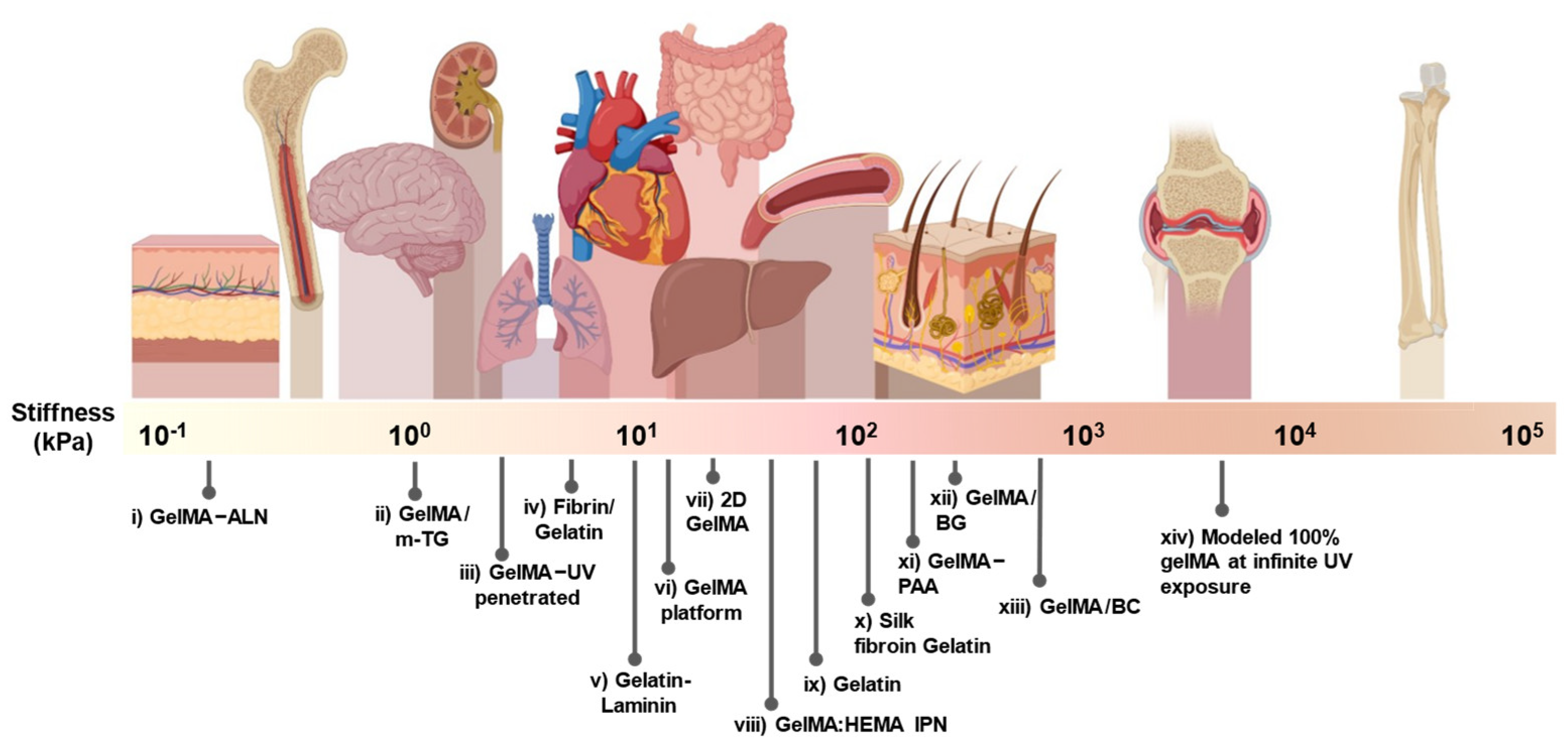
  154. Liu, L.; Li, X.; Shi, X.; Wang, Y. Injectable Alendronate-Functionalized GelMA Hydrogels for Mineralization and Osteogenesis. RSC Adv. 2018, 8, 22764–22776. [Google Scholar] [CrossRef] [PubMed] [Green Version]
  155. Kim, C.; Young, J.L.; Holle, A.W.; Jeong, K.; Major, L.G.; Jeong, J.H.; Aman, Z.; Han, D.-W.; Hwang, Y.; Spatz, J.P.; et al. Stem Cell Mechanosensation on Gelatin Methacryloyl (GelMA) Stiffness Gradient Hydrogels. Ann. Biomed. Eng. 2020, 48, 893–902. [Google Scholar] [CrossRef] [PubMed]
  156. Wachendörfer, M.; Schräder, P.; Buhl, E.M.; Palkowitz, A.L.; Ben Messaoud, G.; Richtering, W.; Fischer, H. A Defined Heat Pretreatment of Gelatin Enables Control of Hydrolytic Stability, Stiffness, and Microstructural Architecture of Fibrin–Gelatin Hydrogel Blends. Biomater. Sci. 2022, 10, 5552–5565. [Google Scholar] [CrossRef]
  157. Sun, Y.; Deng, R.; Ren, X.; Zhang, K.; Li, J. 2D Gelatin Methacrylate Hydrogels with Tunable Stiffness for Investigating Cell Behaviors. ACS Appl. Bio Mater. 2019, 2, 570–576. [Google Scholar] [CrossRef]
  158. Kilic Bektas, C.; Hasirci, V. Cell Loaded GelMA: HEMA IPN Hydrogels for Corneal Stroma Engineering. J. Mater. Sci. Mater. Med. 2020, 31, 2. [Google Scholar] [CrossRef]
  159. Karimi, A.; Navidbakhsh, M. Material Properties in Unconfined Compression of Gelatin Hydro-gel for Skin Tissue Engineering Applications. Biomed. Eng. Biomed. Tech. 2014, 59, 6. [Google Scholar]
  160. Singh, Y.P.; Bandyopadhyay, A.; Mandal, B.B. 3D Bioprinting Using Cross-Linker-Free Silk–Gelatin Bioink for Cartilage Tissue Engineering. ACS Appl. Mater. Interfaces 2019, 11, 33684–33696. [Google Scholar] [CrossRef]
  161. Serafim, A.; Tucureanu, C.; Petre, D.-G.; Dragusin, D.-M.; Salageanu, A.; Van Vlierberghe, S.; Dubruel, P.; Stancu, I.-C. One-Pot Synthesis of Superabsorbent Hybrid Hydrogels Based on Methacrylamide Gela-tin and Polyacrylamide. Effortless Control of Hydrogel Properties through Composition Design. New J. Chem. 2014, 38, 3112–3126. [Google Scholar] [CrossRef]
  162. Moghanian, A.; Portillo-Lara, R.; Sani, E.S.; Konisky, H.; Bassir, S.H.; Annabi, N. Synthesis and characterization of osteoinductive visible light-activated adhesive composites with antimicrobial properties. J. Tissue Eng. Regen. Med. 2020, 14, 66–81. [Google Scholar] [CrossRef]
  163. Gu, L.; Li, T.; Song, X.; Yang, X.; Li, S.; Chen, L.; Liu, P.; Gong, X.; Chen, C.; Sun, L. Preparation and Characterization of Methacrylated Gelatin/Bacterial Cellulose Composite Hydrogels for Cartilage Tissue En-gineering. Regen. Biomater. 2020, 7, 195–202. [Google Scholar] [CrossRef]
  164. Schuurman, W.; Levett, P.A.; Pot, M.W.; van Weeren, P.R.; Dhert, W.J.A.; Hutmacher, D.W.; Melchels, F.P.W.; Klein, T.J.; Malda, J. Gelatin-Methacrylamide Hydrogels as Potential Biomaterials for Fabrication of Tissue-Engineered Cartilage Constructs: Gelatin-Methacrylamide Hydrogels as Potential Bio-materials for Fabrication. Macromol. Biosci. 2013, 13, 551–561. [Google Scholar] [CrossRef]
Figure 1. Overview schematic of gelatin actuators.
Actuators 12 00063 g001
Figure 2. Bending actuators. (A) Mechanically actuated gelatin bending actuators with Nitinol wire and crosslinked with microbial transglutaminase ([64], original image). (B) pH-sensitive swelling bilayer gelatin actuator in water before and after deformation [41]. (Images (A,B) are licensed under CC BY 4.0 (http://creativecommons.org/licenses/by/4.0/ (accessed on 30 October 2022))).
Actuators 12 00063 g002
Figure 3. Pneumatically-stimulated bending actuators. (A) Biodegradable actuator under non-pressurized and pressurized states [66]. (B) PneuNet actuators made of gelatin and glycerol are used as grippers to apply a force that picks up objects [23,28]. (Images (A,B) are licensed under CC BY 4.0 (http://creativecommons.org/licenses/by/4.0/ (accessed on 30 October 2022))).
Actuators 12 00063 g003
Figure 4. Electromechanically-stimulated gelatin actuators. (A) The degree of deflection and the electrode distance of 4 cm for gelatin actuators or graphite/gelatin actuators with increasing electric field strength [44]. (B) Gelatin can also be used as a sensor. All five fingers of the glove are 3D printed with gelatin sensor networks and can return separate strain responses [28]. (Images (A,B) are licensed under CC BY 4.0 (http://creativecommons.org/licenses/by/4.0/ (accessed on 30 October 2022))).
Actuators 12 00063 g004
Figure 5. Twisting gelatin actuators and plant-inspired actuation. (A) Solvent actuated twisting actuation in edamame seedpods with clay composite twisting actuation scheme [86]. (B) Conifer pinecone bending motion when dried out to when fully hydrated (original images) with clay composite bending actuation scheme [86]. (C) Thermally stimulated shape memory gelatin hydrogels twisting actuators (Adapted) [87]. (Images (AC) are licensed under CC BY 4.0 (http://creativecommons.org/licenses/by/4.0/ (accessed on 30 October 2022))).
Actuators 12 00063 g005
Figure 7. Gelatin foam enhancement. (A) Microwave-assisted fabrication of gelatin foams before and after microwaving at 700 W for 30 secs, with a view of the foam structure [116]. (B) The chemical structure and electric field/sunlight highly enhanced the uranium-adsorbing mechanism of the wood-mimetic directional macro-porous MXene-based hydrogel [117]. (C) (i) Bubbles generated by focusing a laser pulse into a 6 wt% gelatin gel supersaturated with dissolved air and (ii) finite-amplitude spherical oscillations of a bubble in the 6 wt% gelatin gel under 28 kHz ultrasound irradiation; the scale bar represents 100 μm [118]. (Images (AC) are licensed under CC BY 4.0 (http://creativecommons.org/licenses/by/4.0/ (accessed on 30 October 2022))).
Actuators 12 00063 g007
Figure 8. Gelatin used for actuator coatings. (A) Schematic depicting the benefits of gelatin-based coatings and their applications. (B) The protocol followed for the encapsulation of probiotic cells in the gelatin-glutaraldehyde (GTA) matrix [138]. (C) Summary of the cellular uptake of gelatin-coated liposomes [7]. (D) Mechanical robust gelatin-CNTs/CFRP composite [139]. (Images (BD) are licensed under CC BY 4.0 (http://creativecommons.org/licenses/by/4.0/ (accessed on 30 October 2022))).
Actuators 12 00063 g008
Figure 9. Depiction of gelatin tunability by comparing its stiffnesses against different tissue stiffness. (i) Alendronate-functionalized GelMA [154], (ii) GelMA/m-TG [121], (iii) GelMA–UV penetrated [155], (iv) Fibrin/gelatin [156], (v) Enzyme crosslinked gelatin-laminin hybrid [33], (vi) GelMA based platforms [33], (vii) 2D Photo-crosslinked GelMA [157], (viii) GelMA and poly(2-hydrox-ethyl methacrylate) (pHEMA) interpenetrating network (IPN) hydrogels [158], (ix) Gelatin of different concentrations [159], (x) Silk fibroin-Gelatin Bio-ink [160], (xi) GELMA-polyacrylamide (PAA) [161], (xii) GelMA/doped bioactive glass (BG) [162], (xiii) GelMA/bacterial cellulose (BC) [163], and (xiv) Theoretical value modeled for 100% GelMA gel at infinite exposure time [164]. Created with BioRender.com (accessed on 30 October 2022).
Actuators 12 00063 g009
Disclaimer/Publisher’s Note: The statements, opinions and data contained in all publications are solely those of the individual author(s) and contributor(s) and not of MDPI and/or the editor(s). MDPI and/or the editor(s) disclaim responsibility for any injury to people or property resulting from any ideas, methods, instructions or products referred to in the content.

