Article

New Molecular Data on Filaria and its Wolbachia from Red Howler Monkeys (Alouatta macconnelli) in French Guiana—A Preliminary Study

by
Younes Laidoudi
1,2,
Hacène Medkour
1,2,
Anthony Levasseur
1,2,
Bernard Davoust
1,2 and
Oleg Mediannikov
1,2,*
1
IRD, AP-HM, Microbes, Evolution, Phylogeny and Infection (MEPHI), IHU Méditerranée Infection, 19–21, Bd Jean Moulin, 13005 Marseille, France
2
Aix Marseille Univ, 19–21, Bd Jean Moulin, 13005 Marseille, France
*
Author to whom correspondence should be addressed.
Pathogens 2020, 9(8), 626; https://doi.org/10.3390/pathogens9080626
Submission received: 10 June 2020 / Revised: 28 July 2020 / Accepted: 29 July 2020 / Published: 31 July 2020
(This article belongs to the Special Issue Animal Parasitic Diseases)

Abstract

:
Previous studies have reported filarial parasites of the genus Dipetalonema and Mansonella from French Guiana monkeys, based on morphological taxonomy. In this study, we screened blood samples from nine howler monkeys (Alouatta macconnelli) for the presence of filaria and Wolbachia DNA. The infection rates were 88.9% for filaria and 55.6% for wolbachiae. The molecular characterization, based on the 18S gene of filariids, revealed that A. macconnelli are infected with at least three species (Mansonella sp., Brugia sp. and an unidentified Onchocercidae species.). Since the 18S and cox1 generic primers are not very effective at resolving co-infections, we developed ITS genus-specific PCRs for Mansonella and Brugia genus. The results revealed coinfections in 75% of positives. The presence of Mansonella sp. and Brugia sp. was also confirmed by the 16S phylogenetic analysis of their associated Wolbachia. Mansonella sp., which close to the species from the subgenus Tetrapetalonema encountered in New World Monkeys, while Brugia sp. was identical to the strain circulating in French Guiana dogs. We propose a novel ITS1 Brugia genus-specific qPCR. We applied it to screen for Brugia infection in howler monkeys and 66.7% were found to be positive. Our finding highlights the need for further studies to clarify the species diversity of neotropics monkeys by combining molecular and morphological features. The novel Brugia genus-specific qPCR assays could be an effective tool for the surveillance and characterization of this potential zoonosis.

Graphical Abstract

1. Introduction

Filariasis unites diseases are caused by arthropod-borne filariids and nematodes belonging to the Onchocercidae family. Several species can be encountered in human and animals with some zoonotic aspects. Morphologically, the adult filariids are long, string-like, white-to-cream-colored worms [1]. They appear to be capable of living inside various tissues and cavities outside the gastrointestinal tract. Once mature, the adult females produce blood or cutaneous microfilariae, where they are available to arthropod vectors [2]. Species having a predilection for subcutaneous tissues are less or completely avirulent in comparison to those found in cavities, such as Dipetalonema species (D. gracile, D. graciliformis, D. caudispina, D. robini and D. freitasi, D. vanhoofi), Macacanema formosana where they induce serious disease manifestations such as pleuritis, fibrinopurulent peritonitis and fibrinous adhesion, resulting in the entrapment of worms [3,4]. Furthermore, species found in the circulatory system (e.g., Dirofilaria immitis and D. pongoi, Edesonfilaria malayensis), as well as those present in the lymphatic system, such as Brugian filariids (B. malayi, B. pahangi, B. timori and B. tupaiae) and Wuchereria bancrofti, disrupt blood and lymphatic drainage, leading to serious and often irreversible vascular damage [4,5,6,7,8,9]. These filariids, along with Onchocerca volvulus, the agent of river blindness, constitute the most thread-like filarial worms and have affected up to 893 million people in 49 countries worldwide [10].
Several filariids of the subfamilies Onchocercinae and Dirofilariinae are associated with an endosymbiotic intracellular bacterium of the genus Wolbachia [11], which is present in all developmental stages of filariids that harbor Wolbachia, leading to their long-term survival [12]. The parasites’ endosymbiotic Wolbachia are implicated in severe inflammatory-mediated filarial diseases [13,14,15,16]. Anti-wolbachial therapies, based on the administration of antibiotics, are known to be effective against the most common filariasis caused by Brugia spp., i.e., W. bancrofti, Mansonella perstans and D. immitis [17,18,19]. The Wolbachia-filaria relationship is species-specific, wherein each filariid has a specific genotype of Wolbachia [11], thus providing an additional target suitable for the diagnosis of filarial infections [20], especially when occurring in dead-end hosts, as is the case in D. immitis in human and cats [21,22]. Recently, the simultaneous detection of both filarial and wolbachial DNAs from infected hosts is used as an improvement tool for the diagnosis of filarial infections [23,24,25].
Filariasis is one of the most neglected tropical diseases selected, but it is included in the Mass Drug Administration (MDA) program to achieve its elimination by 2020 [26,27,28]. Human filariasis was almost eliminated from Latin America [29,30]. Thanks to the MDA program, river blindness (onchocerciasis caused by O. volvulus) transmission is currently limited to the Amazon rainforest on the Venezuelan–Brazilian border, while the lymphatic filariasis caused by W. bancrofti only occurs in four countries: Brazil, the Dominican Republic, Guyana, and Haiti [31]. Another human sympatric filariasis caused by M. ozzardi and M. perstans occurs today in a small foci in South America (Amazon Basin, Yucatan, Panama and Haiti) [32,33,34]. In Latin America, domestic and wild animals seem to be the foci of some neglected filariasis potentially zoonotic such as Brugia guyanensis (Orihel 1964) from the lymphatic system of the coatimundi (Nasua nasua vittata) in French Guiana [35] and some unidentified Brugian filariids in dogs and ring-tailed coatis (Nasua nasua) [25,36], and the zoonotic canine filariasis (e.g., D. immitis and Acanthocheilonema reconditum) from Brazil and French Guiana [25,37].
New world monkeys are a diverse group of arboreal primates inhabiting the tropical forest environments of southern Mexico, Central and South America [38]. These primates are the natural hosts for several filariids belonging to the genus Dipetalonema and Mansonella, where they are often present as co-infected [3,39]. Howler, monkeys (Alouatta spp., Atelidae, Primata) have a wide distribution, from Mexico to northern Argentina. Only a few species of this group have been genetically characterized [40]. The red howler monkey (Alouatta macconnelli, Linnaeus 1766—Elliot 1910) is one of eight species of primates found in the French Guiana forest [41]. They are medium sized (10 kg) and about 84 cm (head and body) with a prehensile tail [38]. They live in small groups of four to eight individuals. The primary forest in the canopy high strata is often frequented by these primates who are mainly found in the north of South America and the Amazonia (Suriname, Guyana, Trinidad, French Guiana, Venezuela and Brazil). Their diet is low in energy (leaves and sometimes fruits and seeds) [40]. Population density is estimated to be 13 individuals/km2 along the Approuague River, which is the location in which we conducted our investigation [42]. Nowadays, little molecular data are available on filarial parasites in howler monkeys from French Guiana. The aims of the present study are mainly to determine, at the molecular level, the presence of filarial parasites and the status of their endosymbiotic Wolbachia in red howler monkeys. To this end, we examined blood samples obtained from a game that was hunted by the natives of French Guiana [43].

2. Results

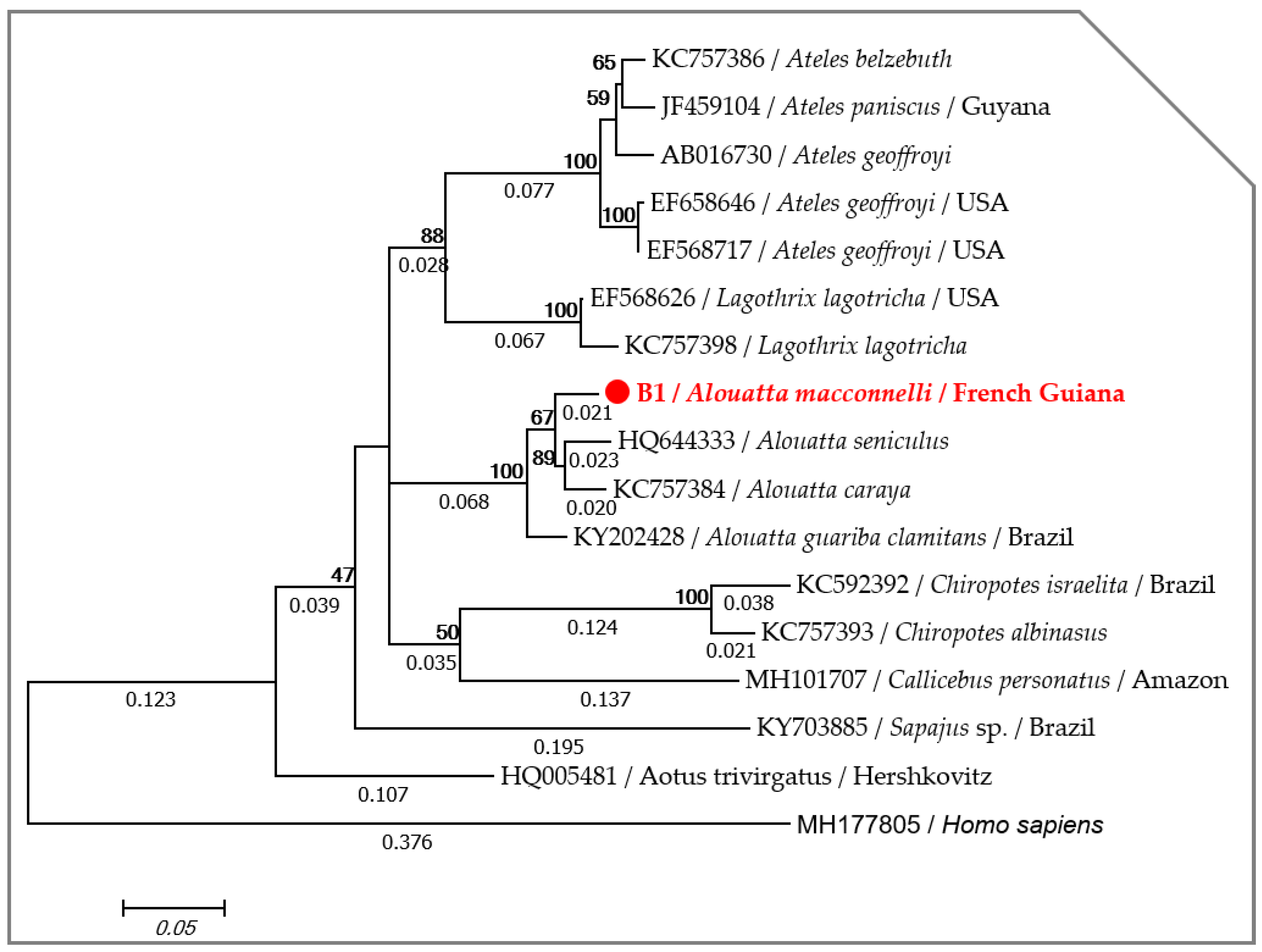
2.1. Host Identification

Folmer’s primers allowed for the amplification of DNA sequences from all blood samples, but despite several attempts, a high-quality DNA sequence of the vertebrate cox1 gene was only obtained in one from among the nine samples tested, suggesting the presence of a non-specific amplification from the latter. The partial nucleotide sequence (558 bp) of the cox1 gene obtained in this study was deposited in the GenBank under accession number MT193011. Blast analysis showed that the cox1 sequence of howler monkeys in our study had an identity of 96.06% with Alouatta seniculus (HQ644333), 95.88% with Alouatta caraya (KC757384) and 95.34% with Alouatta guariba (KY202428) and a query cover of 100%. Accordingly, the phylogenetic analysis using the Maximum Likelihood (ML) method showed that the specimen of howler monkeys (Alouatta macconnelli) is monophyletic with other Alouatta species (Figure 1).

2.2. Molecular Screening for Filarial and Wolbachia DNAs in Howler Monkeys

Filarial and Wolbachia DNAs were detected by qPCR assays in eight out of nine samples tested and six out of nine samples tested, which correspond to a frequency of infection of 88.9% and 66.7% for filaria and Wolbachia, respectively. This is the first molecular report of filaria and its Wolbachia from the howler monkeys of French Guiana.

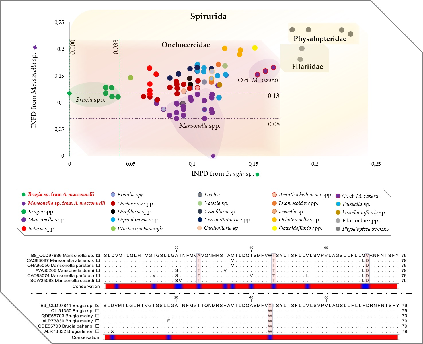
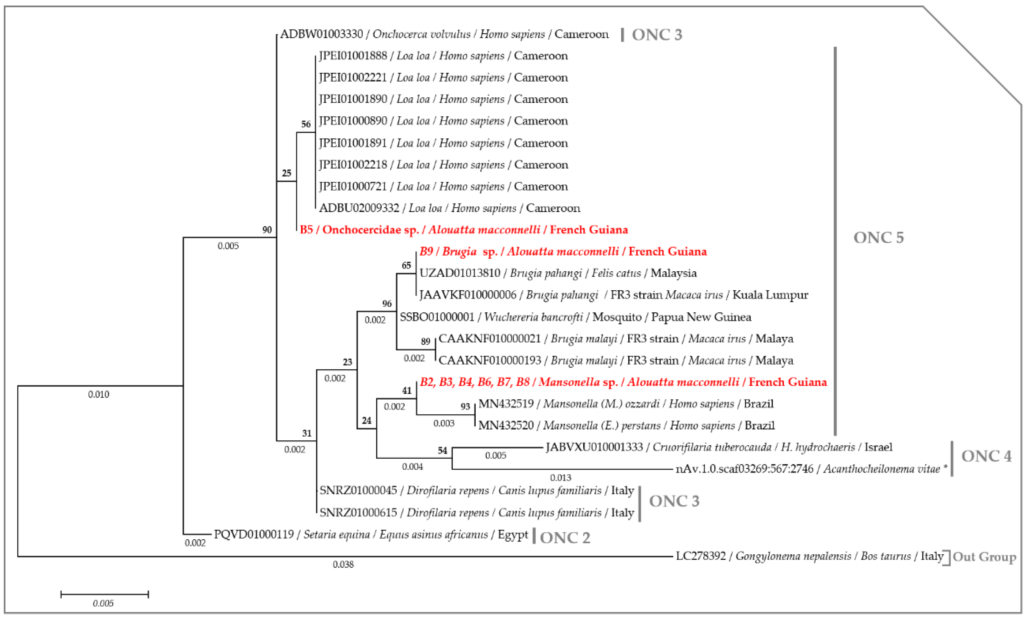
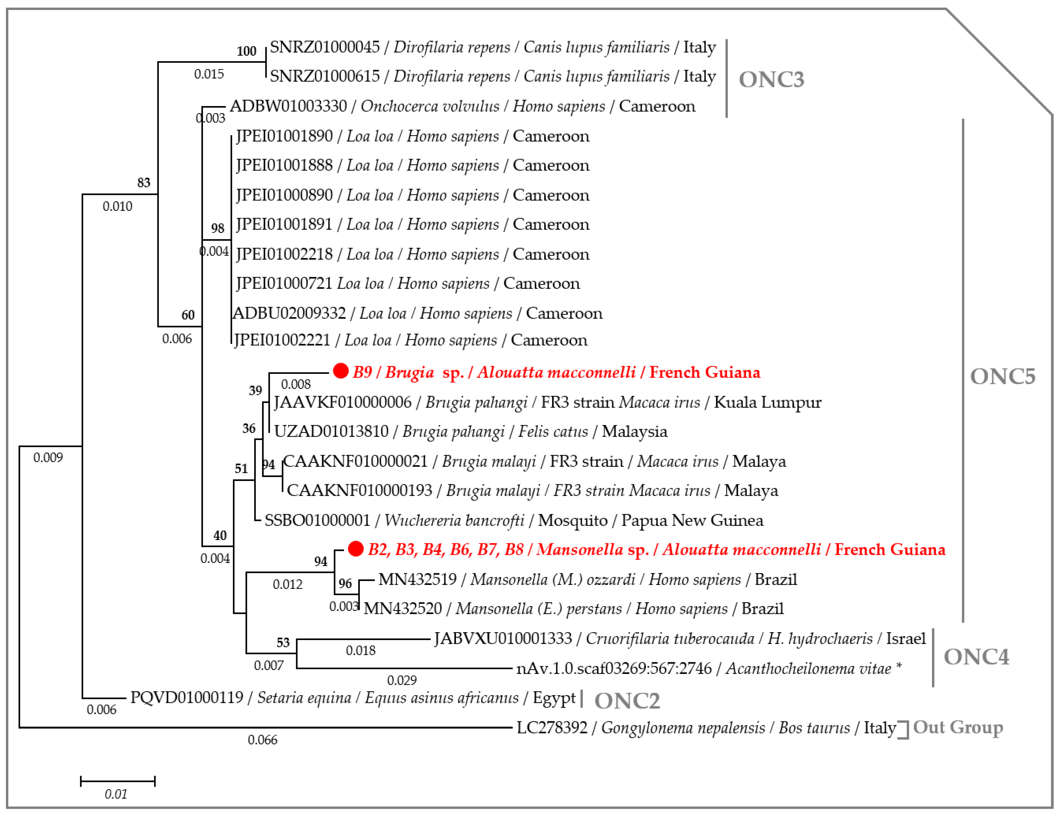
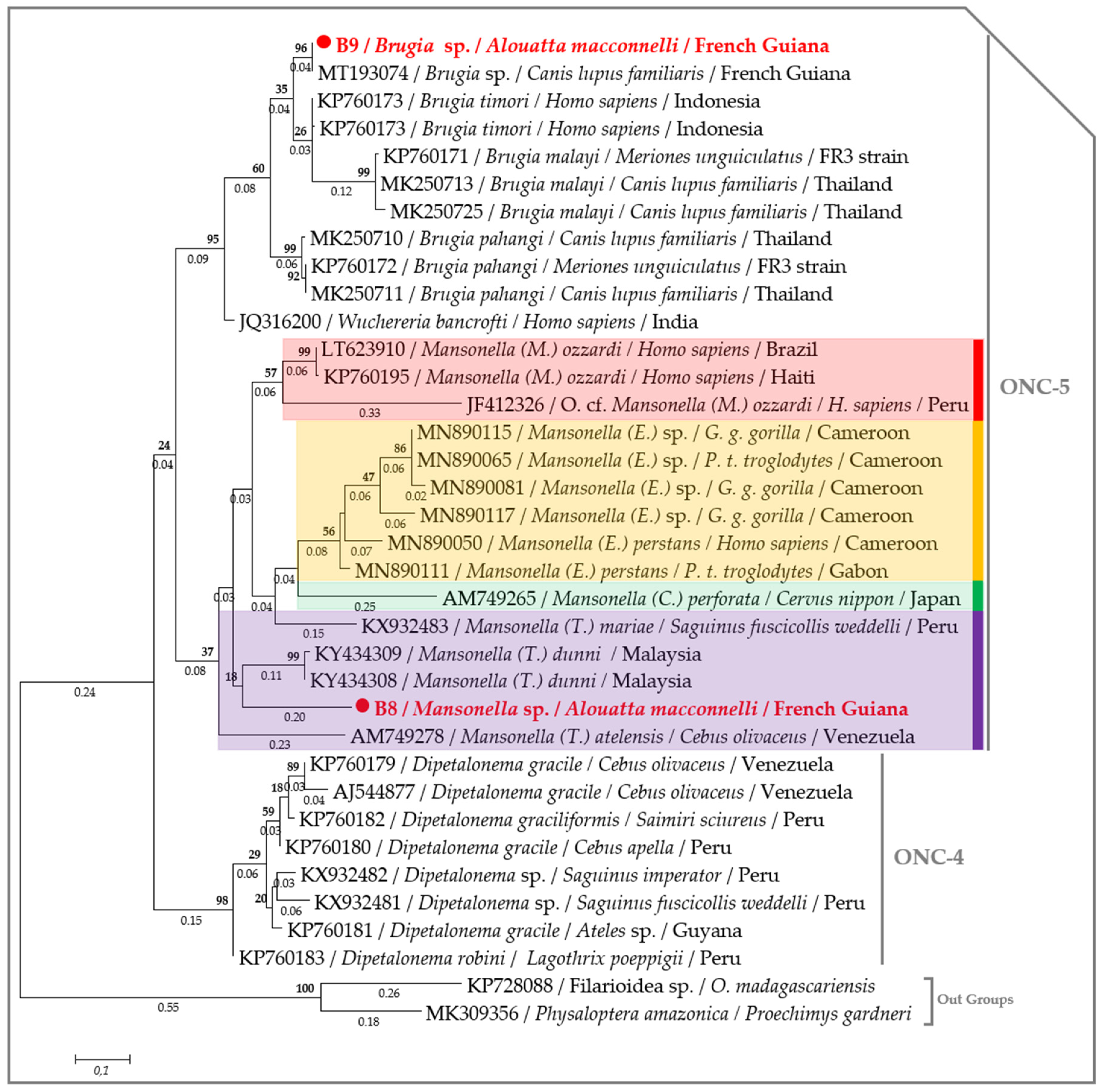
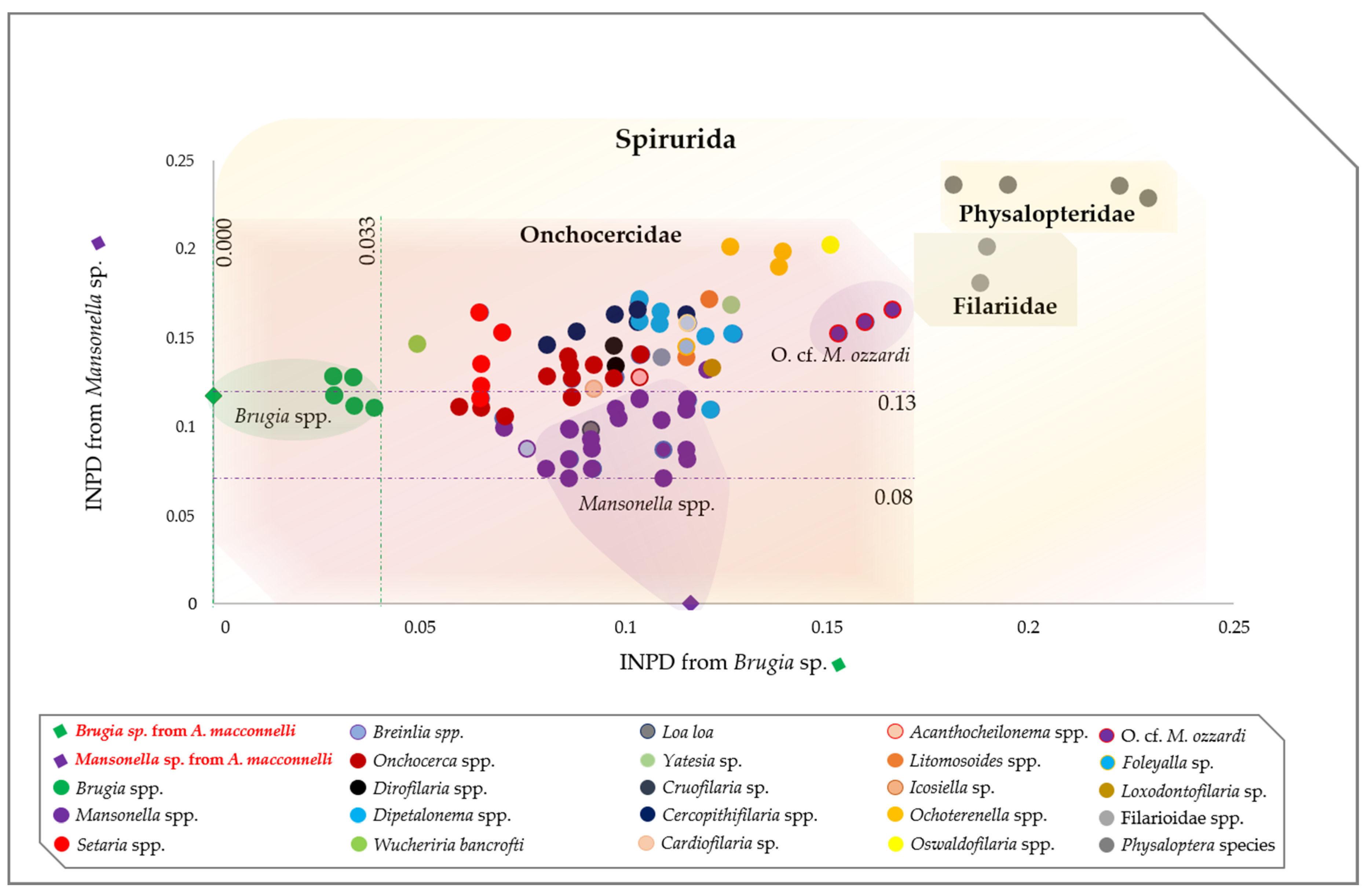
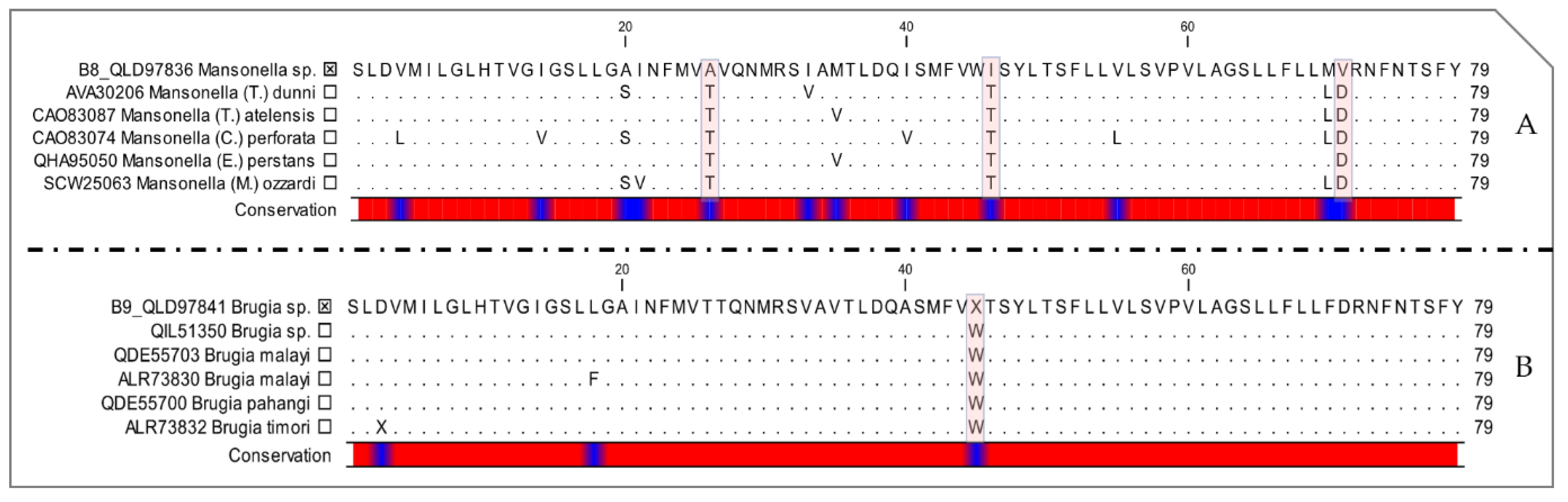
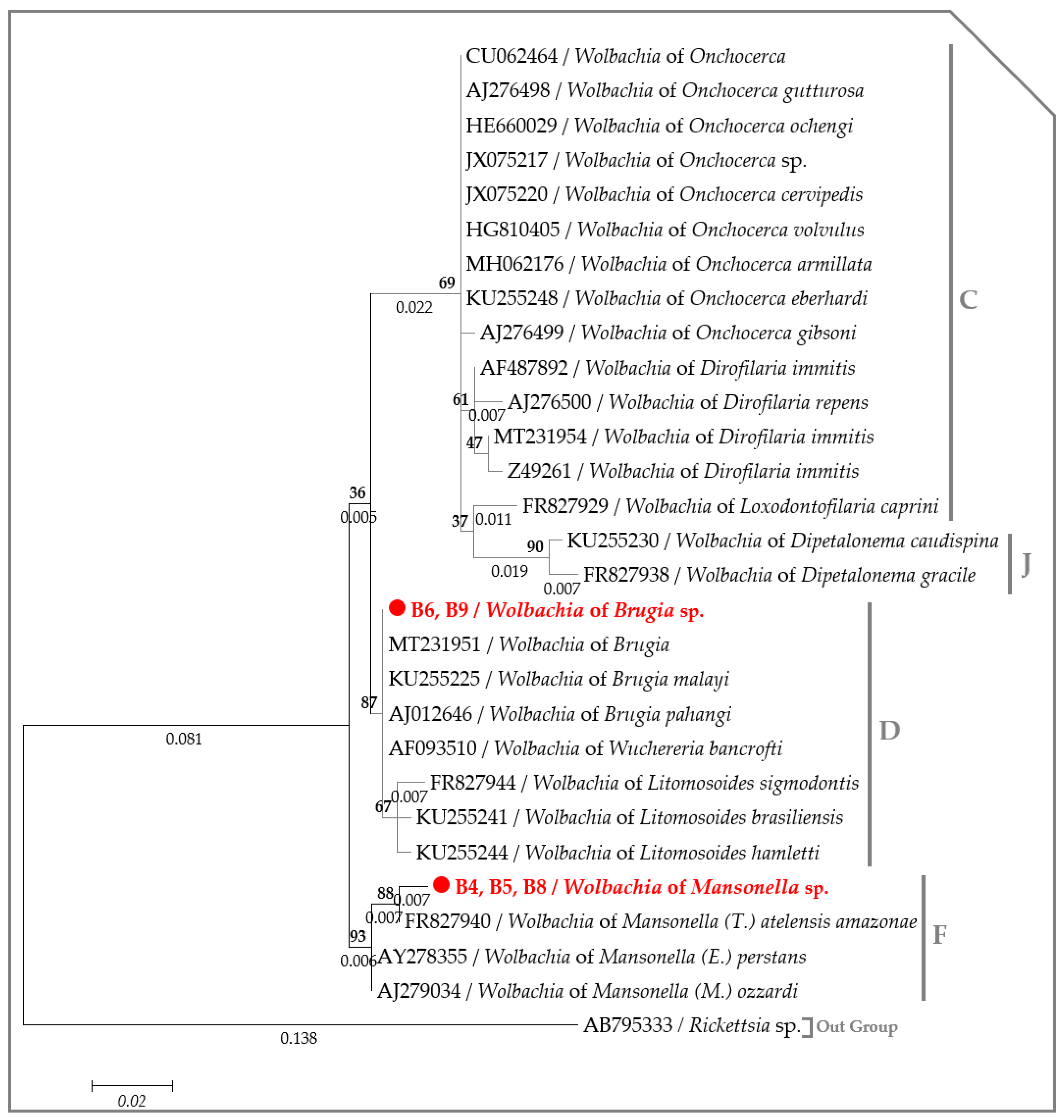
2.3. Molecular Characterization of Filarial Species

To identify filaria detected by qPCR. we performed standard polymerase chain reaction (PCR) screening with primers targeting the small subunit rRNA (18S), the internal transcribed spacer 1 (ITS1) and the cytochrome c oxidase subunit I (cox1) genes. A nearly full-length DNA sequence of the 18S rRNA gene was obtained from all eight samples, was positive in qPCR and was split into three isolates according to the blast results. (i) Six sequences were obtained from the monkeys B2, B3, B4, B6, B7 and B8. These amplicon sequences were identical to each other, showing an identity and query cover of 100% with Dipetalonema sp. (DQ531723) isolated from an owl monkey (Aotus nancymaae) captured in Peru and 99.6% of identification with the Mansonella species (MN432520, MN432519). (ii) One 18S sequence obtained from sample B5 was very close to the Onchocercidae members (Onchocerca cervicalis: DQ094174, and Loa loa: DQ094173), where the identification was 99.9% and 100% of the query cover. Further sequence comparisons showed that the Adenine and Thymine mutated into Cytosine at the position 304 and 879 with O. cervicalis (DQ094174) and L. loa (DQ094173), respectively (Figure S1). (iii) One sequence from sample B9 showed an identification of 100% with B. malayi (AF036588) and 99.9% with Brugia sp. (MN795087), isolated from dogs in French Guiana.
Mansonella genus-specific PCR, based on the amplification of the ITS1, allowed us to obtain ITS sequences of Mansonella sp. from seven monkeys (B2, B3, B4, B5, B6, B7 and B8). They were almost identical and displayed an identity ranging from 83.47% to 93.49% and a query cover ranging from 62% to 83% with Mansonella species (M. ozzardi: KR952332, M. perstans: MN432520, M. mariae: AB362562, M. streptocerca: KR868771, M. dunni: KY434312 and Mansonella sp.: MN821052). Furthermore, Brugia sp. was identified in five samples (B2, 3, 4, 7 and 9) using the Brugia-specific qPCR and ITS sequences were obtained for four of them. These sequences were similar and were close to the Brugia species, wherein the identity ranged from 88.81% to 91.98% with B. malayi (JQ327147, EU419333) and from 89.10% to 91.19% with B. pahangi (EU373633, EU419348).
Primers targeting the cox1 gene amplified the expected DNA amplicon size from all the filaria-positive samples. However, only two sample (B8 and B9) sequences provided good quality electropherograms. Several overlapping peaks (double peaks) within samples B2, B3, B4, B5, B6 and B7 suggested co-infection with two or more filarial species. Blast analysis showed that the specimen amplified from monkey B8 had an identity of 88.2% with Mansonella perstans (MN890111). While the cox1 sequence amplified from monkey B9 was very close to Brugian filariids, with an identity of 99.6% with Brugia sp. (MT193074), isolated from dogs in French Guiana, 95.4% with Brugia timori (AP017686) and 94.9% with Brugia malayi (MN564741).
Phylogenetic analysis using the maximum likelihood method of the 18S rRNA gene showed that howler monkeys from French Guiana are infected with at least three filarial species belonging to the Onchocercidae clade, namely ONC 5. The 18S sequences amplified from monkeys B2, 3, 4, 6, 7 and 8 clustered in a separate branch with Mansonella species, while the sequence obtained from monkey B5 appeared paraphyletic with respect to L. loa (ADBU02009332) and O. volvulus (ADBW01003330), suggesting an unknown onchocercid. Finally, the sequence from monkey B5 clustered with the B. pahangi strain (UZAD01013810 and JAAVKF010000006) (Figure 2).
The ML tree, based on the concatenated rRNA sequences (18S and ITS1), showed that the specimens amplified from monkeys B2, 3, 4, 6, 7 and 8 clustered with other monophyletic species of the genus Mansonella, while the specimen amplified from monkey B9 clustered with the Brugia species (Figure 3). Interestingly, the cox1 phylogram replicated the same results, though with a greater degree of accuracy. The species amplified in this study belong to the clade 5 of the Onchocercidae family. More precisely, the species amplified from monkey B8 belong to the genus Mansonella and the subgenus Tetrapetalonema encountered in New World Primates [44], while the species from monkey B9 clustered with Brugia sp. (MT193074), isolated from dogs in French Guiana [45] and are monophyletic with other Brugian filariids (Figure 4). Interspecific nucleotide distances (IND) of the cox1 sequences ranged between 0.08 and 0.13 between Mansonella sp. from the monkey B8 and most species from the genus Mansonella (MN890075, MN890115, MN890111 and KY434309), while the IND ranged from 0 to 0.03 between Brugia sp. amplified from monkey B9 and Brugian filariids (Figure 5, Table S1).
Importantly, the cox1 DNA sequences were aligned correctly to the reference mitogenome of M. ozzardi (KX822021) [45], and when translated, there were no stop codons in the amino acid sequences, suggesting the absence of co-amplified numts. Finally, translated protein sequences of the cytochrome c oxidase subunit I (COI) showed three amino acid changes between Mansonella sp. from monkey B8 and the other Mansonella species from GenBank, namely, from threonine to alanine, threonine to isoleucine and aspartic acid to valine (Figure 6A). While Brugia sp. from monkey B9 showed a deletion of one amino acid instead of tryptophan, in comparison to Brugian filariids from GenBank (Figure 6B).
A partial DNA sequence of the Wolbachia 16S gene (295 bps) was obtained from five out of six samples that tested positive for Wolbachia DNA through the qPCR. Three identical sequences revealed 99.32% identity with Wolbachia of M. atelensis amazonae (FR827940) and 98.64% with both Wolbachia of M. perstans (AY278355) and M. ozzardi (AJ279034). These sequences were obtained from filaria-positive monkeys, including monkey B4, which was co-infected with Mansonella sp. and Brugia sp., monkey B5 co-infected with an unidentified Onchocercidae species and Mansonella sp. and monkey B8, which was mono-infected with Mansonella sp. The two remaining sequences were amplified from two filaria-positive samples, one for Mansonella sp. (B6) and the other for Brugia sp. (B9). These sequences were identical with each other and were 100% identical with all Wolbachia genotypes associated to Brugia species (CP050521, CP034333, AJ012646 and MT231956). Accordingly, the ML inference indicates that the Wolbachia genotype from monkeys B4, 5 and 8 belong to the Clade F of Wolbachia lineage infecting Mansonella species, while the genotype obtained from monkeys B6 and B9 clustered together with Wolbachia endosymbiont of Brugian filariids within Clade D of the Wolbachia lineage (Figure 7).
Finally, by combining all the molecular results for filaria and Wolbachia detection, we concluded six cases (75%) of co-infections in monkeys, including Mansonella sp.—Brugia sp. co-infection in five and Mansonella sp.—unidentified Onchocercidae species in one. Two other monkeys (25%) presented mono-infections, one with Mansonella sp. and the other with Brugia sp. (Table 1).

3. Discussion

This is the first molecular report of filaria and Wolbachia infection from red howler monkeys (Alouatta macconnelli, Linnaeus 1766—Elliot 1910) in French Guiana. These monkeys were morphologically considered as a distinct species from A. seniculus and they are not a subspecies [46]. Our data confirmed that, molecularly, both species can be distinguished by their cox1 sequences. The wide distribution of howler monkeys (from Mexico to northern Argentina) constitutes a non-negligible reservoir for zoonotic disease [43] and should be monitored. Our study is limited in the number of species and samples, due to the difficulties encountered in the field. The number of monkeys tested was much lower than those tested in Reference [47], where 1353 free-ranging mammals, including 114 howler monkeys (A. seniculus) and 84 red handed tamarins (Saguinus midas) from the neotropical primary rainforest in French Guiana were studied for haemoparasites and microfilariae. However, the prevalence of filarial infection we recorded using molecular assays is close to that reported in tamarins and howler monkeys using blood smear, where the infection rates were 80% and 92% of filaria infections (Dipetalonema and Mansonella (Tetrapetalonema) species), respectively [47]. Our data indicate that the prevalence of filarial infection was higher than that of sloths, anteaters and porcupines in French Guiana, where the infection rate of 40% was reported using blood smears test [47]. The higher prevalence observed in monkeys may be related to the lower host specificity of filariids [48] and/or similar biotope of potential vectors [49]. Another hypothesis is that the lifestyle of these animals increases the risk of vector-borne disease transmission between infected and non-infected individuals in the monkey colony. Therefore, the highest mixed-infection detected in our study corroborates previous reports [50], but it is still unknown whether it is geographical or host-specific. Several species of filariids are reported from a wide range of neo-tropical primates based on morphological taxonomy (Table 2). Most of them belong to the genus Dipetalonema and Mansonella (Tetrapetalonema). However, data in DNA barcoding of these species is lacking.
The use of two (or more) different molecular markers for species delimitation remained necessary for the accurate identification of nematode species [51]. In the present study, our molecular approach, based on generic and genus specific primers, permits the detection and characterization of filarial infections and resolved the co-infections. This is due to the ability of ITS genus-specific PCR assays to separately amplify DNA amplicons depending on their specificity. Filarial nematodes could be misclassified when the 18S gene is used alone as a barcode. This gene is often limited to the genus level and has proven to be inconclusive for the molecular taxonomy of nematodes [52], while the ITS 1 gene appears to be a satisfactory barcode in resolving taxonomic relationships among species [53,54,55]. Furthermore, as suggested by previous authors [56], the use of partitioned concatenated DNA sequences enables the accurate identification of filarial nematodes. We used both the 18S and the partitioned concatenated rRNA (18S and ITS1) gene, which confirmed the presence of at least three potential new species from clade 5 of the Onchocercidae family present in howler monkeys in French Guiana, including Mansonella sp., Brugia sp. and an unidentified Onchocercidae species.
The cox1 gene enabled the accurate identification of the Mansonella species from wild non-human primates from Cameroon and Gabon [57], and has been proven to be a satisfactory discrimination between filarial species. This gene was described by its low nucleotide distances (from 0 to 0.02) within filarial species [58] and a larger variation between congeneric species (i.e., 0.098 to 0.2) [58,59]. In the present study, we used two different phylogenetic methods for the analysis of cox1, together with the alignment of COI protein sequences, which confirmed that species from monkeys B8 and B9 clustered, respectively, with Mansonella Tetrapetalonema subgenus and Brugia species, with the distance ranging between 0.02 and 0.2, suggesting unidentified or potential new species from these genera.
Wolbachia are host-specific, and each genotype is associated with a specific filarial species [11,60]. Bacterial genotype-specific identification was previously proposed for the speciation of Brugia parasites that infect humans [9]. Several studies showed the utility of the specific detection of Wolbachia in determining the subject as infected or not with filarial species (e.g., D. immitis, D. repens, B. pahangi and B. malayi) from domestic animals [14,21,23,24,25,61,62]. Accordingly, the phylogenetic analysis of the Wolbachia 16S DNA sequences demonstrated the presence of two bacterial genotypes belonging to the supergroup F and D encountered in Mansonella and Brugia species, thus corroborating with filaria phylogenies. The inconsistency between the bacterial genotype and filaria species was observed in monkey B6. The presence of Mansonella sp. and Wolbachia of Brugia sp. DNAs highlights a co-infection with both filarial species. However, the absence of Wallachia of Mansonella sp. could be explained by a weaker infection density in this species, while the absence of Brugia sp. DNA, despite the presence of its Wolbachia, could be result to an amicrofilaremic infection due to single sex infection, an earlier infection stage or any other causes. Such inconsistencies were previously reported between Brugia and Dirofilaria species in dogs [63]. Wolbachia-filaria interactions within co-infected hosts are not well understood. Despite the presence of both parasites in co-infected dogs with D. immitis and D. repens, the single detection of Wolbachia of D. immitis is frequent [24] and may result in an unexplained suppression effect on the production of D. immitis microfilariae induced by the presence of D. repens [64,65].
Our findings extend the presence of Brugia sp. and an unidentified Onchocercidae species to the New World Monkeys (e.g., Alouatta macconnelli). Several species of filariae have been described from these primates and they all belong to the genus Dipetalonema or Mansonella subgenus Tetrapetalonema [4] (Table 2). The genus Dipetalonema (Diesing 1861) is restricted to non-human primates (NHPs) of the neotropics, according to the phylogenetic study conducted by Lefoulon et al. [56]. Adult worms are prevalent in the serous cavities of the hosts. A high species diversity of this genus was observed in a wide range of New World monkeys. D. gracile (Rudolphi 1819), D. graciliformis (Freitas 1964) and D. caudispina (Molin 1858) are the main species found in Guiana monkeys, using a morphological taxonomy (Table 2).
The subgenus Mansonella (Tetrapetalonema) is one of the five subgenera derived from the genus Mansonella. Adult filariids are small, slender and can be found in subcutaneous tissues. The Tetrapetalonema subgenus comprises 13 species (Table 2), which have been restricted to platyrrhine (neotropical) primates [66]. Human mansonellensiasis across South America regions are caused by M. ozzardi type species of Mansonella (Mansonella) subgen. n. [44,45] causing fever, pruritis, arthralgias, headache, rashes, lymphadenopathy, edema, and pulmonary symptoms and a common eosinophilia mainly associated with corneal lesions [67,68,69,70]. M. perstans type species of Mansonella (Esslingeria, Chabaud and Bain 1976) subgen. n. [44] is another agent of human mansonellensiasis in some neotropical regions of Central and South America that causes the bung-eye diseases [71]. These species have been found in both humans and non-human primates [4,44]. However, the possibility that the Mansonella sp. we have detected here is one of the 13 Mansonella (Tetrapetalonema) species or a new species from this subgenus cannot be ruled out in the absence of morphological identification.
Brugia spp. are incidental filariids that parasitize non-human vertebrates [72]. The classical brugian filariids involved in lymphatic filariasis are found in Asia, while species reported from North and South America constitute the most zoonotic species of this genus [73]. In Latin America, Brugia sp. infection was reported from the ring-tailed coatis (Nasua nasua nasua) in Brazil [36], Brugia guyanensis from the lymphatic system of the coatimundi (Nasua nasua vittata) in British Guiana [35] and Brugia sp. from domestic dogs in French Guiana [25]. Our findings indicate that Brugia sp. detected from howler monkeys is the same as that recently detected in domestic dogs [25]. Unlike Asian primates in which infection with B. malayi and B. pahangi has been reported [74], Brugian filariid has not been reported in neotropical primates [75]. Cases of human infection by Brugia sp. have been reported in several localities (Amazon, Peru, Colombia) in South America, but the reservoir of the parasites is unknown [72,73]. However, the possibility that the Brugia sp. we detected from howler monkeys and dogs in our previous study [25] is of the same species circulating in humans cannot be ruled out in the absence of molecular data.

4. Materials and Methods

4.1. Samples and Ethic Statement

In January 2016, we obtained samples from howler monkeys that were legally hunted by two Amerindian hunters for family consumption of meat. The International Union for Conservation of Nature conservation status for this species is a “least concern” [83,84]. The hunters applied the provisions of the prefectural decree regulating the quotas of species that can be taken by a person in the department of Guiana (No. 583/DEAL of 12 April 2011). The hunt took place in the deep forest (4°01′39.5″ N 52°31′32.5″ W), near the Approuague River, 50 km from the village of Regina. We were able to examine corpses of nine hunted howler monkeys (five females and four males). Blood was collected by a heart-puncture in sterile tubes containing Ethylene-Diamine-Tetra-Acetic acid (EDTA) and was kept in a cooler before being frozen at −20 °C until further analysis.

4.2. DNA Extraction

Genomic DNA was extracted from 200 µL of each blood samples. The extraction was performed using QIAGEN DNA tissues kit (QIAGEN, Hilden, Germany) following the manufacturer’s recommendations. Two lysis steps were applied before the extraction procedure: (i) mechanical lyses performed on FastPrep-24™ 5G homogenizer using high speed stirring for 40 s in the presence of glass powder, (ii) enzymatic digestion of proteins with buffer G2 and proteinase K for 12 h at 56 °C. The extracted DNA was eluted in a total volume of 100 µL and stored at −20 °C.

4.3. Host Identification

The universal cox1 DNA barcoding region of metazoans [85] was targeted using the degenerated primers of Folmer, as described elsewhere [86]. The PCR products were purified, sequenced and edited, as described below, and were then aligned against cox1 sequences of Alouatta spp. (HQ644333, KC757384, KY202428), Ateles spp. (AB016730, KC757386, JF459104, EF658646, EF568717), Callicebus personatus (MH101707), Chiropotes israelita (KC592392, KC757393), Lagothrix lagotricha (EF568626, KC757398), Sapajus spp. (KY703885) and Aotus trivirgatus (HQ005481) as representative New World monkeys [46]. The sequence (MH177805) of human cox1 was used as an out-group. Finally, the Hasegawa-Kishino-Yano (+G, +I) [87] was selected as a best fit model according to the Akaike Information Criterion (AIC) option in MEGA6 [88]. The maximum likelihood (ML) phylogenetic inference was used with 1000 bootstrap replicates to generate the phylogenetic tree using the same software.

4.4. Molecular Screening for Filaria and Wolbachia

First, all blood samples were screened for the presence of filaria and Wolbachia DNAs using, respectively, the pan-filarial [Pan-fil 28S] and pan-Wolbachia [All-Wol 16S] qPCRs, as described elsewhere [24].

4.5. Molecular Characterization of Filariids and their Associated Wolbachia Using Generic Primers

Samples positive for filaria and Wolbachia by qPCR were subjected to amplification and sequencing analysis using the pan-Nematoda-18S primers [61] and pan-filarial cox1 based PCR [Pan-fil cox1] [24] to generate 1127–1155 bp and 509 bp from the filarial 18S and cox1 genes, respectively. The third PCR system [W16S-Spec] PCR [89] was used to amplify 438 bp from the 16S gene of Wolbachia spp. (Table 3).

4.6. Molecular Characterization of Filariids Using Genus Specific PCR Assays

4.6.1. Design of Oligonucleotides

In order to complete the molecular characterization of filariids detected by the 18S and cox1 genes, we targeted the Internal Transcribed Spacer 1 (ITS1) gene to design genus-specific PCR assays targeting Brugia and Mansonella species. The choice for this gene was based on the following criteria: a higher divergence between filarial species especially among Brugia species [90], its tandem repeat that increases PCR sensitivity [91] and its availability in the GenBank database for these species. Three PCR assays were designed by the alignment of ITS1 sequences of Brugia sp. (HE856316), B. malayi (EU419346, JQ327149), B. timori (AF499132), B. pahangi (EU373628), M. ozzardi (MN432519, LT623912, AF228559), M. perstans (MN432520, KJ631373, EU272184) and M. mariae (KX932484) against 33 sequences (data not showed) from a representative member of Onchocercidae using the MUSCLE application within DNAstar software [92]. Three genus specific PCR systems were proposed (Table 3). This includes two PCRs: one specific for Brugia spp. [Brug-gen-spec] and the other specific for Mansonella spp. [Manso-gen-spec], and qPCR system [Brug-gen-spec qPCR] targeting Brugia spp.
Assay specificity was confirmed in silico and in vitro for each system, as described elsewhere [24]. Briefly, the in silico validation was conducted using Primer-BLAST [93]. Genomic DNA of M. perstens was used to validate the PCR for Mansonella, while the B. malayi DNA was used to validate both the qPCR and PCR for Brugia spp. Moreover, all PCR assays were challenged against the genomic DNA of filariids other than Brugia and Mansonella, as well as several nematodes, arthropods, vertebrate hosts (e.g., human, monkey, donkey, horse, cattle, mouse and dog) and laboratory-maintained colonies [24].

4.6.2. Amplification, Sequencing and Run Protocol

All blood samples from howler monkeys were screened for the presence of Mansonella and Brugia DNA using the genus specific PCR. The PCR reactions were carried out in a total volume of 50 µL, comprising 25 µL of AmpliTaq Gold master mix (Thermo Fisher Scientific, Saint Herblain, France), 18 µL of ultrapure water free of DNAse-RNAse, 1 µL of each primer and 5 µL of genomic DNA. PCR reactions were run under the following protocol: the incubation step at 95 °C for 15 min, 40 cycles of one minute at 95 °C, 30 s for the annealing at a different melting temperature for each PCR assays (Table 3), and 72 °C of elongation step (Table 3) with a final extension step of five minutes at 72 °C. PCR reactions were performed in a Peltier PTC-200 model thermal cycler (MJ Research Inc., Watertown, MA, USA).
DNA amplicons generated throughout each PCR reaction were purified using NucleoFast® 96 PCR DNA purification plate (Macherey Nagel EURL, Hoerdt, France). Purified DNAs were subjected to the second amplification using the BigDye™ Terminator v3.1 Cycle Sequencing Kit (Perkin Elmer Applied Biosystems, Foster City, CA, USA), then the BigDye PCR products were purified on the Sephadex G-50 Superfine gel filtration resin prior to sequencing on the ABI Prism 3130XL (Applied Biosystems, Courtaboeuf, France).

4.6.3. Molecular Screening for Brugia

In order to reveal the infection rate of Brugia spp., all the samples were subjected to the amplification using the genus-specific qPCR. The qPCR reaction was performed in a total volume of 20 µL including 5 μL of DNA template, 10 μL of Master Mix Roche (Eurogentec France, Angers, France), 3 µL of ultra-purified water DNAse-RNAse free and 0.5 µL of each primer, UDG and each probe. The TaqMan reaction of both systems was run using the same cycling conditions. This included two hold steps at 50 °C and 95 °C for 2 and 15 min, respectively, followed by 40 cycles of two steps each (f 95 °C for 30 s and 60 °C for 30 s). The qPCR reaction was performed in a CFX96 Real-Time system (Bio-Rad Laboratories, Foster City, CA, USA).

4.7. Phylogenetic Analysis

First, nucleotide sequences of the filarial cox1, 18S and ITS1 genes, as well as the 16S gene of Wolbachia, were assembled and edited by Chromas-Pro 2.0.0 (http://technelysium.com.au/wp/chromaspro/). The absence of co-amplification of nuclear mitochondrial genes (numts) was verified by aligning the obtained cox1 sequences with the Mansonella ozzardi mitogenome (KX822021) [45]. Furthermore, ambiguities in the sequence chromatograms, stop codons and indels were visually verified, as recommended in Reference [94]. All the sequences were subjected separately to a preliminary analysis using Basic Local Alignment Search Tool (BLAST) [95].
Both the nuclear 18S rRNA alone or concatenated with the ITS1 (if amplified) gene from each filarial species generated through the present study were separately aligned against the previously published sequences from the complete rRNA sequences or draft/complete genomes from the Onchocercidae clade ONC2, ONC3, ONC4 and ONC5 [56]. While, the cox1 sequences were aligned against the representative members of the clade ONC4 and ONC5 encountered in primates [56]. The Wolbachia 16S DNA sequences were aligned against the representative members of Wolbachia lineages (C, D, F and J) infecting filarial parasites [11,16]. MAFFT alignment was performed on the concatenated nuclear (18S rRNA and ITS1) sequences using DNAstar software [92], while the 18S, the cox1 and the 16S DNA sequences were aligned using ClustalW application within Bioedit v.7.2.5. [96]. The Akaike Information Criterion (AIC) option in MEGA6 [88] was used to establish the best nucleotide substitution model adapted to each sequence alignment. The Kimura 2-parameter model (+G) [97] was used to generate the 18S and the 16S trees, while the Tamura 3-parameter model (+I) [98] and the General Time Reversible model (+G, +I) [98] were, respectively, used for the concatenated rRNA (18S and ITS1) and the cox1 alignments. A maximum likelihood (ML) phylogenetic inference was used with 1000 bootstrap replicates to generate the phylogenetic tree in MEGA6 [88]. Gongylonema nepalensis (LC278392) rRNA sequence, both Filarioidea species (KP728088) and Physaloptera amazonica (MK309356) cox1 sequences and the 16S DNA sequence of Rickettsia sp. (AB795333) were used as out groups to root the trees.
In addition, we generated another cox1 alignment, including the representative members of all the Onchocercidae clades (ONC1, ONC2, ONC3, ONC4 and ONC5) [56]. Two Filariidae and four Physalopteridae sequences were included as out-groups. The interspecific nucleotide pairwise distance (IND) was used to estimate the evolutionary divergence between cox1 sequences among Onchocercidae. Standard error was obtained by a bootstrap procedure with 1000 replicates. Analyses were inferred on MEGA6 software [88], based on the Maximum Composite Likelihood model [99]. A scatter chart based on the IND between Onchocercidae members and the cox1 sequences generated in the present study was drowned using XLSTAT Addinsoft version 4.1 (XLSTAT 2019: Data Analysis and Statistical Solution for Microsoft Excel, Paris, France).
Finally, COI protein sequences of Brugia species (Protein Id: QIL51350, QDE55703, ALR73830, QDE55700 and ALR73832) and those of Mansonella species (Protein Id: CAO83087, QHA95050, AVA30206, CAO83074 and SCW25063) were retrieved from the GenBank database and aligned against the COI sequences obtained from monkeys B9 and B8, respectively. The alignment was performed using the ClustalW application within Bioedit v.7.2.5. [96]. Amino acids conservation between the COI sequences from this study comparatively to GenBank sequences was visualized on the CLC Sequence Viewer 7 (CLC Bio Qiagen, Aarhus, Denmark).

5. Conclusions

In this study, we phylogenetically describe filarial parasites belonging to three distinct genera: Mansonella sp. Brugia sp. and an unidentified Onchocercidae species. Funding extends the presence of Brugia sp. and the unidentified Onchocercidae species to Guiana monkeys. In addition, phylogenetic analyses highlight the necessity of completing the classification of filariasis of neo-tropical monkeys by combining morphological and molecular-based identification for an integrative taxonomical perspective. Filaria associated Wolbachia can be used as diagnostic markers since they are genus specific endosymbionts. Regarding the presence of Brugia sp. in Guiana monkeys, the same genotype circulates in French Guiana dogs, suggesting host diversity of this filariids. We therefore developed a novel qPCR assay that could be useful for the surveillance of brugian filariasis in vectors, animals, and humans. Further studies will be needed to shed light on the life cycle, epidemiology and circulation of this potentially zoonotic parasite.

Supplementary Materials

The following are available online at https://www.mdpi.com/2076-0817/9/8/626/s1, Figure S1: 18S sequences alignment showing the nucleotide conservation of the unidentified Onchocercidae species obtained from howler monkey against the GenBank sequences of O. volvulus and L. loa, Table S1: Estimates of the evolutionary divergence between the cytochrome c oxidase subunit I (cox1) sequences of Mansonella sp. and Brugia sp. obtained in this study comparatively with Onchocercidae members from GenBank database.

Author Contributions

Conceptualization: B.D., Y.L., H.M., O.M.; Formal analysis and investigation: Y.L., H.M., B.D., A.L.; Writing—original draft preparation: B.D., Y.L.; Writing-review: O.M.; Supervision: O.M. and B.D. All authors have read and agreed to the published version of the manuscript.

Funding

This study was supported by the Institut Hospitalo-Universitaire (IHU) Méditerranée Infection, the National Research Agency under the program “Investissements d’avenir”, reference ANR-10-IAHU-03, the Région Provence-Alpes-Côte d’Azur and European funding FEDER PRIMI.

Acknowledgments

We especially thank Christophe B., Amélie V., and Coarasi S. for their significant help in providing samples.

Conflicts of Interest

The authors declare no conflict of interest.

References

  1. Pritt, S.; Cohen, K.; Sedlacek, H. Parasitic Diseases. In The Laboratory Rabbit, Guinea Pig, Hamster, and Other Rodents, 2nd ed.; American College of Laboratory Animal Medicine; Academic Press: London, UK, 2012; pp. 415–446. ISBN 9780123809209. [Google Scholar]
  2. Anderson, R.C. Nematode parasites of vertebrates. Their development and transmission; C.A.B. International: Wallingford, UK, 1992; p. 578. [Google Scholar]
  3. Strait, K.; Else, J.G.; Eberhard, M.L. Parasitic Diseases of Nonhuman Primates. In Nonhuman Primates in Biomedical Research, 2nd ed.; Academic Press Elsevier Inc.: London, UK, 2012; Volume 2, pp. 197–297. ISBN 9780123813664. [Google Scholar]
  4. Orihel, T.C.; Seibold, H.R. Nematodes of the bowel and tissues. In Pathology of Simian Primates; Fiennes, R.N., Ed.; Karger Publisher: Basel, Switzerland, 1972; pp. 1366–1368. [Google Scholar]
  5. Gardiner, C.H.; Nold, J.B.; Sanders, J.E. Diagnostic exercise. Edesonfilaria malayensis infection in two cynomolgus monkeys. Lab. Anim. Sci. 1982, 32, 601–602. [Google Scholar]
  6. Lowenstine, L.J.; Osborn, K.G. Respiratory system diseases of nonhuman primates. In Nonhuman Primates in Biomedical Research, 2nd ed.; Academic Press Elsevier Inc.: London, UK, 2012; Volume 2, pp. 413–481. ISBN 9780123813664. [Google Scholar]
  7. Bennuru, S.; Nutman, T.B. Lymphangiogenesis and lymphatic remodeling induced by filarial parasites: Implications for pathogenesis. PLoS Pathog. 2009, 5, e1000688. [Google Scholar] [CrossRef] [PubMed] [Green Version]
  8. Bowman, D.D.; Atkins, C.E. Heartworm biology, treatment, and control. Vet. Clin. Small Anim. Pract. 2009, 39, 1127–1158. [Google Scholar] [CrossRef]
  9. Fischer, P.; Supali, T.; Maizels, R.M. Lymphatic filariasis and Brugia timori: Prospects for elimination. Trends Parasitol. 2004, 20, 351–355. [Google Scholar] [CrossRef] [PubMed]
  10. World Health Organization. Lymphatic Filariasis. Available online: https://www.who.int/news-room/fact-sheets/detail/lymphatic-filariasis (accessed on 12 May 2020).
  11. Bandi, C.; Anderson, T.J.C.; Genchi, C.; Blaxter, M.L. Phylogeny of Wolbachia in filarial nematodes. Proc. R. Soc. B Biol. Sci. 1998, 265, 2407–2413. [Google Scholar] [CrossRef] [Green Version]
  12. Martin, C.; Gavotte, L. The bacteria Wolbachia in filariae, a biological Russian dolls’ system: New trends in antifilarial treatments. Parasite 2010, 17, 79–89. [Google Scholar] [CrossRef] [Green Version]
  13. Taylor, M.J. Wolbachia in the inflammatory pathogenesis of human filariasis. Ann. N. Y. Acad. Sci. 2003, 990, 444–449. [Google Scholar] [CrossRef]
  14. Dingman, P.; Levy, J.K.; Kramer, L.H.; Johnson, C.M.; Lappin, M.R.; Greiner, E.C.; Courtney, C.H.; Tucker, S.J.; Morchon, R. Association of Wolbachia with heartworm disease in cats and dogs. Vet. Parasitol. 2010, 170, 50–60. [Google Scholar] [CrossRef]
  15. Bouchery, T.; Lefoulon, E.; Karadjian, G.; Nieguitsila, A.; Martin, C. The symbiotic role of Wolbachia in Onchocercidae and its impact on filariasis. Clin. Microbiol. Infect. 2013, 19, 131–140. [Google Scholar] [CrossRef] [Green Version]
  16. Hise, A.G.; Gillette-Ferguson, I.; Pearlman, E. The role of endosymbiotic Wolbachia bacteria in filarial disease. Cell. Microbiol. 2004, 6, 97–104. [Google Scholar] [CrossRef] [Green Version]
  17. Debrah, L.B.; Phillips, R.O.; Pfarr, K.; Klarmann-Schulz, U.; Opoku, V.S.; Nausch, N.; Owusu, W.; Mubarik, Y.; Sander, A.L.; Lämmer, C.; et al. The efficacy of doxycycline treatment on Mansonella perstans infection: An open-label, randomized trial in Ghana. Am. J. Trop. Med. Hyg. 2019, 101, 84–92. [Google Scholar] [CrossRef] [PubMed]
  18. Sulaiman, W.A.W.; Kamtchum-Tatuene, J.; Mohamed, M.H.; Ramachandran, V.; Ching, S.M.; Lim, S.M.S.; Hashim, H.Z.; Mat, L.N.I.; Hoo, F.K.; Basri, H. Anti-Wolbachia therapy for onchocerciasis & lymphatic filariasis: Current perspectives. Indian J. Med. Res. 2019, 149, 706–714. [Google Scholar]
  19. McCall, J.W.; Genchi, C.; Kramer, L.; Guerrero, J.; Dzimianski, M.T. Heartworm and Wolbachia: Therapeutic implications. Vet. Parasitol. 2008, 158, 204–214. [Google Scholar] [CrossRef]
  20. Punkosdy, G.A.; Dennis, V.A.; Lasater, B.L.; Tzertzinis, G.; Foster, J.M.; Lammie, P.J. Detection of serum IgG antibodies specific for Wolbachia surface protein in Rhesus monkeys infected with Brugia malayi. J. Infect. Dis. 2001, 184, 385–389. [Google Scholar] [CrossRef] [PubMed] [Green Version]
  21. Turb, M.E.; Zambon, E.; Zannoni, A.; Russo, S.; Gentilini, F. Detection of Wolbachia DNA in blood for diagnosing filaria-associated syndromes in cats. J. Clin. Microbiol. 2012, 50, 2624–2630. [Google Scholar] [CrossRef] [Green Version]
  22. Simón, F.; Siles-Lucas, M.; Morchón, R.; González-Miguel, J.; Mellado, I.; Carretón, E.; Montoya-Alonso, J.A. Human and animal dirofilariasis: The emergence of a zoonotic mosaic. Clin. Microbiol. Rev. 2012, 25, 507–544. [Google Scholar] [CrossRef] [Green Version]
  23. Maia, C.; Altet, L.; Serrano, L.; Cristóvão, J.M.; Tabar, M.D.; Francino, O.; Cardoso, L.; Campino, L.; Roura, X. Molecular detection of Leishmania infantum, filariae and Wolbachia spp. in dogs from southern Portugal. Parasit. Vectors 2016, 9, 170. [Google Scholar] [CrossRef] [Green Version]
  24. Laidoudi, Y.; Davoust, B.; Varloud, M.; Niang, E.H.A.; Fenollar, F.; Mediannikov, O. Development of a multiplexed qPCRs-based approach for the diagnosis of Dirofilaria immitis, D. repens, Acanthocheilonema reconditum. Parasites Vectors Dev. 2019, 13, 319. [Google Scholar] [CrossRef]
  25. Laidoudi, Y.; Marié, J.L.; Tahir, D.; Watier-Grillot, S.; Mediannikov, O.; Davoust, B. Detection of canine vector-borne filariasis and their Wolbachia endosymbionts in French Guiana. Microorganisms 2020, 8, 770. [Google Scholar] [CrossRef]
  26. Ichimori, K.; King, J.D.; Engels, D.; Yajima, A.; Mikhailov, A.; Lammie, P.; Ottesen, E.A. Global programme to eliminate lymphatic filariasis: The processes underlying programme success. PLoS Negl. Trop. Dis. 2014, 8, e3328. [Google Scholar] [CrossRef]
  27. Webster, J.P.; Molyneux, D.H.; Hotez, P.J.; Fenwick, A. The contribution of mass drug administration to global health: Past, present and future. Philos. Trans. R. Soc. B Biol. Sci. 2014, 369, 20130434. [Google Scholar] [CrossRef] [PubMed]
  28. World Health Organization. Lymphatic Filariasis: Practical Entomology—A Handbook for National Elimination Programmes; No. WHO/HTM/NTD/PCT/2013.10.; World Health Organization: Geneva, Switzerland, 2013; p. 92. [Google Scholar]
  29. Hotez, P.J.; Bottazzi, M.E.; Franco-Paredes, C.; Ault, S.K.; Periago, M.R. The neglected tropical diseases of Latin America and the Caribbean: A review of disease burden and distribution and a roadmap for control and elimination. PLoS Negl. Trop. Dis. 2008, 2, e300. [Google Scholar] [CrossRef] [PubMed] [Green Version]
  30. Schneider, M.C.; Aguilera, X.P.; da Silva Junior, J.B.; Ault, S.K.; Najera, P.; Martinez, J.; Requejo, R.; Nicholls, R.S.; Yadon, Z.; Silva, J.C.; et al. Elimination of neglected diseases in Latin America and the Caribbean: A mapping of selected diseases. PLoS Negl. Trop. Dis. 2011, 5, e964. [Google Scholar] [CrossRef] [PubMed]
  31. Pan American Health Organisation and World Health Organisation. Latin American countries gather to work towards the elimination of lymphatic filariasis (elephantiasis). Available online: https://www.paho.org/hq/index.php?option=com_content&view=article&id=8784:2013-paises-latinoamerica-reunen-avanzar-hacia-eliminacion-filariasis-linfatica-elefantiasis&Itemid=40282&lang=en (accessed on 22 June 2020).
  32. Raccurt, C.P. Mansonella ozzardi and its vectors in the New World: An update with emphasis on the current situation in Haiti. J. Helminthol. 2018, 92, 655–661. [Google Scholar] [CrossRef] [PubMed]
  33. Ta-Tang, T.H.; Luz, S.L.B.; Merino, F.J.; De Fuentes, I.; López-Vélez, R.; Almeida, T.A.P.; Lanza, M.; Abrahim, C.M.M.; Rubio, J.M. Atypical Mansonella ozzardi microfilariae from an endemic area of Brazilian amazonia. Am. J. Trop. Med. Hyg. 2016, 95, 629–632. [Google Scholar] [CrossRef] [Green Version]
  34. Tavares da Silva, L.B.; Crainey, J.L.; Ribeiro da Silva, T.R.; Suwa, U.F.; Vicente, A.C.P.; Fernandes de Medeiros, J.; Pessoa, F.A.C.; Luz, S.L.B. Molecular verification of new world Mansonella perstans parasitemias. Emerg. Infect. Dis. 2017, 23, 545–547. [Google Scholar] [CrossRef] [Green Version]
  35. Orihel, T.C. Brugia guyanensis sp. n. (Nematoda: Filarioidea) from the Coatimundi (Nasua nasua vittata) in British Guiana. J. Parasitol. 1964, 50, 115–118. [Google Scholar] [CrossRef]
  36. Moraes, M.F.D.; da Silva, M.X.; Magalhães-Matos, P.C.; de Albuquerque, A.C.A.; Tebaldi, J.H.; Mathias, L.A.; Lux Hoppe, E.G. Filarial nematodes with zoonotic potential in ring-tailed coatis (Nasua nasua Linnaeus, 1766, Carnivora: Procyonidae) and domestic dogs from Iguaçu National Park, Brazil. Vet. Parasitol. Reg. Stud. Rep. 2017, 8, 1–9. [Google Scholar] [CrossRef]
  37. De Argôlo, E.G.G.; Reis, T.; Fontes, D.A.T.; Gonçalves, E.C.; Giese, E.G.; de Vasconcelos Melo, F.T.; dos Santos, J.N.; Furtado, A.P. Canine filariasis in the Amazon: Species diversity and epidemiology of these emergent and neglected zoonoses. PLoS ONE 2018, 13, 1–9. [Google Scholar] [CrossRef] [Green Version]
  38. Eisenberg, J.F.; Redford, K.H. Alouatta. In Mammals of the Neotropics. The Central Neotropics: Ecuador, Perú, Bolivia, Brazil; University of Chicago Press: Chicago, IL, USA, 1999; Volume 3, pp. 265–266. [Google Scholar]
  39. Stuart, M.; Pendergast, V.; Rumfelt, S.; Pierberg, S.; Greenspan, L.; Glander, K.; Clarke, M. Parasites of wild howlers (Alouatta spp.). Int. J. Primatol. 1998, 19, 493–512. [Google Scholar] [CrossRef]
  40. Cortés-Ortiz, L.; Bermingham, E.; Rico, C.; Rodríguez-Luna, E.; Sampaio, I.; Ruiz-García, M. Molecular systematics and biogeography of the Neotropical monkey genus, Alouatta. Mol. Phylogenet. Evol. 2003, 26, 64–81. [Google Scholar] [CrossRef]
  41. Groves, C. Primate taxonomy. In The International Encyclopedia of Biological Anthropology; John Wiley & Sons: Hoboken, NJ, USA, 2018; Volume 1. [Google Scholar] [CrossRef]
  42. Stevenson, P.R. Potential determinants of the abundance of woolly monkeys in neotropical forests. In The Woolly Monkey: Behavior, Ecology, Systematics, and Captive Research; Pablo, R., Ed.; Springer: New York, NY, USA, 2014; ISBN 9781493906970. [Google Scholar]
  43. Medkour, H.; Davoust, B.; Levasseur, A.; Mediannikov, O. Molecular evidence of Leishmania infantum and Leishmania guyanensis in red howler monkey (Alouatta seniculus) from French Guiana. Vector Borne Zoonotic Dis. 2019, 19, 896–900. [Google Scholar] [CrossRef] [PubMed]
  44. Bain, O.; Mutafchiev, Y.; Junker, K.; Guerrero, R.; Martin, C.; Lefoulon, E.; Uni, S. Review of the genus Mansonella Faust, 1929 sensu lato (Nematoda: Onchocercidae), with descriptions of a new subgenus and a new subspecies. Zootaxa 2015, 3918, 151–193. [Google Scholar] [CrossRef] [PubMed] [Green Version]
  45. Crainey, J.L.; Marín, M.A.; Da Silva, T.R.R.; De Medeiros, J.F.; Pessoa, F.A.C.; Santos, Y.V.; Vicente, A.C.P.; Luz, S.L.B. Mansonella ozzardi mitogenome and pseudogene characterisation provides new perspectives on filarial parasite systematics and CO-1 barcoding. Sci. Rep. 2018, 8, 6158. [Google Scholar] [CrossRef] [Green Version]
  46. Ruiz-García, M.; Cerón, Á.; Sánchez-Castillo, S.; Rueda-Zozaya, P.; Pinedo-Castro, M.; Gutierrez-Espeleta, G.; Shostell, J.M. Phylogeography of the mantled howler monkey (Alouatta palliata; Atelidae, Primates) across its geographical range by means of mitochondrial genetic analyses and new insights about the phylogeny of Alouatta. Folia Primatol. 2018, 88, 421–454. [Google Scholar] [CrossRef]
  47. De Thoisy, B.; Michel, J.C.; Vogel, I.; Vié, J.C. A survey of hemoparasite infections in free-ranging mammals and reptiles in French Guiana. J. Parasitol. 2000, 86, 1035–1040. [Google Scholar] [CrossRef]
  48. Klein, A.; Strube, C.; Radespiel, U.; Springer, A.; Zimmermann, E. Differences in infection patterns of vector-borne blood-stage parasites of sympatric Malagasy primate species (Microcebus murinus, M. ravelobensis). Int. J. Parasitol. Parasites Wildl. 2019, 10, 59–70. [Google Scholar] [CrossRef]
  49. Lainson, R.; Shaw, J.J.; Fraiha, H.; Miles, M.A.; Draper, C.C. Chagas’s disease in the amazon basin: I. Trypanosoma cruzi infections in silvatic mammals, triatomine bugs and man in the state of North Brazil. Trans. R. Soc. Trop. Med. Hyg. 1979, 73, 193–204. [Google Scholar] [CrossRef]
  50. Petit, G.; Bain, O.; Roussilhon, C. Deux nouvelles filaires chez un singe, Saimiri sciureus, au Guyana. Ann. Parasitol. Hum. Comp. 1985, 60, 65–81. [Google Scholar] [CrossRef] [Green Version]
  51. De León, G.P.-P.; Nadler, S.A. What we don’t recognize can hurt us: A plea for awareness about cryptic species. J. Parasitol. 2010, 96, 453–464. [Google Scholar] [CrossRef]
  52. De Ley, P.; Tandingan De Ley, I.; Morris, K.; Abebe, E.; Mundo-Ocampo, M.; Yoder, M.; Heras, J.; Waumann, D.; Rocha-Olivares, A.; Burr, A.H.J.; et al. An integrated approach to fast and informative morphological vouchering of nematodes for applications in molecular barcoding. Philos. Trans. R. Soc. B Biol. Sci. 2005, 360, 1945–1958. [Google Scholar] [CrossRef] [PubMed] [Green Version]
  53. Blaxter, M.; Mann, J.; Chapman, T.; Thomas, F.; Whitton, C.; Floyd, R.; Abebe, E. Defining operational taxonomic units using DNA barcode data. Philos. Trans. R. Soc. B Biol. Sci. 2005, 360, 1935–1943. [Google Scholar] [CrossRef] [PubMed]
  54. Meldal, B.H.M.; Debenham, N.J.; De Ley, P.; De Ley, I.T.; Vanfleteren, J.R.; Vierstraete, A.R.; Bert, W.; Borgonie, G.; Moens, T.; Tyler, P.A.; et al. An improved molecular phylogeny of the Nematoda with special emphasis on marine taxa. Mol. Phylogenet. Evol. 2007, 42, 622–636. [Google Scholar] [CrossRef] [PubMed]
  55. Nuchprayoon, S.; Junpee, A.; Poovorawan, Y.; Scott, A.L. Detection and differentiation of filarial parasites by universal primers and polymerase chain reaction-restriction fragment length polymorphism analysis. Am. J. Trop. Med. Hyg. 2005, 73, 895–900. [Google Scholar] [CrossRef] [Green Version]
  56. Lefoulon, E.; Bain, O.; Bourret, J.; Junker, K.; Guerrero, R.; Cañizales, I.; Kuzmin, Y.; Satoto, T.B.T.; Cardenas-Callirgos, J.M.; de Souza Lima, S.; et al. Shaking the tree: Multi-locus sequence typing usurps current Onchocercid (Filarial Nematode) phylogeny. PLoS Negl. Trop. Dis. 2015, 9, 1–19. [Google Scholar] [CrossRef]
  57. Gaillard, C.M.; Pion, S.D.; Hamou, H.; Sirima, C.; Bizet, C.; Lemarcis, T.; Rodrigues, J.; Esteban, A.; Peeters, M.; Mpoudi Ngole, E.; et al. Detection of DNA of filariae closely related to Mansonella perstans in faecal samples from wild non-human primates from Cameroon and Gabon. Parasit. Vectors 2020, 13, 313. [Google Scholar] [CrossRef]
  58. Ferri, E.; Barbuto, M.; Bain, O.; Galimberti, A.; Uni, S.; Guerrero, R.; Ferté, H.; Bandi, C.; Martin, C.; Casiraghi, M. Integrated taxonomy: Traditional approach and DNA barcoding for the identification of filarioid worms and related parasites (Nematoda). Front. Zool. 2009, 6, 1. [Google Scholar] [CrossRef] [Green Version]
  59. Casiraghi, M.; Anderson, T.J.C.; Bandi, C.; Bazzocchi, C.; Genchi, C. A phylogenetic analysis of filarial nematodes: Comparison with the phylogeny of Wolbachia endosymbionts. Parasitology 2001, 1, 93–103. [Google Scholar] [CrossRef] [Green Version]
  60. Taylor, M.J.; Hoerauf, A. Wolbachia bacteria of filarial nematodes. Parasitol. Today 1999, 15, 437–442. [Google Scholar] [CrossRef]
  61. Laidoudi, Y.; Ringot, D.; Watier-Grillot, S.; Davoust, B.; Mediannikov, O. A cardiac and subcutaneous canine dirofilariosis outbreak in a kennel in central France. Parasites 2019, 26, 72. [Google Scholar] [CrossRef]
  62. Rossi, M.I.D.; Aguiar-Alves, F.; Santos, S.; Paiva, J.; Bendas, A.; Fernandes, O.; Labarthe, N. Detection of Wolbachia DNA in blood from dogs infected with Dirofilaria immitis. Exp. Parasitol. 2010, 126, 270–272. [Google Scholar] [CrossRef] [PubMed]
  63. Satjawongvanit, H.; Phumee, A.; Tiawsirisup, S.; Sungpradit, S.; Brownell, N.; Siriyasatien, P.; Preativatanyou, K. Molecular analysis of canine filaria and its Wolbachia endosymbionts in domestic dogs collected from two animal university hospitals in Bangkok metropolitan region, Thailand. Pathogens 2019, 8, 114. [Google Scholar] [CrossRef] [PubMed] [Green Version]
  64. Monica, A.; Matei, I.A.; Amico, G.D.; Bel, L.V.; Dumitrache, M.O.; Modrý, D.; Mihalca, A.D. Dirofilaria immitis and D. repens show circadian co-periodicity in naturally co-infected dogs. Parasit Vectors 2017, 10, 116. [Google Scholar]
  65. Mircean, M.; Ionică, A.M.; Mircean, V.; Györke, A.; Codea, A.R.; Tăbăran, F.A.; Taulescu, M.; Dumitrache, M.O. Clinical and pathological effects of Dirofilaria repens and Dirofilaria immitis in a dog with a natural co-infection. Parasitol. Int. 2017, 66, 331–334. [Google Scholar] [CrossRef] [PubMed]
  66. Eberhard, M.L.; Orihel, T.C. The genus Mansonella (syn. Tetrapetalonema): A new classification. Ann. Parasitol. Hum. Comp. 1984, 59, 483–496. [Google Scholar] [CrossRef] [Green Version]
  67. Carme, B.; de Thoisy, B.; Motard, A.; Aznar, C.; Vié, J.C. Parasitoses humaines et mammifères sauvages en Guyane Française. Med. Trop. 2000, 60, 223–231. [Google Scholar]
  68. Vianna, L.M.M.; Martins, M.; Cohen, M.J.; Cohen, J.M.; Belfort, R. Mansonella ozzardi corneal lesions in the Amazon: A cross-sectional study. BMJ Open 2012, 2, e001266. [Google Scholar] [CrossRef] [Green Version]
  69. Garrido, C.; Campos, M. First report of presumed parasitic keratitis in Indians from the Brazilian Amazon. Cornea 2000, 19, 817–819. [Google Scholar] [CrossRef]
  70. Cohen, J.M.; Santana Ribeiro, J.A.; Martins, M. Ocular manifestations in mansonelliasis. Arq. Bras. Oftalmol. 2008, 71, 167–171. [Google Scholar]
  71. Baird, J.K.; Neafie, R.C.; Connor, D.H. Nodules in the conjunctiva, bung-eye, and bulge-eye in Africa caused by Mansonella perstans. Am. J. Trop. Med. Hyg. 1988, 38, 553–557. [Google Scholar] [CrossRef]
  72. Paniz-Mondolfi, A.E.; Gárate, T.; Stavropoulos, C.; Fan, W.; González, L.M.; Eberhard, M.; Kimmelstiel, F.; Sordillo, E.M. Zoonotic filariasis caused by novel Brugia sp. Nematode, United States, 2011. Emerg. Infect. Dis. 2014, 20, 1248–1250. [Google Scholar] [CrossRef] [PubMed] [Green Version]
  73. Orihel, T.C.; Beaver, P.C. Zoonotic Brugia infections in North and South America. Am. J. Trop. Med. Hyg. 1989, 40, 638–647. [Google Scholar] [CrossRef] [PubMed]
  74. Orihel, T.C.; Eberhard, M.L. Zoonotic filariasis. Clin. Microbiol. Rev. 1998, 11, 366–381. [Google Scholar] [CrossRef] [PubMed] [Green Version]
  75. Chalifoux, L.V. Filariasis, New World Primates. In Nonhuman Primates I. Monographs on Pathology of Laboratory Animals; Jones, T.C., Mohr, U., Hunt, R.D., Eds.; Springer: Berlin/Heidelberg, Germany, 1993; pp. 206–214. [Google Scholar]
  76. Notarnicola, J.; Jiménez, F.A.; Gardner, S.L. A new species of Dipetalonema (Filarioidea: Onchocercidae) from ateles chamek from the beni of Bolivia. J. Parasitol. 2007, 93, 661–667. [Google Scholar] [CrossRef] [Green Version]
  77. Notarnicola, J.; Pinto, C.M.; Navone, G.T. Host occurrence and geographical distribution of Dipetalonema spp. (Nematoda: Onchocercidae) in neotropical monkeys and the first record of Dipetalonema gracile in Ecuador. Comp. Parasitol. 2008, 75, 61–68. [Google Scholar] [CrossRef]
  78. Chabaud, A.G. Le genre Dipetalonema Diesing 1861; Essai de classification. Ann. Parasitol. Hum. Comp. 1952, 27, 250–285. [Google Scholar] [CrossRef] [Green Version]
  79. Bain, O.; Petit, G.; Rosales-Loesener, L. Filaires de singes sud-américains. Bull. Muséum Natl. Histoire Nat. Sect. A Zool. Biol. Ecologie Anim. 1986, 8, 513–542. [Google Scholar]
  80. Bain, O.; Diagne, M.; Muller, R. Une cinquième filaire du genre Dipetalonema, parasite de singes sud-américains. Ann. Parasitol. Hum. Comp. 1987, 62, 262–270. [Google Scholar] [CrossRef] [Green Version]
  81. Esslinger, J.H. Dipetalonema obtusa (McCoy, 1936) comb. n. (Filarioidea: Onchocercidae) in colombian primates, with a description of the adult. J. Parasitol. 1966, 52, 498–502. [Google Scholar] [CrossRef]
  82. Vanderhoeven, E.A.; Notarnicola, J.; Agostini, I. First record of Dipetalonema robini Petit, Bain & Roussilhon 1985 (Nematoda: Onchocercidae) parasitizing sapajus nigritus in northeastern argentina. Mastozoologia Neotropical. 2017, 24, 483–488. [Google Scholar]
  83. UICN France. La Liste rouge des espèces menacées en France; Muséum National d’Histoire Naturelle: Paris, France, 2015; p. 12. [Google Scholar]
  84. Boubli, J.P.; di Fiore, A.; Mittermeier, R.A.; dos Santos, M. Alouatta macconnelli. The IUCN Red List of Threatened Species 2020, e.T40642A17926817. Downloaded on 22 July 2020. [CrossRef]
  85. Folmer, O.; Black, M.; Hoeh, W.; Lutz, R.; Vrijenhoek, R. DNA primers for amplification of mitochondrial Cytochrome C oxidase subunit I from diverse metazoan invertebrates. Mol. Mar. Biol. Biotechnol. 1994, 3, 294–299. [Google Scholar] [PubMed]
  86. Meyer, C.P. Molecular systematics of cowries (Gastropoda: Cypraeidae) and diversification patterns in the tropics. Biol. J. Linn. Soc. 2003, 79, 401–459. [Google Scholar] [CrossRef] [Green Version]
  87. Hasegawa, M.; Kishino, H.; Yano, T. Dating of the human-ape splitting by a molecular clock of mitochondrial DNA. J. Mol. Evol. 1985, 22, 160–174. [Google Scholar] [CrossRef]
  88. Tamura, K.; Stecher, G.; Peterson, D.; Filipski, A.; Kumar, S. MEGA6: Molecular evolutionary genetics analysis version 6.0. Mol. Biol. Evol. 2013, 30, 2725–2729. [Google Scholar] [CrossRef] [Green Version]
  89. Fong, M.Y.; Noordin, R.; Lau, Y.L.; Cheong, F.W.; Yunus, M.H.; Idris, Z.M. Comparative analysis of ITS1 nucleotide sequence reveals distinct genetic difference between Brugia malayi from Northeast Borneo and Thailand. Parasitology 2013, 140, 39–45. [Google Scholar] [CrossRef] [Green Version]
  90. McNulty, S.N.; Mitreva, M.; Weil, G.J.; Fischer, P.U. Inter and intra-specific diversity of parasites that cause lymphatic filariasis. Infect. Genet. Evol. 2013, 14, 137–146. [Google Scholar] [CrossRef] [Green Version]
  91. Burland, T.G. DNASTAR’s Lasergene sequence analysis software. Methods Mol. Biol. 2000, 132, 71–91. [Google Scholar]
  92. Ye, J.; Coulouris, G.; Zaretskaya, I.; Cutcutache, I.; Rozen, S.; Madden, T.L. Primer-BLAST: A tool to design target-specific primers for polymerase chain reaction. BMC Bioinformatics 2012, 13, 134. [Google Scholar] [CrossRef] [Green Version]
  93. Werren, J.H.; Windsor, D.M. Wolbachia infection frequencies in insects: Evidence of a global equilibrium? Proc. R. Soc. B Biol. Sci. 2000, 267, 1277–1285. [Google Scholar] [CrossRef] [Green Version]
  94. Song, H.; Buhay, J.E.; Whiting, M.F.; Crandall, K.A. Many species in one: DNA barcoding overestimates the number of species when nuclear mitochondrial pseudogenes are coamplified. Proc. Natl. Acad. Sci. USA 2008, 105, 13486–13491. [Google Scholar] [CrossRef] [Green Version]
  95. Altschul, S.F.; Gish, W.; Miller, W.; Myers, E.W.; Lipman, D.J. Basic local alignment search tool. J. Mol. Biol. 1990, 215, 403–410. [Google Scholar] [CrossRef]
  96. Hall, T.; Biosciences, I.; Carlsbad, C. BioEdit: An important software for molecular biology. GERF Bull. Biosci. 2011, 2, 60–61. [Google Scholar]
  97. Kimura, M. A simple method for estimating evolutionary rates of base substitutions through comparative studies of nucleotide sequences. J. Mol. Evol. 1980, 16, 111–120. [Google Scholar] [CrossRef] [PubMed]
  98. Tamura, K. Estimation of the number of nucleotide substitutions when there are strong transition-transversion and G+C-content biases. Mol. Biol. Evol. 1992, 9, 678–687. [Google Scholar]
  99. Tamura, K.; Nei, M.; Kumar, S. Prospects for inferring very large phylogenies by using the neighbor-joining method. Proc. Natl. Acad. Sci. USA 2004, 101, 11030–11035. [Google Scholar] [CrossRef] [Green Version]
Figure 1. Phylogram generated by maximum likelihood method from 17 partial (521 bp) cox1 sequences showing the position of Alouatta macconnelli through the neotropics monkeys. A discrete Gamma distribution was used to model evolutionary rate differences among the sites (5 categories (+G, parameter = 0.4575)). The rate variation model allowed for some sites to be evolutionarily invariable ([+I], 57.2649% sites). Likelihood was −2676.5239. Numbers above and below the branches display the nod statistics and branch length, respectively. Geographical location (when available) and GenBank accession numbers are indicated in each node.
Pathogens 09 00626 g001
Figure 2. Phylogram generated by Maximum Likelihood (ML) method based on 24 partial (941 bps) rRNA sequences showing the position of filariids from howler monkeys Onchocercidae clades (ONC). A discrete Gamma distribution was used to model evolutionary rate differences among the sites (5 categories (+G, parameter = 0.1000)). The likelihood was −1770.1752. Numbers above and below the branches display nod statistics and branch lengths, respectively. Geographical location (when available) and GenBank accession numbers are indicated in each node. (*) indicates sequences retrieved from the Worm parasites database.
Pathogens 09 00626 g002
Figure 3. Phylogram generated by ML method based on 24 partitioned concatenated rRNA sequences (18S ad ITS1) showing the position of Brugia sp. and Mansonella sp. through Onchocercidae clades (ONC). The total length was 1221 bp, the rate variation model allowed for some sites to be evolutionarily invariable ([+I], 29.0648% sites). Likelihood was −3034.4989. Numbers above and below the branches display nod statistics and branch lengths, respectively. Geographical location (when available) and GenBank accession numbers are indicated in each node. (*) indicates sequences retrieved from Worm parasites database.
Pathogens 09 00626 g003
Figure 4. Phylogram generated by ML method based on 36 cox1 partial sequences (266 bp) showing the position of Brugia sp. and Mansonella sp. through Onchocercidae clades (ONC). A discrete Gamma distribution was used to model evolutionary rate differences among the sites (five categories (+G, parameter = 0.4964)). The rate variation model allowed for some sites to be evolutionarily invariable ([+I], 0.000% sites). The likelihood was −2194.0587. Numbers above and below the branches display nod statistics and branch lengths, respectively. Host, geographical location (when available) and GenBank accession numbers are indicated in each node. Mansonella species are color-coded according to their subgenus.
Pathogens 09 00626 g004
Figure 5. Scatter chart showing the interspecific pairwise distance between the cox1 sequences of Brugia sp. (abscissa) and Mansonella sp. (ordinate) from A. macconnellii and the representative members of Onchocercidae clades. The analyses involved 112 partial (266 bp) cox1 sequences with a total of 216 positions in the final dataset. All positions containing gaps and missing data were eliminated.
Pathogens 09 00626 g005
Figure 6. Cytochrome c oxidase subunit I protein sequences (COI) alignment showing the conservation of amino acid within (A) Mansonella spp., (B) Brugia spp. Protein Id and species name are indicated for each sequence. Selected boxes represent species obtained in this study.
Pathogens 09 00626 g006
Figure 7. Phylogram generated by the maximum likelihood method based on 29 nucleotide sequences of the partial (295 bp) 16S gene showing the position of Wolbachia of Brugia sp. and Mansonella sp. through Wolbachia of filarial nematodes. The likelihood was −777.8125. A discrete Gamma distribution was used to model evolutionary rate differences among the sites (5 categories (+G, parameter = 0.2802)). Numbers above and below the branches display nod statistics and branch lengths, respectively. Filarial host and GenBank accession numbers are indicated in each node.
Pathogens 09 00626 g007
Table 1. Results of molecular assays used for the identification of filariids and their associated Wolbachia in the blood of red howler monkeys from French Guiana.
Sample CodeFilarial DNAWolbachia DNADecision
FilariidsITS genus-specific PCRsWolbachia 16S-specific PCRs
28S qPCR18S PCRCOI PCRMansonella spp. PCRBrugia spp. PCRBrugia spp. qPCRWolbachia 16S qPCRWolbachia 16S PCRCombined Assays
B1N/AN/AN/AN/AN/ANeg.Neg.N/ANegative.
B2Pos.Mansonella sp.
[MT336169]
O/PMansonella sp.
[MT341515]
N/APos.Neg.N/AMansonella sp. + Brugia sp.
B3Pos.Mansonella sp.
[MT336170]
O/PMansonella sp.
[MT341516]
Brugia sp.
[MT341511]
Pos.Pos.O/PMansonella sp. + Brugia sp.
B4Pos.Mansonella sp.
[MT336171]
O/PMansonella sp.
[MT341517]
Brugia sp.
[MT341512]
Pos.Pos.W-Mansonella sp.
[MT231961]
Mansonella sp. + Brugia sp.
B5Pos.unidentified Onchocercidae species
[MT336175]
O/PMansonella sp.
[MT341518]
N/ANeg.Pos.W-Mansonella sp.
[MT231962]
Mansonella sp. + unidentified Onchocercidae species
B6Pos.Mansonella sp.
[MT336172]
O/PMansonella sp.
[MT341519]
N/ANeg.Pos.W-Brugia sp.
[MT231964]
Mansonella sp. + Brugia sp.
B7Pos.Mansonella sp.
[MT336173]
O/PMansonella sp.
[MT341520]
Brugia sp.
[MT341513]
Pos.Neg.N/AMansonella sp. + Brugia sp.
B8Pos.Mansonella sp.
[MT336174]
Mansonella sp.
[MT724663]
Mansonella sp.
[MT341521]
N/ANeg.Pos.W-Mansonella sp.
[MT231963]
Mansonella sp.
B9Pos.Brugia sp.
[MT336168]
Brugia sp.
[MT724693]
N/ABrugia sp.
[MT341514]
Pos.Pos.W-Brugia sp.
[MT231965]
Brugia sp.
N/A: no amplification, O/P: overlapping peaks on the electropherograms, Pos: positive reaction, Neg: negative reaction, W-Mansonella sp.: Wolbachia endosymbiont of Mansonella sp., W-Brugia sp.: Wolbachia endosymbiont of Brugia sp. GenBank accession numbers are given in square brackets.
Table 2. Filarial parasites and host diversity from neotropic monkeys.
GeneraSpeciesHostReferences
Mansonella (Faust, 1929), Mansonella (Tetrapetalonema) comb. n. (Faust 1935)Mansonella (T.) marmosetae (Faust 1935)Saguinus geoffroyi, Saimiri oerstedii oerstedii, Ateles paniscus, Saimiri boliviensis, Saimiri sciureus and Alouatta spp.[44,66,67,76]
Mansonella (T.) zakii (Nagaty 1935)Leontopithecus (= Leontocebus) rosalia
Mansonella (T.) panamensis (McCoy 1936)Cebus capucinus, Saimiri oerstedii oerstedii, Aotus lemurinus zonalis, C. apella and A. trivirgatus
Mansonella (T.) atelensis atelensis
(McCoy 1935)
Ateles geoffroyi, A. fusciceps rufiventris
Mansonella (T.) atelensis amazonae
(Bain and Guerrero 2015)
Cebus olivaceus
Mansonella (T.) parvum (McCoy 1936)Cebus capucinus, Saimiri oerstedii oerstedii
Mansonella (T.) obtusa (McCoy 1936)Cebus capucinus, C. capucinus, C. albifrons, Saimiri oerstedii oerstedii
Mansonella (T.) tamarinae
(Dunn and Lambrecht 1963)
Saguinus (= Tamarinus) nigricollis
Mansonella (T.) barbascalensis
(Esslinger and Gardiner 1974)
Aotus trivirgatus
Mansonella (T.) mystaxi (Eberhard 1978)Saguinus mystax mystax
Mansonella (T.) saimiri (Esslinger 1981)Saimiri sciureus
Mansonella (T.) peruviana
(Bain, Petit and Rosales-Loesener 1986)
Saimiri sciureus
Mansonella (T.) colombiensis (Esslinger 1982)Saimiri sciureus, Cebus apella
Mansonella (T.) mariae
(Petit, Bain and Roussilhon 1985)
Saimiri sciureus
Dipetalonema (Diesing 1861)D. gracile (Rudolphi 1819)Saimiri sciureus, Cebus albifrons, A. geoffroyi, Aotus lemurinus, Ateles chamek, Ateles fusciceps, Ateles geoffroyi, Ateles paniscus, Cebus apella, Cebus capucinus, Cebus spp., Lagothrix lagothricha, Saguinus mystax, Saguinus nigricollis, Saimiri oerstedii, Saimiri sciureus, Saimiri sciureus, Sapajus macrocephalus, B. arachnoïdes, L. rosalia, Leontopithecus chrysopygus, Saguinus bicolor, Cebus albifrons[76,77,78,79,80,81,82]
D. graciliformis (Freitas 1964)Saguinus midas
D. robini (Petit et al. 1985)Saimiri sciureus, Sapajus nigritus, Saimiri boliviensis, Cebus spp.
D. freitasi (Bain, Diagne and Muller 1987)Cebus capucinus
D. caudispina (Molin 1858)Alouatta seniculus, Ateles paniscus, Brachyteles arachnoides, Cebus albifrons, Cebus apella, Lagothrix lagotricha, Leontopithecus rosalia, Saimiri sciureus, Saimiri sciureus, Sapajus macrocephalus
D. obtusa (McCoy 1936)Cebus albifron, Cebus capucinus
D. yatesi (Julians 2007)Ateles chamek
Species in bold are occurring in French Guiana monkeys.
Table 3. The primers and probes used in this study.
System NameTarget GenePrimer and Probe NameSequence (5′–3′)Amplicon Size (bp)Tm/Elongation TimeAssay SpecificityRef.
Pan-fil 28S qPCR-based systemLSU rRNA (28S)qFil-28S-FTTGTTTGAGATTGCAGCCCA15160 °C/30”Filariids[24]
qFil-28S-P6FAM-CAAGTACCGTGAGGGAAAGT-TAMRA
qFil-28S-RGTTTCCATCTCAGCGGTTTC
All-Wol 16S qPCR-based system16S rRNA geneall.Wol.16S.301-FTGGAACTGAGATACGGTCCAG17761 °C/30”Wolbachia
all.Wol.16S.347-P6FAM-AATATTGGACAATGGGCGAA-TAMRA
all.Wol.16S.478-RGCACGGAGTTAGCCAGGACT
16S W-SpecW-SpecfCATACC TATTCGAAGGGATAG43860 °C/1’[89]
W-SpecrAGCTTCGAGTGAA ACCAATTC
Brug-gen-spec qPCRInternal Transcribed Spacer 1 (ITS1)Brug.ITS.f.260AGCGATAGCTTAATTAATTTTACCATTT16161 °C/30”Brugia spp.This study
Brug.ITS.p.3076FAM- GCATTTATGCTAGATATGCTACCAA-TAMRA
Brug.ITS.r.421CCACCGCTAAGAGTTAAAAAAATT
Brug-gen-spec PCRFil.ITS.f:GAACCTGCGGAAGGATCA417–44154 °C/30”
Brug.ITS.rCCACCGCTAAGAGTTAAAAAAATT
Manso-gen-spec PCRFil.ITS.f:GAACCTGCGGAAGGATCA333–34555 °C/30”Mansonella spp.
Manso.ITS.rTGTGTATTTATTTGTTGGTAGCATATT
SSU rRNA (18S)Fwd.18S.631TCGTCATTGCTGCGGTTAAA1127–115554 °C/1’30”Nematoda[61]
Rwd.18S.1825rGGTTCAAGCCACTGCGATTAA
Pan-fil cox1
PCR
Cytochrome c oxidase subunit 1 gene (cox1)Fwd.957ATRGTTTATCAGTCTTTTTTTATTGG50952 °C/1’Filariids[24]
Rwd.1465GCAATYCAAATAGAAGCAAAAGT
dg-Folmer’s primersdgLCO-1490GGTCAACAAATCATAAAGAYATYGG70844 °C/40”Metazoans[86]
dgHCO-2198TAAACTTCAGGGTGACCAAARAAYCA

