Article

Genomic Analysis of Aeromonas veronii C198, a Novel Mcr-3.41-Harboring Isolate from a Patient with Septicemia in Thailand

by
Rujirat Hatrongjit
1,
Anusak Kerdsin
2,*,
Dan Takeuchi
3,
Thidathip Wongsurawat
4,5,
Piroon Jenjaroenpun
4,5,
Peechanika Chopjitt
2,
Parichart Boueroy
2,
Yukihiro Akeda
3,6 and
Shigeyuki Hamada
3
1
Department of General Sciences, Faculty of Science and Engineering, Chalermphrakiat Sakon Nakhon province campus, Kasetsart University, Sakon Nakhon 47000, Thailand
2
Faculty of Public Health, Chalermphrakiat Sakon Nakhon province campus, Kasetsart University, Sakon Nakhon 47000, Thailand
3
Research Institute for Microbial Diseases, Osaka University, Osaka 565-0871, Japan
4
Department of Biomedical Informatics, University of Arkansas for Medical Sciences, Little Rock, AR 72205, USA
5
Division of Bioinformatics and Data Management for Research, Department of Research and Development, Faculty of Medicine Siriraj Hospital, Mahidol University, Bangkok 10700, Thailand
6
Department of Infection Control and Prevention, Graduate School of Medicine, Osaka University, Osaka 565-0871, Japan
*
Author to whom correspondence should be addressed.
Pathogens 2020, 9(12), 1031; https://doi.org/10.3390/pathogens9121031
Submission received: 17 October 2020 / Revised: 5 December 2020 / Accepted: 8 December 2020 / Published: 9 December 2020
(This article belongs to the Section Human Pathogens)

Abstract

:
The resistance of Gram-negative bacteria to colistin, mediated by plasmid-borne mcr genes, is an emerging public health concern. The complete genome sequence (4.55 Mb) of a clinical isolate of Aeromonas veronii biovar veronii obtained from a patient with septicemia was determined using short-read and long-read platforms. This isolate (C198) was found to harbor a novel mcr-3 gene, designated mcr-3.41. Isolate C198 revealed adjacent mcr-3.41 and mcr-3-like genes. It contained one chromosome and two plasmids, both of which encoded a RepB replication protein. Other antimicrobial resistance genes, including blacphA3, blaOXA-12, tetA, rsmA, and adeF, were also present. Isolate C198 was resistant to amoxicillin–clavulanate, ampicillin–sulbactam and tetracycline, and showed intermediate resistance to trimethoprim–sulfamethoxazole. The isolate was susceptible to piperacillin–tazobactam, carbapenem, third-generation cephalosporins, fluoroquinolones, chloramphenicol, and aminoglycosides. Putative virulence genes in the C198 genome encoded type II, III, and VI secretion systems; type IV Aeromonas pili; and type I fimbria, flagella, hemagglutinin, aerolysin, and hemolysins. Multilocus sequence typing revealed a novel sequence type (ST), ST720 for C198. Phylogenetic analysis of the single nucleotide polymorphisms in C198 demonstrated that the strain was closely related to A. veronii 17ISAe. The present study provides insights into the genomic characteristics of human A. veronii isolates.

1. Introduction

Aeromonas veronii is a Gram-negative bacterium found in a variety of environmental niches, including water, seafood, meat, and vegetables, and occasionally in the feces of healthy individuals [1,2]. It can cause several diseases in humans, including wound infections, pneumonia, hemolytic uremic syndrome, peritonitis, biliary sepsis, and septicemia [2,3]. One study showed that A. veronii was more common in patients with acute gastroenteritis (35.7%) than in patients with extra-intestinal infections (5.9%) [4]. A retrospective study of bacteremia caused by monomicrobial Aeromonas spp. at a medical center in southern Taiwan from 2004 to 2011 revealed that A. veronii (32.7%), A. dhakensis (31.4%), A. caviae (28.1%), and A. hydrophila (6.5%) were the principal disease-causing agents [5].
The growing trend in plasmid-mediated resistance to antimicrobial classes of clinical importance arose from the rapid dissemination of resistance genes in humans and animals. Plasmid-borne antimicrobial resistance determinants have been detected in Aeromonas spp. isolated from freshwater, animals, and even humans [6]. The plasmid-borne genes mcr-110 confer resistance to colistin, which is administered as a last-line therapeutic intervention for the treatment of Gram-negative bacterial infections [7]. Among the mcr genes, mcr-3 has been disseminated globally in species in both the orders Enterobacterales and Aeromonas, with the latter serving as a potential reservoir for this gene [8].
Here, we sequenced the complete genome of an A. veronii isolate carrying an mcr-3 gene that was isolated from the blood of a patient with septicemia in Thailand. We also investigated the genomic characteristics, plasmids, antimicrobial resistance, and antimicrobial susceptibility of this isolate.

2. Results and Discussion

2.1. General Characteristic of A. veronii C198

In 2016, a Gram-negative bacterium was isolated from a blood specimen of a 68-year-old male with septicemia who was admitted to a tertiary hospital in southern Thailand. He was treated with ceftriaxone, and he eventually recovered. Based on the results of a matrix-assisted laser desorption/ionization time-of-flight mass spectrometry (MALDI-TOF MS) analysis, the isolate, which was named strain C198, was determined to be A. veronii. Analysis of its genome sequence using average nucleotide identity (ANI) and Kraken2 assigned the isolate to the biovar veronii, out of the two A. veronii biovars (veronii and sorbria). The ANI value between strain C198 and the type strain of A. veronii CECT 4257T was 96.15%, above the species cut-off level of 95%.
A vast majority (95.4%) of the Aeromonas spp. infections in humans are caused by only four species: A. caviae (37.26%), A. dhakensis (23.49%), A. veronii (21.54%), and A. hydrophila (13.07%) [1]. Although the global impact of Aeromonas in human infections is unknown, a study in California revealed that the annual incidence of Aeromonas infections was 10.5 cases per million people [1]. The estimated incidence of Aeromonas bacteremia in France was 0.66 cases per million people [9]. Another study showed that the incidence of bacteremia in Taiwan was 76 cases per one million people [10]. In Thailand, Aeromonas spp. have been clinically isolated from human cases of bacteremia and peritonitis [11,12,13].

2.2. Genome Features

The whole-genome sequence of A. veronii C198 is composed of 4,575,001 bp (N50 = 4,550,752 bp) with 58.6% GC content. The final genomic assembly had three contigs, one circular chromosome (4,550,752 bp), and two circular plasmids (13,923 and 10,326 bp). The genome was predicted to contain 4223 genes, including 4065 coding sequences (CDS), 11 5S rRNA genes, 10 16S rRNA genes, 10 23S rRNA genes, 123 tRNA genes, and 4 ncRNA genes. There were no clustered regularly interspaced short palindromic repeats. Tekedar et al. (2019) reported a comparison of 41 publicly available A. veronii genomes from various sources. Their genome size ranged from 4.28 to 4.95 Mb, and their G + C ratios ranged from 58.1 to 58.9%. Of these 41 genomes, only strain AVNIH1 had a plasmid [2].
The two plasmids of the A. veronii C198 isolate are 13,923 and 10,326 bp in length, encoding 15 and 12 proteins, respectively. The larger plasmid contains a tetA gene that was predicted to encode the tetracycline efflux MFS transporter, Tet (A). Both plasmids carried genes encoding AAA family ATPases, EamA family transporters, relaxases, Tn3-like element TnAs1 family transposases, integrase domain-containing proteins, type II toxin-antitoxin system RelE/ParE family toxins, cysteine hydrolases, ribbon–helix–helix CopG family proteins, GGDEF domain-containing proteins, sel1 repeat family proteins, and transcriptional regulators. The gene encoding the plasmid replication protein RepB was also present in both plasmids. PlasmidFinder and PLACNETw did not identify the Inc and MOB replicon types, respectively.

2.3. Phylogenetic Analysis

Multilocus sequence typing (MLST) revealed that the C198 isolate was a novel sequence type (ST), termed ST720 (gyrB: 126, groL: 154, gltA: 354, metG: 16, ppsA: 551, recA: 374). As shown in Figure 1, ST720 was significantly related to ST485 (gyrB: 126, groL: 154, gltA: 354, metG: 16, ppsA: 353, and recA: 374), an isolate obtained from seafood in China in 2014. There was a single locus variant in ppsA between these two sequence types (STs).
The single nucleotide polymorphism (SNP) phylogeny demonstrated that C198 was closely related to a fish isolate (17ISAe) in Korea (Figure 2). There are four highly conserved genetic subgroups for this: (1) USA dairy cattle isolates and an isolate from Pamvotida Lake, Greece; (2) strain ML09-123 (USA) and strain TH0426 (catfish isolate) from China; (3) human isolates (strains CECT4257T, CCM4359, and AER397) from the USA and a sediment isolate from China (B565); and (4) surface water isolates from the USA and Germany [2]. As shown in Figure 2, our phylogenetic analysis revealed nine clusters represented by the following strains: (1) C198 (our isolate); (2) China HX3; (3) USA ML09-123, genetic subgroup 2 described by Tekedar et al. (2019); (4) Greece pamvotica, genetic subgroup 1 of Tekedar et al. (2019); (5) Thailand NK07; (6) China ZJ12-3; (7) USA AVNIH1; (8) India FC951; and (9) a type strain CECT4257T of genetic subgroup 3 [2]. Interestingly, human A. veronii isolates are distributed throughout the phylogenetic tree, which means they are genetically diverse.

2.4. Antimicrobial Susceptibility and Resistance Genes

Antimicrobial susceptibility testing revealed that A. veronii C198 was resistant to amoxicillin–clavulanate, ampicillin–sulbactam, and tetracycline, but showed intermediate resistance to trimethoprim–sulfamethoxazole (Table 1). It was susceptible to piperacillin–tazobactam, carbapenem, third-generation cephalosporins, fluoroquinolones, chloramphenicol, and aminoglycosides (Table 1). Some studies have reported that A. veronii is usually resistant to amoxicillin–clavulanate and is susceptible to carbapenem, third- and fourth-generation cephalosporins, aminoglycosides, monobactam, and fluoroquinolones [1,8,14,15]. The minimal inhibitory concentration (MIC) of colistin for our isolate was 2 µg/mL, which could not be interpreted as susceptible, intermediate, or resistant by either the Clinical Laboratory Standards Institute (CLSI) (M45) or EUCAST guidelines due to the lack of a clinical breakpoint for colistin in Aeromonas spp. However, this MIC was similar to those of A. veronii strains 172, Z2-7, and w55, which were also 2 µg/mL [8,14,15].
Whole-genome sequencing revealed the presence of mcr-3, blacphA3, blaOXA-12, tetA, rsmA, and adeF genes, which are likely responsible for the observed resistance to various antimicrobials (Table 1 and Table 2). Most of these resistance genes are located on the chromosome, except for tetA, which is located on the larger (13,923 bp) plasmid. The presence of blacphA3 and blaOXA-12 may contribute to the observed resistance to ampicillin–sulbactam and amoxicillin–clavulanate. However, these genes do not confer resistance to piperacillin–tazobactam, third-generation cephalosporins, or carbapenem, because our isolate was susceptible to these antimicrobials. The tetA gene likely contributed to tetracycline resistance, and mcr-3.3 may be responsible for the observed MIC of colistin (2 μg/mL).
Comparison of the antimicrobial resistance genes in our isolate to those in other A. veronii isolates revealed the presence of mcr-3 in the genomes of isolates from China, Korea, and the USA (Table 2). The pattern of antimicrobial resistance genes in C198 was similar to those in strains 126-14 and HX3, which were isolated from humans and alligators, respectively, in China; only the tetA gene was different. Almost all isolates contained blacphA, blaOXA-12, adeF, rsmA, and EF-Tu mutant (R234F). This suggests that these genes are conserved in A. veronii. The current study revealed that mcr-3 genes were present in some, but not all, A. veronii genomes (Table 2), indicating that A. veronii does not inherently carry the mcr-3 gene. However, A. veronii may be a reservoir for the dissemination of mcr-3 genes to other bacteria, as previously reported [14,16].
The genetic organization surrounding mcr-3 in the C198 isolate was examined. As shown in Figure 3, the mcr-3 and mcr-3-like genes were adjacent to each other. Similar findings have been reported for two adjacent mcr-3 genes in other Aeromonas spp., including A. veronii isolates [8,14]. Among the previously evaluated genes, only mcr-3.3 conferred colistin resistance, whereas mcr-3-like genes did not affect the MIC of colistin [14]. Both the mcr-3 and mcr-3-like genes of the C198 isolate were located on the chromosome and were flanked by diacylglycerol kinase (dgk) and ISAeca5 family transposase genes downstream, and an EamA family transporter and the IS3 family transposase genes upstream (Figure 3). This is similar to the gene arrangement observed in isolate HX3. A common gene found in all mcr-3-harboring A. veronii isolates is dgk, which is located downstream of the mcr-3-like gene in C198 (Figure 3).
The mcr-3 variant in our C198 isolate was analyzed using ResFinder and CARD, and these two tools identified different variants: it was identified as mcr-3.25 by ResFinder and as mcr-3.3 by CARD. Collectively, the GenBank database then assigned our mcr-3 variant as a novel mcr, named mcr-3.41 (accession no. MBA2799562.1). The deduced amino-acid sequence of MCR-3.41 (A. veronii C198) showed 99.81, 99.44, and 95.55% identity to MCR-3.25 (A. veronii 126-14), MCR-3.3 (A. veronii 172), and MCR-3.4 (E. coli ECCTRSRTH07), respectively. MCR-3.41 differs from MCR-3.25 in one amino acid position (A154T) (Supplementary File S1). All of these data indicated that the MCR-3.41 was more closely related to MCR-3.25 than to MCR-3.3. The phylogenetic tree of all mcr-3 protein sequences is shown in Figure 4. Our novel mcr-3.41 was clustered with mcr-3.25 according to the alignment analysis result as described above. An analysis of the mcr-3-like genes among strains C198, 172, and HX3 demonstrated 100% identity among C198, 126-14, and HX3, and 99.81% identity between C198 and 172 (Supplementary File S1).

2.5. Virulence Factors

The virulence genes detected in the C198 isolate included genes encoding type II, III, and VI secretion systems, tap type IV pili, type I fimbria, flagella, hemagglutinin, aerolysin, and hemolysins (Table 1). A. veronii possesses a type II secretion system that exports hydrolytic enzymes, hemolysin, and aerolysin [1,2]. Type VI systems are known to inject protein effectors, such as G repeat proteins (VgrG) and hemolysin-co-regulated protein (Hcp), directly into the cytosol of the target cell [1]. However, another study reported that seven of nine human A. veronii isolates lack a type III system, indicating that specific system may not be crucial for its virulence in mammals [2].
Hemolysins, including aerolysin, are capable of forming pores in the cell membrane, leading to the osmotic lysis of target cells [1]. In addition, aerolysins may alter the permeability of blood cells and other eukaryotic cells, resulting in cell lysis [17]. Tap type IV pili, type I fimbria, flagella, and hemagglutinin are involved in host cell adhesion [1,17].
The pathogenesis of A. veronii infection is complex and is an area of active investigation. A recent study identified several virulence factors that were positively correlated with the pathogenicity of A. veronii [18]. These virulence genes contribute significantly to the development of infections, and A. veronii isolates carrying more virulence genes were more virulent in mice [17,18].

3. Materials and Methods

3.1. Bacterial Strain

A laboratory-based surveillance program for the detection of emerging species of antimicrobial-resistant bacteria in 11 hospitals (Sakon Nakhon, Nakhon Phanom, Surin, Udonthani, Mukdahan, Bueng Kan, Chumporn, Surat Thani, Tak, Phayao, and Bangkok) within representative provinces in Thailand has been ongoing since 2016. The A. veronii strain described in this study was isolated from the blood of a patient with septicemia admitted to a tertiary hospital in southern Thailand.

3.2. Antimicrobial Susceptibility Testing

A. veronii C198 cells were incubated on blood agar for 24 h at 37 °C. Identification was carried out using matrix-assisted laser desorption/ionization time-of-flight (MALDI-TOF) mass spectrometry (MALDI Sepsityper; Bruker Daltonics, Bremen, Germany). Antimicrobial susceptibility of Aeromonas spp. was assessed with the broth microdilution method using 17 antimicrobial agents (amoxicillin–clavulanate, ampicillin–sulbactam, piperacillin–tazobactam, cefepime, cefotaxime, cefoxitin, ceftazidime, ceftriaxone, ertapenem, meropenem, imipenem, aztreonam, gentamicin, amikacin, tetracycline, ciprofloxacin, levofloxacin, trimethoprim–sulfamethoxazole, chloramphenicol, and colistin) in accordance with the 2016 Clinical Laboratory Standards Institute (CLSI) guidelines [19]. The antimicrobial susceptibility profile (resistant, intermediate, or susceptible) was interpreted according to the CLSI-M45-A2 (2016) guidelines [19].

3.3. Whole-Genome Sequencing

Bacterial DNA was extracted using the ZymoBIOMICS DNA Kit (Zymo Research, Irvine, CA, USA) and sequenced on the Oxford Nanopore Technologies (ONT) and Illumina MiSeq platforms. The rapid barcoding protocol was followed for ONT-based DNA sequencing using the SQK-RBK004 kit without selecting DNA size to preserve plasmid DNA. The libraries were sequenced using a single R9.4.1/FLO-MIN106 flow cell on a MinION Mk1B sequencer. The raw data were demultiplexed using Guppy v3.4.5 (Oxford Nanopore Technologies (ONT), Oxford, UK), specifying the high-accuracy model (-c dna_r9.4.1_450 bps_hac.cfg). The ONT adapters were trimmed using Porechop v0.2.4 (https://github.com/rrwick/Porechop). Quality control for the ONT reads was performed using NanoPlot v1.28.1 (https://github.com/wdecoster/NanoPlot).
A sequencing library was generated using the NEBNext Ultra II DNA Library Prep Kit for Illumina (New England Biolabs, Ipswich, MA, USA) according to the manufacturer’s recommendations. We applied Fastp v0.19.5 [20] for quality filtering of Illumina reads. Adapters were trimmed using Skewer v0.2.2 [21]. Quality checking of the reads was performed using FastQC v0.11.8 (https://www.bioinformatics.babraham.ac.uk/ projects/fastqc/). Hybrid assemblies of ONT and Illumina data were generated using Unicycler v0.4.8 [22], and the quality of the genome sequences was checked using QUAST v5.0.2 [23]. The circular DNA structures of the bacterial chromosome and This is a free program. No company owns. We used it via website, so, I added the reference is enough.plasmids were computationally produced using Unicycler software. Genome sequences were submitted to the NCBI Prokaryotic Genome Annotation Pipeline v4.12 for validation. Default parameters were used for all software, unless otherwise specified.

3.4. Analysis of the Whole-Genome Sequence

The (MALDI-TOF) mass spectrometry species determination was confirmed through ANI [24] and Kraken [25] using the whole-genome sequence. A. veronii CECT4257T (Type strain) was used as a reference genome for the ANI comparisons. Antimicrobial resistance genes were identified using ResFinder 4.0 (https://cge.cbs.dtu.dk/services/ResFinder/) and CARD (https://card.mcmaster.ca/home) [26,27]. Virulence genes were identified using the VFDB database [28]. Plasmid type was determined using PlasmidFinder [29] and PLACNETw [30].
Multilocus sequence typing (MLST) analysis to determine the STs of Aeromonas spp. was performed using PubMLST (https://pubmlst.org/aeromonas/). A Google BURST analysis of the STs was performed using PHYLOViZ 2.0 [31]. Genomic sequences were compared using a reference genome-based SNP strategy using REALPHY [32]. A phylogenetic tree was constructed from the REALPHY data using MEGA-X with the neighbor-joining method (500 bootstrap replicates) by applying the Tamura three-parameter model [33]. The reference genomes of 40 isolates of A. veronii (isolated from humans (n = 16), animals (n = 18), and external environments (n = 6) were downloaded from GenBank for the REALPHY analysis (Table 2). In the case of phylogenetic analysis of all mcr-3 proteins, the tree was constructed using MEGA-X via the neighbor-joining method with 500 bootstrap replicates by applying the Poisson model [33]. The mcr-3 protein sequences used in this study are shown in Supplementary File S2.

3.5. Accession Numbers

The complete sequence was submitted to GenBank under the BioProject accession number: PRJNA525849, BioSample accession number: SAMN15587301, and accession number: JACEGL000000000.

3.6. Ethics

The Human Research Ethics Committee Office of Osaka University reviewed this protocol and approved this study. The ethics approval number was 14468-5. This study was conducted according to the principles of the Declaration of Helsinki.

4. Conclusions

This study revealed the clinical isolate A. veronii obtained from a patient with septicemia contained adjacent mcr-3 and mcr-3-like genes. In addition to these, other antimicrobial resistance genes and virulence genes were also present. Phylogenetic analysis demonstrated that the strain was closely related to the A. veronii strain obtained from a discus fish. Combining ONT and Illumina sequencing data provided insights into the genomic characteristics of human A. veronii isolates.

Supplementary Materials

The following are available online at https://www.mdpi.com/2076-0817/9/12/1031/s1, File S1: Sequence alignment of mcr-3 between our isolate (C198) and others; File S2: The mcr-3 protein sequences used for phylogenetic tree construction.

Author Contributions

Conceptualization, R.H.; A.K.; Y.A. and S.H.; methodology, R.H.; D.T.; T.W.; P.J.; P.C. and P.B.; validation, R.H., A.K. and S.H.; formal analysis, R.H.; D.T.; T.W. and P.J.; resources, R.H. and A.K.; data curation, A.K.; Y.A. and S.H.; writing—original draft preparation, R.H.; A.K.; YA. and S.H.; writing—review and editing, R.H.; A.K. and S.H.; supervision, A.K. and S.H.; funding acquisition, A.K.; All authors have read and agreed to the published version of the manuscript.

Funding

This research was funded by the Kasetsart University Research and Development Institute, Bangkok, Thailand; the Japan Initiative for Global Research Network on Infectious Diseases of the Ministry of Education, Culture, Sports, and Technology, Japan; and the Japan Agency for Medical Research and Development.

Conflicts of Interest

The authors declare no conflict of interest. The funders had no role in the design of the study; in the collection, analyses, or interpretation of data; in the writing of the manuscript, or in the decision to publish the results.

References

  1. Fernández-Bravo, A.; Figueras, M.J. An update on the genus Aeromonas: Taxonomy, epidemiology, and pathogenicity. Microorganisms 2020, 8, 129. [Google Scholar] [CrossRef] [PubMed] [Green Version]
  2. Tekedar, H.C.; Kumru, S.; Blom, J.; Perkins, A.D.; Griffin, M.J.; Abdelhamed, H.; Karsi, A.; Lawrence, M.L. Comparative genomics of Aeromonas veronii: Identification of a pathotype impacting aquaculture globally. PLoS ONE 2019, 14, e0221018. [Google Scholar] [CrossRef] [PubMed] [Green Version]
  3. Monti, M.; Torri, A.; Amadori, E.; Rossi, A.; Bartolini, G.; Casadei, C.; Frassineti, G.L. Aeromonas veronii biovar veronii and sepsis-infrequent complication of biliary drainage placement: A case report. World J. Clin. Cases 2019, 7, 759–764. [Google Scholar] [CrossRef] [PubMed]
  4. Zhou, Y.; Yu, L.; Nan, Z.; Zhang, P.; Kan, B.; Yan, D.; Su, J. Taxonomy, virulence genes and antimicrobial resistance of Aeromonas isolated from extra-intestinal and intestinal infections. BMC Infect Dis. 2019, 19, 158. [Google Scholar] [CrossRef] [Green Version]
  5. Wu, C.J.; Chen, P.L.; Hsueh, P.R.; Chang, M.C.; Tsai, P.J.; Shih, H.I.; Wang, H.C.; Chou, P.H.; Ko, W.C. Clinical implications of species identification in monomicrobial Aeromonas bacteremia. PLoS ONE 2015, 10, e0117821. [Google Scholar] [CrossRef]
  6. Nwaiwu, O.; Aduba, C.C. An in silico analysis of acquired antimicrobial resistance genes in Aeromonas plasmids. AIMS Microbiol. 2020, 6, 75–91. [Google Scholar]
  7. Anyanwu, M.U.; Jaja, I.F.; Nwobi, O.C. Occurrence and characteristics of mobile colistin resistance (mcr) gene-containing isolates from the environment: A review. Int. J. Environ. Res. Public Health 2020, 17, 1028. [Google Scholar] [CrossRef] [Green Version]
  8. Shen, Y.; Xu, C.; Sun, Q.; Schwarz, S.; Ou, Y.; Yang, L.; Huang, Z.; Eichhorn, I.; Walsh, T.R.; Wang, Y.; et al. Prevalence and genetic analysis of mcr-3-positive Aeromonas species from humans, retail meat, and environmental water samples. Antimicrob. Agents Chemother. 2018, 62, e00404-18. [Google Scholar] [CrossRef] [Green Version]
  9. Lamy, B.; Kodjo, A.; Laurent, F. Prospective nationwide study of Aeromonas infections in France. J. Clin. Microbiol. 2009, 47, 1234–1237. [Google Scholar] [CrossRef] [Green Version]
  10. Wu, C.J.; Chen, P.L.; Tang, H.J.; Chen, H.M.; Tseng, F.C.; Shih, H.I.; Hung, Y.P.; Chung, C.H.; Ko, W.C. Incidence of Aeromonas bacteremia in Southern Taiwan: Vibrio and Salmonella bacteremia as comparators. J. Microbiol. Immunol. Infect 2014, 47, 145–148. [Google Scholar] [CrossRef] [Green Version]
  11. Turner, P.; Willemse, C.; Phakaudom, K.; Zin, T.W.; Nosten, F.; McGready, R. Aeromonas spp. bacteremia in pregnant women, Thailand-Myanmar border, 2011. Emerg. Infect Dis. 2012, 18, 1522–1523. [Google Scholar] [CrossRef] [PubMed]
  12. Chompoonuch, S.; Wangsomboonsiri, W.; Wongprasit, P.; Sungkanuparph, S.; Phakdeekitcharoen, B. Aeromonas hydrophila sepsis with septic embolism and rhabdomyolysis in a chronic iron overload haemodialysis patient treated with deferoxamine. NDT Plus 2009, 2, 303–305. [Google Scholar] [CrossRef] [PubMed]
  13. Janma, J.; Linasmita, P.; Changsirikulchai, S. A report of peritonitis from Aeromonas sobria in a peritoneal dialysis (PD) patient with necrotizing fasciitis. J Med Assoc Thai 2015, 98 (Suppl. S10), 150–153. [Google Scholar]
  14. Ling, Z.; Yin, W.; Li, H.; Zhang, Q.; Wang, X.; Wang, Z.; Ke, Y.; Wang, Y.; Shen, J. Chromosome-mediated mcr-3 variants in Aeromonas veronii from chicken meat. Antimicrob. Agents Chemother. 2017, 61, e01272-17. [Google Scholar] [CrossRef] [Green Version]
  15. Wang, X.; Zhai, W.; Wang, S.; Shen, Z.; Wang, Y.; Zhang, Q. A novel transposon, Tn6518, mediated transfer of mcr-3 variant in ESBL-producing Aeromonas veronii. Infect. Drug Resist. 2020, 13, 893–899. [Google Scholar] [CrossRef] [Green Version]
  16. Tekedar, H.C.; Arick, M.A., II; Hsu, C.Y.; Thrash, A.; Blom, J.; Lawrence, M.L.; Abdelhamed, H. Identification of antimicrobial resistance determinants in Aeromonas veronii strain MS-17-88 recovered from channel catfish (Ictalurus punctatus). Front Cell Infect Microbiol. 2020, 10, 348. [Google Scholar] [CrossRef]
  17. Gonçalves Pessoa, R.B.; de Oliveira, W.F.; Marques, D.; Dos Santos Correia, M.T.; de Carvalho, E.; Coelho, L. The genus Aeromonas: A general approach. Microb. Pathog. 2019, 130, 81–94. [Google Scholar] [CrossRef]
  18. Li, T.; Raza, S.; Yang, B.; Sun, Y.; Wang, G.; Sun, W.; Qian, A.; Wang, C.; Kang, Y.; Shan, X. Aeromonas veronii infection in commercial freshwater fish: A potential threat to public health. Animals 2020, 10, 608. [Google Scholar] [CrossRef] [Green Version]
  19. Clinical Laboratory Standard Institute. Methods for Antimicrobial Dilution and Disk Susceptibility Testing of Infrequently Isolated or Fastidious Bacteria, 3rd ed.; CLSI Document M45-A2; CLSI: Wayne, PA, USA, 2016. [Google Scholar]
  20. Chen, S.; Zhou, Y.; Chen, Y.; Gu, J. Fastp: An ultra-fast all-in-one FASTQ preprocessor. Bioinformatics 2018, 34, i884–i890. [Google Scholar] [CrossRef]
  21. Jiang, H.; Lei, R.; Ding, S.W.; Zhu, S. Skewer: A fast and accurate adapter trimmer for next-generation sequencing paired-end reads. BMC Bioinform. 2014, 15, 182. [Google Scholar] [CrossRef]
  22. Wick, R.R.; Judd, L.M.; Gorrie, C.L.; Holt, K.E. Unicycler: Resolving bacterial genome assemblies from short and long sequencing reads. PLoS Comput. Biol. 2017, 13, e1005595. [Google Scholar] [CrossRef] [PubMed] [Green Version]
  23. Gurevich, A.; Saveliev, V.; Vyahhi, N.; Tesler, G. QUAST: Quality assessment tool for genome assemblies. Bioinformatics 2013, 29, 1072–1075. [Google Scholar] [CrossRef] [PubMed]
  24. Richter, M.; Rosselló-Móra, R.; Glöckner, F.O.; Peplies, J. JSpeciesWS: A web server for prokaryotic species circumscription based on pairwise genome comparison. Bioinformatics 2016, 32, 929–931. [Google Scholar] [CrossRef] [PubMed]
  25. Wood, D.E.; Salzberg, S.L. Kraken: Ultrafast metagenomic sequence classification using exact alignments. Genome. Biol. 2014, 15, R46. [Google Scholar] [CrossRef] [Green Version]
  26. Zankari, E.; Hasman, H.; Cosentino, S.; Vestergaard, M.; Rasmussen, S.; Lund, O.; Aarestrup, F.M.; Larsen, M.V. Identification of acquired antimicrobial resistance genes. J. Antimicrob. Chemother. 2012, 67, 2640–2644. [Google Scholar] [CrossRef]
  27. Alcock, B.P.; Raphenya, A.R.; Lau, T.; Tsang, K.K.; Bouchard, M.; Edalatmand, A.; Huynh, W.; Nguyen, A.-L.V.; Cheng, A.A.; Liu, S.; et al. CARD 2020: Antibiotic resistome surveillance with the comprehensive antibiotic resistance database. Nucleic Acids Res. 2020, 48, D517–D525. [Google Scholar] [CrossRef]
  28. Liu, B.; Zheng, D.; Jin, Q.; Chen, L.; Yang, J. VFDB 2019: A comparative pathogenomic platform with an interactive web interface. Nucleic Acids Res. 2019, 47, D687–D692. [Google Scholar] [CrossRef]
  29. Carattoli, A.; Zankari, E.; García-Fernández, A.; Voldby Larsen, M.; Lund, O.; Villa, L.; Møller Aarestrup, F.; Hasman, H. In silico detection and typing of plasmids using PlasmidFinder and plasmid multilocus sequence typing. Antimicrob. Agents Chemother. 2014, 58, 3895–3903. [Google Scholar] [CrossRef] [Green Version]
  30. Vielva, L.; de Toro, M.; Lanza, V.F.; de la Cruz, F. PLACNETw: A web-based tool for plasmid reconstruction from bacterial genomes. Bioinformatics 2017, 33, 3796–3798. [Google Scholar] [CrossRef]
  31. Nascimento, M.; Sousa, A.; Ramirez, M.; Francisco, A.P.; Carriço, J.A.; Vaz, C. PHYLOViZ 2.0: Providing scalable data integration and visualization for multiple phylogenetic inference methods. Bioinformatics 2017, 33, 128–129. [Google Scholar] [CrossRef]
  32. Bertels, F.; Silander, O.K.; Pachkov, M.; Rainey, P.B.; van Nimwegen, E. Automated reconstruction of whole-genome phylogenies from short-sequence reads. Mol. Biol. Evol. 2014, 31, 1077–1088. [Google Scholar] [CrossRef] [PubMed]
  33. Kumar, S.; Stecher, G.; Li, M.; Knyaz, C.; Tamura, K. Mega X: Molecular Evolutionary Genetics Analysis across Computing Platforms. Mol. Biol. Evol. 2018, 35, 1547–1549. [Google Scholar] [CrossRef] [PubMed]
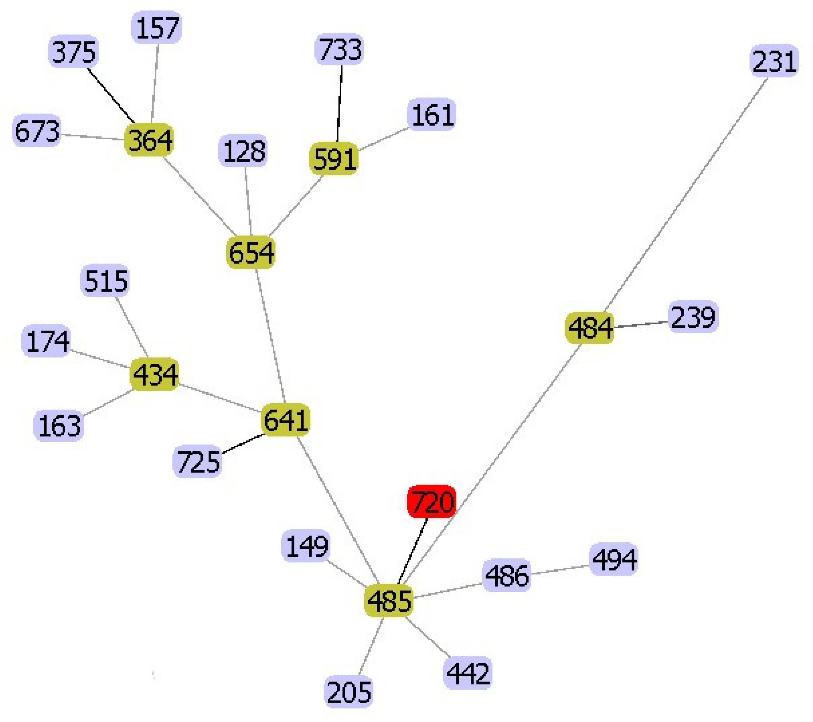
Figure 1. goeBURST analysis of Aeromonas veronii C198 (ST720). Other numbers indicate the other sequence types (STs).
Pathogens 09 01031 g001
Figure 2. Whole-genome phylogeny analysis of A. veronii. A phylogenetic tree was generated using REALPHY and visualized with MEGA-X software using the neighbor-joining method. Nine major clusters are shown, separated by dashed lines and numbers. Blue boxes indicate the four subgroups described by Tekedar et al. (2019), and the red box indicates our isolate and related isolates. Escherichia coli K12 was used as an outgroup.
Pathogens 09 01031 g002
Figure 3. Comparison of the genetic organization surrounding the mcr-3 and mcr-3-like genes in five A. veronii isolates. The schematic shows the genes flanking the mcr-3 and mcr-3-like genes in these isolates.
Pathogens 09 01031 g003
Figure 4. Unrooted neighbor-joining tree based on amino acid sequences of all MCR-3 like variants described. The phylogenetic tree was generated and visualized with MEGA-X software using the neighbor-joining method with 500 bootstraps after applying the Poisson model.
Pathogens 09 01031 g004
Table 1. Characteristics of the A. veronii C198 isolate used in this study.
CharacteristicsA. veronii C198
Sequence TypeST720
Antimicrobial susceptibility (μg/mL) *
Amoxicillin–clavulanic acid32/16 (R)
Ampicillin–sulbactam32/16 (R)
Piperacillin–tazobactam2/4 (S)
Cefepime0.25 (S)
Cefotaxime0.25 (S)
Cefoxitin1 (S)
Ceftazidime0.5 (S)
Ertapenem0.5 (S)
Imipenem0.125 (S)
Meropenem0.125 (S)
Amikacin4 (S)
Gentamicin1 (S)
Tetracycline32 (R)
Ciprofloxacin0.25 (S)
Levofloxacin0.25 (S)
Chloramphenicol2 (S)
Trimethoprim–sulfamethoxazole2/38 (S)
Colistin2 (ND **)
Antimicrobial and resistance genes
β-lactamblacphA3
blaOXA-12
TetracyclinetetA
Colistinmcr-3
Fluoroquinolone and tetracyclineadeF
Fluoroquinolone, diaminopyrimidine, and phenicolrsmA
elfamycinEF-Tu (R234F) #
Virulence genes
AdherenceFlagella
Mannose-sensitive hemagglutinin
Tap type IV pili
Type I fimbria
Secretion systemType II secretion system
Type III secretion system
Type VI secretion system
ToxinsAerolysin
Hemolysin III
Hemolysin, HlyA
Thermostable hemolysin
R: resistant, S: susceptible, I: intermediate. * Susceptibility to antimicrobials was performed using broth microdilution. ND **: no clinical breakpoint for colistin in Aeromonas spp. by either the Clinical Laboratory Standards Institute (CLSI) (M45) or EUCAST guidelines. # Substitution of amino acid at position 234 in EF-Tu.
Table 2. Comparison of antimicrobial resistance genes in A. veronii isolates from various specimens.
Strain Accession No.SourceCountry MCRcphAOXA-12OXA-21TEMSHVAAC(6’)AAC(3)aadAAPH(3’)-IaAPH(6)-IdqnrtetmphAfloRdfrAsularr-3catadeFrsmAqacEdelta1EF-Tu Mutant
C198JACEGL000000000HumanThailand3.41A3+ A ++ R234F
126-14PPTE00000000HumanChina3.25A3+ D ++ R234F
MGYG-HGUT-02529CABMOE000000000HumanChina3.3A3++ IIa A16 A A151 B3+++R234F
ZJ12-3UETM00000000HumanChina3.3A3++ IIa A16 A A151 B3+++R234F
AVNIH1CP014774HumanUSA A3+ 134Iic, Ib10IIgA2 B4D, E++A121, 2 +++R234F
AVNIH2LRBO00000000HumanUSA A3+ E ++ R234F
AER39AGWT00000000HumanUSA A3+ ++ R234F
AMC35AGWW00000000HumanUSA A3+ ++ R234F
BVH46NKWS01000000HumanUSA A3+ ++ R234F
FDAARGOS_632CP044060HumanUSA A3+ ++ R234F
AER397AGWV00000000HumanUSA A6+ ++ R234F
CCM4359MRZR00000000HumanUSA A6+ ++
CECT4257TCDDK00000000HumanUSA A6+ ++
FC951PKSR00000000HumanIndia A3+ ++ R234F
VBF557LXJN00000000HumanIndia A3 R234F
312MRHDQ00000000HumanBrazil A3+ ++ R234F
BC88CAAKNH000000000HumanAustralia A7+ ++ R234F
UDRT09JAAQQM000000000FishThailand A3+ 116 S2E ++ R234F
CNRT12JAAQQN000000000FishThailand A3+ ++ R234F
NK07JAAQQQ000000000FishThailand A3+ A2 A+ A121 +++R234F
TH0426CP012504CatfishChina A3+ ++ R234F
CB51CP015448FishChina A3+ + R234F
JC529CP058912FishChina A3+ ++ R234F
A8-AHPCP046407FishIndia A3+ + ++ R234F
Ae52BDGY00000000GoldfishSri-Lanka A3+ A2++ A, D+ A121 II+++
AG-5.28.6NNSE00000000FishGreece A3+ ++
PDBNMUS00000000FishGreece A3+ ++
B48WSFS00000000FishBrazil A3+ ++ R234F
ML09-123PPUW00000000CatfishUSA + ++ R234F
MS-17-88RAWX00000000CatfishUSA3, 7.1A3+ D +A3 B7++ R234F
TTU2014-141AMELKKD00000000CattleUSA A3+ E ++
TTU2014-115ASCLKJS00000000CattleUSA A3+ ++ R234F
Z2-7UETI00000000PorkChina3.3A3+ D A151 +++R234F
HX3CP040717AlligatorChina3.3A3+ D ++ R234F
17ISAeCP028133Discus fishKorea3.6, 3.8A3+ Ib-cr A A11+ +++R234F
CCM7244MRZQ00000000Surface waterGermany A6+ ++ R234F
AK236NKXF00000000Lake waterFrance A3+ ++
ARB3JRBE00000000Pond waterJapan A6+ ++
KLG5CAAKNL000000000RiverEngland A3+ ++ R234F
B565NC_015424SedimentChina A6+ ++ R234F
PamvoticaMRUI00000000SedimentGreece A3+ ++ R234F
+: means present; blank means absent.
Publisher’s Note: MDPI stays neutral with regard to jurisdictional claims in published maps and institutional affiliations.