Article

Molecular Characteristics of Rickettsia in Ticks Collected along the Southern Border of Mongolia

1
Department of Global and Community Health, George Mason University, Fairfax, VA 22030, USA
2
United States Army Medical Research Institute of Infectious Diseases (USAMRIID), Frederick, MD 21702, USA
3
Johns Hopkins School of Medicine, Baltimore, MD 21218, USA
4
Department of Clinical Sciences, College of Veterinary Medicine, North Carolina State University, Raleigh, NC 27606, USA
5
National Center for Zoonotic Diseases, Ulaanbaatar 16000, Mongolia
*
Author to whom correspondence should be addressed.
Pathogens 2020, 9(11), 943; https://doi.org/10.3390/pathogens9110943
Submission received: 20 October 2020 / Revised: 4 November 2020 / Accepted: 9 November 2020 / Published: 13 November 2020
(This article belongs to the Special Issue Virulence Mechanisms of Rickettsiae)

Abstract

:
Tick-borne infections are a significant threat to public health, particularly in regions where individuals frequently enter tick habitats. Roughly 26% of the population in Mongolia practice nomadic pastoralism and are considered at high risk of exposure to ticks and the diseases they carry. This study tested ticks from Mongolia’s southern border for Rickettsia spp. to better understand the epidemiology of tick-borne diseases in the region. Dermacentor nuttalli and Hyalomma asiaticum ticks (n = 4022) were pooled and tested for Rickettsia spp. by real-time PCR. Melt-curve analyses and Sanger sequencing were used to identify Rickettsia species. Approximately 64% of the 786 tick pools tested positive for Rickettsia bacteria. Melt curve analyses identified four different Rickettsia species circulating in these tick pools. Amplicon sequencing of the ompA gene identified Rickettsia spp. that closely resembled R. raoultii and R. sibirica. Dermacentor nuttalli ticks from Govi-Altai had the highest maximum likelihood estimation infection rate 48.4% (95% CI: 41.7–56.5%), while Hyalomma asiaticum collected in Omnogovi had a rate of 7.6% (95% CI: 6.2–9.2%). The high detection of Rickettsia suggests a substantial risk of infection in southern Mongolia. Further studies are necessary to investigate the clinical burden of tick-borne diseases in Mongolia.

1. Introduction

Rickettsial infections are on the rise globally and pose an emerging threat to human health [1,2,3]. Transmission and infection occur after arthropods such as ticks and fleas suck blood from a host, causing mild to fatal illnesses (ex. spotted fevers and typhus) characterized by non-specific fever, myalgia nausea, and rash [4]. Spotted fever group (SFG) rickettsial organisms are gram-negative obligate intracellular coccoid-shaped bacteria that can infect a variety of mammalian species, including livestock and humans. Tick-borne SFG Rickettsia are distinguished from the Rickettsia typhus group (TG) by vector, clinical presentation, and the presence of the outer membrane protein ompA, which is absent in the TG Rickettsia [5]. The epizootiology of Rickettsia spp. is further complicated by transovarial and transstadial transmission within the vector tick species [6].
Roughly 26% of Mongolia’s population of three million residents engage in traditional pastoral herding. This subset of the population is at an increased risk of exposure to both zoonotic and vector-borne infectious diseases [7,8,9]. Tick-borne rickettsioses in particular have a significant impact on the health of this at-risk population: peak tick bite rates occur during economically productive months (increased risk of disease), and low healthcare-seeking rates despite the presence of symptoms delay treatment [10]. Tick-borne diseases result in additional economic losses incurred from illness in livestock [8].
Multiple Rickettsia species, including R. raoultii, R. sibirica sibirica, R. sibirica mongolitimonae, R. heilongjiangensis, and “Candidatus R. tarasevichiae”, have been identified in Dermacentor spp., Ixodes persulcatus, and Haemaphysalis concinna ticks collected in and around Mongolia [3,8,11,12,13,14]. Of these, only R. raoultii (scalp eschar and neck lymphadenopathy after tick bite, or SENLAT) and R. sibirica sibirica (Siberian tick typhus) are known to cause human disease [15]. Characterization of Rickettsia species is also important due to serious clinical diseases in neighboring countries [16,17,18,19,20,21]. A previous study by our group collected and tested Dermacentor nuttalli and Hyalomma asiaticum tick pools for the presence of Crimean-Congo hemorrhagic fever virus by real-time RT-PCR [9]. Here, we utilized this same sample set to assess the presence and distribution of different Rickettsia spp. in Mongolia.

2. Results

A total of 4022 ticks [Dermacentor nuttalli (n = 2396) and Hyalomma asiaticum (n = 1626)] were collected across southern Mongolia. Livestock sampling resulted in the collection of 592 D. nuttalli (25.7%) and 1028 H. asiaticum (63.2%) partially fed ticks, with the remaining 2402 ticks collected directly from the environment. A total of 467 D. nuttalli and 319 H. asiaticum tick pools, sorted by collection method and location, were included for testing. Initial testing by real-time PCR found 64% of tick pools tested positive (505/786) for Rickettsia spp. with D. nuttalli and H. asiaticum detection rates of 86% and 33%, respectively. The highest Rickettsia spp. pool detection rate was observed in Govi-Altai at 95% (195/204). Table 1 depicts a summary of the tick collection locations, species identification, and testing results.
Overall, maximum likelihood estimates (MLE) found Rickettsia spp. present at an average prevalence of 33.2% (95% CI: 30.1–36.2%) in Dermacentor ticks, and 7.4% (95% CI: 6.1–8.9%) Hyalomma ticks collected along the southern border (Figure 1). Bayankhongor had an 82% prevalence (55/67 positive tick pools) with an MLE of 30.1% (95% CI: 22.9–37.3%) and a minimum infection rate (MIR) of 16.5% (95% CI: 12.5–20.4%). In contrast, Omnogovi aimag had the highest percentage of Rickettsia-positive Hyalomma ticks, with a pool-positive rate of 33% (96/289), an MLE of 7.59% (95% CI: 6.24–9.16%), and MIR of 6.5% (95% CI: 5.2–7.6%). No association was observed in detection rates when examining H. asiaticum (p = 0.48) and D. nuttalli (p = 0.27) removed from livestock, compared to those collected from the environment.
Melt curve analysis of the amplicon generated at the end of the real-time PCR reaction can differentiate some species of Rickettsia based on the impact nucleic acid compositional differences have on strand binding kinetics. Analysis of these melt curves identified at least eight distinct curves, suggesting a wide diversity of Rickettsia spp. in circulation in the Mongolian tick population. Thirty samples were selected for Sanger sequencing based on the melt curve analysis, tick species, and geographic distribution (Supplementary Table S1). An approximate 212 base pair segment of ompA was amplified, sequenced, and BLAST-identified. Rickettsia raoultii (n = 13), R. sibirica mongolitimonae (n = 6), and R. sibirica (n = 2) were detected. One isolate from Dornogovi, located in southeastern Mongolia, shared 97.66% identity with R. sibirica mongolitimonae (MK922654) and was detected in a pool of H. asiaticum ticks removed from livestock (Figure 2).

3. Discussion

Rickettsia spp. circulate at a high rate within native tick species in Mongolia, presenting a significant health risk to pastoralist populations in close contact with their environment. The highest MLE rate of 48.4% (95% CI: 41.7–56.5%) was observed in Dermacentor ticks from the Govi-Altai region. Additionally, an MLE rate of 7.6% (95% CI: 6.2–9.2%) was observed in Hyalomma ticks collected in Omnogovi, warranting further testing. Overall, a large percentage of D. nuttalli pools (86%) tested positive for Rickettsia spp. by real-time PCR, and nearly all the ticks tested from the Govi-Altai region tested positive. Melt curve analysis found a high amount of Rickettsia spp. diversity; ompA sequencing identified four species of Rickettsia (R. raoultii, R. sibirica mongolitimonae, R. sibirica, and one species closely related to R. sibirica mongolitimonae) known to cause human disease. Utilizing melt curve analysis in tandem with Sangar sequencing allowed our team to detect multiple circulating Rickettsia species without requiring extensive sequencing of positive samples.
Speck and colleagues (2012) found prevalence rates of R. raoultii (82%) and R. sibirica (4%) in Dermacentor nuttalli ticks in northern Mongolia; 5% of the identified Rickettsia spp. were not able to be assigned to a specific tick species [14]. Most infected ticks were found in the Selenge and Khentii aimags, with R. sibirica being found exclusively in ticks from Arkhangai aimag [14]. PCR analysis of ticks collected at Sino-Russian and Sino-Mongolian borders found a 53.4% prevalence of Rickettsia phylogenetically belonging to R. raoultii [20]. Boldbaatar and colleagues (2017) detected a 15.1% prevalence of R. raoultii in D. nuttalli in Dornogovi (a southern aimag), while higher maximum likelihood estimates (MLEs) were found in the northern aimags of Selenge and Tov [7]. This study found an MLE of 39.6 (95% CI: 17.4–61.3%) in Dornogovi among Dermacentor ticks; however, this finding is limited given the sample size (12 pools). Hyalomma ticks collected from the same area had a much lower MLE, 4.6 (95% CI: 2.1–9.2%), with only 6/27 pools testing positive. Furthermore, previous work in Dornogovi has indicated serological evidence of Rickettsia exposure in both herders and livestock [8]. The high detection rates observed in this study, paired with previously published findings from elsewhere in Mongolia, indicate that the distribution of ticks harboring pathogenic rickettsia is ubiquitous throughout the country, representing a major public health concern.
Continued vector surveillance is necessary, especially in the Govi-Altai region, which had the highest detection rate in sampled ticks for this study. Enhanced serological and syndromic surveillance are also needed to determine the clinical burden of SFG Rickettsia in Mongolia, paying particular attention to pastoral herding populations. Such studies will help characterize the relationship between the high detection rates of Rickettsia found in Mongolian ticks and their impact on both human and animal health. Considering the severity of the clinical symptoms of Rickettsia isolates reported in neighboring China and Russia, it is possible that these same pathogens may also be circulating in Mongolia.

4. Materials and Methods

4.1. Sample Collection, Study Location, and Processing

Questing environmental ticks and ticks removed from livestock were collected in 2013 and 2014 by the National Center for Zoonotic Diseases (Ulaanbaatar, Mongolia) from five aimags in southern Mongolia (Khovd, Govi-Altai, Bayankhongor, Omnogovi, and Dornogovi;). Adult Dermacentor nuttalli (n = 2396) and Hyalomma asiaticum (n = 1626) ticks were pooled into 2011 pools based on tick identity, sex, geographic location, and collection method (livestock vs. environment). Tick species were determined based on morphological classification, using local keys. Tick pools were homogenized (SPEX SamplePrep MiniG® 1600 tissue homogenizer (Metuchen, NJ, USA)) and total nucleic acid extracted (TRIzol LS® reagent, KingFisher Flex Purification System, MagMax 96 for MicroArrays Total RNA Isolation Kit (ThermoFisher Scientific, Waltham, MA, USA)). These homogenates were further pooled for testing (786 pools of 2–6 ticks each). All extracted nucleic acid and homogenized tick pools were stored at −70 °C until testing.

4.2. Rickettsia spp. Testing

Five microliter nucleic acid pools were tested in duplicate for Rickettsia spp. utilizing a real-time PCR assay with melt curve analysis targeting the 23s–5s ITS region with 0.4 μM (final concentration) primers Rick23-5 F (5′- AGCTCGATTGATTTACTTTGCTG -3′) and Rick23-5 R (5′- CCACCAAGCTAGCAATACAAA-3′) and SsoAdvanced SYBR Green Supermix (Bio-Rad, Hercules, CA, USA) in a 25 μL reaction [22]. Cycling conditions were 98 °C for 3 min; 40 cycles of (98 °C for 15 s, 62 °C 15 s, and 72 °C for 15 s) were followed by a melt curve analysis of the 75–90 °C range with measurements in 0.5 °C increments. Samples were run on the LightCycler 480 (Roche, Indianapolis, IN, USA). Melt curve analysis was used as a rationale to identify candidates for sequencing, based on amplicon melt temperatures potentially indicating different Rickettsia spp.: R. amblyomma (78 °C), R. bellii (76.5 °C), R. canadensis (77.5 °C), R. conorii (77.5 °C), R. montanesis (77 °C), R. parkeri (78 °C), R. typhi (75.5 °C), R. rickettsii (77 or 78 °C), R. rhipicephali (78 °C), R. felis (78 °C), “Candidatus R. amblyommii” (78.5 °C), R. honei (78 °C), and R. raoultii (78 °C) [22].
A 212 base pair amplicon of ompA was amplified and sequenced using the Big Dye Direct Sanger Sequencing Kit (ThermoFisher Scientific) for samples selected based on the melt curve analysis. Amplification used the primers Rick-ompA-F (5′-TGTAAAACGACGGCCAGTGCTTTATTCACCACCTCAAC) and Rick-ompA-R (5′- CAGGAAACAGCTATGACCTRATCACCACCGTAAGTAAAT) modified for the Big Dye Direct Sanger Sequencing kit (underlined sequence). Sequences from the forward and reverse primers were assembled and analyzed using CLC Genomics Workbench v10.1.2. (Qiagen, Hilden, Germany) Amplicon sequences, not including the primer sequences, were deposited into GenBank (#MW013059-MW013079).

4.3. Statistical Analysis

Maximum likelihood estimates (MLE) and minimum infection rates (MIR) were calculated to estimate the likelihood of pathogen detection from pooled samples based on laboratory findings, both of which are common measurements used when examining pooled samples. Chi-square statistic was used to determine significance (p < 0.05) in Rickettsia detection rates by species between ticks removed from livestock and those collected from the environment.

5. Conclusions

Rickettsial pathogens have a complex disease ecology, with a wide distribution of hosts that includes mammals, humans, and ectoparasites. Public health campaigns can be used to increase awareness and inform populations within these regions of the potential risk ticks play, especially among pastoralist who have regular contact with livestock. This study found an alarming proportion of adult ticks carrying bacterial species belonging to the SFG Rickettsia. Increased syndromic surveillance, particularly in southern Mongolia and among high-risk populations, is needed to further characterize the epidemiology of tick-borne diseases in Mongolia.

Supplementary Materials

The following are available online at https://www.mdpi.com/2076-0817/9/11/943/s1, Table S1: ompA sequences and accession numbers.

Author Contributions

M.E.v.F. designed study, conducted biological analysis and sequencing, drafted manuscript, and performed statistical analysis; M.A.V. and J.W.K. conducted DNA extraction, biological analysis, and sequencing. C.A. and B.L. drafted manuscript and conducted biological analysis; B.Q. drafted manuscript, provided controls and protocols, and assisted with optimization of lab assays; K.M.H. performed statistical analysis and data visualization; U.B. and B.J. collected samples and organized original dataset; R.J.S. designed study, coordinated efforts, and drafted manuscript. All authors have read and agreed to the published version of the manuscript.

Funding

The laboratory work was funded in part by the Global Emerging Infections Surveillance (GEIS) Section of the Armed Forces Health Surveillance Branch (AFHSB) research plans (ProMIS P00123-16-RD and P0017-17-RD) through USAMRIID and (ProMIS P0113-19-AF) through AFRIMS. This material was further supported by the Naval Medical Logistics Command under Contract No. N62645-18-D-5058.

Acknowledgments

The authors would like to gratefully thank all the men and women of the Mongolia whose contributions directly or indirectly made this work possible. Thanks to Susana Padilla, Lucas Bagley, and Andrew Haddow for assistance in sorting, identifying and tick homogenization. Opinions, interpretations, conclusions, and recommendations are those of the authors and are not necessarily endorsed by the U.S. Army.

Conflicts of Interest

The authors declare no conflict of interest.

References

  1. Abdad, M.Y.; Abou Abdallah, R.; Fournier, P.E.; Stenos, J.; Vasoo, S. A Concise Review of the Epidemiology and Diagnostics of Rickettsioses: Rickettsia and Orientia spp. J. Clin. Microbiol. 2018, 56. [Google Scholar] [CrossRef] [PubMed] [Green Version]
  2. Blanton, L.S. The Rickettsioses: A Practical Update. Infect. Dis. Clin. N. Am. 2019, 33, 213–229. [Google Scholar] [CrossRef] [PubMed]
  3. Parola, P.; Paddock, C.D.; Socolovschi, C.; Labruna, M.B.; Mediannikov, O.; Kernif, T.; Abdad, M.Y.; Stenos, J.; Bitam, I.; Fournier, P.-E.; et al. Update on tick-borne rickettsioses around the world: A geographic approach. Clin. Microbiol. Rev. 2013, 26, 657–702. [Google Scholar] [CrossRef] [PubMed] [Green Version]
  4. Galanakis, E.; Bitsori, M. When to think of Rickettsia. Pediatr. Infect. Dis. J. 2019, 38, S20–S23. [Google Scholar] [CrossRef]
  5. Pérez-Osorio, C.E.; Zavala-Velázquez, J.E.; Arias León, J.J.; Zavala-Castro, J.E. Rickettsia felis as emergent global threat for humans. Emerg. Infect. Dis. 2008, 4, 1019–1023. [Google Scholar] [CrossRef]
  6. Moore, T.C.; Pulscher, L.A.; Caddell, L.; von Fricken, M.E.; Anderson, B.D.; Gonchigoo, B.; Gray, G.C. Evidence for transovarial transmission of tick-borne rickettsiae circulating in Northern Mongolia. PLoS Negl. Trop. Dis. 2018, 12, e0006696. [Google Scholar] [CrossRef] [Green Version]
  7. Boldbaatar, B.; Jiang, R.R.; von Fricken, M.E.; Lkhagvatseren, S.; Nymadawa, P.; Baigalmaa, B.; Wang, Y.; Anderson, B.D.; Jiang, J.; Gray, G.C. Distribution and molecular characteristics of rickettsiae found in ticks across Central Mongolia. Parasites Vectors 2017, 10, 61. [Google Scholar] [CrossRef] [Green Version]
  8. von Fricken, M.E.; Lkhagvatseren, S.; Boldbaatar, B.; Nymadawa, P.; Weppelmann, T.A.; Baigalmaa, B.O.; Andersonbg, B.D.; Rellerbg, M.E.; Lantosh, P.M.; Graybg, G.C. Estimated seroprevalence of Anaplasma spp. and spotted fever group Rickettsia exposure among herders and livestock in Mongolia. Acta Trop. 2018, 177, 179–185. [Google Scholar] [CrossRef]
  9. Voorhees, M.A.; Padilla, S.L.; Jamsransuren, D.; Koehler, J.W.; Delp, K.L.; Adiyadorj, D.; Baasandagwa, U.; Jigjav, B.; Olschner, S.P.; Minogue, T.D.; et al. Crimean-Congo Hemorrhagic Fever Virus, Mongolia, 2013–2014. Emerg. Infect. Dis. 2018, 24, 2202–2209. [Google Scholar] [CrossRef]
  10. Lkhagvatseren, S.; Hogan, K.M.; Boldbaatar, B.; von Fricken, M.E.; Anderson, B.D.; Pulscher, L.A.; Caddell, L.; Nymadawa, P.; Gray, G.C. Discrepancies between self-reported tick bites and evidence of tick-borne disease exposure among nomadic Mongolian herders. Zoonoses Public Health 2019, 66, 480–486. [Google Scholar] [CrossRef]
  11. Byambaa, B. Nature-focal rickettsioses in Mongolia. Two decades of Russian-Mongolian scientific collaboration. Vestn Ross Akad Med Nauk 2008, 7, 44–45. [Google Scholar]
  12. Pulscher, L.A.; Moore, T.C.; Caddell, L.; Sukhbaatar, L.; von Fricken, M.E.; Anderson, B.D.; Gonchigoo, B.; Gray, G.C. A cross-sectional study of small mammals for tick-borne pathogen infection in northern Mongolia. Infect. Ecol. Epidemiol. 2018, 8, 1450591. [Google Scholar] [CrossRef] [PubMed]
  13. Sandagdorj, N.; Punsantsogvoo, M.; Davaasuren, P.; Enkhtaivan, B.; Battsetsegn, B.; Banzragch, B. Molecular biological detection of emerging tick-borne zoonotic pathogens in ixodid tick species. Mong. J. Agric. Sci. 2015, 13. [Google Scholar] [CrossRef] [Green Version]
  14. Speck, S.; Derschum, H.; Damdindorj, T.; Dashdavaa, O.; Jiang, J.; Kaysser, P.; Jigjav, B.; Nyamdorj, E.; Baatar, U.; Munkhbat, E.; et al. Rickettsia raoultii, the predominant Rickettsia found in Mongolian Dermacentor nuttalli. Ticks Tick Borne Dis. 2012, 3, 227–231. [Google Scholar] [CrossRef]
  15. Angelakis, E.; Pulcini, C.; Waton, J.; Imbert, P.; Socolovschi, C.; Edouard, S.; Dellamonica, P.; Raoult, D. Scalp eschar and neck lymphadenopathy caused by Bartonella henselae after Tick Bite. Clin. Infect. Dis. Off. Publ. Infect. Dis. Soc. Am. 2010, 50, 549–551. [Google Scholar] [CrossRef] [Green Version]
  16. Jia, N.; Zheng, Y.C.; Jiang, J.F.; Ma, L.; Cao, W.C. Human infection with Candidatus Rickettsia tarasevichiae. N. Engl. J. Med. 2013, 369, 1178–1180. [Google Scholar] [CrossRef] [Green Version]
  17. Jia, N.; Zheng, Y.C.; Ma, L.; Huo, Q.B.; Ni, X.B.; Jiang, B.G.; Chu, Y.L.; Jiang, R.R.; Jiang, J.F.; Cao, W.C. Human infections with Rickettsia raoultii, China. Emerg. Infect. Dis. 2014, 20, 866–868. [Google Scholar] [CrossRef]
  18. Li, H.; Fu, X.Y.; Jiang, J.F.; Liu, R.X.; Li, R.; Zheng, Y.C.; Qi, W.J.; Liu, W.; Cao, W.C. Severe illness caused by Rickettsia sibirica subspecies sibirica BJ-90 infection, China. Emerg. Microbes Infect. 2017, 6, e107. [Google Scholar] [CrossRef] [Green Version]
  19. Li, H.; Zhang, P.H.; Huang, Y.; Du, J.; Cui, N.; Yang, Z.D.; Tang, F.; Fu, F.X.; Li, X.-M.; Cui, X.-M.; et al. Isolation and Identification of Rickettsia raoultii in Human Cases: A Surveillance Study in 3 Medical Centers in China. Clin. Infect. Dis. 2018, 66, 1109–1115. [Google Scholar] [CrossRef]
  20. Liu, L.; Chen, Q.; Yang, Y.; Wang, J.; Cao, X.; Zhang, S.; Li, H.; Hou, Y.; Wang, F.; Xu, B. Investigations on Rickettsia in Ticks at the Sino-Russian and Sino-Mongolian Borders, China. Vector Borne Zoonotic Dis. 2015, 15, 785–789. [Google Scholar] [CrossRef]
  21. Liu, Q.H.; Walker, D.H.; Zhou, G.F. Serologic survey for antibodies to Rickettsia sibirica in Inner Mongolia, People’s Republic of China. Ann. N. Y. Acad. Sci. 1990, 590, 237–242. [Google Scholar] [CrossRef] [PubMed]
  22. Lado, P.; Qurollo, B.; Williams, C.; Junge, R.; Klompen, H. The microbiome of Haemaphysalis lemuris (Acari: Ixodidae), a possible vector of pathogens of endangered lemur species in Madagascar. Ticks Tick Borne Dis. 2018, 9, 1252–1260. [Google Scholar] [CrossRef] [PubMed]
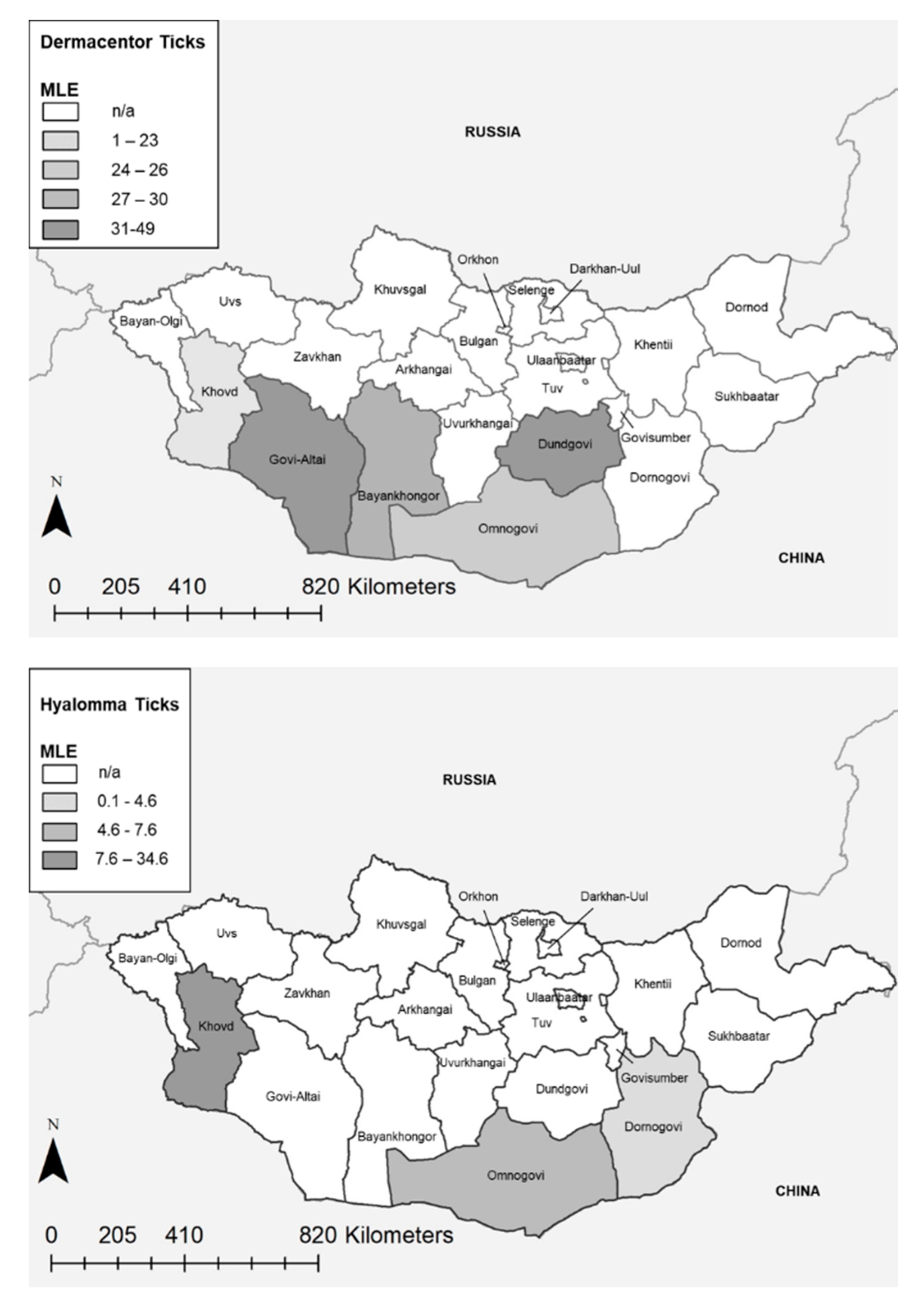
Figure 1. Maximum likelihood estimates (MLE) by Aimag for Dermacentor (above) and Hyalomma (below) ticks.
Pathogens 09 00943 g001
Figure 2. Sequence analysis of ompA gene fragment. Rickettsia ompA sequences from the Mongolian tick samples (stared, in red) were aligned with ompA sequences from multiple Rickettsia species found in GenBank. A phylogenetic tree (Neighbor Joining, Jukes-Cantor) highlights the genetic diversity of the detected Rickettsia species.
Pathogens 09 00943 g002
Table 1. Maximum likelihood estimates and minimum infection rate by region based on qPCR results, including 95% confidence intervals.
ProvinceGenusPositive Pools (%)Total TicksMLEMIR
PointLowHighPointLowHigh
BayankhongorDermacentor55/67 (82%)33430.122.937.316.512.520.4
DornogoviDermacentor10/12 (83%)5839.617.461.317.27.527.0
Govi-AltaiDermacentor195/204 (96%)105848.941.255.818.416.120.8
KhovdDermacentor34/46 (74%)23823.216.430.414.39.818.7
OmnogoviDermacentor107/138 (78%)70826.921.630.815.112.517.8
DornogoviHyalomma6/27 (22%)1444.62.19.24.21.07.4
KhovdHyalomma2/3 (66%)1034.67.369.720.00.044.8
OmnogoviHyalomma96/289 (33%)14727.66.29.26.55.27.8
TotalDermacentor401/467 (86%)239633.230.136.216.715.218.2
TotalHyalomma104/319 (33%)16267.46.18.96.45.27.6
Publisher’s Note: MDPI stays neutral with regard to jurisdictional claims in published maps and institutional affiliations.

