Article

Molecular Identification of Zoonotic Parasites of the Genus Anisakis (Nematoda: Anisakidae) from Fish of the Southeastern Pacific Ocean (Off Peru Coast)

by
Renato Aco Alburqueque
1,2,†,
Marialetizia Palomba
1,3,4,†,
Mario Santoro
4 and
Simonetta Mattiucci
1,3,*
1
Department of Public Health and Infectious Diseases, Section of Parasitology, Sapienza University of Rome, P.le Aldo Moro, 5, 00185 Rome, Italy
2
Faculty of Veterinary Medicine and Zootechnics, Universidad Peruana Cayetano Heredia, Av. Honorio Delgado 430, Lima 31, Peru
3
Laboratory Affiliated to Istituto Pasteur Italia-Fondazione Cenci-Bolognetti, Viale Regina Elena, 291, 00161 Roma, Italy
4
Department of Integrative Marine Ecology, Stazione Zoologica Anton Dohrn, Villa Comunale, 80121 Naples, Italy
*
Author to whom correspondence should be addressed.
These authors contributed equally to this work.
Pathogens 2020, 9(11), 910; https://doi.org/10.3390/pathogens9110910
Submission received: 13 October 2020 / Revised: 30 October 2020 / Accepted: 1 November 2020 / Published: 3 November 2020
(This article belongs to the Special Issue Animal Parasitic Diseases)

Abstract

:
The study aims to perform, for the first time, the molecular identification of anisakid larvae in commercial fish from the Southeastern Pacific Ocean off the Peru coast, and to provide data on their infection level by fishing ground, fish host, and site of infection. Fish specimens (N = 348) from the northern and the central coast of Peru were examined for parasites. The fish fillets were examined by the UV-press method. Anisakis spp. larvae (N = 305) were identified by mtDNA cox2 sequences analysis and by the ARMS-PCR of the locus nas10 nDNA. Two hundred and eighty-eight Anisakis Type I larvae corresponded to Anisakis pegreffii, whereas 17 Anisakis Type II larvae clustered in a phylogenetic lineage distinct from Anisakis physeteris deposited in GenBank, and corresponding to a phylogenetic lineage indicated as Anisakis sp. 2, previously detected in fish from both Pacific and Atlantic waters. Anisakis pegreffii was found to infect both the flesh and viscera, while Anisakis sp. 2 occurred only in the viscera. The average parasitic burden with A. pegreffii in the examined fish species from the two fishing grounds was significantly higher than that observed with Anisakis sp. 2. The results obtained contribute to improve the knowledge on the distribution and occurrence of Anisakis species in Southeastern Pacific waters and their implications in seafood safety for the local human populations.

1. Introduction

Industrial fishery represents an important economic sector in Peru. With an estimated production of around 7.4 million tons per year, it is a relevant food resource, and thus offers employment for the coastal populations of the Southeastern Pacific Ocean [1]. In Peru, the industrial fishery has been traditionally based on marine pelagic species, mainly constituted by anchovy (Engraulis ringens), as well as in other fish species such as jack mackerel (Trachurus murphyi), chub mackerel (Scomber japonicus), and Pacific bonito (Sarda chilensis) [1]. However, in recent years, there has been an effort to diversify the fishery into other resources including the jumbo squid (Dosidicus gigas), dolphinfish (dorado/perico) (Coryphaena hippurus), palm ruff (Seriolella violacea), and Pacific pomfret (Brama japonica) [1]. These last fish species are well distributed along the Peruvian coast, and they also represent the most highly consumed seafood by local populations [2]. In these terms, the higher demand for seafood products, more often consumed even raw, raise the concern about seafood safety and quality [3]. Among the parasites affecting seafood products, the presence of Anisakis spp. larvae have an impact for the food safety and human health [4]. The zoonotic implications associated with these parasites are a major concern, and their presence in seafood products, even when worms are dead, may significantly lower their aesthetic appeal [4,5].
To date, nine species are so far included in the genus Anisakis [4]. They are heteroxenous parasites of marine organisms, in which crustaceans (krill) act as first intermediate hosts, fishes and squids as intermediate and/or paratenic hosts and, finally, cetaceans serve as definitive ones [4]. The larval stages of Anisakis spp. commonly infect the viscera and musculature of many teleost species [4,6]. These parasites represent a public health concern causing anisakiasis, an emerging fishborne parasitic zoonosis, originating from the consumption of raw or insufficiently thermally processed seafood, carrying alive larvae [4,7,8]. In some South American countries such as Peru, Chile, Ecuador, or Colombia, the higher risk for anisakiasis has been associated to the consumption of traditional raw fish-based dishes, such as ceviche, or insufficiently thermally processed seafood (i.e., salted or marinated) [9,10,11]. Despite those feeding habits, a few human cases have been so far reported in South American countries, mostly from Peru and Chile [12,13,14,15]. However, in those cases, the etiological agent was not identified at the species level. Along the Peruvian coast, the presence of Anisakis larvae infecting several fish species with commercial importance (e.g., S. japonicus, T. murphyi, S. chilensis, C. hippurus, Merluccius gayi peruanus, B. japonica, and S. violacea) [16,17] has been reported. Likewise, the adult stage of Anisakis spp. has been found in two cetacean species from the same geographic area, Lagenorhynchus obscurus and Phocoena spinipinnis [18,19]. Some authors [20,21,22] have pointed out that the level of parasitic infection with Anisakis spp. larvae observed in T. murphy, S. japonicus, and other pelagic marine fish from Peruvian and Chilean fishing grounds, can be used as biological tags for the identification of fish stocks along the southeastern coast of the Pacific Ocean [23,24,25].
Unfortunately, those reports of Anisakis spp. larvae were so far based on morphologic features of larval stages, lacking certainty in their taxonomic identification, and jeopardizing the knowledge of the biodiversity and distribution of the species of Anisakis in the understudied region. Indeed, larval stages of Anisakis spp. cannot be recognized at the species level by morphological characters; thereby, the amount of information concerning Peruvian waters is still poor and requires further investigation. It has been underlined that the precise identification of parasites of the genus Anisakis is essential for understanding their distribution and epidemiology [4].
The present study aimed to (i) identify Anisakis spp. larvae from different economically important fish species collected from two fishing grounds of the Southeastern Pacific Ocean (off the Peru coast) using a genetic-molecular approach; (ii) improve the epidemiological data of Anisakis species genetically identified for local fisheries, according to fish species, fishing ground, and site of infection.

2. Results

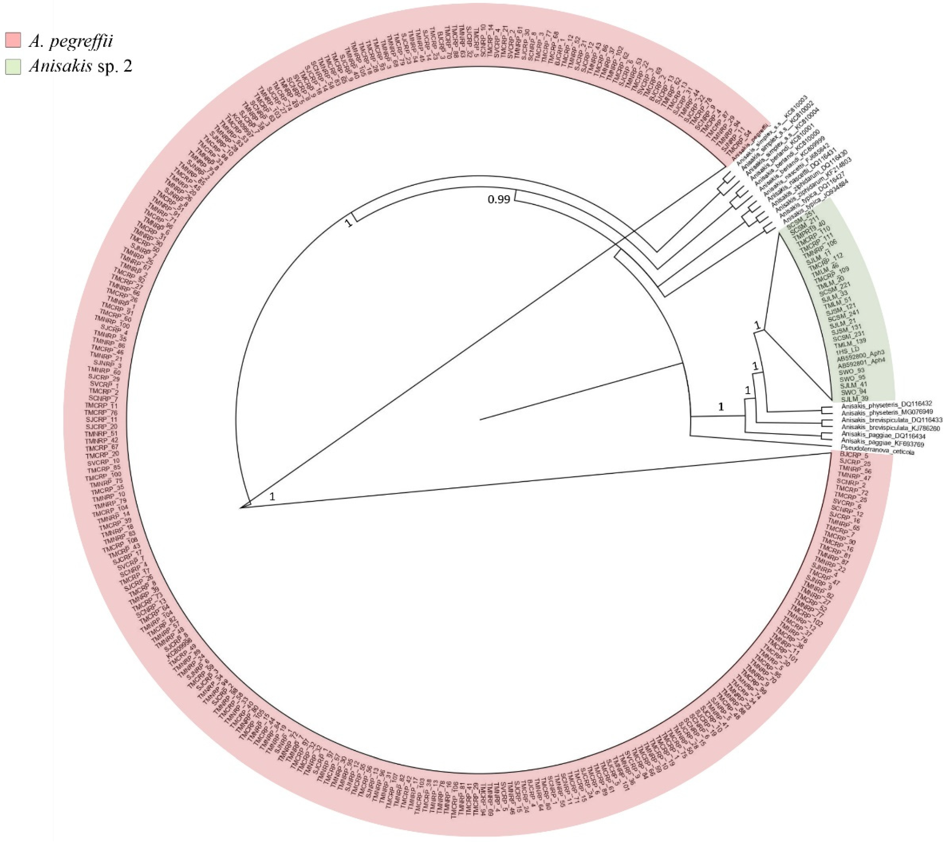
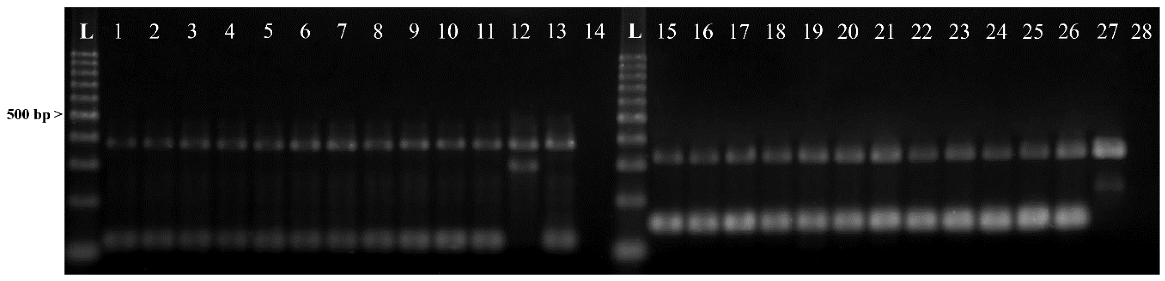
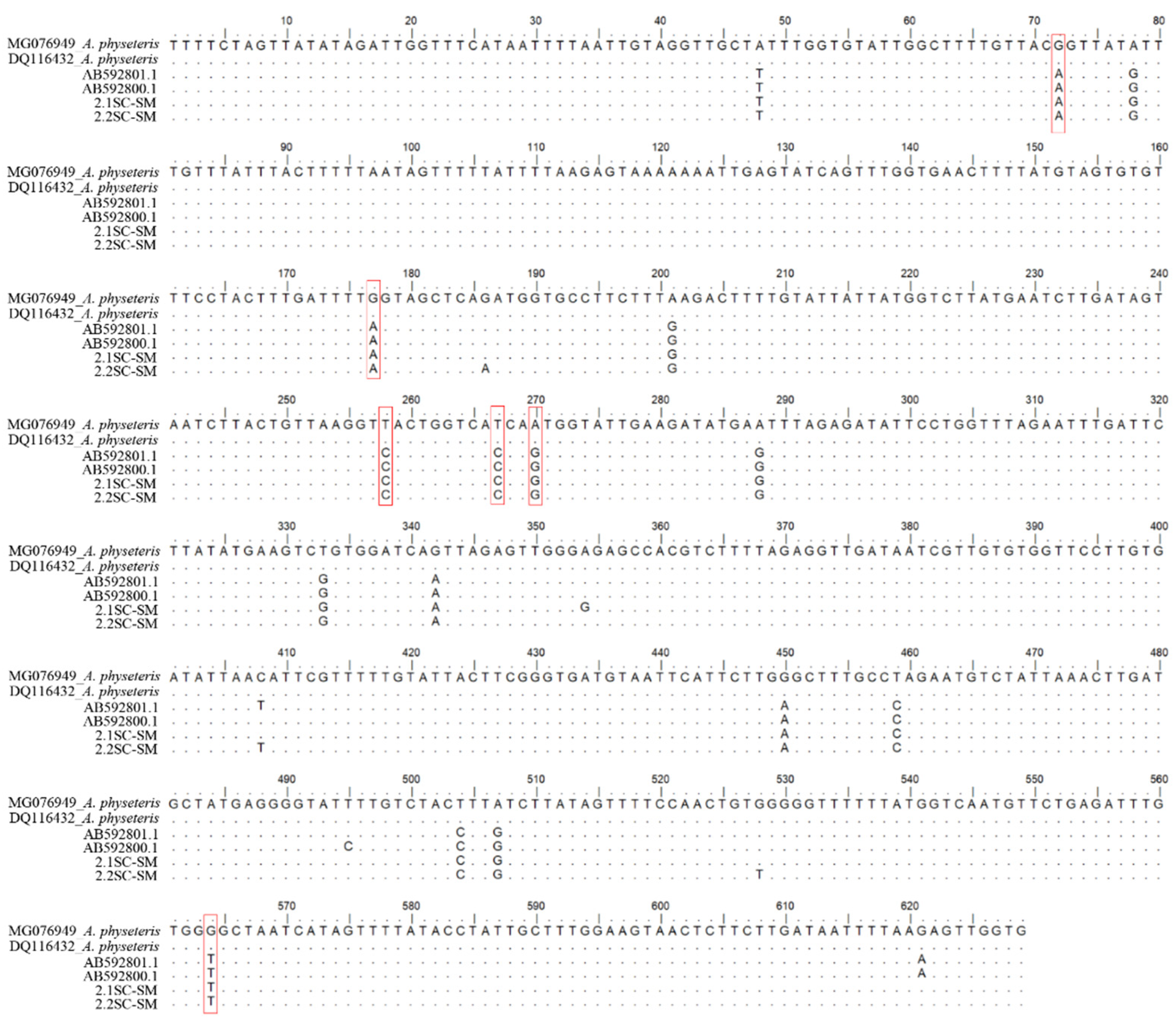
2.1. Identification of Anisakis spp.

Anisakis spp. larvae (N = 305) collected from the examined fish species were first assigned morphologically (sensu Berland, 1961) to the larval morphotype Type I (N = 288) and to the Type II (N = 17). The overall results obtained of the 348 examined fish are reported in Table 1.
According to the sequence analysis at the mtDNA cox2 gene locus, 288 Anisakis specimens were identified as A. pegreffii. Indeed, the specimens matched the 98–100% with the A. pegreffii sequences previously deposited in GenBank [26,27] (Table 2).
The phylogenetic tree resulted from the BI analysis (Figure 1) showed that those sequences obtained at the mtDNA cox2 gene from those Anisakis Type I larvae clustered in a well-supported phylogenetic lineage with a 100% posterior probability value, with the sequences of A. pegreffii, previously obtained and deposited in GenBank at that gene.
In addition, all the specimens identified as A. pegreffii (N = 288) by mtDNA cox2 sequences analysis, showed a genotype pattern belonging to the A. pegreffii species at the tetra-primer ARMS-PCR method, having the diagnostic nucleotide positions at 373 and 117 bp as those given by Palomba et al. [31] for the species A. pegreffii. Further, no heterozygote genotypes at those positions were found in the samples of Anisakis spp. tested in this study (Figure 2).
The sequences obtained at the mtDNA cox2 gene locus from the Type II larvae (N = 17) were quite distinct (96% of genetic similarity) from the reference sequences of A. physeteris deposited in GenBank (i.e., DQ116432; MG076949; MG076948; AB592798; KY595213) [26,32]; indeed, they showed six fixed base substitutions at the nucleotide position 73, 177, 258, 267, 270 and 564 (Figure 3) in comparison with those of A. physeteris. Instead, those same sequences (N = 17) had a 99.48% of genetic similarity with respect to the mtDNA cox2 sequences AB592801 and AB592800 of a genotype of A. physeteris (s.l.) previously deposited in GenBank by Murata et al. [32], and indicated by the authors as corresponding to a possible distinct lineage from A. physeteris (s.l.) [32]. The phylogenetic inference (BI) here obtained (Figure 1), showed that the sequences from those Anisakis Type II larvae are indeed forming a well-supported clade, with a high posterior probability value, including also those two sequences AB592801 and AB592800 (indicated as Aph3 and Aph4 isolates in Murata et al. [32]). In addition, the same sequences of Type II larvae here obtained are clustering in the same clade (Figure 1) with those previously obtained (indicated as SWO93; SWO94 and SWO95) from Anisakis Type II larvae detected in the swordfish Xiphias gladius from equatorial Atlantic waters [33,34], and indicated as Anisakis sp. 2, as likely corresponding to a new genotype of A. physeteris (s.l.) (Table 2).
The mtDNA cox2 sequences obtained for the species of A. pegreffii are deposited in GenBank under the following Accession numbers: MW074865 and MW074866; while those of Anisakis sp. 2 under the following Accession numbers: MW074867 and MW074868.

2.2. Distribution of Anisakis spp. by Fishing Ground, Fish Species, and Site of Infection

The overall prevalence of A. pegreffii from the two fishing grounds along the Peruvian coast was 29.1% (CI 24.5–34.0) and the mean abundance was 0.83 (±2.22); whereas, Anisakis sp. 2 larvae showed a significantly lower prevalence (6.0% (3.6–9.5)) and mean abundance (0.06 (±0.24)) values. A. pegreffii larvae were detected both in the viscera and flesh of the examined fish. The two Anisakis species co-infected the same individual fish in the viscera of jack mackerel, chub mackerel, and Pacific bonito. No larvae of Anisakis sp. 2 were found to infect the fish fillets. Data on the prevalence and mean abundance of A. pegreffii and Anisakis sp. 2 larvae by the site of infection (viscera cavity and flesh) of the fish species sampled from the two fishing grounds off the Peruvian coast are given in Table 3.
The overall levels of infection with A. pegreffii in the T. murphyi from the northern coast of Peru exhibited the highest parasitic load, both in prevalence (60.0%) and mean abundance (5.25 ± 5.44), in comparison with the sample from the central coast that showed low parasitic infection rate (P = 39.4%; A= 1.09 ± 1.09). Significant differences in terms of prevalence were observed (p = 0.001); however, no significant differences concerning the mean abundance (p = 0.45) were found.
The parasitic load values with A. pegreffii in S. japonicus caught off the northern (P = 26.1 and A = 0.28) and central (P = 24.6 and A = 0.46) coast of Peru, showed no significant differences between fishing grounds (p = 0.40 for prevalence and p = 0.35 for abundance). Further, the levels of infection with Anisakis sp. 2 from the northern (P = 4.3 and A = 0.04) and central (P = 7.2 and A = 0.07) also showed no significant differences between both areas (p = 0.30 for prevalence and p = 0.15 for abundance). Regarding the other fish species, in S. violacea and B. japonica, caught off the central coast of Peru, only A. pegreffii larvae were identified. The distribution of Anisakis spp. larvae genetically identified from fish species aforementioned along the Peruvian coast are given in Figure 4.
Concerning the site of infection, the parasitic load of A. pegreffii larvae was significantly higher in the musculature of the jack mackerel (P = 30, A = 0.50) than that observed in the chub mackerel from the northern coast (P = 4.3, A = 0.04) (respectively p = 0.001 for prevalence, and p = 0.03 for abundance) (Table 3). Despite the average smaller size of the jack mackerel and chub mackerel, the fish length (TL) did not differ significantly between the two fishing areas (p = 0.65 for the northern coast and p = 0.57 for the central coast. A significant correlation (r2 = 0.18, p = 0.0001) between the fish length and the number of larvae resulted in a Spearman correlation analysis, with the total number of larvae increasing with the fish length. In particular, a significant correlation was recorded between fish length and the number of larvae infecting the flesh (r2 = 0.19, p = 0.0001), and those found in the viscera (r2 = 0.11, p= 0.0001). Furthermore, a significant correlation was recorded between the value of the parasitic abundance observed in the flesh with those detected in the viscera (r2 = 0.36, p = 0.0001).

3. Discussion

The marine ecosystem off the Peruvian coast represents an important area of exploitable fish biomass of the Southeastern Pacific Ocean region. This area currently yields more than 20 times the tonnage of fishery landings produced by other comparable regional large marine ecosystems that operate under a similar dynamic context, and is characterized by basic primary production [35,36]. The large concentration of pelagic fishes influences the patterns of abundance, spatial distribution, and trophic ecology of a great diversity of top-level predators, including marine mammals and sea birds [37,38], which are definitive hosts of anisakid nematodes [6]. For this reason, the knowledge about the composition and diversity of anisakid parasites distribution in different fish species from this geographic area enhances the possibility of the use of these parasites as indicators of fish host biology, feeding behavior as well as fish stocks composition and migration, and host phylogenetics and systematics [39]. Besides, the presence of nematodes belonging to the genus Anisakis, with their zoonotic potential, puts at risk of infection the coastal human populations of Peru, due to their high rate of consumption of raw or undercooked seafood products [4,8,11].
The zoonotic species A. pegreffii, identified for the first time in these areas by molecular markers, was detected in commercially important fish species caught in two fishing grounds along the Southeastern Pacific Ocean region off the Peruvian coast. This parasite proved to be the predominant anisakid species, and highly capable of infecting the fish fillets. The finding of A. pegreffii in T. murphyi, S. japonicus, S. chiliensis, S. violacea, and B. japonica represents new host records for this parasite species, and it enlarges its range of distribution to the eastern coast of Pacific Ocean waters from off the Peruvian coast. In addition, in this study, we have further demonstrated the utility of the nas10 nDNA nuclear gene as a molecular tool in the identification of the species of the A. simplex (s.l.) complex [31].
The data on parasitic burden here observed for the species A. pegreffii resulted congruent with previous data for the same parasite collected in other sea basins included in the geographical range of the species. For example, in the Mediterranean Sea, A. pegreffii represents the most common anisakid species, and it is found in all pelagic and demersal fish species, often with infection levels comparable to the present findings [40,41,42]. Levsen et al. [5] mentioned that A. pegreffii seems to occur occasionally in waters of the northern hemisphere (Northeast Atlantic Ocean). The distribution of A. pegreffii is also widespread in the Austral region, between 30° S and 60° S, both in the larval and adult stages [4]. A high prevalence of the parasite species in fish from the East China Sea, the Taiwanese Sea, and the Korean Sea has been also observed [43,44,45,46,47]. Along the coast of Peru, the presence of small cetaceans belonging to the family of Delphinidae is frequently reported. These taxa are listed amongst the main definitive hosts for the species of the A. simplex (s.l.) complex in the austral and boreal hemisphere [4,48]. These cetacean species would play their role as definitive hosts of A. pegreffii in the Southeastern Pacific Ocean region (off the coast of Peru). Indeed, some authors reported the presence of Anisakis sp. infecting the stomach of some delphinids, such as the dusky dolphin, Burmeister’s porpoise, and the long-beaked common dolphin (Delphinus capensis) [18,49,50]; however, no identification to species level was performed by those authors. Garcia-Godo et al. [51] also pointed out that the feeding habits of these cetaceans are based mainly on pelagic fish species.
In the present study, the sequences analysis of the mtDNA cox2 and their Bayesian inference have shown the possible existence of a further phylogenetic lineage, here indicated as Anisakis sp. 2. This genotype, represented by the Anisakis Type II larvae collected from the jack mackerel, chub mackerel, and Pacific bonito, seems to be very closely related to the species of A. physeteris. This phylogenetic lineage corresponds to the same detected by Murata et al. [32] from Japanese waters, as well as to the same taxon previously discovered by allozyme data in the swordfish from the equatorial area [33,52], and there named as Anisakis sp. 2, distinct from A. physeteris (s.l.). The specimens indicated Anisakis sp. 2 detected in the present study resulted to be indeed a divergent lineage at the mtDNAcox2 level. Anisakis sp. 2 is clustering in the same main clade formed by A. physeteris, A. brevispiculata and A. paggiae. Similar results were gathered by Murata et al. [32]. This finding seems to suggest that Anisakis sp. 2 would represent a new gene pool in the A. physeteris (s.l.) complex of species. However, the genetic investigation of further nuclear and mitochondrial gene loci is needed to clarify its phylogenetic status and taxonomic position as included in the A. physeteris (s.l.) complex of species.
The finding of A. physeteris (s.l.) larvae in T. murphyi, S. japonicus, and S. chiliensis from the northern and central coast of Peru also represents new record. The main definitive host of A. physeteris species is the sperm whale (Physeter microcephalus), even if adults of the species A. physeteris have been also identified in other physeterid, such as Kogia spp. [53]. Adults showing the same genotype of Anisakis sp. 2 have been also found at the adult stage in an individual of sperm whale from the central Atlantic Ocean Mattiucci et al. (unpublished). Along the Peruvian coast, the presence of the sperm whale has been also reported [48]. Céspedes et al. [54] have detected 50% of prevalence of Anisakis Type II larvae in the jumbo squid from the southern coast of Peru, suggesting that it could be an intermediate/paratenic host in the life cycle of the parasite in this geographic area, since it represents a prey item of sperm whale, a main definitive host of the A. physeteris (s.l.) [4].
A significant difference was found in the infection level regarding the fishing grounds and the site of infection with both Anisakis species. For instance, the overall parasite burden of A. pegreffii in jack mackerel reported on the central coast of Peru was significantly lower than on the northern coast of Peru (Table 3). A. pegreffii exhibited higher values of infection than Anisakis Type II larvae of all fish specimens from the two fishing grounds; the overall higher infection levels are generally similar to those previously reported in European hake (Merluccius merluccius) from the Mediterranean Sea [55,56]. Furthermore, A. pegreffii larvae occurred in the viscera and flesh of the fish species; whereas, the larvae of Anisakis sp. 2 were located in the viscera. The relative proportion of A. pegreffii larvae infecting the fish flesh was 5.3% of the total number of larvae collected, which was lower than those detected in the visceral cavity or embedded on the surface of the visceral organs. Similar proportions have been also observed in previous studies carried out on the species A. pegreffii [56,57].
Likewise, jack mackerel from the northern coast of Peru showed statistically significant higher prevalence in comparison with those from the central coast. This difference in the prevalence of infection could be explained by the larger body length recorded for the fish obtained off the northern coast compared to the batch originating from the central one. In all the fish samples examined, larger fish tended to show a higher abundance of the parasite. The higher prevalence in larger fish could be explained by parasite accumulation during life [58], considering that Anisakis spp. larvae accumulate in the fish host, where they may remain an undetermined time. The correlation between the size of the fish and the parasitic burden is a direct consequence of the time that the fish spent feeding on its prey [4]. The jack mackerel of the northern region recorded higher levels of infection with A. pegreffii compared to the fish of the same size range obtained from the central fishing ground. Indeed, Cipriani et al. [56] noted that the level of infection by different species of Anisakis varies depending on the fishing ground of the fish host. The infection levels of Anisakis species observed in a given geographic area can also be affected by several drivers that shape the population size of the intermediate/paratenic hosts that participate in the life cycle of the parasite [4,59,60]. However, not only biotic factors, but also abiotic parameters, such as sea water temperature and salinity, influence the biogeography and infection dynamics of Anisakis species [4].
Since the Peruvian fishery is one of the most important economic activities on the Pacific coast of South America, this study also offers a crucial food safety background for assessing the risk associated with those parasites in seafood products. The fillets, as an edible part of the fish, represent the real risk of the consumer when harboring zoonotic Anisakis spp. larvae. This aspect raises in importance especially for the coastal population along the Southeastern Pacific Ocean coast (Colombia, Ecuador, Peru, and Chile) due to their feeding habits of eating dishes based on raw fish. The fish species mainly consumed are the jack mackerel, chub mackerel, and the Pacific bonito. They are all used in preparation of raw-based fish dishes such as “ceviche” and marinated products. For this reason, the exact information on the Anisakis larvae location (site of the infection) in the fish host assumes a great importance in a detailed risk assessment for the industry to alert and protect consumers. The presence of A. pegreffii on the edible parts of the fish from Peruvian waters not only could represent a zoonosic risk (human anisakiasis) if alive larvae are ingested, but even dead larvae could provoke allergic symptoms in sensitized patients [8]. Indeed, A. pegreffii is well known to provoke gastric, intestinal and gastroallergic anisakiasis [4,61]. In addition, massive infections, such as those observed in larger fish, can reduce the market appeal to the consumers. Therefore, an accurate examination of the fish flesh allows a real assessment of the parasitic infection levels to evaluate the real threat of human infection. Finally, the correct genetic/molecular identification of the anisakid nematodes involved in the fish infection represents the basis for any epidemiological survey intended to identify the zoonotic species involved and enhances the possibility to provide an accurate human risk assessment.

4. Materials and Methods

4.1. Fish Samplings

A total of 348 fish specimens were collected from two fishing grounds along the Peruvian coast, spanning from July 2017 to February 2018. A total of 113 fish specimens (T. murphyi, S. japonicus, and S. chiliensis) were sampled from off the coast of the district of Piura, located on the northern region of Peru (NRP) (04°54′ S, 81°21′ W), whereas 235 fish (T. murphyi, S. japonicus, S. violacea, and B. japonica) from off the coast of the district of Lima, located in the central region of Peru (CRP) (12°09′ S, 77°28′ W). The fish were obtained directly from fishing craft; they were immediately frozen and shipped by a refrigerated truck to the Laboratory of Parasitology at the Faculty of Veterinary Medicine & Animal Science of “Universidad Peruana Cayetano Heredia”. The fish were then kept frozen at −20 °C, until their parasitological examination.

4.2. Parasitological Analysis

The fish were measured (total body length, TL, 5 mm accuracy) and weighed (total body weight, TW, in g) before inspection for nematode larvae. The fish were gutted, and the viscera of each individual were separated and then carefully observed with a stereomicroscope (Leica DVM6S, Germany). Each fish was manually filleted on both the left- and right-side flesh (butterfly fillets) and placed into clear plastic bags. The samples were then pressed to 1–2 mm thick layers in a hydraulic pressing device and stored overnight at −20 °C for subsequent UV-based detection of larvae, optimizing the quantification of the whole larvae [5,57,62]. Larvae were counted and assigned to the genus level according to diagnostic morphological keys [63], using an optical microscope (Leica DM750, Germany). Finally, the collected larvae were washed in saline solution, fixed in 70% ethanol, and delivered to the Laboratory of Parasitology, Department of Public Health and Infection Diseases of “Sapienza-University in Rome”, where molecular identification was performed.

4.3. Genetic Identification of Larval Nematodes

A subsample (N = 305 Anisakis spp. Larvae, out of the total 421 collected), corresponding to 72% of the larvae recovered in all the fish specimens from the different fishing grounds, was randomly selected for the identification study by the direct sequence analysis of mitochondrial gene locus mtDNA cox2, (629 bp) [27] which allowed us to distinguish all the species so far included in the genus. However, because the Southeastern Pacific Ocean would be a geographic area where the sibling species of the A. simplex (s.l.) complex (i.e., A. simplex (s.s.), A. pegreffii, A. berlandi) would occur in sympatry [4], the analysis of a nuclear marker is important for the detection of possible hybrid genotypes between those species. In this regard, the analysis of a nuclear marker, i.e., the nas10 nDNA, recently discovered as a diagnostic genetic marker between the three Anisakis species of the A. simplex (s.l.) complex [31], was also analyzed. In particular, the ARMS-PCR assay of the nas10 nDNA [31] was applied to the same specimens previously identified as A. pegreffii by mtDNA cox2 sequences analysis.
The samples fixed in alcohol were first washed in PBS and distilled water before the DNA extraction. The total DNA of each single larvae was extracted using the Quick-gDNATM Miniprep Kit (ZYMO RESEARCH). The mitochondrial cytochrome C oxidase subunit II (cox2) gene was amplified using the primers 211F (5′-TTTTCTAGTTATATAGATTGRTTYAT-3′) and 210R (5′- CACCAACTCTTAAAATTA TC-3′) [26,27]. Polymerase chain reaction (PCR) was carried out according to the previously described procedures [27]. The sequences obtained at the mtDNA cox2 for the larval nematodes were compared with those obtained in other previous studies for the same gene and deposited in GenBank: A. simplex (s.s.) (DQ116426), A. pegreffii (JQ900761), A. berlandi (KC809999), A. typica (DQ116427), A. ziphidarum (DQ116430), A. nascettii (FJ685642), A. physeteris (DQ116432), A. brevispiculata (DQ116433) and A. paggiae (DQ116434).
ARMS-PCR assay of the nuclear metallopeptidase nas10 nDNA locus was performed in two PCR reactions simultaneously. The PCR reaction was amplified using the set-1 primers: Out-F1 (5′-TATGGCAAATATTATTATCGTA-3′), Out-R1 (5′-TATTTCCGACAGCAAACAA-3′), In-F1 (5′-GCATTGTACACTTCGTATATT-3′ and In-R1 (5′-ATTTCTYCAGCAATCGTAAG-3′) and, for the set-2 primers, we used the following: Out-F2: (5′-GAAAGACAGGTTCATCTCA-3′), Out-R1 (5′-TATTTCCGACAGCAAACAA-3′), In-F2 (5′-AACGGATATGAATGATCCC-3′), In-R2 (5′-HAAATGAAAGTAGAAAGAATTTAC-3′). PCR conditions and procedures were reported in Palomba et al. [31]. The PCR products were separated by electrophoresis using agarose gel (2.5%) and stained with 20-fold dilutions of the stock (10,000×) solution of Diamond™ Nucleic Acid Dye (Promega, Madison, Wisconsin, USA) for the visualization. The distinct banding patterns were detected by the use of ultraviolet transillumination and the sizes of the fragments were determined by comparison with a 100 bp DNA ladder marker (Promega).
A Bayesian inference (BI) tree, based on mtDNA cox2 sequences obtained on the Anisakis spp. larvae from different host species and fishing grounds were elaborated including sequences of the same species previously studied and deposited in GenBank. The analysis was performed by MrBayes3.1 [28] using the GTR + G substitution model as implemented in jModeltest2.1 [29]. The parameter for the selected model was G = 0.131, calculated with the Akaike information criterion (AIC) [30].

4.4. Statistical Analysis of the Parasitic Infection Parameters

Parameters describing parasite infection, i.e., prevalence (P%) and mean abundance (A) of the infection with the detected species of Anisakis were calculated using software Quantitative Parasitology 3.0 [64]. Sterne’s exact 95% confidence limits (or adjusted Wald’s for N > 1000) were calculated for prevalence and bootstrap 95% confidence limits (2000 bootstrap replications) for mean abundance, following Bush et al. [65], Rozsa et al. [66] and Reiczigel [67]. Comparison between the levels of infection of Anisakis spp. larvae were calculated by Quantitative Parasitology 3.0 [64] using Fisher’s exact test or exact unconditional test (prevalence—depending on sample size) [68] and bootstrap two-sample t-test (mean abundance). Differences were considered significant when p < 0.05. Differences in fish body length (TL) between the two fishing areas were analyzed with t-tests or Kruskal–Wallis tests, depending on data distribution after testing for normality, separately for the jack mackerel and chub mackerel. Spearman rank tests were run to analyze the relationships between the fish host body size (TL) and the overall abundance with Anisakis spp. larvae in the viscera and the flesh separately.

Author Contributions

Conceptualization, R.A.A., M.P. and S.M.; methodology, R.A.A., M.P. and S.M.; formal analysis, R.A.A., M.P. and S.M.; investigation, R.A.A., M.P. and S.M.; writing—original draft preparation, R.A.A., M.P., M.S. and S.M.; writing—review and editing, R.A.A., M.P., M.S. and S.M.; supervision, S.M.; project administration and funding acquisition, S.M. All authors have read and agreed to the published version of the manuscript.

Funding

This research received no external funding

Acknowledgments

This study was supported by a grant from the Master’s program in Aquatic Animal Health at Universidad Peruana Cayetano Heredia subsidized by CONCYTEC-PERU (Consejo Nacional de Ciencia, Tecnología e Innovación Tecnológica), and by a Grant to the first Author from the “Organizzazione internazionale italo-latina americana” (IILA) for his fellowship at “Sapienza - University of Rome” to perform parasitological ad molecular analysis within the Project: “Molecular epidemiology and distribution of zoonotic anisakid parasites in fish from the coast of Peru: Implications on health and food quality”. S. Mattiucci carried out part of this research work with a grant from Istituto Pasteur Italia-Fondazione Cenci Bolognetti, Italy.

Conflicts of Interest

The authors declare no conflict of interest.

References

  1. Food and Agriculture Organization of the United Nations (FAO). The State of World Fisheries and Aquaculture 2016: Contributing to Food Security and Nutrition for All; Food & Agriculture Org: Rome, Italy, 2016; pp. 80–95. ISBN 978-925-109-185-2. [Google Scholar]
  2. Sanchez, D.N.; Gallo-Seminario, M. Status of and trends in the use of small pelagic fish species for reduction fisheries and for human consumption in Peru. In Fish as Feed Inputs for Aquaculture: Practices, Sustainability and Implications; Hasan, M.R., Halwart, M., Eds.; FAO: Rome, Italy, 2009; pp. 325–369. ISBN 978-92-5-106419-1. [Google Scholar]
  3. Fayer, R. Introduction and Public Health Importance of Foodborne Parasites. In Biology of Foodborne Parasites, 1st ed.; Xiao, L., Ryan, U., Feng, Y., Eds.; CRC Press: Boca Raton, FL, USA, 2015; Volume 1, pp. 2–20. ISBN 978-146-656-885-3. [Google Scholar]
  4. Mattiucci, S.; Cipriani, P.; Levsen, A.; Paoletti, M.; Nascetti, G. Molecular Epidemiology of Anisakis and Anisakiasis: An Ecological and Evolutionary Road Map. Adv. Parasitol. 2018, 99, 93–263. [Google Scholar] [CrossRef]
  5. Levsen, A.; Svanevik, C.S.; Cipriani, P.; Mattiucci, S.; Gay, M.; Hastie, L.C.; Bušelić, I.; Mladineo, I.; Horst, K.; Ostermeyer, U.; et al. A survey of zoonotic nematodes of commercial key fish species from major European fishing grounds—Introducing the FP7 PARASITE exposure assessment study. Fish. Res. 2018, 202, 4–21. [Google Scholar] [CrossRef]
  6. Mattiucci, S.; Nascetti, G. Advances and trends in the molecular systematics of anisakid nematodes, with implications for their evolutionary ecology and host-parasite co-evolutionary processes. Adv. Parasitol. 2008, 66, 47–148. [Google Scholar] [CrossRef] [PubMed]
  7. Chai, J.Y.; Murrell, K.D.; Lymbery, A.J. Fish-borne parasitic zoonoses: Status and issues. Int. J. Parasitol. 2005, 35, 1233–1254. [Google Scholar] [CrossRef] [PubMed]
  8. Audicana, M.T.; Kennedy, M.W. Anisakis simplex: From Obscure Infectious Worm to Inducer of Immune Hypersensitivity. Clin. Microbiol. Rev. 2008, 21, 360–379. [Google Scholar] [CrossRef] [PubMed] [Green Version]
  9. Cabrera, R.; Suárez-Ognio, L. Probable emergencia de anisakiosis por Anisakis physeteris durante el fenómeno El Niño 1997–1998 en la costa peruana. Parasitol. Latinoam. 2002, 57, 166–170. [Google Scholar] [CrossRef]
  10. Cabrera, R.; Trillo-Altamirano, M.D.P. Anisakidosis: Una zoonosis parasitaria marina desconocida o emergente en el Perú? Rev. Gastroenterol. Peru 2004, 24, 335–342. [Google Scholar]
  11. Eiras, J.C.; Pavanelli, G.C.; Takemoto, R.M.; Nawa, Y. Fish-borne nematodiases in South America: Neglected emerging diseases. J. Helminthol. 2018, 92, 649–654. [Google Scholar] [CrossRef]
  12. Barriga, J.; Salazar, F.; Barriga, E. Anisakiasis: Presentación de un caso y revisión de la literatura. Rev. Gastroenterol Peru 1999, 19, 317–323. [Google Scholar] [PubMed]
  13. Mercado, R.; Torres, P.; Gil, L.C.; Goldin, L. Anisakiasis en una paciente portadora de una pequeña hernia hiatal. Caso clinico. Rev. Med. Chil. 2006, 134, 1562–1564. [Google Scholar] [CrossRef] [Green Version]
  14. Weitzel, T.; Sugiyama, H.; Yamasaki, H.; Ramirez, C.; Rosas, R.; Mercado, R. Human infections with Pseudoterranova cattani, Chile. Emerging Infect. Dis. 2015, 21, 1874–1875. [Google Scholar] [CrossRef] [PubMed] [Green Version]
  15. Menghi, C.I.; Gatta, C.L.; Arias, L.E.; Santoni, G.; Nicola, F.; Smayevsky, J.; Degese, M.F.; Krivokapich, S.J. Human infection with Pseudoterranova cattani by ingestion of “ceviche” in Buenos Aires. Argentina. Rev. Argent. Microbiol. 2020, 52, 118–120. [Google Scholar] [CrossRef] [PubMed]
  16. Pérez, I.; Chávez, A.; Casas, E. Presencia de formas parasitarias en peces comerciales del mar peruano. Rev. Investig. Vet. Peru 1999, 10, 34–38. [Google Scholar] [CrossRef] [Green Version]
  17. Llerena, C.; Chávez, A.; Casas, E. Frecuencia de larvas Diphyllobothriidae y larvas Anisakidae en peces marinos comerciales del terminal pesquero de ventanilla-callao. Rev. Investig. Vet. Peru 2001, 12, 58–62. [Google Scholar] [CrossRef]
  18. Van Waerebeek, K.; Reyes, J.C.; Alfaro, J. Helminth parasites and phoronts of dusky dolphins Lagenorhynchus obscurus (Gray, 1828) from Peru. Aquat. Mamm. 1993, 19, 159–169. [Google Scholar]
  19. Reyes, J.C.; Wan Waerebeek, K. Aspects of the biology of Burmeister’s porpoise from Peru. In Scientific Committee Special Meeting Report: Biology of the Phocoenids No. 16 (Report of the International Whaling Commission, Special Issue); Bjorge, A., Donovan, G.P., Eds.; International Whaling Commission: Cambridge, UK, 1995; pp. 349–364. ISBN 978-090-697-529-9. [Google Scholar]
  20. Oliva, M.E. Metazoan parasites of the jack mackerel Trachurus murphyi (Teleostei, Carangidae) in a latitudinal gradient from South America (Chile and Perú). Parasite 1999, 6, 223–230. [Google Scholar] [CrossRef] [Green Version]
  21. George-Nascimento, M. Geographical variations in the jack mackerel Trachurus symmetricus murphyi populations in the southeastern pacific ocean as evidenced from the associated parasite communities. J. Parasitol. 2000, 86, 929–932. [Google Scholar] [CrossRef]
  22. Muñoz, G.; Olmos, V. Revisión bibliográfica de especies endoparásitas y hospedadoras de sistemas acuáticos de Chile. Rev. Biol. Mar. Oceanogr. 2008, 43, 173–245. [Google Scholar] [CrossRef]
  23. Gerlotto, F.; Gutiérrez, M.; Bertrand, A. Insight on population structure of the Chilean jack mackerel (Trachurus murphyi). Aquat. Living Resour. 2012, 25, 341–355. [Google Scholar] [CrossRef] [Green Version]
  24. Oliva, M.E. Is Anisakis simplex s.l. a biological marker for stock identification of Strangomera bentincki from Chile? J. Fish. Biol. 2013, 83, 412–416. [Google Scholar] [CrossRef]
  25. George-Nascimento, M.; Oliva, M. Fish population studies using parasites from the Southeastern Pacific Ocean: Considering host population changes and species body size as sources of variability of parasite communities. Parasitology 2015, 142, 25–35. [Google Scholar] [CrossRef]
  26. Valentini, A.; Mattiucci, S.; Bondanelli, P.; Webb, S.C.; Mignucci-Giannone, A.A.; Colom-Llavina, M.M.; Nascetti, G. Genetic relationships among Anisakis species (Nematoda: Anisakidae) inferred from mitochondrial cox-2 sequences, and comparison with allozyme data. J. Parasitol. 2006, 92, 156–166. [Google Scholar] [CrossRef]
  27. Mattiucci, S.; Cipriani, P.; Webb, S.C.; Paoletti, M.; Marcer, F.; Bellisario, B.; Gibson, D.I.; Nascetti, G. Genetic and morphological approaches distinguish the three sibling species of the Anisakis simplex species complex, with a species designation as Anisakis berlandi sp. for A. simplex sp. C (Nematoda: Anisakidae). J. Parasitol. 2014, 100, 199–214. [Google Scholar] [CrossRef]
  28. Huelsenbeck, J.P.; Ronquist, F. Bayesian Analysis of Molecular Evolution Using MrBayes. In Statistical Methods in Molecular Evolution. Statistics for Biology and Health, 1st ed.; Nielsen, R., Ed.; Springer: New York, NY, USA, 2005; pp. 183–226. ISBN 978-0-387-27733-2. [Google Scholar]
  29. Darriba, D.; Taboada, G.; Doallo, R.; Posada, D. jModelTest 2: More models, new heuristics and parallel computing. Nat. Methods 2012, 9, 772. [Google Scholar] [CrossRef] [PubMed] [Green Version]
  30. Posada, D.; Buckley, T.R. Model Selection and Model Averaging in Phylogenetics: Advantages of Akaike Information Criterion and Bayesian Approaches over Likelihood Ratio Tests. Syst. Biol. 2004, 53, 793–808. [Google Scholar] [CrossRef]
  31. Palomba, M.; Paoletti, M.; Webb, S.; Nascetti, G.; Mattiucci, S. A novel nuclear marker and development of an ARMS-PCR assay targeting the metallopeptidase 10 (nas 10) locus to identify the species of the Anisakis simplex (s.l.) complex (Nematoda, Anisakidae). Parasite 2020, 27, 39:1–39:9. [Google Scholar] [CrossRef]
  32. Murata, R.; Suzuki, J.; Sadamasu, K.; Kai, A. Morphological and molecular characterization of Anisakis larvae (Nematoda: Anisakidae) in Beryx splendens from Japanese waters. Parasitol. Int. 2011, 60, 193–198. [Google Scholar] [CrossRef]
  33. Garcia, A.; Mattiucci, S.; Damiano, S.; Santos, M.N.; Nascetti, G. Metazoan parasites of swordfish, Xiphias gladius (Pisces: Xiphiidae) from the Atlantic Ocean: Implications for host stock identification. ICES J. Mar. Sci. 2011, 68, 175–182. [Google Scholar] [CrossRef] [Green Version]
  34. Mattiucci, S.; Garcia, A.; Cipriani, P.; Santos, M.N.; Nascetti, G.; Cimmaruta, R. Metazoan parasite infection in the swordfish, Xiphias gladius, from the Mediterranean Sea and comparison with Atlantic populations: Implications for its stock characterization. Parasite 2014, 21, 35. [Google Scholar] [CrossRef] [PubMed] [Green Version]
  35. Bakun, A.; Weeks, S.J. The marine ecosystem off Peru: What are the secrets of its fishery productivity and what might its future hold? Prog. Oceanogr. 2008, 79, 290–299. [Google Scholar] [CrossRef]
  36. Oerder, V.; Colas, F.; Echevin, V.; Codron, F.; Tam, J.; Belmadani, A. Peru-Chile upwelling dynamics under climate change. J. Geophys. Res. Oceans 2015, 120, 1152–1172. [Google Scholar] [CrossRef] [Green Version]
  37. Jahncke, J.; Checkley, D.M., Jr.; Hunt, G.L., Jr. Trends in carbon flux to seabirds in the Peruvian upwelling system: Effects of wind and fisheries on population regulation. Fish. Oceanogr. 2004, 13, 208–223. [Google Scholar] [CrossRef] [Green Version]
  38. Passuni, G.; Barbraud, C.; Chaigneau, A.; Demarcq, H.; Ledesma, J.; Bertrand, A.; Castillo, R.; Perea, A.; Mori, J.; Viblanc, V.A.; et al. Seasonality in marine ecosystems: Peruvian seabirds, anchovy, and oceanographic conditions. Ecology 2016, 97, 182–193. [Google Scholar] [CrossRef]
  39. Barber, I.; Hoare, D.; Krause, J. Effects of parasites on fish behaviour: A review and evolutionary perspective. Rev. Fish. Biol. Fish. 2000, 10, 131–165. [Google Scholar] [CrossRef]
  40. Farjallah, S.; Slimane, B.B.; Busi, M.; Paggi, L.; Amor, N.; Blel, H.; Said, K.; D’Amelio, S. Occurrence and molecular identification of Anisakis spp. from the North African coasts of Mediterranean Sea. Parasitol. Res. 2008, 102, 371–379. [Google Scholar] [CrossRef]
  41. Meloni, M.; Angelucci, G.; Merella, P.; Siddi, R.; Deiana, C.; Orru, G.; Salati, F. Molecular characterization of Anisakis larvae from fish caught off Sardinia. J. Parasitol. 2011, 97, 908–914. [Google Scholar] [CrossRef]
  42. Mattiucci, S.; Cimmaruta, R.; Cipriani, P.; Abaunza, P.; Bellisario, B.; Nascetti, G. Integrating parasite data and host genetic structure in the frame of an holistic approach for stock identification in Mediterranean Sea fish species. Parasitology 2014, 142, 90–108. [Google Scholar] [CrossRef]
  43. Chou, Y.Y.; Wang, C.S.; Chen, H.G.; Chen, H.Y.; Chen, S.N.; Shih, H.H. Parasitism between Anisakis simplex (Nematoda: Anisakidae) third-stage larvae and the spotted mackerel Scomber australasicus with regard to the application of stock identification. Vet. Parasitol. 2011, 177, 324–331. [Google Scholar] [CrossRef]
  44. Setyobudi, E.; Jeon, C.H.; Lee, C.H.; Seong, K.B.; Kim, J.H. Occurrence and identification of Anisakis spp. (Nematoda: Anisakidae) isolated from chum salmon (Oncorhynchus keta) in Korea. Parasitol. Res. 2011, 108, 585–592. [Google Scholar] [CrossRef]
  45. Bak, T.J.; Jeon, C.H.; Kim, J.H. Occurrence of anisakid nematode larvae in chub mackerel (Scomber japonicus) caught off Korea. Int. J. Food Microbiol. 2014, 191, 149–156. [Google Scholar] [CrossRef]
  46. Chen, H.Y.; Shih, H.H. Occurrence and prevalence of fish-borne Anisakis larvae in the spotted mackerel Scomber australasicus from Taiwanese waters. Acta Trop. 2015, 145, 61–67. [Google Scholar] [CrossRef]
  47. Kong, Q.; Fan, L.; Zhang, J.; Akao, N.; Dong, K.; Lou, D.; Ding, J.; Tong, Q.; Zheng, B.; Chen, R.; et al. Molecular identification of Anisakis and Hysterothylacium larvae in marine fishes from the East China Sea and the Pacific coast of central Japan. Int. J. Food Microbiol. 2015, 199, 1–7. [Google Scholar] [CrossRef]
  48. Reyes, J.C. Ballenas, Delfines y otros Cetáceos del Perú. Una Fuente de Información, 1st ed.; Squema Ediciones: Lima, Perú, 2009; pp. 70–150. [Google Scholar]
  49. Tantalean, M.; Cabrera, R. Algunos helmintos de la marsopa espinosa, Phocoena spinipinnis de la Reserva Nacional de Paracas, Perú. Parasitología 1999, 23, 57–58. [Google Scholar] [CrossRef]
  50. Van Bressem, M.F.; Van Waerebeek, K.; Montes, D.; Kennedy, S.; Reyes, J.C.; García–Godos, I.; Ontón, K.; Alfaro, J. Diseases, lesions and malformations in the long-beaked common dolphin Delphinus capensis from the Southeast Pacific. Dis. Aquat. Org. 2006, 68, 149–165. [Google Scholar] [CrossRef] [Green Version]
  51. García-Godos, I.; Van Waerebeek, K.; Reyes, J.C.; Alfaro, J.; Arias-Schreiber, M. Prey occurrence in the stomach of four small cetacean species in Peru. LAJAM 2007, 6, 171–183. [Google Scholar] [CrossRef] [Green Version]
  52. Mattiucci, S.; Paoletti, M.; Borrini, F.; Palumbo, M.; Palmieri, R.M.; Gomes, V.; Casati, A.; Nascetti, G. First molecular identification of the zoonotic parasite Anisakis pegreffii (Nematoda: Anisakidae) in a paraffin-embedded granuloma taken from a case of human intestinal anisakiasis in Italy. BMC Infect. Dis. 2011, 11, 82. [Google Scholar] [CrossRef]
  53. Santoro, M.; Di Nocera, F.; Iaccarino, D.; Cipriani, P.; Guadano Procesi, I.; Maffucci, F.; Hochscheid, S.; Blanco, C.; Cerrone, A.; Galiero, G.; et al. Helminth parasites of the dwarf sperm whale Kogia sima (Cetacea: Kogiidae) from the Mediterranean Sea, with implications on host ecology. Dis. Aquat. Org. 2018, 129, 175–182. [Google Scholar] [CrossRef]
  54. Céspedes, R.E.; Iannacone, J.; Salas, A. Helmintos parásitos de Dosidicus gigas “Pota” eviscerada en Arequipa, Perú. Ecol. Apl. 2011, 10, 1–11. [Google Scholar] [CrossRef] [Green Version]
  55. Mattiucci, S.; Abaunza, P.; Ramadori, L.; Nascetti, G. Genetic identification of Anisakis larvae in European hake from Atlantic and Mediterranean waters for stock recognition. J. Fish. Biol. 2004, 65, 495–510. [Google Scholar] [CrossRef]
  56. Cipriani, P.; Sbaraglia, G.; Palomba, L.; Giulietti, L.; Bellisario, B.; Bušelić, I.; Mladineo, I.; Cheleschi, R.; Nascetti, G.; Mattiucci, S. Anisakis pegreffii (Nematoda: Anisakidae) in European anchovy Engraulis encrasicolus from the Mediterranean Sea: Considerations in relation to fishing ground as a driver for parasite distribution. Fish. Res. 2018, 202, 59–68. [Google Scholar] [CrossRef]
  57. Cipriani, P.; Acerra, V.; Bellisario, B.; Sbaraglia, G.L.; Cheleschi, R.; Nascetti, G.; Mattiucci, S. Larval migration of the zoonotic parasite Anisakis pegreffii (Nematoda: Anisakidae) in European anchovy, Engraulis encrasicolus: Implications to seafood safety. Food Control. 2016, 59, 148–157. [Google Scholar] [CrossRef] [Green Version]
  58. Valero, A.; Martín-Sánchez, J.; Reyes-Muelas, E.; Adroher, F.J. Larval anisakids parasitizing the blue whiting, Micromesistius poutassou, from Motril Bay in the Mediterranean region of southern Spain. J. Helminthol. 2000, 74, 361–364. [Google Scholar] [CrossRef] [Green Version]
  59. Valero, A.; del Mar López-Cuello, M.; Benítez, R.; Adroher, F.J. Anisakis spp. in European hake, Merluccius merluccius (L.) from the Atlantic off north-west Africa and the Mediterranean off southern Spain. Acta Parasitol. 2006, 51, 209–212. [Google Scholar] [CrossRef]
  60. Suzuki, J.; Murata, R.; Hosaka, M.; Araki, J. Risk factors for human Anisakis infection and association between the geographic origins of Scomber japonicus and anisakid nematodes. Int. J. Food Microbiol. 2010, 137, 88–93. [Google Scholar] [CrossRef]
  61. Mattiucci, S.; Fazii, P.; De Rosa, A.; Paoletti, M.; Megna, A.S.; Glielmo, A.; De Angelis, M.; Costa, A.; Meucci, C.; Calvaruso, V.; et al. Anisakiasis and gastroallergic reactions associated with Anisakis pegreffii infection, Italy. Emerg. Infect. Dis. 2013, 19, 496–499. [Google Scholar] [CrossRef]
  62. Karl, H.; Leinemann, M. A fast and quantitative detection method for nematodes in fish fillets and fishery products. Arch. Lebensmittelhyg. 1993, 44, 105–128. [Google Scholar]
  63. Berland, B. Nematodes from some Norwegian marine fishes. Sarsia. 1961, 2, 1–50. [Google Scholar] [CrossRef]
  64. Reiczigel, J.; Rozsa, L. Quantitative Parasitology 3.0. Budapest. Distributed by the authors. Available online: http://www.zoologia.hu/qp/ (accessed on 24 April 2020).
  65. Bush, A.O.; Lafferty, K.D.; Lotz, J.M.; Shostak, A.W. Parasitology meets ecology on its own terms: Margolis et al. revisited. J. Parasitol. 1997, 83, 575–583. [Google Scholar] [CrossRef] [PubMed]
  66. Rozsa, L.; Reiczigel, J.; Majoros, G. Quantifying parasites in samples of hosts. J. Parasitol. 2000, 86, 228–232. [Google Scholar] [CrossRef]
  67. Reiczigel, J. Confidence intervals for the binomial parameter: Some new considerations. Stat. Med. 2003, 22, 611–621. [Google Scholar] [CrossRef]
  68. Reiczigel, J.; Abonyi-Tóth, Z.; Singer, J. An exact confidence set for two binomial proportions and exact unconditional confidence intervals for the difference and ratio of proportions. Comput. Stat. Data Anal. 2008, 52, 5046–5053. [Google Scholar] [CrossRef]
Figure 1. Bayesian inference (BI) circular tree-based on mtDNA cox2 gene sequences of A. pegreffii and Anisakis sp. 2 (larvae obtained from fish species sampled in two fishing ground off the Peruvian coast). The analysis was performed by MrBayes 3.1 [28], using the GTR + G substitution model, as implemented in jModeltest2.1 [29]; the parameter for the selected model was G = 0.131, calculated with Akaike information criterion (AIC) [30]. For the Bayesian analysis, four incrementally heated Markov chains (using default heating values), were run for 1,000,000 generations, sampling the Markov chains at intervals of 100 generations. Numbers at the nodes are posterior probabilities. Pseudoterranova ceticola was used as an outgroup.
Pathogens 09 00910 g001
Figure 2. Products (genotypes) of tetraprimer ARMS-PCR obtained at the nas10 nDNA locus on larvae of A. pegreffii identified in the present study, using the set-1primers, showing: specimen No. 1–10: A. pegreffii genotype (bands size: 373-117 bp); 11: positive control of A. pegreffii; 12: positive control of Anisakis simplex (s.s.) (bands size: 373-296 bp); 13: positive control of Anisakis berlandi (bands size: 373-117 bp); 14: negative control. Using the set-2 primers, showing: specimen No. 15–24: A. pegreffii genotype (band size: 321-148 bp); 25: positive control of A. pegreffii; 26: positive control of A. simplex (s.s.) (band size: 321-148 bp); 27: positive control of A. berlandi (bands size: 321-216 bp); 28: negative control; L: 100 bp ladder.
Pathogens 09 00910 g002
Figure 3. Alignment of mtDNA cox2 sequences (629 bp) of Anisakis sp. 2 here sequenced with respect to those previously deposited in GenBank by Murata et al. [32] (AB592800; AB592801), and with respect to the consensus sequences of Anisakis physeteris (i.e., MG076949; DQ116432) previously obtained at the same gene. The red columns show the fixed substitutions at the nucleotide positions between the two genotypes of A. physeteris (s.l.).
Pathogens 09 00910 g003
Figure 4. Overall distribution of A. pegreffii and Anisakis sp. 2 larvae genetically identified in fish species from two fishing grounds (NRP and CRP) of the southeastern Pacific waters (off the Peru coast).
Pathogens 09 00910 g004
Table 1. Number of fish examined (N) from the northern coast (NRP) and central coast (CRP) of Peru, recorded with their total body length, weight, and the total number of Anisakis spp. larvae collected (Ncoll), and genetically identified (Nid).
NMean total body length ± SD (Min-Max) (mm)Mean Weight ± SD (Min-Max) (g)NcollNid
NRP (4°54’ S, 81°21’ W)
Trachurus murphyi21337.71 ± 22.85 (282–375)309.29 ± 30.91 (267–350)117106
Scomber japonicus47275.27 ± 22.9 (236–345)253.3 ± 38.56 (192–350)1515
Sarda chiliensis45419.33 ± 20.52 (376–473)1240.3 ± 226.9 (310–1700)2020
Total152141
CRP (12°09′ S, 77°28′ W)
Trachurus murphyi100257.82 ± 33.6 (243–383)298.17 ± 35.72 (220–460)212112
Scomber japonicus70294.07 ± 30.88 (236–358)281.67 ± 52.04 (192–460)4237
Seriolella violacea35282.67 ± 31.24 (241–379)275.23 ± 31.7 (189–346)1010
Brama japonica30329.25 ± 29.34 (283–381)310.62 ± 21.89 (285–382)55
Total269164
Table 2. Specimens of A. pegreffii and Anisakis sp. 2 genetically identified in fish species from the Southeastern Pacific waters (off the coast of Peru). NRP: Northern region of Peru. CRP: Central Region of Peru.
A. pegreffiiAnisakis sp. 2
mtDNA cox2nas10 nDNAmtDNA cox2
Trachurus murphyii
NRP1051051
CRP1081084
Scomber japonicus
NRP13132
CRP32325
Sarda chiliensis
NRP15155
Seriolella violacea
CRP1010-
Brama japonica
CRP55-
Total28828817
Table 3. Parasitic infection levels with Anisakis spp. larvae, genetically identified in fish species from two fishing grounds along the Pacific coast of Peru. Abbreviations: P—prevalence (± 95% CI) and A—mean abundance (± SD). NRP = Northern coast of Peru; CRP = Central coast of Peru.
Fishing Area/Fish SpeciesAnisakis pegreffiiAnisakis sp. 2
OverallVisceraMusculatureOverall/Viscera
P (%)AP (%)AP (%)AP (%)A
NRP (4°54’ S, 81°21’ W)
Trachurus murphyi60.0 (0.14–0.41)5.25 ± 5.4460.0 (0.36–0.81)4.75 ± 4.9130.0 (0.12–0.54)0.50 ± 0.895.0 (0.01–0.25)0.05 ± 0.22
Scomber japonicus26.1 (0.36–0.81)0.28 ± 0.5021.7 (0.11–0.36)0.24 ± 0.484.3 (0.01–0.15)0.04 ± 0.214.3 (0.01–0.15)0.04 ± 0.21
Sarda chiliensis22.7 (0.12–0.38)0.32 ± 0.6722.7 (0.12–0.38)0.32 ± 0.67--11.4 (0.04–0.25)0.11 ± 0.32
CRP (12°09’ S, 77°28’ W)
Trachurus murphyi39.4 (0.30–0.50)1.09 ± 2.3938.4 (0.29–0.49)0.94 ± 2.1314.1 (0.02–0.14)0.15 ± 0.394.0 (0.01–0.10)0.04 ± 0.20
Scomber japonicus24.6 (0.15–0.37)0.46 ± 0.9824.6 (0.15–0.37)0.39 ± 0.775.8 (0.02–0.14)0.07 ± 0.317.2 (0.02–0.16)0.07 ± 0.26
Seriolella violacea17.6 (0.07–0.35)0.32 ± 0.8117.6 (0.07–0.35)0.32 ± 0.81----
Brama japonica17.2 (0.06–0.36)0.17 ± 0.3817.2 (0.06–0.36)0.17 ± 0.38----
Publisher’s Note: MDPI stays neutral with regard to jurisdictional claims in published maps and institutional affiliations.

