Article

Faecal Indicator Bacteria and Pseudomonas aeruginosa in Marine Coastal Waters: Is there a Relationship?

MARE – Marine and Environmental Sciences Centre, ESTM, Politécnico de Leiria, 2520-630 Peniche, Portugal
*
Author to whom correspondence should be addressed.
Pathogens 2020, 9(1), 13; https://doi.org/10.3390/pathogens9010013
Submission received: 19 November 2019 / Revised: 6 December 2019 / Accepted: 18 December 2019 / Published: 21 December 2019
(This article belongs to the Section Waterborne/Foodborne/Airborne Pathogens)

Abstract

:
To estimate the quality of coastal waters, European Union Directive 2006/7/EC provides guidelines to assess levels of faecal bacteria, including Escherichia coli and intestinal enterococci. These microbiological criteria are based on studies that determine the risk of bathers having diseases caused by enteric bacteria, not necessarily measuring the potential danger associated with the presence of nonenteric pathogens. The association between the presence of faecal contaminant indicators and nonenteric pathogenic microorganisms has not been well defined yet. The purpose of this study is to establish a relationship between Pseudomonas aeruginosa and microbiological indicators of faecal contamination. Presence of microbiological contamination in the coastal waters near the sewage treatment plant (STP) of Peniche (Portugal) was confirmed (P. aeruginosa 135.8 Colony Forming Unit/100 mL, Escherichia coli 1100.1 Most Probable Number/100 mL, intestinal enterococci 2685.9 MPN/100 mL) with much lower levels in the areas located south of the STP, along the main water coastal current (beach 1: 0.7 CFU/100 mL, 16.5 MPN/100 mL, 100.5 MPN/100 mL; beach 2: 0.3 CFU/100 mL, 74.0 MPN/100 mL, 145.9 MPN/100 mL, respectively). Analysis of Pearson’s correlation revealed a strong positive correlation between E. coli and P. aeruginosa, suggesting E. coli as an indicator of P. aeruginosa presence.

1. Introduction

Beaches and coastal areas provide pleasant recreational environments, leading to an increase in tourism demand that has stimulated the development of these areas [1]. Consequently, intensified anthropogenic pressures on beaches are affecting the environmental quality, recreational experience, and human well-being [2]. Pollution of coastal areas is now an increasing concern for public health organisations and the general public [3]. High quality standards must be met in these coastal waters, so that people can appreciate them without compromising their health [4,5].
The monitoring programs that are in place to assess the quality of bathing waters at the microbiological level are based on counts of bacterial indicators related to faecal contamination, such as Escherichia coli and intestinal enterococci [6]. Regular monitoring of bathing water is an absolute need in order to protect the health of bathers and ensure high water quality. Members of the European Union states apply the European Bathing Water Directive (Directive 2006/7/EC) [7] to monitor and characterize the quality of coastal bathing waters. This directive defines criteria for categorizing bathing water in four different levels—poor, sufficient, good, or excellent—based on microbial quality, by specifying the acceptable counts of E. coli and intestinal enterococci, which are monitored as microbiological parameters [8,9,10].
Although broadly used, traditional faecal indicators may be insufficient for determining the risk of disease from nonenteric pathogens involved in skin, eye, respiratory tract, nose, ear, and throat diseases [11]. These indicators were based on conclusions from epidemiological studies that implicate the enteric bacteria E. coli and enterococci in cases of gastroenteritis associated with swimming. However, the majority of recently reported cases of infectious diseases linked with coastal bathing waters are of nonenteric origin [12], creating a requirement for nonenteric indicator organisms that must be additionally evaluated. Opportunistic bacteria, such as Pseudomonas aeruginosa [13], might better serve to indicate the occurrence of some of the nonfaecal pathogens [12], especially in bathing waters [14]. Taking into account the results found in a study from Mariño and coworkers, where a positive correlation was found between skin infections and the presence of P. aeruginosa in seawater [14], this opportunistic bacteria [13] might better serve to indicate the occurrence of some nonfaecal pathogens in bathing waters [12].
Being rod-shaped, gram-negative, facultative aerobic bacterium, P. aeruginosa has minimal survival requirements [15,16] and a remarkable adaptation ability towards a variety of environmental conditions, being able to thrive in soil, water, animal hosts, hospital settings, soap, and even distilled water [17,18,19,20]. P. aeruginosa, as the origin of an extensive variety of infections, is a main cause of illness in immunocompromised individuals [21]. Some examples are endocarditis, urinary tract infections, pneumonia, gastrointestinal infections, and meningitis, and it is also a leading cause of septicaemia [19]. P. aeruginosa is the pathogen mostly implicated in folliculitis and ear infections (otitis externa) acquired by contact with recreational waters containing the bacterium [19,22,23].
The aims of this study are to detect and enumerate P. aeruginosa in the coastal waters of Peniche (Portugal), and to establish a relationship between this opportunistic pathogen and the faecal contamination indicators (intestinal enterococci and E. coli).

2. Results

Intestinal enterococci and E. coli were enumerated (MPN/100 mL), and P. aeruginosa samples were counted (CFU/100 mL) at three sampling sites (the sewage treatment plant (STP), beach 1, and beach 2), for 6 months (Table 1).
The higher loads of both faecal indicators and P. aeruginosa counts were obtained from the sewage treatment plant (STP) sampling site, where the water can be classified as having poor quality, and the values of microbiological indicators are higher when compared to the values established by law [1]. The same assessment can be made for the remaining two sites, beach 1 and beach 2, having exceeded the safety limits, although the difference between the criteria threshold and the actual value obtained was lower.
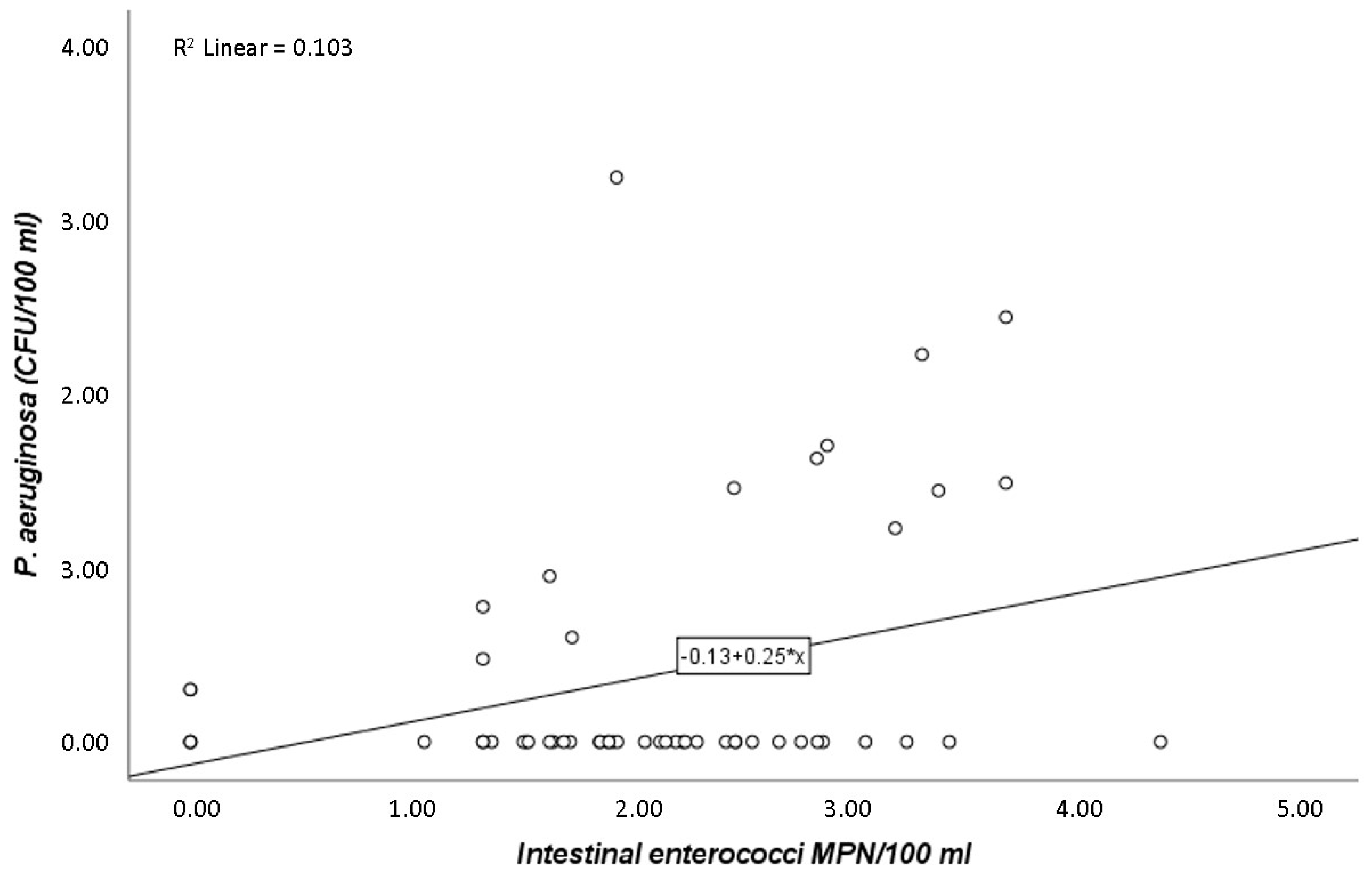
Pearson’s correlation coefficient (r) showed that P. aeruginosa, when correlated only with E. coli, presented a strong positive association (r = 0.7; p-value ≤ 0.05). Thus, variations of P. aeruginosa are explained in 47.3% of cases by E. coli (Figure 1). Equally significant, but with a much less pronounced tendency, was the relationship described between P. aeruginosa and intestinal enterococci (r = 0.3; p-value ≤ 0.05) (Figure 2).
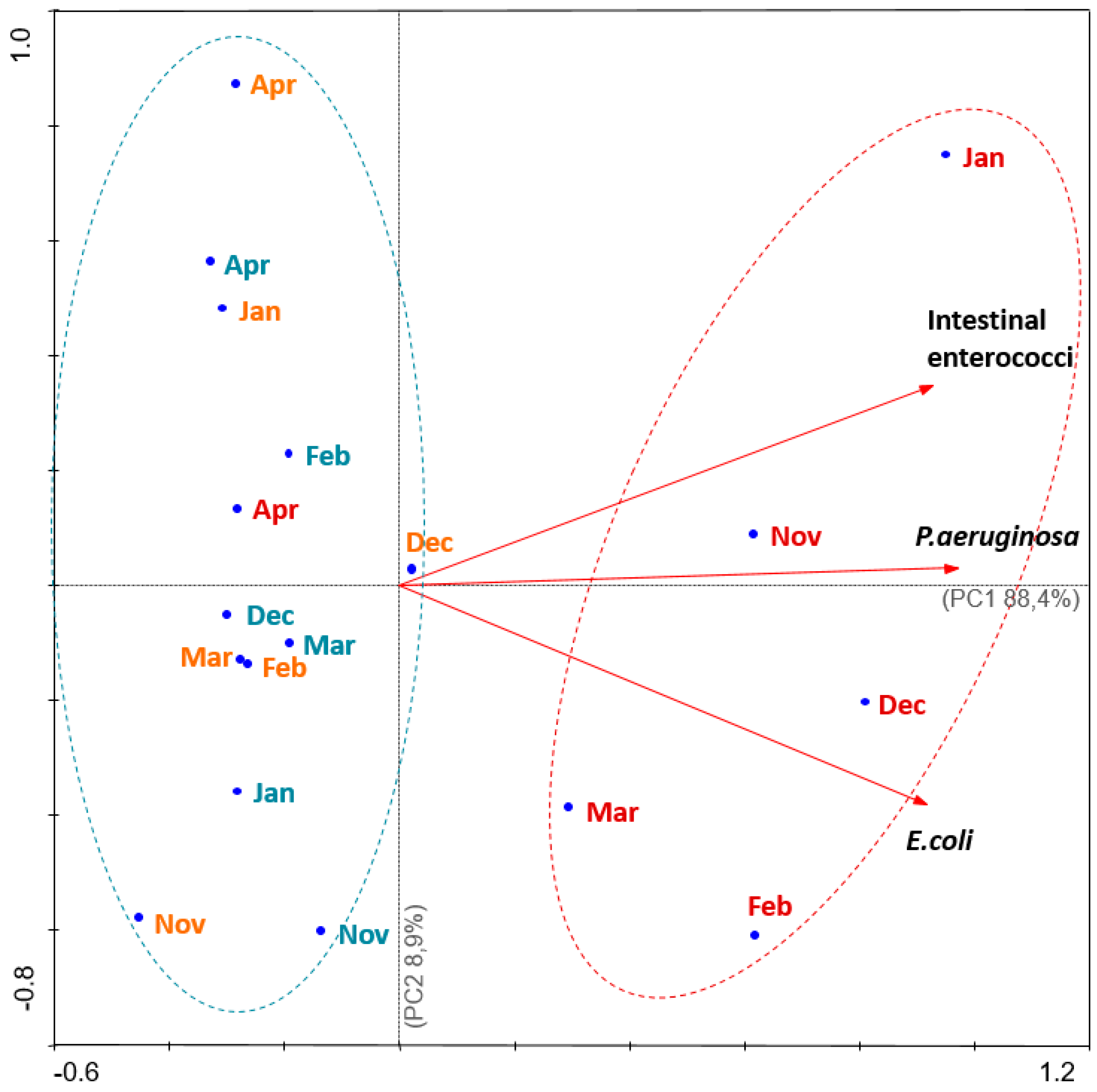
First and second principal components (PC1 and PC2, respectively) describe 97.3% of the original information and reinforce the differences between the characteristics of the sampling sites (Figure 3), with PC1 being the most significant component, expressing 88.4% of the total variance. In addition, the Principal Component Analysis biplot demonstrated a pattern of contamination characterized by STP in winter months (red group, Figure 3). On the other hand, there are beach 1 and beach 2, which show lower contamination patterns (Figure 3). This explains the positive and strong correlation between P. aeruginosa and the faecal bacteria indicators (intestinal enterococci and E. coli), clearly demonstrated by the position of the vectors, such as equal direction and the formation of an acute angle (Figure 3).

3. Discussion

Directive 2006/7/EC of the European Parliament and of the council from 15 February 2006, regarding the management of bathing water quality, provide clear guidelines to assess inland, coastal, and transitional water quality by the enumeration of microbiological parameters, such as intestinal enterococci and E. coli, indicating the potential presence of microbiological hazards [7]. In view of this, the main goal of the present study was to understand if there was a relationship between Pseudomonas aeruginosa and the faecal contamination indicators (intestinal enterococci and E. coli). Our results showed not only high numbers of intestinal enterococci and E. coli, but also the presence of P. aeruginosa in the water samples collected near the sewage treatment plant (STP) (Table 1, Figure 3). Previous studies reported high loads of E. coli and enterococci that exceeded the acceptable levels in the same coastal waters [24], and high numbers of bacteria were detected in limpets in this same spot [25]. In Europe, before coastline dischargement, sewage can receive secondary (organic matter removal) or tertiary (nutrients and bacteria removal) treatment. However, in about half of the European countries, most of the sewage treatment plants comprise only primary and secondary treatment [26], which can explain the high rate of faecal bacteria enumerated from the STP site (Figure 3).
Because E. coli has an exclusively faecal origin, its occurrence in water endorses faecal contamination of the analysed water samples [10]. In this case, such contamination may originate from discharges from the STP (Figure 3). Literature shows that there is an epidemiological connection between gastrointestinal diseases among bathers and poor water quality [2], and sewage pollution of water bodies carries a risk to human health through waterborne pathogens [27]. Vergine et al. showed that the counting capability of Colilert-18 presented a significantly higher recovery of E. coli than the classical membrane filtration method when evaluating wastewater samples from different origins and levels of treatment. Thus, Colilert-18 is an effective method for enumerating E. coli at the STP site, which contains a mix of coastal waters and treated wastewaters [28]. Another study showed that Enterolert-E detected similar proportions of Enterococcus species in marine waters compared with a standard reference method (Environmental Protection Agency Method 1600) [29].
It is known that P. aeruginosa can be found at locations near the runoff of treatment plants [30], resulting in contaminated waters that are frequently described as a vehicle for infections [16,31,32]. In the present study, there were mean counts of 135.8 CFU/100 mL of P. aeruginosa in the water collected near the STP runoff. In one month, there was a maximum of 1800 CFU/100 mL, which is a very high and alarming number (Table 1, Figure 3). Lower counts had been reported before at locations upstream of sewage treatment plants, where the P. aeruginosa counts were 2–33 CFU/100 mL, while at a downstream site, the level was 350 CFU/100 mL in an emergency raw sewage overflow pipe [33].
The most relevant conclusion of this study is that E. coli counts are statistically significant in explaining the variations of P. aeruginosa. Intestinal enterococci and E. coli have been the only mandatory parameters for the assessment of the water quality at bathing sites [34], and because of this, it was expected that the numbers of these coliforms would correlate (being statistically significant) with the presence of the pathogen. Accordingly, our results revealed that E. coli numbers correlate with P. aeruginosa presence, despite the nonenteric nature of this pathogen. E. coli was earlier described as the best-known indicator of pathogenic microbes (although of faecal origin) in water [10], which is in accordance with the findings of the present study. Additionally, Pearson’s correlation between P. aeruginosa and E. coli is 0.687, and variations of P. aeruginosa are explained in 47.3% of cases by E. coli (Figure 1). This result points to a strong correlation, which leads to a positive and important relationship between them. When it comes to intestinal enterococci, this indicator parameter does not explain the numbers of P. aeruginosa, suggesting that intestinal enterococci may not be a good indicator of P. aeruginosa presence, since its correlation had a much less pronounced tendency (r = 0.3; p-value ≤ 0.05) (Figure 2). Curiously, previous studies have advised that introduction of intestinal enterococci as a quality parameter could cause higher failures of microbiological standards, leading to more cases of beach closures [35]. On the other hand, some authors suggest that intestinal enterococci may be better indicators of human health dangers in bathing coastal waters rather than E. coli [36], which is not in accordance with the present results.
The statistically significant relationships between P. aeruginosa and E. coli (Figure 1) and between P. aeruginosa and intestinal enterococci (Figure 2) are also represented in the PCA analysis (Figure 3). It is showed that the three locations under study can be separated into two major areas: one area was heavily contaminated (STP), where the counts of the microorganisms were very high; and a second area was less contaminated (beach 1 and beach 2), where the enumerations were lower. The information obtained from the PCA analysis (Figure 3) is consistent with the statistical significance of the relationship of the parameters in the study (P. aeruginosa and E. coli, Figure 1; P. aeruginosa and intestinal enterococci, Figure 2). This can still be explained by the discharged waters from the STP and their probably poorly disinfection. Corroborating this, Silva et al. [25] performed microbiological analysis of limpet samples near the same STP site and other surrounding areas. Their findings suggest that limpets near the STP discharges have higher levels of E. coli compared with the other two sites in the geographical area, with lower levels of the same bacteria.
Beach 1 and beach 2 are places of recreational activity that are visited by bathers. During the months under study, the quality of water was evaluated and considered poor (Table 1). However, this was not representative of the bathing season, where the presence of more bathers is expected, along with the increase of water temperatures. It is reported that elevated water temperatures in summer stimulate P. aeruginosa growth [32], which can explain the relatively low numbers of this pathogen during the set of months analysed, namely at the end of autumn, in winter, and in early spring (Beach 1: mean of 0.7 CFU/100 mL, maximum 8 CFU/100 mL; Beach 2: mean of 0.3 CFU/100 mL, maximum of 5 CFU/100 mL). The exposure conditions and the dermal route of infection by P. aeruginosa are not well-defined [23]; nonetheless, it is known that even lower concentrations of this pathogen can cause illness in both normal and immunocompromised humans [19]. P. aeruginosa in untreated recreational waters has been linked with otitis externa in bathers [37,38], as well as other health problems. For instance, otitis externa may occur when levels of P. aeruginosa exceed 11 CFU/100 mL, which was demonstrated in a study by Strathman and coworkers [30]. Two outbreaks of this disease were documented in three lakes, revealing the presence of P. aeruginosa in both water samples and patient ear swabs. The concentrations of the bacteria varied from 2–37 CFU/100 mL to 311–736 CFU/100 mL in the third lake [30]. Other study corroborating the same infection pattern of otitis externa also highlighted the amplified risk with the exposure period to the contaminated waters. In this case, 69% of the water samples had only 4 CFU/L of P. aeruginosa [39]. These cases confirm that even lower levels of P. aeruginosa in bathing waters may be associated with an enhanced risk of ear infections, particularly otitis externa. This leads to a reasonable concern in monitoring bathing waters near polluted sites, such as near sewage treatment plants, where high levels of faecal bacteria have been found, especially as a positive correlation has already been established in seawater between skin infections and the presence of P. aeruginosa [14].
Moreover, there is still a lack of information available regarding the correlation between faecal indicator bacteria and pathogenic microorganism concentrations [40]. Additionally, Zabed and colleagues [41] pointed out that the enumeration of these faecal contaminants may be insufficient to assess the risk of nonenteric pathogens, such as Staphylococcus aureus and P. aeruginosa, which have been detected in recreational waters.
In the present research, a positive correlation was observed between E. coli and P. aeruginosa numbers, so these results reinforce the information given by the official quality indicator parameters (for intestinal enterococci and E. coli). However, the authors believe it is advisable to add the enumeration of P. aeruginosa to the quality control of coastal waters, as this is a nonenteric pathogen, and the observed positive correlation might not always be true, especially in the summer, when the numbers of P. aeruginosa might reach even higher values than those observed in the present study. Additionally, the relationship between E. coli and P. aeruginosa numbers should be studied in colder coastal waters, such as northern countries. Because E. coli is mesophilic and P. aeruginosa is psychrophilic, it is possible that the survival time of E. coli might be reduced compared to the survival time of P. aeruginosa in the natural environment.

4. Materials and Methods

4.1. Study Area and Sample Collection

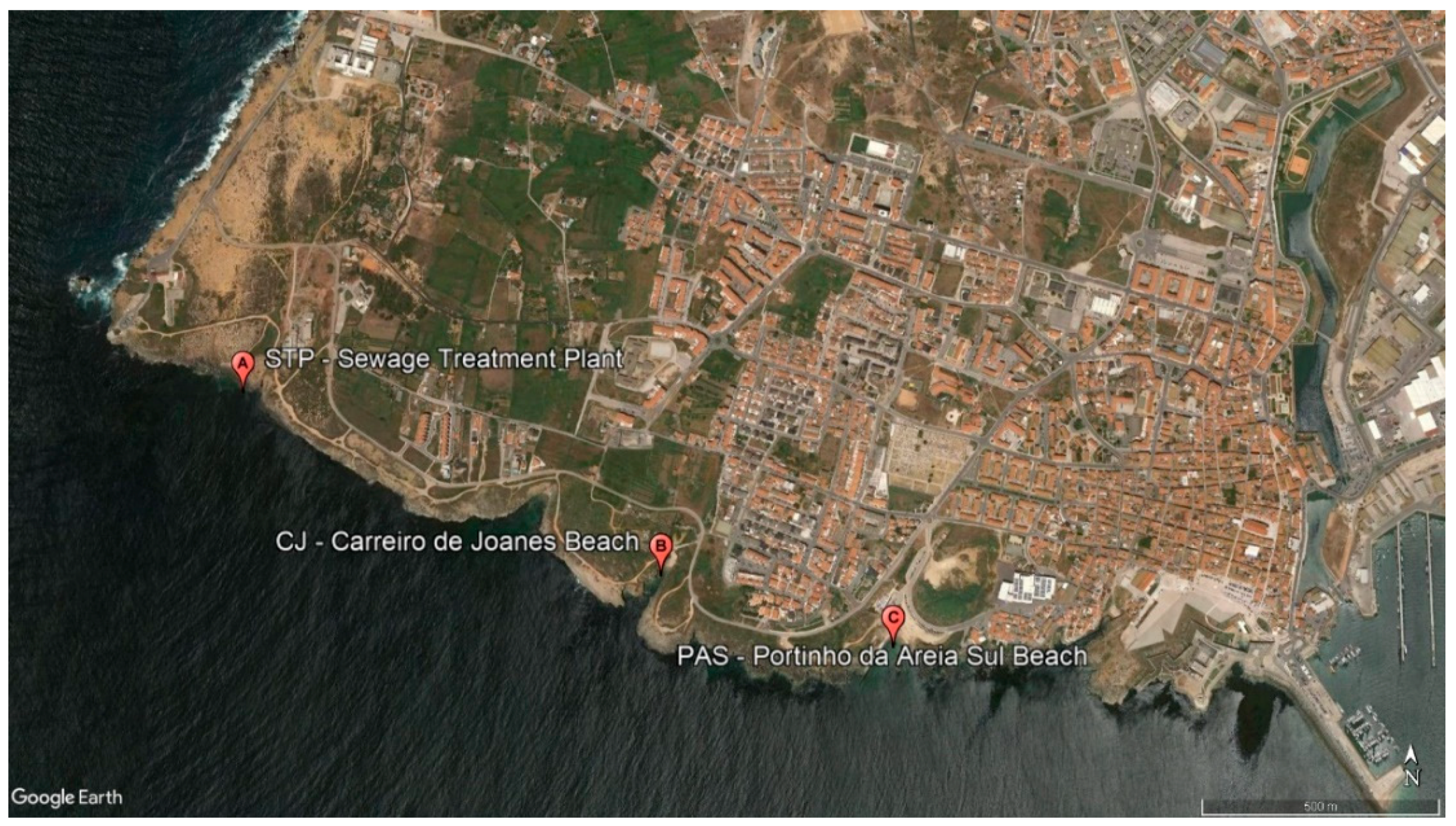
Water samples (1 litre) were collected during low tide, approximately 30 cm below the water level, at three sampling sites in Peniche south coast (Portugal) for six consecutive months: November, December, January, February, March, and April. The first site was chosen because of its close proximity with the sewage treatment plant runoff (STP; 39.358447°, −9.405446°). To understand if there is contamination and this is scattered through the currents along the south coast, two more spots were chosen: beach 1—Carreiro de Joanes beach (CJ; 39.354758°, −9.394529°); and beach 2—Portinho da Areia Sul beach (PAS; 39.353452°, −9388739°), approximately 1.03 km and 1.55 km southeast from STP, respectively (Figure 4). A total of 54 samples (Table 1) were duly packed in thermal bags and transported to the laboratory. Samples were analysed within 6 hours of completion.

4.2. Enumeration of E. coli and Intestinal Enterococci

The Colilert-18 (ISO 9308-2:2012, IDEXX Laboratories, Inc., Westbrook, ME, USA) and Enterolert-E (ISO 7899-1, IDEXX Laboratories, Inc., Westbrook, USA) MPN method procedures were executed according to the company instructions (IDEXX Laboratories, Inc., Westbrook, ME, USA) for the enumeration of Escherichia coli and intestinal enterococci, respectively. Three dilutions were performed (1:10; 50:50; 9:10) due to the suspicion of the high microbiological load. Briefly, 100 mL of each diluted sample and one vial of Colilert-18 and Enterolert-E were added to sterile vessels, capped and shaken to dissolve the reagent, the content was transferred to a 97-well Quanti-Tray (IDEXX Laboratories, Inc., Westbrook, USA), heat-sealed in the Quanti-Tray Sealer (IDEXX Laboratories, Inc., Westbrook, ME, USA), and incubated at 35 ± 0.5 °C for 18 hours (Colilert-18) or 41 ± 0.5 °C for 24 hours (Enterolert-E). After incubation, Quanti-Trays were observed under ultra-violet (UV) light (365 nm) and the fluorescent cells were counted and referred to the MPN table provided by IDEXX for the enumeration of E. coli and intestinal enterococci. Microbiological counts were reported as MPN/100 mL.

4.3. Enumeration of Pseudomonas aeruginosa

For the detection and enumeration of P. aeruginosa in coastal water samples, the procedure described in ISO 16266:2006 was followed [42]. Briefly, 100 mL of each sample (no dilution, 1:10, 1:100) were filtered through a membrane filter of 0.45 μm and incubated on Cetrimide Agar (Biokar, France) plates, supplemented with 1% of glycerol (Himedia, India), at 36 ± 2 °C for 44 ± 4 h. Fluorescent colonies were considered confirmed for P. aeruginosa and reported as CFU/100 mL.

4.4. Statistical Analysis

Prior to statistical analysis, and to minimize the dominant effect of exceptional counts, enumerations of intestinal enterococci, E. coli, and P. aeruginosa were changed to log (x + 1) [43]. To evaluate the strength of the correlations between P. aeruginosa and intestinal enterococci, and between P. aeruginosa and E. coli, Pearson’s correlation coefficients (r) were determined [44]. All calculations were performed with IBM® SPSS® Statistics version 26 software (Copyright IBM Corp. © 1989–2019, Armonk, New York 10504-1722, USA). Results were considered significant at p-value ≤ 0.05. In addition, in order to identify patterns in the relation between intestinal enterococci, E. coli, and P. aeruginosa and the sampling sites, principal component analysis (PCA) was performed with CANOCO version 4.5 package (Copyright Petr Smilauer © 2012–2019, Ithaca, New York 14850, USA) [45]. This multivariate technique was used to identify the components (that is, the core variables) that explain the correlations within the measured data. Therefore, by means of PCA, it was possible to achieve associations between variables, reducing the dimension of the original data. The information provided by the principal components highlight the most meaningful parameters, describing the whole data matrix and affording data reduction with minimum loss of original information. Through a linear combination analysis, the positions of original variables in the diagram represent their relevantly interrelations. Thus, principal components effectively represent the original measured data. As a result, if the variables are closely positioned, their correlation is strong and positive. In contrast, if the variables are in an opposite position, then those variables are negatively correlated. Hence, graphical representation of PCA, which plots simultaneously the objects and the variables, is very useful to detect possible associations between variables and objects. Moreover, the association between objects and variables can be determined depending on their relationship and proximity within each group. In this study, PCA was executed to assess distribution patterns based on the parameters under study (P. aeruginosa, E. coli, intestinal enterococci) and the month-spot sampling. Although the results for the first two components (PC1 and PC2) were presented, the others were also analysed.

Author Contributions

Conceptualisation, A.P.J., C.N.A., M.J.R.; methodology, A.P.J., M.J.R.; software, S.M.; formal analysis, S.M.; investigation, A.P.J.; resources, C.N.A., M.J.R; writing—A.P.J.; writing—review and editing, C.N.A., M.J.R., S.M.; supervision, C.N.A., M.J.R. All authors have read and agreed to the published version of the manuscript.

Funding

This research had the support of Fundação para a Ciência e Tecnologia (FCT), through the strategic project UID/MAR/04292/2019 granted to MARE.

Conflicts of Interest

The authors declare no conflict of interest.

References

  1. Semeoshenkova, V.; Newton, A.; Contin, A.; Greggio, N. Development and application of an Integrated Beach Quality Index (BQI). Ocean Coast. Manag. 2017, 143, 74–86. [Google Scholar] [CrossRef]
  2. Mansilha, C.R.; Coelho, C.A.; Heitor, A.M.; Amado, J.; Martins, J.P.; Gameiro, P. Bathing waters: New directive, new standards, new quality approach. Mar. Pollut. Bull. 2009, 58, 1562–1565. [Google Scholar] [CrossRef] [PubMed]
  3. Palmer, C.J.; Tsai, Y.L.; Lang, A.L.; Sangermano, L.R. Evaluation of colilert-marine water for detection of total coliforms and Escherichia coli in the marine-environment. Appl. Environ. Microbiol. 1993, 59, 786–790. [Google Scholar] [PubMed]
  4. Bartram, J.; Rees, G. Monitoring Bathing Waters—A Practical Guide to the Design and Implementation of Assessments and Monitoring Programmes; WHO: Geneva, Switzerland, 2000; ISBN 0419243801. [Google Scholar]
  5. Aragones, L.; Lopez, I.; Palazon, A.; Lopez-Ubeda, R.; Garcia, C. Evaluation of the quality of coastal bathing waters in Spain through fecal bacteria Escherichia coli and Enterococcus. Sci. Total Environ. 2016, 566, 288–297. [Google Scholar] [CrossRef] [PubMed]
  6. Baudart, J.; Servais, P.; De Paoli, H.; Henry, A.; Lebaron, P. Rapid enumeration of Escherichia coli in marine bathing waters: Potential interference of non-target bacteria. J. Appl. Microbiol. 2009, 107, 2054–2062. [Google Scholar] [CrossRef] [PubMed] [Green Version]
  7. European Union. Directive 2006/7/EC of the European Parliament and of the Council of 15 February 2006 concerning the management of bathing water quality and repealing Directive 76/160/EEC. Off. J. Eur. Union 2006, L 064, 37–51.
  8. López Martínez, I.; Álvarez Díaz, C.; Gil Díaz, J.L.; Revilla Cortezón, J.A.; Juanes, J.A. The European bathing water directive: Application and consequences in quality monitoring programs. J. Environ. Monit. 2010, 12, 369–376. [Google Scholar] [CrossRef]
  9. López, I.; Álvarez, C.; Gil, J.L.; Revilla, J.A. Methodology to elaborate the bathing water profile on urban beaches, according to the requirements of the European Directive 2006/7/EC: The case of Santander beaches (Spain). Water Sci. Technol. 2013, 68, 1037–1047. [Google Scholar] [CrossRef]
  10. Tiwari, A.; Niemela, S.I.; Vepsalainen, A.; Rapala, J.; Kalso, S.; Pitkanen, T. Comparison of Colilert-18 with miniaturised most probable number method for monitoring of Escherichia coli in bathing water. J. Water Health 2016, 14, 121–131. [Google Scholar] [CrossRef] [Green Version]
  11. Napier, M.D.; Haugland, R.; Poole, C.; Dufour, A.P.; Stewart, J.R.; Weber, D.J.; Varma, M.; Lavender, J.S.; Wade, T.J. Exposure to human-associated fecal indicators and self-reported illness among swimmers at recreational beaches: A cohort study. Environ. Health 2017, 16, 103–118. [Google Scholar] [CrossRef] [Green Version]
  12. Mohammed, R.L.; Echeverry, A.; Stinson, C.M.; Green, M.; Bonilla, T.D.; Hartz, A.; McCorquodale, D.S.; Rogerson, A.; Esiobu, N. Survival trends of Staphylococcus aureus, Pseudomonas aeruginosa, and Clostridium perfringens in a sandy South Florida beach. Mar. Pollut. Bull. 2012, 64, 1201–1209. [Google Scholar] [CrossRef] [PubMed]
  13. Gellatly, S.L.; Hancock, R.E.W. Pseudomonas aeruginosa: New insights into pathogenesis and host defenses. Pathog. Dis. 2013, 67, 159–173. [Google Scholar] [CrossRef] [PubMed] [Green Version]
  14. Mariño, F.J.; Moriñigo, M.A.; Martinez-Manzanares, E.; Borrego, J.J. Microbiological-epidemiological study of selected marine beaches in Malaga (Spain). Water Sci. Technol. 1995, 31, 5–9. [Google Scholar] [CrossRef]
  15. Trautmann, M.; Lepper, P.M.; Haller, M. Ecology of Pseudomonas aeruginosa in the intensive care unit and the evolving role of water outlets as a reservoir of the organism. Am. J. Infect. Control 2005, 33, S41–S49. [Google Scholar] [CrossRef] [PubMed]
  16. Ngwa, G.A.; Schop, R.; Chow, J.; Lukic, L.; McKague, K. Comparative detection and recovery of Pseudomonas aeruginosa by membrane filtration and a Most Probable Number technique. J. Microbiol. Methods 2017, 133, 76–81. [Google Scholar] [CrossRef] [PubMed]
  17. Hardalo, C.; Edberg, S.C. Pseudomonas aeruginosa: Assessment of risk from drinking water. Crit. Rev. Microbiol. 1997, 23, 47–75. [Google Scholar] [CrossRef]
  18. Quinn, J.P. Pseudomonas aeruginosa infections in the intensive care unit. Semin. Respir. Crit. Care Med. 2003, 24, 61–68. [Google Scholar] [CrossRef] [Green Version]
  19. Mena, K.D.; Gerba, C.P. Risk Assessment of Pseudomonas aeruginosa in Water. Rev. Environ. Contam. Toxicol. 2009, 201, 71–115. [Google Scholar]
  20. Bedard, E.; Prevost, M.; Deziel, E. Pseudomonas aeruginosa in premise plumbing of large buildings. Microbiologyopen 2016, 5, 937–956. [Google Scholar] [CrossRef]
  21. Fazeli, H.; Akbari, R.; Moghim, S.; Narimani, T.; Arabestani, M.R.; Ghoddousi, A.R. Pseudomonas aeruginosa infections in patients, hospital means, and personnel’s specimens. J. Res. Med. Sci. 2012, 17, 332–337. [Google Scholar]
  22. Zichichi, L.; Asta, G.; Noto, G. Pseudomonas aeruginosa folliculitis after shower/bath exposure. Int. J. Dermatol. 2000, 39, 270–273. [Google Scholar] [CrossRef] [PubMed]
  23. Roser, D.J.; den Akker, B.; Boase, S.; Haas, C.N.; Ashbolt, N.J.; Rice, S.A. Pseudomonas aeruginosa dose response and bathing water infection. Epidemiol. Infect. 2014, 142, 449–462. [Google Scholar] [CrossRef] [PubMed]
  24. Rodrigues, M.J.; Martins, K.; Garcia, D.; Ferreira, S.M.F.; Gonçalves, S.C.; Mendes, S.; Lemos, M.F.L. Using the mini-VIDAS® Easy Salmonella protocol to assess contamination in transitional and coastal waters. Arch. Microbiol. 2016, 198, 483–487. [Google Scholar] [CrossRef] [PubMed]
  25. Silva, C.S.E.; Novais, S.C.; Simões, T.; Caramalho, M.; Gravato, C.; Rodrigues, M.J.; Maranhão, P.; Lemos, M.F.L. Using biomarkers to address the impacts of pollution on limpets (Patella depressa) and their mechanisms to cope with stress. Ecol. Indic. 2018, 95, 1077–1086. [Google Scholar] [CrossRef]
  26. Cabral-Oliveira, J.; Pratas, J.; Mendes, S.; Pardal, M.A. Trace Elements in Edible Rocky Shore Species: Effect of Sewage Discharges and Human Health Risk Implications. Hum. Ecol. Risk Assess. 2015, 21, 135–145. [Google Scholar] [CrossRef]
  27. Harwood, V.J.; Staley, C.; Badgley, B.D.; Borges, K.; Korajkic, A. Microbial source tracking markers for detection of fecal contamination in environmental waters: Relationships between pathogens and human health outcomes. FEMS Microbiol. Rev. 2014, 38, 1–40. [Google Scholar] [CrossRef] [Green Version]
  28. Vergine, P.; Salerno, C.; Barca, E.; Berardi, G.; Pollice, A. Identification of the faecal indicator Escherichia coli in wastewater through the β-D-glucuronidase activity: Comparison between two enumeration methods, membrane filtration with TBX agar, and Colilert ®-18. J. Water Health 2017, 15, 209–217. [Google Scholar] [CrossRef]
  29. Ferguson, D.M.; Griffith, J.F.; McGee, C.D.; Weisberg, S.B.; Hagedorn, C. Comparison of enterococcus species diversity in marine water and wastewater using Enterolert and EPA method 1600. J. Environ. Public Health 2013, 2013, 1–6. [Google Scholar] [CrossRef] [Green Version]
  30. Strathmann, M.; Horstkott, M.; Koch, C.; Gayer, U.; Wingender, J. The River Ruhr—An urban river under particular interest for recreational use and as a raw water source for drinking water: The collaborative research project ‘Safe Ruhr’—Microbiological aspects. Int. J. Hyg. Environ. Health 2016, 219, 643–661. [Google Scholar] [CrossRef]
  31. Havelaar, A.H.; During, M.; Delfgouvanasch, E.H.M. Comparative-study of membrane filtration and enrichment media for the isolation and enumeration of Pseudomonasaeruginosa from sewage, surface-water, and swimming pools. Can. J. Microbiol. 1985, 31, 686–692. [Google Scholar] [CrossRef]
  32. Warburton, D.W.; Bowen, B.; Konkle, A. The survival and recovery of Pseudomonas aeruginosa and its effect upon Salmonellae in mater—Methodology to test bottled water in Canada. Can. J. Microbiol. 1994, 40, 987–992. [Google Scholar] [CrossRef] [PubMed]
  33. Seyfried, P.L.; Cook, R.J. Otitis externa infections related to Pseudomonas aeruginosa levels in five Ontario lakes. Can. J. Public Health 1984, 75, 83–91. [Google Scholar] [PubMed]
  34. Valente, M.S.; Pedro, P.; Alonso, M.C.; Borrego, J.J.; Dionísio, L. Are the defined substrate-based methods adequate to determine the microbiological quality of natural recreational waters? J. Water Health 2010, 8, 11. [Google Scholar] [CrossRef] [PubMed]
  35. Noble, R.; Moore, D.; Leecaster, M.; McGee, C.; Weisberg, S. Comparison of total coliform, fecal coliform, and enterococcus bacterial indicator response for ocean recreational water quality testing. Water Res. 2003, 37, 1637–1643. [Google Scholar] [CrossRef]
  36. Kay, D.; Bartram, J.; Prüss, A.; Ashbolt, N.; Wyer, M.D.; Fleisher, J.M.; Fewtrell, L.; Rogers, A.; Rees, G. Derivation of numerical values for the World Health Organization guidelines for recreational waters. Water Res. 2004, 38, 1296–1304. [Google Scholar] [CrossRef] [PubMed]
  37. Hoadley, A.W.; Knight, D.E. External otitis among swimmers and nonswimmers. Arch. Environ. Health 1975, 30, 445–448. [Google Scholar] [CrossRef]
  38. Reid, T.M.; Porter, I.A. An outbreak of otitis externa in competitive swimmers due to Pseudomonas aeruginosa. J. Hyg. 1981, 86, 357–362. [Google Scholar] [CrossRef] [Green Version]
  39. Van Asperen, I.A.; de Rover, C.M.; Schijven, J.F.; Oetomo, S.B.; Schellekens, J.F.P.; van Leeuwen, N.J.; Colle, C.; Havelaar, A.H.; Kromhout, D.; Sprenger, M.W.J. Risk of otitis externa after swimming in recreational fresh water lakes containing Pseudomonas aeruginosa. BMJ 1995, 311, 1407–1410. [Google Scholar] [CrossRef]
  40. Zhang, Q.; Eichmiller, J.J.; Staley, C.; Sadowsky, M.J.; Ishii, S. Correlations between pathogen concentration and fecal indicator marker genes in beach environments. Sci. Total Environ. 2016, 573, 826–830. [Google Scholar] [CrossRef]
  41. Zabed, H.; Suely, A.; Faruq, G.; Sahu, J.N. Water quality assessment of an unusual ritual well in Bangladesh and impact of mass bathing on this quality. Sci. Total Environ. 2014, 472, 363–369. [Google Scholar] [CrossRef]
  42. ISO 16266 Water quality—Detection and enumeration of Pseudomonas aeruginosa—Method by membrane filtration. Available online: https://www.iso.org/obp/ui/#iso:std:iso:16266:ed-1:v1:en (accessed on 6 December 2019).
  43. Legendre, L.; Legendre, P. Écologie numérique. Tome 1: Le traitement multiple des données écologiques; Collection d′Écologie no 12 XIV. Masson; Paris et les Presses de l′Université du Québec: Paris, France, 1979; p. 197. [Google Scholar]
  44. Zar, J.H. Biostatistical Analysis, 5th ed.; Pearson Prentice-Hall: Upper Saddle River, NJ, USA, 2010. [Google Scholar]
  45. Ter Braak, C.J.F.; Smilauer, P. CANOCO Reference Manual and User’s Guide to Canoco for Windows—Software for Canonical Community Ordination; Version 4; Microcomputer Power: Ithaca, NY, USA, 1998. [Google Scholar]
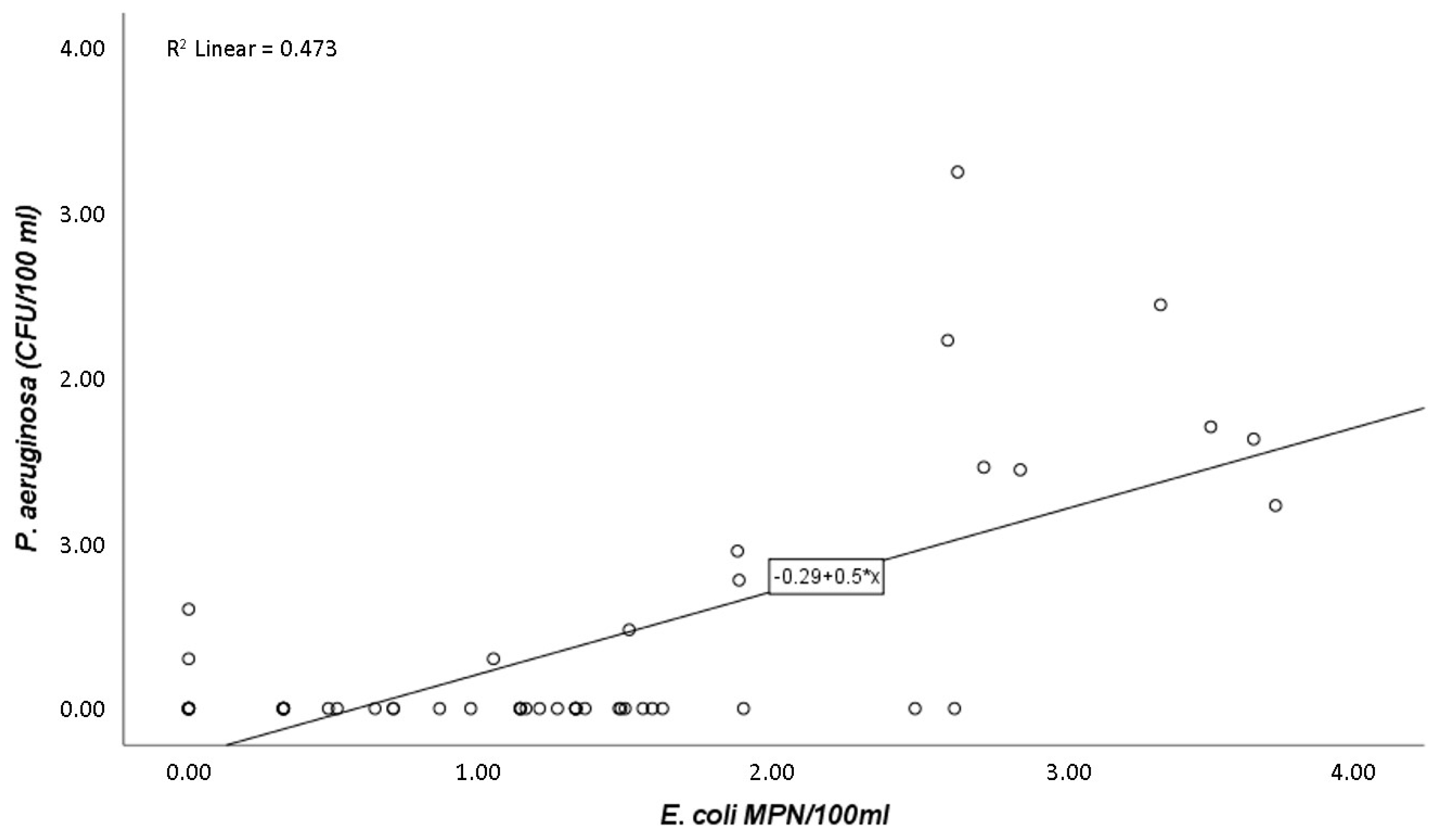
Figure 1. Scatter diagram indicating the relationship between P. aeruginosa (CFU/100 mL) and E. coli (MPN/100 mL) counts (r = 0.7; p-value ≤ 0.05).
Pathogens 09 00013 g001
Figure 2. Scatter diagram indicating the relationship between P. aeruginosa (CFU/100 mL) and intestinal enterococci (MPN/100 mL) counts (r = 0.3; p-value ≤ 0.05).
Pathogens 09 00013 g002
Figure 3. Principal component analysis (PCA) biplot of intestinal enterococci, E. coli, P. aeruginosa, and the sampling sites over the 6 months of the experiment. Note: months represented in red—sewage treatment plant (STP); months represented in blue—beach 1; months represented in orange—beach 2.
Pathogens 09 00013 g003
Figure 4. South coast of Peniche (Portugal), showing the sampling sites: (A) sewage treatment plant (STP; 39.358447°, −9.405446°), (B) beach 1 (CJ; 39.354758°, −9.394529°), and (C) beach 2 (PAS; 39.353452°, −9388739°). Image generated from Google Earth Pro software.
Pathogens 09 00013 g004
Table 1. Means, sample size (n), minimum, and maximum counts for P. aeruginosa, E. coli, and intestinal enterococci from the three sampling sites: the sewage treatment plant (STP), beach 1 (Carreiro de Joanes, CJ), and beach 2 (Portinho da Areia Sul beach, PAS).
STPCJ (Beach 1)PAS (Beach 2)
MeannMinimumMaximumMeannMinimumMaximumMeannMinimumMaximum
P. aeruginosa(CFU/100 mL)135.8180.01800.00.7180.08.00.3180.05.0
E. coli(MPN/100 mL)1100.1160.05172.016.5180.074.014.1180.075.0
Intestinal enterococci(MPN/100 mL)2685.9180.024196.0100.5180.0455.8145.9180.0720.2