Article

Phenotypic and Genotypic Characterization of Staphylococcus aureus Isolated from Patients with Chronic Furunculosis and Osteomyelitis from Northwestern Poland

by
Aleksandra Wcisłek
1,
Joanna Jursa-Kulesza
1,
Helena Masiuk
1,
Bartłomiej Grygorcewicz
2,
Beata Hukowska-Szematowicz
3,4,
Piotr Prowans
5,
Paweł Ziętek
6 and
Danuta Kosik-Bogacka
7,*
1
Independent Medical Microbiology Laboratory, Pomeranian Medical University in Szczecin, Powstanców Wielkopolskich 72, 70-204 Szczecin, Poland
2
Department of Genomics and Forensic Genetics, Pomeranian Medical University in Szczecin, Powstanców Wielkopolskich 72, 70-204 Szczecin, Poland
3
Institute of Biology, Laboratory of Infectious Biology, Molecular Biology and Immunology, University of Szczecin, Wąska 13, 71-412 Szczecin, Poland
4
Molecular Biology and Biotechnology Center, University of Szczecin, Wąska 13, 71-412 Szczecin, Poland
5
Department of Plastic, Endocrine and General Surgery, Pomeranian Medical University in Szczecin, 1 Unii Lubelskiej Street, 71-252 Szczecin, Poland
6
Autonomous Knee Surgery Unit, Pomeranian Medical University in Szczecin, 1 Unii Lubelskiej Street, 71-252 Szczecin, Poland
7
Department of Biology, Parasitology and Pharmaceutical Botany, Pomeranian Medical University in Szczecin, Powstanców Wielkopolskich 72, 70-204 Szczecin, Poland
*
Author to whom correspondence should be addressed.
Pathogens 2025, 14(9), 923; https://doi.org/10.3390/pathogens14090923
Submission received: 9 July 2025 / Revised: 8 September 2025 / Accepted: 9 September 2025 / Published: 12 September 2025

Abstract

Staphylococcus aureus, a Gram-positive coccus commonly found in the environment, is indeed a common cause of both superficial and deep infections. The aim of the study was to determine the virulence factors of S. aureus characteristic of chronic infections, including chronic furunculosis and chronic osteomyelitis. Phenotypic characteristics of the bacteria (ability to produce hemolysis, clumping factor, and coagulase; antibiotic susceptibility) and genotypic characteristics (presence of genes responsible for the production of enzymes and toxins; ability to form biofilm) were examined. The real-time PCR method was used to determine the presence of virulence genes. Biofilm production was confirmed using the crystal violet method. Antibiotic and chemotherapeutic susceptibility tests were performed using the disk diffusion method. In 90% of cases, S. aureus strains possessed the following virulence genes: clfA, clfB, spa, cna, eap, hlgA, hlgB, hlg, hld, bap, bbp, ebpS, fib, fnbA, fnbB, and pvl. A total of 82% of S. aureus strains showed susceptibility to methicillin (MSSA), whereas 12% of strains were susceptible to methicillin and simultaneously resistant to macrolides, lincosamides, and streptogramin B, including 10.5% with inducible resistance and 1.5% with constitutive resistance (MSSA/MLSB). In addition, 1.5% were methicillin-resistant S. aureus (MRSA) and susceptible to the remaining antimicrobial agents. The predominance of MSSA in the etiology of chronic furunculosis and chronic osteomyelitis was observed. It has been demonstrated that MSSA possesses a similar set of virulence genes to MRSA and that MSSA is responsible for most cases of chronic osteomyelitis and furunculosis. The findings indicate that S. aureus possesses numerous virulence factors that play a key role in the processes of adhesion to and proliferation within host cells.

1. Introduction

Staphylococcus aureus is a Gram-positive, facultative anaerobic bacterium [1], which colonizes the prenares of approximately 20–30% of healthy individuals and up to 40–50% of healthcare personnel, who can act as reservoirs and vectors for transmission in clinical settings [2]. This opportunistic pathogen can cause multiple human infections, such as impetigo, folliculitis, furuncles, carbuncles and cellulitis, toxin-mediated diseases, such as food poisoning, scalded skin syndrome, and toxic shock syndrome, and systemic and life-threatening conditions for endocarditis, pulmonary infections (e.g., necrotizing pnuemonia), deep-seated abscesses, meningitis, urinary tract, and bloodstream infections [3].
The mortality rate of S. aureus bloodstream infections reaches 30% [4] and most commonly is associated with the emergence of methicillin-resistant S. aureus (MRSA) strains, representing a therapeutic challenge. MRSA is prevalent in several hospitals (healthcare-associated methicillin-resistant S. aureus, HA-MRSA), particularly in Europe, Asia, and the United States. A significant concern is the high level of resistance among strains isolated from hospital-acquired infections to antibiotics and chemotherapeutics, including tetracyclines, aminoglycosides, macrolides, and lincosamides. In addition, these strains frequently exhibit resistance to fluoroquinolones, chloramphenicol, trimethoprim/sulfamethoxazole, fusidic acid, and rifampicin [5]. The World Health Organization and the Centers for Disease Control and Prevention continuously monitor the increasing resistance of S. aureus to antibiotics and chemotherapeutics [6,7]. Community-associated MRSA (CA-MRSA) strains are generally characterized by higher virulence but lower antimicrobial resistance and often infect individuals without significant predisposing risk factors for infection [8].
Methicillin-susceptible S. aureus (MSSA) is an important etiological agent of dermatitis and soft-tissue inflammation, including chronic furunculosis and chronic osteomyelitis. MSSA strains frequently exhibit higher virulence compared to MRSA, as the acquisition and maintenance of antibiotic resistance determinants in MRSA impose a metabolic fitness cost that may attenuate its pathogenic potential. Nevertheless, MSSA demonstrates numerous additional traits that confer enhanced resilience against environmental stressors [5,9]. One such characteristic is the ability to produce biofilm [10]. By protecting bacterial cells, biofilm formation significantly reduces the effectiveness of antibiotics and/or chemotherapeutics. Treatment of chronic S. aureus infections is challenging, and the pathogenesis of these infections is still poorly understood. The problem of chronic infections concerns patients diagnosed with metabolic diseases, including diabetes, as well as patients after orthopedic procedures in which S. aureus infection has occurred [11]. In some patients, the lack of clinically significant impairments that could affect the chronic nature of the inflammatory process was found. In the available literature, there is a limited number of S. aureus infections differentiated in terms of their clinical course. Therefore, the aim of the study was to determine the virulence factors of S. aureus that contribute to chronic infections.
Staphylococcus aureus isolated from chronic furunculosis and chronic osteomyelitis were selected for analysis. Accordingly, phenotypic characteristics of the bacteria (ability to produce hemolysis, clumping factor, and coagulase; antibiotic susceptibility) and genotypic characteristics (presence of genes responsible for the production of enzymes and toxins; ability to form biofilm) were examined. The analysis included genes encoding surface proteins (microbial surface components recognizing adhesive matrix molecules, MSCRAMM). These proteins are involved in adhesion to host tissues, weakening of the immune response, and promotion of biofilm formation. This group includes staphylococcal protein A (SpA), collagen-binding protein (Cna), fibronectin-binding proteins A (fnbA) and B (fnbB), and extracellular adherence protein (eap) [12,13]. Clumping factor A (ClfA) is the major staphylococcal fibrinogen (Fg)-binding protein that binds to fibrinogen in plasma, facilitating bacterial aggregation and immune evasion [14]. Other MSCRAMM components include elastin-binding protein (ebpS), fibrinogen-binding protein (Efb), and bone sialoprotein-binding protein (bbp). These structures can bind to molecules such as collagen (primarily via Cna), fibronectin (via FnbAB), and fibrinogen (via ClfAB and Fib), thereby enabling immune evasion and subsequent infection development [15]. Expression of the genes encoding these proteins is particularly elevated during the early stages of biofilm formation. A surface protein involved in biofilm production by S. aureus on all surfaces is biofilm-associated protein (Bap). In addition, genes responsible for toxin production were also included in the analysis. S. aureus produces and secretes multiple toxins with diverse roles in pathogenesis, particularly in immune evasion and modulation of the immune response [16]. These include four hemolysins, α (hlgA), β (hlgB), γ (hlg), and δ (hld), as well as Panton–Valentine leukocidin (PVL) [17,18].

2. Materials and Methods

2.1. Bacterial Isolates

In the present study, S. aureus strains (n = 67) from the collection of the Independent Medical Microbiology Laboratory of the Pomeranian Medical University in Szczecin (PUM) were analyzed. All strains were isolated from samples collected from boils and from bone with surrounding tissue excised during surgery in patients with chronic furunculosis (n = 43) and chronic osteomyelitis (n = 24) from northwestern Poland, respectively. The study was conducted in accordance with the Declaration of Helsinki and was approved by the Ethics Committee of Pomeranian Medical University (protocol code KB-0012/112/17, date 2 October 2017) for studies involving humans. The study was conducted between 2009 and 2020.
Analyzed strains were stored at −80 °C in tryptic soy broth (Becton Dickinson, Franklin Lakes, NJ, USA) supplemented with anhydrous glycerol (Chempur, Piekary Śląskie, Poland) at a volume ratio of 9:1. Before testing, the strains were subcultured onto Columbia agar enriched with 5% sheep blood (BioMérieux, Craponne, France) and incubated for 24 h at 37 °C. A three-phase streaking pattern was applied to ensure regular metabolic activity. Control S. aureus strains used in the experiments included ATCC 29213 and ATCC 6538 from the American Type Culture Collection (ATCC) [19,20].

2.2. Identification of Microorganisms

All strains grown on Columbia agar with 5% sheep blood exhibited beta-hemolysis. Species determination was conducted using biochemical tests (e.g., the Vitek 2 Compact System) (BioMérieux, Craponne, France) and basic preliminary microbiological diagnostic assays (e.g., agglutination tests and detection of the clumping factor, CF). The test for bound coagulase (clumping factor, CF) was performed using an agglutination test (Staphytect Plus, Oxoid, Basingstoke, UK) during the routine microbiological diagnostics.

2.3. Isolation of Chromosomal DNA

All strains were cultured onto Brain Heart Infusion (BHI) agar medium (BTL, Warsaw, Poland) and incubated at 37 °C for 18 h. DNA extraction was performed using the Genomic Mini AX Bacteria + SPIN kit (A&A Biotechnology, Gdańsk, Poland) according to the manufacturer’s instructions. Approximately 50–100 ng of genomic DNA per reaction was used.
Bacterial colonies from BHI agar were suspended in 1 mL of sterile water in an Eppendorf tube and vortexed vigorously for 30 s using a Vortex Lab Dancer (Binovo, Legnica, Poland). The tubes were then centrifuged at 13,000 rpm for one minute in an Eppendorf MiniSpin Plus centrifuge (Eppendorf SE, Hamburg, Germany). After centrifugation, the supernatant was discarded, and the sediment was resuspended in PBS buffer. Subsequently, 10 µL of lysozyme and 5 µL of mutanolysin were added to the suspension, which was incubated for 20 min at 50 °C in an Eppendorf Thermomixer 5436 (Eppendorf SE, Hamburg, Germany).
Following this, 20 µL of proteinase K and 200 µL of L1.4 buffer were added and mixed, and the mixture was incubated for an additional 10 min at 50 °C with vigorous shaking. The tubes were then vortexed for 20 s and centrifuged at 12,000 rpm for 45 s. Next, the supernatant was applied to Mini AX Spin columns and centrifuged at 12,000 rpm for 45 s. The columns were then transferred to 2 mL collection tubes included in the kit and washed sequentially with 600 µL of W1 buffer, followed by centrifugation at 12,000 rpm for 45 s, and then with 500 µL of W2 buffer, followed by centrifugation under the same conditions. Finally, the columns were transferred to 2 mL Eppendorf tubes, and DNA was eluted by adding 140 µL of elution buffer and centrifuging at 13,000 rpm for 60 s. The elution buffer combined with the isolated bacterial DNA settled at the bottom of the tube after centrifugation. Total bacterial DNA was used for subsequent stages of the study.

2.4. Real-Time PCR

A quantitative polymerase chain reaction (real-time PCR) technique was used to identify virulence genes in the bacterial genome. The primers were designed and made by Sigma-Aldrich (Merck, Poznań, Poland) based on [21,22,23,24] (Table 1). The primers in lyophilized form were dissolved in the manufacturer’s specified volume of sterile water, obtaining an initial concentration of 100 µmol. The next step was to obtain a working concentration of 10 µmol by diluting them in a ratio of 1:10. In this study, the following primers were used.
A real-time PCR kit (RT-HS PCR Mix SYBR A®, A&A Biotechnology, Gdańsk, Poland) was used in the study. The kit consisted of Master Mix and sterile water. The composition of Master Mix is shown below as follows:
  • Taq DNA polymerase: 0.1 U/µL;
  • MgCl2: 4 mM;
  • dNTPs: 0.5 mM;
  • 2 x reaction buffer with SYBR Green.
DNA was placed in a 96-well plate (Bio-Rad, Hercules, CA, USA), and then primers, sterile water, and Master Mix were added. The reaction mixture consisted (per sample) of the following:
  • ➢ DNA: 1 µL;
  • ➢ Starter F: 2 µL;
  • ➢ R starter: 2 µL;
  • ➢ Sterile water: 2.5 µL;
  • ➢ Master Mix: 7.5 µL.
The PCR was carried out using the CFX Connect™ Real-time System (BioRad, Hercules, CA, USA), with thermal cycles tailored for each gene, ranging from 35 to 39 cycles depending on the virulence gene being amplified. For the staphylococcal protein A (spa), extracellular adherence protein (eap), clumping factor A (clfa) and B (clfb), collagen binding protein (cna), α- (hlgA), β- (hlgB), γ- (hlg), and δ- (hld) hemolysin genes, it was 39 cycles; for the Panton–Valentine leukocidin (pvl) gene, it was 38 cycles; and for the bone sialoprotein (bbp), penicillin-binding proteins (ebps), fibrinogen-binding protein (fib), fibronectin-binding protein a (fnpba) and b (fnpbb), biofilm-associated protein (bap) genes, it was 35 cycles. Thermal profile conditions are given in Table 2. Prior to the experiments, a rigorous calibration of the real-time PCR assays was conducted to ensure accuracy, involving primer specificity checks through gradient PCR and efficiency evaluations, with efficiencies confirmed between 90% and 110%. Calibration curves were then constructed for each gene from serial dilutions of template DNA to enable precise quantification of gene expression, correlating PCR cycle thresholds (Ct) with initial DNA amounts. Gene positivity was accurately determined if Ct values fell within the calibrated range of 15 to 35 cycles and were accompanied by distinct, specific melting peaks, ensuring the detection of the correct PCR products without nonspecific amplifications. Positive results were further validated using both no-template controls and positive controls containing known DNA amounts, reinforcing the reliability of our findings.

2.5. Biofilm Formation Ability of S. aureus Strains

The test of biofilm-generating capacity was conducted according to the procedure described by Kwiatkowski et al. [25]. Each strain was tested three times. The use of crystal violet in this study was dictated by the ability of this dye to stain live and dead bacteria and extracellular matrix products, as confirmed by Oleksy-Wawrzyniak et al. [26]. Similar studies were presented using crystal violet by Kamimura et al. [27] and Yang et al. [28]. In the first step, bacteria were resuspended in Tryptoy Soy Broth, TSB (Becton Dickinson, Franklin Lakes, NJ, USA) and then incubated at 37 °C for 18 h. The multiplied bacteria were diluted 1:200, placed in a 96-well plate in a hothouse at 37 °C, and incubated for 24 h. After incubation, the wells were washed 3 times with sterile saline solution (0.45% NaCl, bioMerieux, Craponne, France), fixed with methanol (Sigma Aldrich, Saint Louis, MO, USA) by adding 200 µL of this alcohol to each well, and left for 20 min at room temperature. The methanol was poured off, and the plate was left to dry. A total of 200 µL of 0.2% (w/v) crystal violet solution (Chempur, Piekary Śląskie, Poland) was poured into each well and incubated for 20 min at room temperature. The crystal violet was washed thoroughly by rinsing the wells 5 times with sterile distilled water. After rinsing, 200 µL of an 8:2 (v/v) ethanol/acetone mixture (P.P.H Stanlab, Gliwice, Poland; Chemland, Stargard, Poland) was poured in, and absorbance was read using an Envision 2104 Multi Reader (Perkin Elmer, Shelton, CT, USA) at λ = 590 nm. The control was three wells with crystal violet without bacteria. The results from the three measurements were averaged, and the value from the control test was subtracted from the individual results.

2.6. Determination of S. aureus Sensitivity to Antibiotics

Antibiotic susceptibility of S. aureus was evaluated according to European Committee on Antimicrobial Susceptibility Testing (EUCAST) guidelines. A 24 h bacterial culture was suspended in saline solution (0.9% NaCl) to obtain a concentration of 0.5 MacFarland turbidity standard. Bacterial suspension was inoculated onto Mueller–Hinton medium (BioMerieux, Craponne, France), where antibiotic discs were applied. The discs contained cefoxitin (30 µg), erythromycin (15 µg), gentamicin (10 µg), clindamycin (2 µg), ciprofloxacin (5 µg), and cotrimoxazole 25 µg (trimethoprim and sulfamethoxazole: 23.75 µg and 1.25 µg, respectively). The antibiograms were incubated for 20 h at 37 °C, and then the zone of inhibition was measured.

2.7. Statistical Analysis of the Results

Statistical analysis of the results was carried out using R version 4.1.1. A chi-square test was used to determine the relationship between the occurrence of a particular resistance phenotype and the induction of a specific infection. The same test was used to analyze gene occurrence versus resistance phenotype. The Kruskal–Wallis test analyzed the relationship between phenotypic resistance and biofilm production.

3. Results

3.1. Phenotypic Assessments

Based on EUCAST guidelines, 82% of S. aureus strains tested showed methicillin susceptibility, while 12% of strains showed methicillin susceptibility with concomitant resistance to macrolides, lincosamides, and streptogramin B, including 10.5% with inducible resistance and 1.5% with constitutive resistance (MSSA/MLSB) (Figure 1). Among the strains tested, 1.5% were MRSA, and in 4.5% of MRSA, constitutive resistance to macrolides, lincosamides, and streptogramin B (MRSA/cMLSB) was simultaneously found. The lack of resistance to gentamicin, ciprofloxacin, and cotrimoxazole among all examined strains was also confirmed.
Our results showed that MSSA strains were responsible for more than 80% of bone infections and cases of furunculosis. Using a chi-square test, we found that MSSA strains were more likely to cause furunculosis (p = 0.0001) and osteomyelitis (p = 0.0001). This may suggest that the clinical picture of these infections is not directly influenced by the susceptibility or resistance of S. aureus strains but most likely linked with the presence of virulence genes that determine production of particular components exacerbating inflammation. In six isolates, the genetic profile revealed the presence of all investigated genes, with the exception of a single missing gene in each case. Among this group, four isolates originated from bone infections and two from furunculosis, of which two strains showed methicillin sensitivity and one strain showed methicillin sensitivity with iMLSB resistance. Since only three S. aureus strains from patients with chronic furunculosis showed the absence of virulence genes, it can be concluded that the strains isolated from furunculosis are more virulent.
Five S. aureus strains isolated from patients with osteomyelitis showed the absence of virulence genes. In particular, one strain lacked three virulence genes, including the bbp, fnpbA, and fib genes encoding bone sialoprotein-binding protein, fibronectin-binding protein, and fibrinogen-binding protein, respectively. This indicates that, in individual cases, the induction of inflammation by S. aureus may be a much more complex process and not directly related to the presence or absence of virulence genes. Other parameters influence inflammation, including the patient’s age and comorbidities.
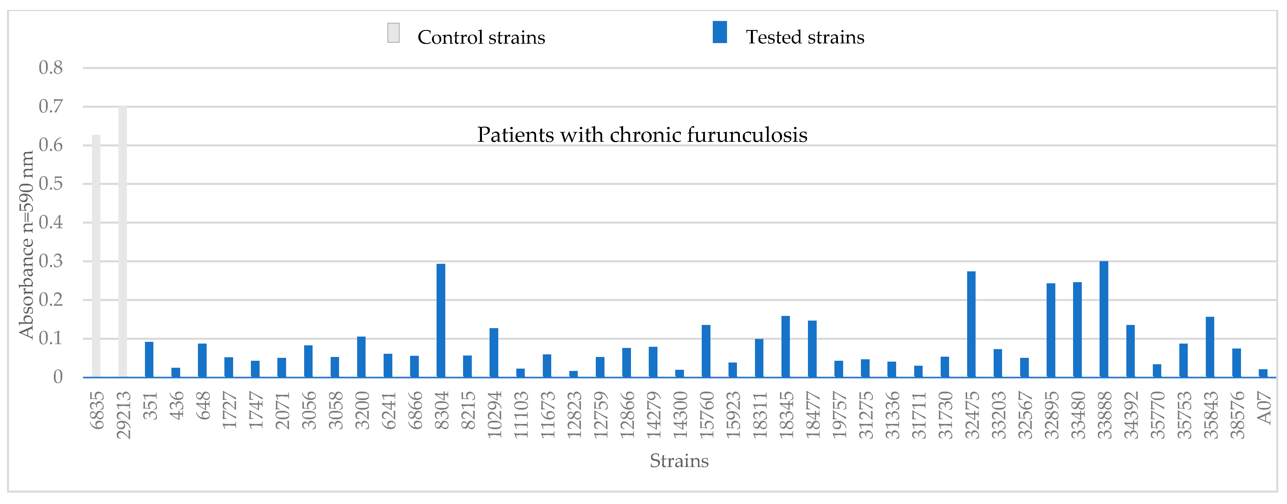
All tested S. aureus strains showed the ability to produce biofilm at different levels. The Kruskal–Wallis test found that MSSA produced more biofilm (p = 0.0183), even though MSSA strains are highly variable in biofilm formation. S. aureus strains that were observed to lack single genes showed little increase in biomass. Only the strain from a patient with chronic furunculosis showed a substantial biomass gain, at 0.27.
Tests conducted using crystal violet indicated that all tested S. aureus strains could produce biofilm. The absorbance value of the tested S. aureus strains ranged from 0.01 to 0.29. For S. aureus strains from patients with chronic furunculosis, a biomass increment ranging from 0.24 to 0.3 was observed. The highest biomass increment values were observed for five strains: 8304, 32475, 33203, 33480, and 33888 (Figure 2).
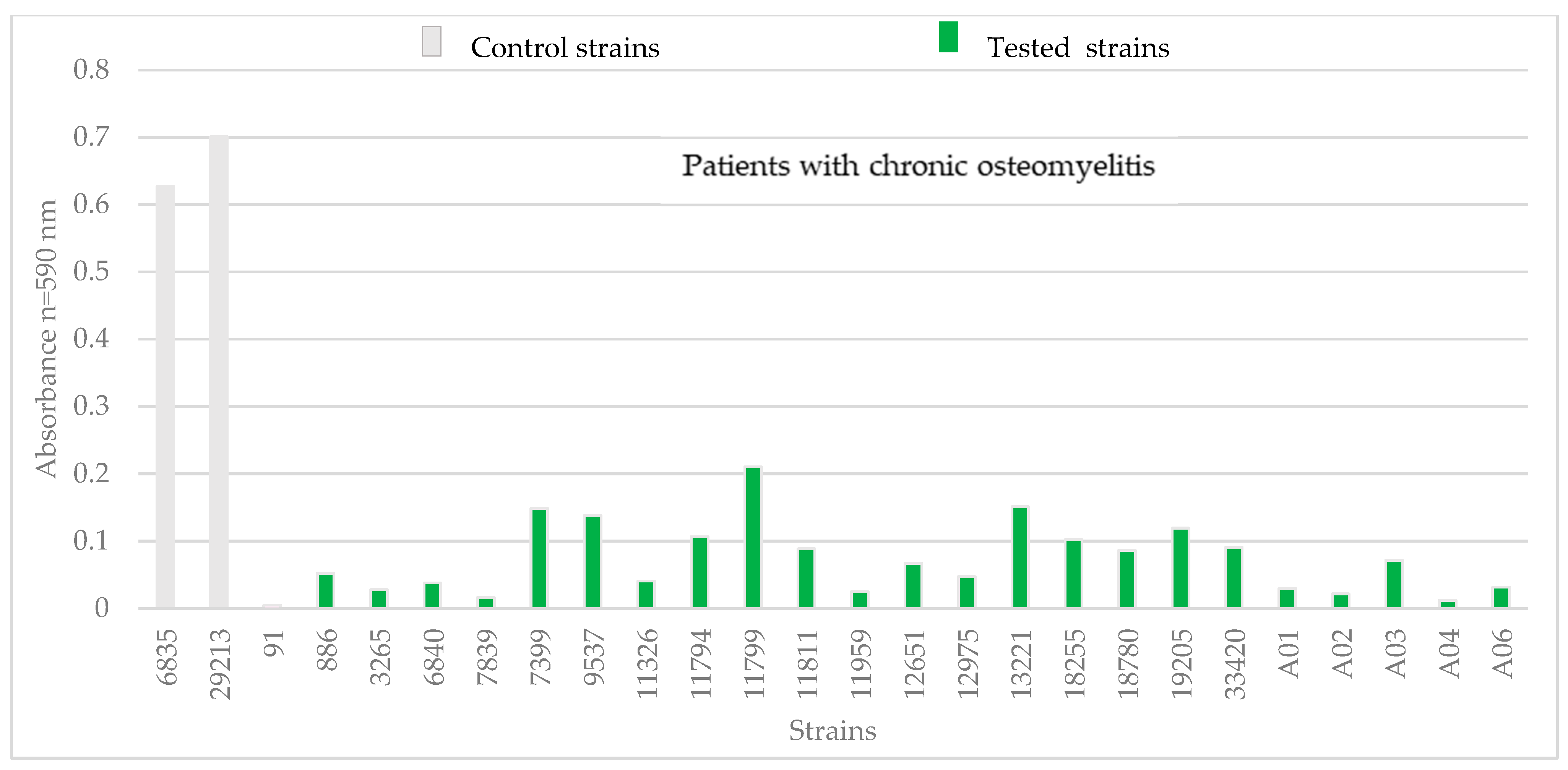
Among S. aureus strains isolated from patients with chronic osteomyelitis, four strains showed the greatest biomass increase: 11799, 7399, 13221, and 9537 (Figure 3). The biomass increment for these strains ranged from 0.14 to 0.21. The control S. aureus strains showed a higher biomass gain than the isolated S. aureus strains. The biomass gain of the two control strains was 0.6 and 0.7.

3.2. Genotypic Assessments

Real-time polymerase chain reaction studies showed that 59 strains possessed the following virulence genes: clfa, clfb, spa, cna, eap, hlgA, hlgB, hlg, hld, bap, bbp, ebps, fib, fnpba, fnpbb, and pvl (Table A1). Eight strains of S. aureus were found to be deficient in single genes, including five strains isolated from patients with chronic osteomyelitis and three strains isolated from patients with chronic furunculosis (Table 3). A chi-square analysis was conducted to investigate the potential relationship between various types of antibiotic resistance (MRSA, iMLSb, cMLSb) and the presence of specific virulence genes. The study included 16 virulence genes, namely, clfa, clfb, spa, cna, eap, hlgA, hlgB, hlg, hld, bap, bbp, ebps, fib, fnpba, fnpbb, and pvl. The analyses revealed no statistically significant associations between the resistances of MRSA, iMLSb, and cMLSb and the presence of most of the studied virulence genes, with p-values equal to 1 for each test.
These results suggest that the resistance and virulence mechanisms in S. aureus may operate independently, without direct links between these traits in the studied population. However, confirming these conclusions requires further research in diverse clinical and epidemiological settings to better understand the potential interactions between virulence genes and antibiotic resistance.

4. Discussion

In a study of S. aureus isolated from chronic osteomyelitis and furunculosis patients, over 80% of the strains were found to be sensitive to methicillin and other tested antibiotics. Approximately 10% of strains were methicillin-susceptible but exhibited resistance to macrolides, lincosamides, and streptogramin B, including 10.5% with inducible resistance and 1.5% with constitutive resistance. Additionally, 4.5% of strains were methicillin-resistant and showed constitutive resistance to macrolides, lincosamides, and streptogramin B, while 1.5% were methicillin-resistant without this combined resistance. All strains tested were susceptible to gentamicin, ciprofloxacin, and cotrimoxazole.
Studies on S. aureus isolated from pediatric patients with bacteremia and/or osteomyelitis identified approximately 77% of strains as methicillin-susceptible environmental isolates [29]. Gomes et al. [30] similarly found MSSA strains to be more prevalent than resistant strains across multiple inflammatory sites, including bone, and highlighted that MSSA infections can nonetheless pose a high risk for severe disease. An et al. [31] reported the isolation of both MRSA and MSSA strains (around 50% each) from bone infections in pediatric musculoskeletal cases, noting that hospitalization duration, outpatient treatment, and disease course were comparable regardless of methicillin resistance status. De la Calle et al. [32] observed that approximately 50% of pneumonia infections were caused by MSSA and 40% by MRSA, with mortality rates above 40% and 50%, respectively. In contrast, Klein et al. [33] found that hospitalization costs for MSSA and MRSA infections in the United States between 2010 and 2014 were similar, and in some cases, MSSA pneumonia treatment costs exceeded those for MRSA infections. Together, these findings from our study and the literature indicate that the infection risk and clinical burden of MSSA strains are comparable to, and in some cases exceed, those associated with MRSA.
Our findings underscore that MSSA strains, despite being methicillin-sensitive, can harbor virulence determinants comparable to MRSA, which is clinically relevant for infection management and antibiotic selection. Worth mentioning is the fact that MSSA can exhibit greater virulence than MRSA, a phenomenon attributed to the lower fitness costs associated with the absence of methicillin resistance. This allows MSSA strains to maintain more efficient growth, higher toxin production, and enhanced persistence in host tissues, which may contribute to the duration and severity of chronic infections [34,35].
Notably, the cfiA and cfiB genes, involved in iron acquisition, have been implicated in the persistence and chronicity of S. aureus infections. These genes facilitate bacterial survival in iron-limited environments, a common condition in chronic infections. Their presence in both MSSA and MRSA strains suggests that virulence potential, rather than methicillin resistance, may be a critical factor in the persistence of chronic infections [36].
The results presented by the ECDC [37] indicate a downward trend in the incidence of MRSA strains, decreasing from 19% in 2015 to 15.5% in 2019. In the present study, we demonstrated that chronic furunculosis and chronic osteomyelitis caused by MSSA occur more frequently than infections caused by resistant strains. Among the MSSA strains in our study, 12% exhibited constitutive and inducible resistance to macrolides, lincosamides, and streptogramin B (cMLSB and iMLSB phenotypes), while 4.5% of MRSA strains showed cMLSB resistance.
Saderi et al. [38] reported that S. aureus strains isolated from abscesses, wounds, and blood showed cMLSB phenotype in 92% of cases and an iMLSB phenotype in 6.3%. Pardo et al. [39] found that 22% of clinical isolates exhibited the cMLSB phenotype and 10% the iMLSB phenotype. In comparison, our study identified 10.5% of MSSA strains with the iMLSB phenotype and only 6% of both MRSA and MSSA strains with the cMLSB phenotype. These variations highlight that antibiotic resistance prevalence depends on the epidemiological context of specific regions and countries [40].
Therapeutic strategies should be based on antimicrobial susceptibility data provided by reference laboratories. In our study, the lack of resistance to cotrimoxazole, gentamicin, or ciprofloxacin was confirmed. Stein et al. [41] reported a treatment success rate using cotrimoxazole for bone infections caused by S. aureus in 66.7% of cases. Nguyen et al. [42] evaluated cotrimoxazole combined with rifampicin for osteomyelitis and infections related to orthopedic implants, finding a 78.6% efficacy compared to rifampicin with linezolid. All isolates examined in this study were susceptible to ciprofloxacin, though epidemiological data from the National Reference Center for Microbial Susceptibility (2019) indicate that approximately 15% of S. aureus strains resist fluoroquinolones. Due to the high percentage of resistant strains, quinolones should be considered as an alternative therapeutic option, used only after microbiological testing to ensure targeted therapy.
Boot et al. [43] explored the use of hyaluronic acid hydrogels loaded with gentamicin and vancomycin to treat infections associated with orthopedic implants, concluding that topical administration of gentamicin yielded the best therapeutic outcomes due to high local drug concentrations. Our study showed that S. aureus strains remain sensitive to gentamicin, suggesting promising therapeutic potential.
It has also been demonstrated that MSSA strains possess a similar repertoire of virulence genes as MRSA and are responsible for most cases of chronic osteomyelitis and furunculosis. Research indicates that S. aureus carries multiple virulence factors critical for adhesion and proliferation on host cells. Bacteria without antibiotic resistance demonstrate alternative strategies to withstand external environmental pressures [44]. One such mechanism is biofilm production, observed in both MSSA and MRSA strains, along with surface proteins that facilitate adhesion to host cells [45]. Biofilms protect bacterial cells, significantly impeding the effectiveness of antibiotics and chemotherapeutics.
In the present study, the analyzed S. aureus strains demonstrated a low capacity for biofilm production. Specifically, 62% of strains exhibited absorbance levels below 0.1, indicating limited biofilm formation. While this level is low, it likely provides sufficient protection against adverse environmental factors, including antibiotic exposure, as noted by Kavanagh et al. [44]. These authors emphasized the role of biofilms in chronic osteomyelitis, highlighting their contribution to the persistence and chronicity of inflammation. Bjarnsholt et al. [46] similarly noted that acute infections caused by planktonic bacteria are easier to treat than those involving biofilms. Biofilms are also implicated in other chronic conditions, such as chronic otitis and cystic fibrosis.
Akiyama et al. [47] isolated S. aureus from carbuncular lesions and cultured the bacteria on plasma- and fibrinogen-coated plates. Electron microscopy after 72 h revealed substantial fibrin deposition and increased bacterial proliferation as early as the fourth hour, underscoring the critical role of fibrinogen and fibrin in bacterial adhesion and biomass production. These findings suggest that S. aureus from furunculosis lesions is capable of biofilm formation.
Biofilm-associated bacteria can evade immune defenses by modulating host immune responses. Paharik and Horswill [48] demonstrated that macrophages near S. aureus biofilms show impaired phagocytic activity, with the clfA gene expression contributing to this inhibition. Moreover, S. aureus in biofilms can kill polymorphonuclear cells (PMNs), further evading phagocytosis and promoting chronic infection. Bhattacharya et. al. [49], corroborated by Paharik and Horswill [48], reported that high levels of Panton–Valentine leukocidin and hemolysins produced by S. aureus regulate NETosis, allowing bacteria to evade neutrophil attacks. Additionally, the production of polysaccharide intercellular adhesin (PIA) by S. aureus aids in avoiding neutrophil phagocytosis.
Recent insights into S. aureus biofilm formation show its crucial role in chronic infections and antibiotic resistance. Biofilms protect bacteria, hinder antibiotic penetration, and enhance tolerance by inducing metabolic dormancy and upregulating efflux pumps. Clinical isolates, especially MRSA, are increasingly resistant to vancomycin and daptomycin due to genetic mutations and biofilm-associated extracellular DNA. Small-colony variants (SCVs) in biofilms further complicate treatment by evading immune responses and antibiotics. Novel strategies like quorum-sensing inhibitors and biofilm-disrupting enzymes are being explored to tackle this resistance, highlighting the need for personalized therapies targeting both planktonic and biofilm states to reduce treatment failures [50,51].
Infections caused by biofilm-embedded S. aureus strains are challenging to treat, with therapies often failing in chronic cases even when strains demonstrate susceptibility to commonly used antimicrobial agents. Furthermore, bacteria within biofilms alter their prophylactic and metabolic profiles, resulting in resistance to antibiotics targeting the cell wall [48]. In this study, both MSSA and MRSA strains exhibited resistance to macrolides, lincosamides, and streptogramin B, supporting their capacity for biofilm-associated antibiotic resistance.
While significant efforts are being made to understand S. aureus virulence mechanisms and their roles in diseases such as osteomyelitis and furunculosis, further research is necessary to elucidate the inflammatory processes driven by these bacteria. The pathogenicity of S. aureus is closely related to the presence of various virulence genes [52]. In the presented study, all S. aureus strains isolated from patients with chronic furunculosis and chronic osteomyelitis were found to possess the following virulence genes: clfa, clfb, spa, eap, hld, bap, ebps, and fnbb. In contrast, 98.5% of the strains harbored the genes hlgB, hlg, hlgA, pvl, cna, and fnbpa, and 97% possessed bbp and fib. This study did not examine the allelic variant of bbp (sdrE), which may be present in their bbp-lacking strain. All S. aureus isolates from infected skin lesions of children with atopic dermatitis showed multiple virulence genes. The hlgA, seu, fnbA, icaA, and sasG genes were found in all isolates, hlg was found in 98.2% of isolates, and cna was found in 70.9% of isolates [53].
Li et al. [52] identified twelve virulence genes in S. aureus causing bloodstream infections, including clfa (100%) and hlgA (99%). It was found that clfa and hlgA were widely present and may play a pivotal role in the pathogenicity of S. aureus. This suggests that adhesion, proliferation, and subsequent development of inflammation by S. aureus strains are facilitated by the presence of multiple virulence factors.
In the present study, in all tested S. aureus strains, the spa gene encoding staphylococcal protein A (Spa) was present. This protein is crucial for typing S. aureus isolates. The spa gene encodes protein A, a critical virulence factor of S. aureus. Protein A binds to the Fc region of IgG antibodies, thereby disrupting opsonization and phagocytosis, which enables the bacteria to evade the host immune response. Additionally, protein A can interfere with immune signaling by interacting with tumor necrosis factor receptor 1 (TNFR1), modulating the inflammatory response. It also functions as an adhesin, facilitating bacterial attachment to host tissues. These combined actions contribute significantly to the pathogenicity of S. aureus and its ability to establish infections [54].
The significance of typing this gene using Based Upon Repeat Pattern (BURP) analysis in epidemiological studies was highlighted by Strommenger et al. [55]. In their study of S. aureus isolates from a reference center (n = 1459), they identified 221 different spa types among the strains studied. Many S. aureus strains have the ability to bind to fibronectin, as evidenced by the presence of the fnbA and fnbB genes. These genes have been found, among others, in S. aureus strains, causing infections associated with orthopedic implants. Giormezis et al. [56] demonstrated the presence of the fnbA gene in 88.9% of S. aureus strains isolated from various patients in three hospitals located in Greece.
The least understood gene is the eap gene, which encodes an extracellular adherence protein involved in binding the bacterial cell to the extracellular matrix of host tissues, making it crucial for colonization. In the present study, the eap gene was found in all tested S. aureus strains. Hussain et al. [57] examined 597 S. aureus isolates from humans and animals and found the eap gene in approximately 98% of strains.
Although the exact role of the extracellular adherence protein in the development of osteomyelitis and furunculosis is not fully understood, its ability to block monocytes and T cells indicates that this protein is not only involved in bacterial adhesion to host cells but also effectively inhibits the host immune response [58]. The collagen-binding protein, encoded by the cna gene, is directly linked to the development of osteomyelitis. This protein enables bacterial cells to bind to the host’s cartilage tissue and plays a key role in septic arthritis, endocarditis, and infections around orthopedic implants [59]. In the current study, one strain isolated from a patient with chronic osteomyelitis lacked the cna gene. Some previous studies have also reported the absence of this gene in strains still capable of causing infection and inflammation [59].
The biofilm-associated protein (Bap) is involved in biofilm production. In this study, all tested strains possessed the bap gene. Interestingly, this gene was present even in S. aureus strains that did not form strong biofilms. Most research on the bap gene has focused on S. aureus strains isolated from cattle, sheep, and goats. For example, Cucarella et al. [60] found the bap gene in only 5% of strains from cattle and did not detect it in human isolates (n = 75).
Cavalcante et al. [53] found that the bbp gene was significantly more present in S. aureus strains from infected skin lesions, with a p-value of 0.0021, suggesting a strong association between bbp and infection-related strains. Additionally, research by Bride et al. [61] indicated that the bbp gene was more prevalent in MSSA strains compared to methicillin-resistant strains, highlighting its potential role in the virulence of MSSA. These findings underscore the significance of the bbp gene in S. aureus pathogenicity, particularly in MSSA strains, and suggest that its presence may correlate with increased virulence.
In this study, three S. aureus strains from patients with chronic osteomyelitis lacked the hlgA, hlgB, and hlg genes, which encode alpha, beta, and gamma hemolysins, respectively. However, all tested strains possessed the hld gene. The gene most directly associated with furunculosis is pvl, which encodes Panton–Valentine leukocidin. Here, all but one strain carried this gene. Masiuk et al. [62] confirmed the role of Panton–Valentine leukocidin in skin and soft tissue infections, particularly furunculosis. The toxin’s ability to lyse leukocytes is a key factor in the inflammatory response.
PVL contributes to the chronicity of S. aureus infections by subtly undermining host defenses. By lysing neutrophils and releasing inflammatory mediators, PVL triggers tissue damage that creates niches for bacterial survival. This persistent inflammation, coupled with partial immune evasion, allows bacteria to persist and recur. Although PVL itself does not directly promote biofilms, the tissue damage it causes can facilitate their formation, further protecting bacteria from clearance and antibiotics. In essence, PVL drives a cycle of tissue injury and immune modulation that favors persistent and recurrent infections [63,64].
The clfa and clfb genes encode clumping factors A and B, which bind fibrinogen to host cells. In the present study, clfa and clfb were found in 99% and 100% of S. aureus strains, respectively. Similar findings were reported by Zmantar et al. [65], who detected clfa in 30% of strains isolated from patients with ear infections, along with several other virulence genes. In the study of Tristan et al. [66], the clfa gene was present in all S. aureus strains isolated from patients with endocarditis and in 95% of strains from patients with osteoarthritis. Similarly, the clfb gene was detected in all isolates from both patient groups.
Bone sialoprotein, encoded by the bbp gene, is related to fibrinogen α, a key component of bone and connective tissue. In the present study, 98.5% of S. aureus strains carried the bbp gene. The absence of bbp was observed in only one strain isolated from a patient with chronic furunculosis. In contrast, Nemati et al. [67] reported that S. aureus strains isolated from healthy and infected poultry lacked the bbp gene.
Tang et al. [26] used PCR to detect the fib gene in S. aureus strains from chickens, goats, and food poisoning samples, finding it present in over 90% of isolates. Similarly, Boden Wästfelt et al. [68] reported the fib gene in 97% of human S. aureus isolates and 100% of bovine isolates, using classical PCR methods. In our study, 97% of S. aureus strains were positive for the fib gene, consistent with previous findings. This suggests possible gene transmission among S. aureus strains, potentially increasing their virulence.
Additionally, the gene encoding elastin-binding protein (ebps) was detected in 100% of the tested S. aureus strains. Azmi et al. [42] found this gene in 84% of MRSA strains isolated from infected wounds, blood, urine, and nasal swabs using classical PCR. These findings underscore the crucial role of ebps in the S. aureus genome and its involvement in the inflammatory process. Overall, these findings highlight the complex interplay of antibiotic resistance, virulence factors, and biofilm formation in S. aureus, underscoring the critical need for targeted therapies guided by susceptibility testing to effectively manage chronic osteomyelitis and furunculosis.
Our study demonstrates that MSSA strains carry a diverse and abundant repertoire of virulence genes, highlighting their clinical significance in infection persistence and management. The significant biofilm production observed in MSSA, together with associations such as the pvl gene with furunculosis, underscores that virulence potential, rather than methicillin resistance alone, is a critical factor in chronic and acute S. aureus infections. These findings emphasize the need for careful monitoring of MSSA strains and consideration of virulence profiles in guiding therapeutic strategies.
This study is part of a larger project investigating the chronicity of Staphylococcus aureus infections, an area still underexplored. Our results show that MSSA strains carry a broad array of virulence genes, yet bacterial characteristics alone do not fully explain persistent infections. Chronicity appears closely linked to patient-specific factors, which vary widely and likely interact with bacterial traits. Future work will integrate patient-derived data and immune responses with strain characteristics to better understand the host–pathogen interactions driving long-lasting infections.

Author Contributions

A.W.: literature search and review, conceptualization, formal analysis, manuscript draft preparation, writing manuscript, investigation, resources, and preparation of manuscript revision. J.J.-K.: conceptualization, formal analysis, writing the manuscript, and preparation of manuscript revision. H.M.: methodology. B.G.: validation and methodology. B.H.-S.: participation in writing the manuscript. P.P.: participation in writing the manuscript. P.Z.: participation in writing the manuscript. D.K.-B.: writing the manuscript, preparing manuscript revision, supervising, and final acceptance of the manuscript. All authors have read and agreed to the published version of the manuscript.

Funding

This research received no external funding.

Institutional Review Board Statement

The study was conducted in accordance with the Declaration of Helsinki and was approved by the Ethics Committee of Pomeranian Medical University (protocol code KB-0012/112/17, date 2 October 2017) for studies involving humans.

Informed Consent Statement

Not applicable. In the study, informed consent was not required as the isolates included in the study were obtained as a result of standard medical care. Written permissions to access the raw data and samples mentioned in the methods were obtained from hospitals authorities.

Data Availability Statement

The data presented in this study are available on request from the corresponding author.

Conflicts of Interest

The authors declare no conflict of interest.

Appendix A

Table A1. The virulence genes in Staphylococcus aureus isolates from patients with chronic furunculosis and chronic osteomyelitis (clfa, clumping factor A; clfb, clumping factor B; spa, protein A; cna, collagen adhesin; eap, extracellular adherence protein; hlgA, alpha-hemolysin; hlgB, beta-hemolysin, hlg, gamma-hemolysin; hld, delta-hemolysin; bap; biofilm-associated protein; bbp, bone sialoprotein-binding protein; ebpS, elastin-binding protein; Efb, fibrinogen-binding protein; fnbA, fibronectin-binding protein A; fnbB, fibronectin-binding protein B; pvl, panton valentine leukocidin; +, indicates the presence of the gene in question; -, indicates an absence of the gene).
Genes
Strain Number
clfaclfbspacnaeaphlgAHlgBhlgHldbapbbpebpSEfbfnbAfnbBPvl
control strains
29213++++++++++++++++
6538++++++++++++++++
strains from patients with chronic osteomyelitis
91++++++-+++++++++
886++++++++++++++++
3265++++++++++++++++
6840++++++++++++++++
7839++++++++++++++++
7399++++++++++++++++
9537++++++++++++++++
11326++++++++++++++++
11794++++++++++++++++
11799++++++++++++++++
11811++++++++++++++++
11959+++++++-++++++++
12651++++++++++++++++
12975++++++++++++++++
13221++++++++++++++++
18255+++++-++++++++++
18780++++++++++++++++
19205++++++++++++++++
33420++++++++++++++++
40034+++-++++++++++++
40675++++++++++++++++
41203++++++++++++++++
43846++++++++++++++++
43901++++++++++++++++
strains from patients with chronic furunculosis
351++++++++++++++++
436++++++++++++++++
648++++++++++++++++
1727++++++++++++++++
1747++++++++++++++++
2071++++++++++++++++
3056++++++++++++++++
3058++++++++++++++++
3200++++++++++++++++
6241++++++++++++++++
6866++++++++++++++++
8304++++++++++++++++
8215++++++++++++++++
10294++++++++++++++++
11103++++++++++++++++
11673++++++++++++++++
12756++++++++++++++++
12823++++++++++++++++
12866++++++++++++++++
14279+++++++++++++++-
14300++++++++++++++++
15760++++++++++++++++
15923++++++++++++++++
18311++++++++++++++++
18345++++++++++++++++
18477++++++++++++++++
19757++++++++++-+++++
31136++++++++++++++++
31275++++++++++++++++
31711++++++++++++++++
31730++++++++++++++++
32475++++++++++++++-+
32567++++++++++++++++
32895++++++++++++++++
33203++++++++++++++++
33480++++++++++++++++
33888++++++++++++++++
34392++++++++++++++++
35753++++++++++++++++
35770++++++++++++++++
35843++++++++++++++++
38576++++++++++++++++
45736++++++++++++++++

References

  1. Touaitia, R.; Mairi, A.; Ibrahim, N.A.; Basher, N.S.; Idres, T.; Touati, A. Staphylococcus aureus: A review of the pathogenesis and virulence mechanisms. Antibiotics 2025, 14, 470. [Google Scholar] [CrossRef]
  2. Gehrke, A.E.; Giai, C.; Gómez, M.I. Staphylococcus aureus adaptation to the skin in health and persistent/recurrent infections. Antibiotics 2023, 12, 1520. [Google Scholar] [CrossRef] [PubMed]
  3. Taylor, T.A.; Unakal, C.G. Staphylococcus aureus infection. In StatPearls. Internet; StatPearls Publishing: Petersburg, FL, USA, 2025. [Google Scholar]
  4. Bai, A.D.; Lo, C.K.L.; Komorowski, A.S.; Suresh, M.; Guo, K.; Garg, A.; Tandon, P.; Senecal, J.; Del Corpo, O.; Stefanova, I.; et al. Staphylococcus aureus bacteraemia mortality: A systematic review and meta-analysis. Clin. Microbiol. Infect. 2022, 28, 1076–1084. [Google Scholar] [CrossRef]
  5. Brdová, D.; Ruml, T.; Viktorová, J. Mechanism of staphylococcal resistance to clinically relevant antibiotics. Drug Resist. Updat. 2024, 77, 101147. [Google Scholar] [CrossRef]
  6. World Health Organization. Antimicrobial Resistance: Global Report on Surveillance. Available online: https://apps.who.int/iris/handle/10665/112642 (accessed on 14 August 2021).
  7. Centers for Disease Control, U. CDC. Antibiotic Resistance Threats in the United States, 2019; Department of Health and Human Services, CDC: Atlanta, GA, USA, 2019. [Google Scholar] [CrossRef]
  8. Kasela, M.; Grzegorczyk, A.; Nowakowicz-Dębek, B.; Malm, A. The prevalence of virulence determinants and antibiotic resistance patterns in methicillin-resistant Staphylococcus aureus in a nursing home in Poland. Pathogens 2021, 10, 427. [Google Scholar] [CrossRef]
  9. Tu, H.; Xu, F.; Cheng, Y.; Pan, Q.; Cai, X.; Wang, S.; Ge, S.; Cao, M.; Su, D.; Li, Y. Proteomic profiling of the endogenous peptides of MRSA and MSSA. PeerJ 2021, 9, e12508. [Google Scholar] [CrossRef]
  10. Chen, L.; Tang, Z.Y.; Cui, S.Y.; Ma, Z.B.; Deng, H.; Kong, W.L.; Yang, L.W.; Lin, C.; Xiong, W.G.; Zeng, Z.L. Biofilm production ability, virulence and antimicrobial resistance genes in Staphylococcus aureus from various veterinary hospitals. Pathogens 2020, 9, 264. [Google Scholar] [CrossRef] [PubMed]
  11. Bae, S.; Kim, E.S.; Kim, H.S.; Yang, E.; Chung, H.; Lee, Y.W.; Jung, J.; Kim, M.J.; Chong, Y.P.; Kim, S.H.; et al. Risk factors of recurrent infection in patients with Staphylococcus aureus bacteremia: A competing risk analysis. Antimicrob. Agents Chemother. 2022, 66, e0012622. [Google Scholar] [CrossRef]
  12. Poolman, J.T.; Torres, V.J.; Missiakas, D.; Welten, S.P.M.; Fernandez, J.; DuMont, A.L.; O’Keeffe, A.; Konstantinov, S.R.; Morrow, B.; Burghout, P.; et al. A SpA+LukAB vaccine targeting Staphylococcus aureus evasion factors restricts infection in two minipig infection models. NPJ Vaccines 2025, 10, 78. [Google Scholar] [CrossRef] [PubMed]
  13. Soltani, E.; Farrokhi, E.; Zamanzad, B.; Shahini Shams Abadi, M.; Deris, F.; Soltani, A.; Gholipour, A. Prevalence and distribution of adhesins and the expression of fibronectin-binding protein (FnbA and FnbB) among Staphylococcus aureus isolates from Shahrekord Hospitals. BMC Res. Notes 2019, 12, 49. [Google Scholar] [CrossRef]
  14. Ghasemian, A.; Najar Peerayeh, S.; Bakhshi, B.; Mirzaee, M. The Microbial Surface Components Recognizing Adhesive Matrix Molecules (MSCRAMMs) Genes among Clinical Isolates of Staphylococcus aureus from Hospitalized Children. Iran. J. Pathol. 2015, 10, 258–264. [Google Scholar]
  15. Ganesh, V.K.; Rivera, J.J.; Smeds, E.; Ko, Y.P.; Bowden, M.G.; Wann, E.R.; Gurusiddappa, S.; Fitzgerald, J.R.; Höök, M. A structural model of the Staphylococcus aureus ClfA-fibrinogen interaction opens new avenues for the design of anti-staphylococcal therapeutics. PLoS Pathog. 2008, 4, e1000226. [Google Scholar] [CrossRef]
  16. Cheung, G.Y.; Otto, M. The potential use of toxin antibodies as a strategy for controlling acute Staphylococcus aureus infections. Expert Opin. Ther. Targets 2012, 16, 601–612. [Google Scholar] [CrossRef]
  17. Bicca, M.R.; Oliveira Pinto, E.; Sousa Filho, W.P.; de Brum, G.F.; da Silva, T.C.; Vizzotto, B.S. Molecular investigation of a furunculosis outbreak at a penitentiary in southern Brazil. Infect. Dis. Health 2025, 30, 284–286. [Google Scholar] [CrossRef]
  18. Linz, M.S.; Mattappallil, A.; Finkel, D.; Parker, D. Clinical Impact of Staphylococcus aureus Skin and Soft Tissue Infections. Antibiotics 2023, 12, 557. [Google Scholar] [CrossRef]
  19. Qi, X.Y.; Qiu, X.S.; Jiang, J.Y.; Chen, Y.X.; Tang, L.M.; Shi, H.F. Microwaves increase the effectiveness of systemic antibiotic treatment in acute bone infection: Experimental study in a rat model. J. Orthop. Surg. Res. 2019, 14, 286. [Google Scholar] [CrossRef] [PubMed]
  20. Kay, W.; Hunt, C.; Nehring, L.; Barnum, B.; Ashton, N.; Williams, D. Biofilm growth on simulated fracture fixation plates using a customized CDC biofilm reactor for a sheep model of biofilm-related infection. Microorganisms 2022, 10, 759. [Google Scholar] [CrossRef] [PubMed]
  21. Kalinka, J.; Hachmeister, M.; Geraci, J.; Sordelli, D.; Hansen, U.; Niemann, S.; Oetermann, S.; Peters, G.; Löffler, B.; Tuchscherr, L. Staphylococcus aureus isolates from chronic osteomyelitis are characterized by high host cell invasion and intracellular adaptation, but still induce inflammation. Int. J. Med. Microbiol. 2014, 304, 1038–1049. [Google Scholar] [CrossRef]
  22. Moraveji, Z.; Tabatabaei, M.; Shirzad Aski, H.; Khoshbakht, R. Characterization of hemolysins of Staphylococcus strains isolated from human and bovine, southern Iran. Iran. J. Vet. Res. 2014, 15, 326–330. [Google Scholar]
  23. Galia, L.; Ligozzi, M.; Bertoncelli, A.; Mazzariol, A. Triplex real-time PCR assay for detection of Staphylococcus aureus, Panton-Valentine Leucocidin and Methicillin Resistance directly from clinical samples. AIMS Microbiol. 2019, 5, 138–146. [Google Scholar] [CrossRef] [PubMed]
  24. Tang, J.; Chen, J.; Li, H.; Zeng, P.; Li, J. Characterization of adhesin genes, staphylococcal nuclease, hemolysis, and biofilm formation among Staphylococcus aureus strains isolated from different sources. Foodborne Pathog. Dis. 2013, 10, 757–763. [Google Scholar] [CrossRef]
  25. Kwiatkowski, P.; Grygorcewicz, B.; Pruss, A.; Wojciuk, B.; Giedrys-Kalemba, S.; Dołęgowska, B.; Zielińska-Bliźniewska, H.; Olszewski, J.; Sienkiewicz, M.; Kochan, E. Synergistic effect of fennel essential oil and hydrogen peroxide on bacterial biofilm. Postepy Dermatol. Alergol. 2020, 37, 690–694. [Google Scholar] [CrossRef]
  26. Oleksy-Wawrzyniak, M.; Junka, A.; Brożyna, M.; Migdał, P.; Kwiek, B.; Nowak, M.; Mączyńska, B.; Bartoszewicz, M. The in vitro ability of Klebsiella pneumoniae to form biofilm and the potential of various compounds to eradicate it from urinary catheters. Pathogens 2021, 11, 42. [Google Scholar] [CrossRef]
  27. Kamimura, R.; Kanematsu, H.; Ogawa, A.; Kogo, T.; Miura, H.; Kawai, R.; Hirai, N.; Kato, T.; Yoshitake, M.; Barry, D.M. Quantitative Analyses of biofilm by using crystal violet staining and optical reflection. Materials 2022, 15, 6727. [Google Scholar] [CrossRef]
  28. Yang, C.H.; Su, P.W.; Moi, S.H.; Chuang, L.Y. Biofilm Formation in Acinetobacter baumannii: Genotype-phenotype correlation. Molecules 2019, 24, 1849. [Google Scholar] [CrossRef] [PubMed]
  29. Kechrid, A.; Pérez-Vázquez, M.; Smaoui, H.; Hariga, D.; Rodríguez-Baños, M.; Vindel, A.; Baquero, F.; Cantón, R.; del Campo, R. Molecular analysis of community-acquired methicillin-susceptible and resistant Staphylococcus aureus isolates recovered from bacteraemic and osteomyelitis infections in children from Tunisia. Clin. Microbiol. Infect. 2011, 17, 1020–1026. [Google Scholar] [CrossRef] [PubMed]
  30. Gomes, R.T.; Lyra, T.G.; Alves, N.N.; Caldas, R.M.; Barberino, M.G.; Nascimento-Carvalho, C.M. Methicillin-resistant and methicillin-susceptible community-acquired Staphylococcus aureus infection among children. Braz. J. Infect. Dis. 2013, 17, 573–578. [Google Scholar] [CrossRef]
  31. An, T.J.; Benvenuti, M.A.; Mignemi, M.E.; Martus, J.; Wood, J.B.; Thomsen, I.P.; Schoenecker, J.G. Similar clinical severity and outcomes for methicillin-resistant and methicillin-susceptible Staphylococcus aureus pediatric musculoskeletal infections. Open Forum Infect. Dis. 2017, 4, ofx013. [Google Scholar] [CrossRef]
  32. De la Calle, C.; Morata, L.; Cobos-Trigueros, N.; Martinez, J.A.; Cardozo, C.; Mensa, J.; Soriano, A. Staphylococcus aureus bacteremic pneumonia. Eur. J. Clin. Microbiol. Infect. Dis. 2016, 35, 497–502. [Google Scholar] [CrossRef] [PubMed]
  33. Klein, E.Y.; Jiang, W.; Mojica, N.; Tseng, K.K.; McNeill, R.; Cosgrove, S.E.; Perl, T.M. National costs associated with methicillin-susceptible and methicillin-resistant Staphylococcus aureus hospitalizations in the United States, 2010–2014. Clin. Infect. Dis. 2019, 68, 22–28. [Google Scholar] [CrossRef]
  34. Bu Lattar, S.M.; Tuchscherr, L.P.; Caccuri, R.L.; Centrón, D.; Becker, K.; Alonso, C.A.; Barberis, C.; Miranda, G.; Buzzola, F.R.; von Eiff, C.; et al. Capsule expression and genotypic differences among Staphylococcus aureus isolates from patients with chronic or acute osteomyelitis. Infect. Immun. 2009, 77, 1968–1975. [Google Scholar] [CrossRef]
  35. Fitzgerald, J.R. Evolution of Staphylococcus aureus during human colonization and infection. Infect. Genet. Evol. 2014, 21, 542–547. [Google Scholar] [CrossRef]
  36. Di Bella, S.; Marini, B.; Stroffolini, G.; Geremia, N.; Giacobbe, D.R.; Campanile, F.; Bartoletti, M.; Alloisio, G.; Di Risio, L.; Viglietti, G.; et al. The virulence toolkit of Staphylococcus aureus: A comprehensive review of toxin diversity, molecular mechanisms, and clinical implications. Eur. J. Clin. Microbiol. Infect. Dis. 2025, 44, 1797–1816. [Google Scholar] [CrossRef]
  37. European Centre for Disease Prevention and Control. Antimicrobial Resistance in the EU/EEA (EARS-Net)—Annual Epidemiological Report for 2019. 2020. Available online: https://www.ecdc.europa.eu/en/publications-data/surveillance-antimicrobial-resistance-europe-2019 (accessed on 31 March 2022).
  38. Saderi, H.; Emadi, B.; Owlia, P. Phenotypic and genotypic study of macrolide, lincosamide and streptogramin B (MLSB) resistance in clinical isolates of Staphylococcus aureus in Tehran, Iran. Med. Sci. Monit. 2011, 17, BR48. [Google Scholar] [CrossRef] [PubMed]
  39. Pardo, L.; Machado, V.; Cuello, D.; Aguerrebere, P.; Seija, V.; Braga, V.; Varela, G. Macrolide-lincosamide-streptogramin B resistance phenotypes and their associated genotypes in Staphylococcus aureus isolates from a tertiary level public hospital of Uruguay. Rev. Argent. Microbiol. 2020, 52, 202–210. [Google Scholar] [CrossRef]
  40. Azmi, K.; Qrei, W.; Abdeen, Z. Screening of genes encoding adhesion factors and biofilm production in methicillin resistant strains of Staphylococcus aureus isolated from Palestinian patients. BMC Genom. 2019, 20, 578. [Google Scholar] [CrossRef] [PubMed]
  41. Stein, A.; Bataille, J.F.; Drancourt, M.; Curvale, G.; Argenson, J.N.; Groulier, P.; Raoult, D. Ambulatory treatment of multidrug-resistant Staphylococcus-infected orthopedic implants with high-dose oral co-trimoxazole (trimethoprim-sulfamethoxazole). Antimicrob. Agents Chemother. 1998, 42, 3086–3091. [Google Scholar] [CrossRef]
  42. Nguyen, S.; Pasquet, A.; Legout, L.; Beltrand, E.; Dubreuil, L.; Migaud, H.; Yazdanpanah, Y.; Senneville, E. Efficacy and tolerance of rifampicin–linezolid compared with rifampicin–cotrimoxazole combinations in prolonged oral therapy for bone and joint infections. Clin. Microbiol. Infect. 2009, 15, 1163–1169. [Google Scholar] [CrossRef] [PubMed]
  43. Boot, W.; Schmid, T.; D’este, M.; Guillaume, O.; Foster, A.; Decosterd, L.; Richards, R.G.; Eglin, D.; Zeiter, S.; Moriarty, T.F. A Hyaluronic acid hydrogel loaded with gentamicin and vancomycin successfully eradicates chronic Methicillin-Resistant Staphylococcus aureus orthopedic infection in a sheep model. Antimicrob. Agents Chemother. 2021, 65, e01840-20. [Google Scholar] [CrossRef]
  44. Kavanagh, N.; Ryan, E.J.; Widaa, A.; Sexton, G.; Fennell, J.; O’Rourke, S.; Cahill, K.C.; Kearney, C.J.; O’Brien, F.J.; Kerrigan, S.W. Staphylococcal osteomyelitis: Disease progression, treatment challenges, and future directions. Clin. Microbiol. Rev. 2018, 31, e00084-17. [Google Scholar] [CrossRef]
  45. McCarthy, H.; Rudkin, J.K.; Black, N.S.; Gallagher, L.; O’Neill, E.; O’Gara, J.P. Methicillin resistance and the biofilm phenotype in Staphylococcus aureus. Front. Cell. Infect. Microbiol. 2015, 5, 1. [Google Scholar] [CrossRef] [PubMed]
  46. Bjarnsholt, T. The role of bacterial biofilms in chronic infections. APMIS 2013, 121, 1–51. [Google Scholar] [CrossRef] [PubMed]
  47. Akiyama, H.; Ueda, M.; Kanzaki, H.; Tada, J.; Arata, J. Biofilm formation of Staphylococcus aureus strains isolated from impetigo and furuncle: Role of fibrinogen and fibrin. J. Dermatol. Sci. 1997, 16, 2–10. [Google Scholar] [CrossRef]
  48. Paharik, A.E.; Horswill, A.R. The Staphylococcal biofilm: Adhesins, regulation, and host response. Microbiol. Spect. 2016, 4, 529–566. [Google Scholar] [CrossRef]
  49. Bhattacharya, M.; Berends, E.T.M.; Chan, R.; Schwab, E.; Roy, S.; Sen, C.K.; Torres, V.J.; Wozniak, D.J. Staphylococcus aureus biofilms release leukocidins to elicit extracellular trap formation and evade neutrophil-mediated killing. Proc. Natl. Acad. Sci. USA 2018, 115, 7416–7421. [Google Scholar] [CrossRef]
  50. Peng, Q.; Tang, X.; Dong, W.; Sun, N.; Yuan, W. A Review of biofilm formation of Staphylococcus aureus and its regulation mechanism. Antibiotics 2022, 12, 12. [Google Scholar] [CrossRef]
  51. Jomehzadeh, N.; Emrani, S.S. Assessment of biofilm formation, antibiotic resistance patterns, and the prevalence of adhesion-related genes in clinical Staphylococcus aureus isolates. Heliyon 2024, 11, e41537. [Google Scholar] [CrossRef] [PubMed]
  52. Li, X.; Fang, F.; Zhao, J.; Lou, N.; Li, C.; Huang, T.; Li, Y. Molecular characteristics and virulence gene profiles of Staphylococcus aureus causing bloodstream infection. Braz. J. Infect. Dis. 2018, 22, 487–494. [Google Scholar] [CrossRef]
  53. Cavalcante, F.S.; Saintive, S.; Carvalho Ferreira, D.; Rocha Silva, A.B.; Guimarães, L.C.; Braga, B.S.; Dios Abad, E.; Ribeiro, M.; Netto Dos Santos, K.R. Methicillin-resistant Staphylococcus aureus from infected skin lesions present several virulence genes and are associated with the CC30 in Brazilian children with atopic dermatitis. Virulence 2021, 12, 260–269. [Google Scholar] [CrossRef]
  54. Foster, T.J. Immune evasion by staphylococci. Nat. Rev. Microbiol. 2005, 3, 948–958. [Google Scholar] [CrossRef] [PubMed]
  55. Strommenger, B.; Braulke, C.; Heuck, D.; Schmidt, C.; Pasemann, B.; Nübel, U.; Witte, W. spa typing of Staphylococcus aureus as a frontline tool in epidemiological typing. J. Clin. Microbiol. 2008, 46, 574–581. [Google Scholar] [CrossRef]
  56. Giormezis, N.; Doudoulakakis, A.; Tsilipounidaki, K.; Militsopoulou, M.; Kalogeras, G.; Stamouli, V.; Kolonitsiou, F.; Petinaki, E.; Lebessi, E.; Spiliopoulou, I. Emergence of a mupirocin-resistant, methicillin-susceptible Staphylococcus aureus clone associated with skin and soft tissue infections in Greece. BMC Microbiol. 2021, 21, 203. [Google Scholar] [CrossRef]
  57. Hussain, M.; Von Eiff, C.; Sinha, B.; Joost, I.; Herrmann, M.; Peters, G.; Becker, K. eap gene as novel target for specific identification of Staphylococcus aureus. J. Clin. Microbiol. 2008, 46, 470–476. [Google Scholar] [CrossRef] [PubMed]
  58. Edwards, A.M.; Bowden, M.G.; Brown, E.L.; Laabei, M.; Massey, R.C. Staphylococcus aureus extracellular adherence protein triggers tnfα release, promoting attachment to endothelial cells via protein A. PLoS ONE 2012, 7, e43046. [Google Scholar] [CrossRef] [PubMed]
  59. Gillaspy, A.F.; Patti, J.M.; Smeltzer, M.S. Transcriptional regulation of the Staphylococcus aureus collagen adhesin gene, cna. Infect. Immun. 1997, 65, 1536–1540. [Google Scholar] [CrossRef]
  60. Cucarella, C.; Solano, C.; Valle, J.; Amorena, B.; Lasa, I.; Penadés, J.R. Bap, a Staphylococcus aureus surface protein involved in biofilm formation. J. Bacteriol. 2001, 183, 2888–2896. [Google Scholar] [CrossRef]
  61. Bride, L.L.; Pereira, M.F.; Barbosa, M.C.; Silva, N.C.; Klein, N.M.; Nascimento, T.C.; Schuenck, R.P. Differences in resistance profiles and virulence genes among methicillin-resistant and methicillin-susceptible Staphylococcus aureus of different lineages at a public tertiary hospital. Rev. Soc. Bras. Med. Trop. 2019, 52, e20190095. [Google Scholar] [CrossRef]
  62. Masiuk, H.; Kopron, K.; Grumann, D.; Goerke, C.; Kolata, J.; Jursa-Kulesza, J.; Giedrys-Kalemba, S.; Bröker, B.M.; Holtfreter, S. Association of recurrent furunculosis with panton-valentine leukocidin and the genetic background of Staphylococcus aureus. J. Clin. Microbiol. 2010, 48, 1527–1535. [Google Scholar] [CrossRef]
  63. Qu, M.D.; Kausar, H.; Smith, S.; Lazar, P.G.; Kroll-Desrosiers, A.R.; Hollins, C., 3rd; Barton, B.A.; Ward, D.V.; Ellison, R.T., 3rd. Epidemiological and clinical features of Panton-Valentine Leukocidin positive Staphylococcus aureus bacteremia: A case-control study. PLoS ONE 2022, 17, e0265476. [Google Scholar] [CrossRef] [PubMed]
  64. Boan, P.; Tan, H.L.; Pearson, J.; Coombs, G.; Heath, C.H.; Robinson, J.O. Epidemiological, clinical, outcome and antibiotic susceptibility differences between PVL positive and PVL negative Staphylococcus aureus infections in Western Australia: A case control study. BMC Infect. Dis. 2015, 15, 10. [Google Scholar] [CrossRef]
  65. Zmantar, T.; Chaieb, K.; Makni, H.; Miladi, H.; Ben Abdallah, F.; Mahdouani, K.; Bakhrouf, A. Detection by PCR of adhesins genes and slime production in clinical Staphylococcus aureus. J. Basic. Microbiol. 2008, 48, 308–314. [Google Scholar] [CrossRef]
  66. Tristan, A.; Benito, Y.; Montserret, R.; Boisset, S.; Dusserre, E.; Penin, F.; Ruggiero, F.; Etienne, J.; Lortat-Jacob, H.; Lina, G.; et al. The signal peptide of Staphylococcus aureus panton valentine leukocidin LukS component mediates increased adhesion to heparan sulfates. PLoS ONE 2009, 4, e5042. [Google Scholar] [CrossRef] [PubMed]
  67. Nemati, M.; Hermans, K.; Devriese, L.A.; Maes, D.; Haesebrouck, F. Screening of genes encoding adhesion factors and biofilm formation in Staphylococcus aureus isolates from poultry. Avian Pathol. 2009, 38, 513–517. [Google Scholar] [CrossRef] [PubMed]
  68. Bodén Wästfelt, M.K.; Flock, J.I. Incidence of the highly conserved fib gene and expression of the fibrinogen-binding (Fib) protein among clinical isolates of Staphylococcus aureus. J. Clin. Microbiol. 1995, 33, 2347–2352. [Google Scholar] [CrossRef] [PubMed]
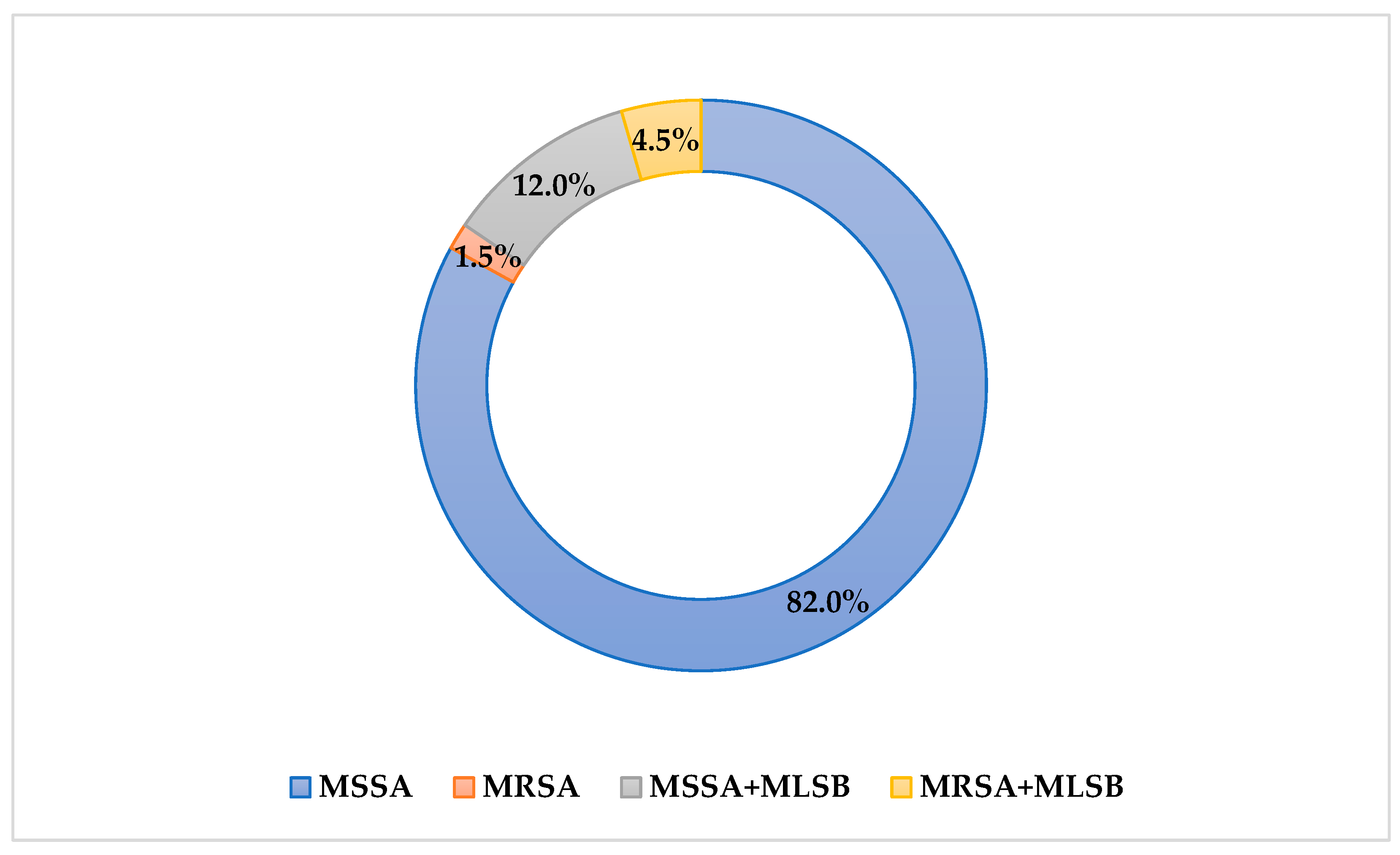
Figure 1. Percentage distribution of resistance phenotypes of the tested S. aureus strains.
Pathogens 14 00923 g001
Figure 2. Absorbance value of S. aureus strains from patients with chronic furunculosis. Strains numbered 6538 and 29213 are control strains.
Pathogens 14 00923 g002
Figure 3. Absorbance value of S. aureus strains from patients with chronic osteomyelitis. Strains numbered 6538 and 29213 are control strains.
Pathogens 14 00923 g003
Table 1. List of primers used in this study related to the experimental procedures.
Gene NamePrimer Sequence 5′-3′Reference
hlgA-FCTGATTACTATCCAAGAAATTCGATTG
hlgA-RCTTTCCAGCCTACTTTTTTATCAGT[22]
hlgB-FGTGCACTTACTGACAATAGTGC
hlgB-RGTTGATGAGTAGCTACCTTCAGT[22]
hld-FAAGAATTTTTATCTTAATTAAGGAAGGAGTG
hld-RTTAGTGAATTTGTTCACTGTGTCGA[22]
hlg-FGTCAYAGAGTCCATAATGCATTTAA
hlg-RCACCAAATGTATAGCCTAAAGTG[22]
spa-FCAGATAACAAATTAGCTGATAAAAACAT
spa-RCTAAGGCTAATGATAATCCACCAAATAC[21]
clfa-FATTGGCGTGGCTTCAGTGCT
clfa-RCGTTTCTTCCGTAGTTGCATTTG[21]
clfb-FGCTGCAAAAATGCAAGATCA
clfb-RTTGCCGCCATAAATGTGTTA[21]
cna-FAAAGCGTTGCCTAGTGGAGA
cna-RAGTGCCTTCCCAAACCTTTT[21]
eap-FAGTCATTGATTACAACAA
eap-RCTTATTAAATGTTAAGCTTG[21]
bap-F
bap-R
GAGCCAAGACAAAGGTGAAG
GTAGCCATAGCACGGAACAT

[24]
fnba–F
fnba–R
TCCGCCGAACAACATACC
TCAAGCACAAGGACCAAT

[24]
fnbb–F
fnbb–R
TCTGCGTTATGAGGATTT
ACAGTAGAGGAAAGTGG

[24]
fib–F
fib–R
AGATGCGAGCGAAGGGTA
TAAACGAAACTAAGTTGACTGC

[24]
ebps–F
ebps–R
GGTGAACCTGAACCGTAG
CTGGCAAGGCGAATAACT

[24]
bbp–F
bbp–R
CTTAGCAGTTCAACAGGGTG
TTGGCTTTATTGTGATGGTC

[24]
pvl–F
pvl-R
AAATGCTGGACAAAACTTCTTGG
TTTGCAGCGTTTTGTTTTCG

[23]
Table 2. Thermal profiles of the real-time PCR relations based on [21,22,23,24].
Stagesspa, eap, clfbcna, clfahlgA, hlgB, hld, hlgPvlbbp, epbs, fibfnbpa, fnbpbBap
Initial denaturation95 °C/
5 min
95 °C/
5 min
94 °C/
7 min
94 °C/
5 min
95 °C/
5 min
95 °C/
5 min
95 °C/
5 min
Denaturation95 °C/
5 min
95 °C/
5 min
94 °C/
1 min
95 °C/
15 s
95 °C/
40 s
95 °C/
40 s
95 °C/
40 s
Annealing50 °C/
1 min
55 °C/
1 min
58 °C/
1 min
60 °C/
30 s
56 °C/
50 s
54 °C/
50 s.
58 °C/
50 s
Elongation72 °C/
1 min
72 °C/
1 min
72 °C/
1 min
60 °C/
30 s
65 °C/
5 s
65 °C/
5 s.
65 °C/
5 s
Final elongation 72 °C/
10 min
72 °C/
10 min
72 °C/
7 min
72 °C/
3 min
72 °C/
50 s
72 °C/
50 s.
72 °C/
50 s
End of the process72 °C/
12 min
72 °C/
12 min
72 °C/
7 min
72 °C/
10 min
72 °C/
10 min
72 °C/
10 min
72 °C/
10 min
Table 3. Frequency of gene carriage among Staphylococcus aureus isolates from patients with chronic furunculosis and chronic osteomyelitis (clfa, clumping factor A; clfb, clumping factor B; spa, protein A; cna, collagen adhesin; eap, extracellular adherence protein; hlgA, alpha-hemolysin; hlgB, beta-hemolysin; hlg, gamma-hemolysin; hld, delta-hemolysin; bap, biofilm-associated protein; bbp, bone sialoprotein-binding protein; ebpS, elastin-binding protein; Efb, fibrinogen-binding protein; fnbA, fibronectin-binding protein A; fnbB, fibronectin-binding protein B; pvl, panton valentine leukocidin; +, indicates the presence of the gene in question; −, indicates an absence of the gene). The virulence genes in S. aureus isolates from patients with chronic furunculosis and chronic osteomyelitis. The table shows only those strains in which the presence of single genes was not observed (cna, collagen adhesin; hlgA, alpha-hemolysin; hlgB, beta-hemolysin; hlg, gamma-hemolysin; hld, delta-hemolysin; bbp, bone sialoprotein-binding protein; fb, fibrinogen-binding protein; fnbB, fibronectin-binding protein; pvl, panton valentine leucocidin).
Gene NameOsteomyelitisFurunculosis
MSCRAMMClfa100100
Clfb100100
spA100100
Can95.8100
Eap100100
Bap100100
Bbp10097.7
ebpS100100
fnbA100100
fnbB10097.7
Fib100100
CytotoxinhlgA95.8100
HlgB95.8100
Hlg95.8100
Hld100100
Pvl10097.7
Disclaimer/Publisher’s Note: The statements, opinions and data contained in all publications are solely those of the individual author(s) and contributor(s) and not of MDPI and/or the editor(s). MDPI and/or the editor(s) disclaim responsibility for any injury to people or property resulting from any ideas, methods, instructions or products referred to in the content.