Article

Optimization of (Dithioperoxo)thiolate-Based Antifungal Agents for Triazole-Resistant Aspergillus Fumigatus

Department of Pharmaceutical Sciences, School of Pharmacy, Marshall University, Huntington, WV 25755, USA
*
Author to whom correspondence should be addressed.
Pathogens 2025, 14(9), 878; https://doi.org/10.3390/pathogens14090878
Submission received: 7 August 2025 / Revised: 25 August 2025 / Accepted: 26 August 2025 / Published: 3 September 2025

Abstract

This investigation on novel antifungal agents featuring a thiol-reactive (dithioperoxo)thiolate chemical nucleus [-NC(S)S-SR] established that the optimal levels of fungal growth inhibition were achieved with thiomethyl-bound derivatives (R = Me). The most efficacious analogs had MIC50/MIC90 values of 2/2 µg/mL and an MIC range of 1 to 2 µg/mL for a ten-member panel of voriconazole-resistant A. fumigatus mutants. Pharmacodynamic studies revealed that the lead (dithioperoxo)thiolates impaired conidial germination and germling development more effectively than voriconazole for the triazole-resistant strain AR-1295. Moreover, glutathione and Cu2+ were shown to have antagonistic interactions, which was attributed to the thiol-reactive, pro-oxidant properties of the (dithioperoxo)thiolates and their metabolic conversion to chelating agents. Cytotoxicity studies further showed that the compounds were less toxic to human fetal kidney cells than squamous carcinoma cells. The collective findings of the investigation indicate that (dithioperoxo)thiolates are effective antifungal agents against A. fumigatus to merit additional research on their therapeutic potential.

1. Introduction

Aspergillus is a genus of conidium-forming filamentous molds that can infiltrate the respiratory tract to spawn invasive mycoses in individuals with deficient fungal immunity [1]. Multiple species of Aspergillus are implicated in aspergillosis, with A. fumigatus being the most common clinical isolate in pulmonary and disseminated infections [2]. Oncology and transplant patients, including hematopoietic stem cell recipients requiring immunosuppressive therapy (e.g., sirolimus), are at greatest risk for disseminated invasive aspergillosis [3]. The primary treatment and prophylaxis for A. fumigatus infections are the triazole antifungals voriconazole, posaconazole, and isavuconazole [3,4]. The Clinical and Laboratory Standards Institute (CLSI) and European Committee on Antimicrobial Susceptibility (EUCAST) have established a resistance breakpoint of ≥2 µg/mL for first-line voriconazole following international surveillance reports that indicated 97.8% of clinical isolates exhibited a MIC of 1 µg/mL or less [5]. However, experts predict up to 15% of patients receiving voriconazole or comparable triazole therapy will require an alternative treatment due to drug intolerance or resistance [6].
To this end, the agricultural use of azole fungicides has largely contributed to the emergence of triazole-resistant A. fumigatus. Resistance is associated with point mutations in the cyp51A gene that weakens the binding of triazole antifungals to the encoded protein target, lanosterol 14α-demethylase [7]. Triazole resistance may also be conferred by the overexpression of efflux transporter genes (e.g., cdr1B) in A. fumigatus [8]. Currently, there are few alternative drugs when these most relied on antifungal therapies are deemed ineffective or contraindicated due to toxicity or an interaction with other cyp3A substrates (e.g., sirolimus). In the event of triazole treatment failure, clinicians will resort to a six- to twelve-week course or longer of liposomal amphotericin B [3]. Salvage therapy with caspofungin or micafungin may then be used for refractory infections [3]. Thus, there is a critical need to develop additional second-line antifungal drugs when triazole-based therapies cannot be administered to aspergillosis patients.
Drug repurposing and repositioning are effective strategies to develop a new treatment from a pre-existing medication with established human pharmacokinetic and safety data. In a recent report, Shanholtzer and coworkers disclosed that fluconazole-resistant Candida auris and Candida glabrata were susceptible to the alcohol sobriety aid disulfiram (Antabuse®) [9]. Moreover, the yeasts were moderately sensitive to the primary metabolite of disulfiram, diethyldithiocarbamate (DETC). Figure 1a depicts the thiol-disulfide exchange between disulfiram and a reducing thiol (e.g., glutathione, cysteine) that generates DETC. Upon release, the metabolite is believed to sequester metal ions (Mn+) such as Zn2+ and Fe3+ as a secondary mechanism of microbial growth inhibition [10,11,12]. Interestingly, co-treatment with Cu2+ salts was shown to decrease the minimal inhibitory concentrations (MICs) of disulfiram and DETC against Candida species but at the cost of reduced fungicidal activity that was attributed to the formation of Cu[DETC]2 [9].
The findings of this research prompted us to evaluate (dithioperoxo)thiolate analogs of disulfiram (Figure 1b) as antifungal agents. We previously reported that (dithioperoxo)thiolates had an optimal MIC range of 0.25 to 1 µg/mL for Candida albicans compared to 2 µg/mL for disulfiram [13]. The structure–activity relationship (SAR) analysis indicated that analogs with short S-alkyl chains were the most efficacious for antifungal activity. The analysis further revealed that N,N-dimethyl and N,N-diethyl substituents conferred the lowest MICs for C. albicans. The primary objective of this investigation was to identify lead (dithioperoxo)thiolates with optimal antifungal activity against triazole-resistant A. fumigatus. In addition, the in vitro antifungal pharmacodynamics and cytotoxic activity were assessed for compounds with optimal MIC50/MIC90 values.

2. Materials and Methods

2.1. Synthesis

Chemicals were obtained from Thermo Fisher Scientific (Waltham, MA, USA) or VWR international (Radnor, PA, USA) and used without further purification. Reactions were monitored by thin-layer chromatography (TLC) using GF254 silica-gel-coated aluminum TLC plates. Compounds were purified using a Biotage® Isolera™ chromatography system (Charlotte, NC, USA) with KP-Sil silica gel columns and elution detection set at 254 nm. 1H and 13C NMR data were recorded on a Bruker AVANCE™ III HD 300 MHz spectrometer (Bruker, Billerica, MA, USA) and reported as δ values in ppm relative to tetramethylsilane (TMS). Coupling constants were recorded in Hz, and multiplicity reported as follows: s (singlet); bs (broad singlet); d (doublet); t (triplet); q (quartet); m (multiplet); dd (doublet of doublets); dt (doublet of triplets); dq (doublet of quartets); and tt (triplet of triplets). Mass spectra were recorded on an Agilent 5977B GCMS (Agilent, Santa Clara, CA, USA).

2.1.1. Preparation of Carbamo(dithioperoxo)thiolates (1a–d); General Procedure

Disulfiram (200 mg, 0.68 mmol) and the appropriate mercaptan (0.74 mmol) were reacted in a seal tube at 60 °C in 2 mL of anhydrous dimethylformamide (DMF) [13,14]. After stirring for 18–24 h, the mixture was cooled to room temperature, diluted with water, and extracted twice with hexanes (2 × 3 mL). The organic layers were combined, washed with water (3 × 2 mL), dried over MgSO4, filtered, and concentrated in vacuo. Silica gel chromatography with 0–10% EtOAc in hexanes provided the pure carbamo(dithioperoxo)thiolates whose characterization data matched that previously reported [13,14].

2.1.2. Preparation of N-Alkylthiophthalimide (2); General Procedure

Sulfuryl chloride (2.4 g, 18 mmol) in 5 mL of CH2Cl2 was slowly added dropwise to a stirring solution of methyldisulfide or ethanethiol (18 mmol) in 20 mL of CH2Cl2 chilled in an ice bath [15]. Upon completion, the ice bath was removed, and the mixture was stirred for 30 min. The solution was transferred to an addition funnel and added dropwise to an ice-chilled suspension of phthalimide (2.4 g, 16.4 mol) in 25 mL CH2Cl2 containing triethylamine (2.2 g, 21.3 mmol). Upon completion, the ice bath was removed and the mixture stirred for 1 h. The solution was then washed twice with deionized water, dried over MgSO4, filtered and evaporated. Recrystallization from boiling hexanes provided the pure N-alkylthiophthalimides whose characterization data matched that previously reported [15,16].
N-methylthiophthalimide (2a): white solid (36%), m.p. 178–179 °C; TLC (SiO2) Rf 0.38 (hexanes:EtOAc 3:1); 1H NMR (300 MHz, CDCl3) δ 8.02–7.72 (m, 4 H), 2.55 (s, 3 H); 13C NMR (75 MHz, CDCl3) δ 168.3, 134.7, 132.3, 124.0, 22.6; MS (EI, 70 eV): m/z (%) = 193 (22) [M], 160 (23), 148 (52), 130 (100), 104 (26).
N-ethylthiophthalimide (2b): white solid (70%), m.p. 115–117 °C; TLC (SiO2) Rf 0.45 (hexanes:EtOAc 3:1); 1H NMR (300 MHz, CDCl3) δ 7.94 (m, 2 H), 7.80 (m, 2 H), 2.93 (dq, J = 13.3, 7.6, 6.5 Hz, 2 H), 1.29 (dq, J = 12.9, 7.0, 5.9 Hz, 3 H); 13C NMR (75 MHz, CDCl3) δ 13C NMR (75 MHz, CDCl3) δ 168.6, 134.6, 132.1, 123.9, 32.7, 13.2; MS (EI, 70 eV): m/z (%) = 207 (5) [M], 148 (100), 130 (100), 104 (40).

2.1.3. Preparation of Carbamo(dithioperoxo)thiolates (1e–i); General Procedure

Carbon disulfide (60 µL, 1.1 mmol) was added dropwise to a stirring solution of amine (0.9 mmol) and KOH powder (51 mg, 1 mmol) in diethyl ether (2 mL) at 0 °C. After 1 h, the solvent was evaporated under reduced pressure. The K+ salt was filtered, washed with ether, and resuspended in benzene (5 mL) containing N-(alkylthio)phthalimide (0.9 mmol). After stirring for 2 h, hexanes was added, the salts were removed by filtration and the filtrate was evaporated. Silica gel chromatography with 0–10% EtOAc in hexanes provided the pure carbamo (dithioperoxo)thiolates whose characterization data matched that previously reported [13,14].
methyl dimethylcarbamo(dithioperoxo)thioate (1e): pale oil (48%); TLC (SiO2) Rf 0.37 (hexanes:EtOAc 9:1); 1H NMR (300 MHz, CDCl3) δ 3.60 (s, 3 H), 3.45 (s, 3 H), 2.51 (s, 3 H); 13C NMR (75 MHz, CDCl3): δ 197.0, 47.1, 41.5, 22.3; MS (EI, 70 eV): m/z (%) = 167 (100) [M], 135 (33), 123 (16), 121 (11), 120 (100).
methyl diethylcarbamo(dithioperoxo)thioate (1f): pale oil (42%); TLC (SiO2) Rf 0.63 (hexanes:EtOAc 9:1); 1H NMR (300 MHz, CDCl3) δ 4.06 (h, J = 6.7 Hz, 2 H), 3.79 (tt, J = 12.5, 6.9 Hz, 2 H), 2.50 (s, 3 H), 1.32 (d, J = 7.9 Hz, 6 H); 13C NMR (75 MHz, CDCl3) δ 195.5, 51.5, 46.9, 22.9, 13.0, 11.5; MS (EI, 70 eV): m/z (%) = 195 (4) [M], 116 (100).
ethyl dimethylcarbamo(dithioperoxo)thioate (1g): oil; 1H NMR (300 MHz, CDCl3) δ 3.64 (s, 3 H), 3.51 (s, 3 H), 2.99–2.82 (m, 2 H), 1.35 (t, J = 7.4 Hz, 3 H); 13C NMR (75 MHz, CDCl3) δ 197.5, 47.2, 41.6, 32.2, 13.7; MS (EI, 70 eV): m/z = 181 [M].
ethyl benzyl(methyl)carbamo(dithioperoxo)thioate (1h): oil; rotamers observed: 1H NMR (300 MHz, CDCl3) δ 7.52–7.05 (m, 5 H), 5.38 (s, 1 H), 5.07 (s, 1 H), 3.50 (s, 1 H), 3.43–3.25 (m, 2 H), 3.06–2.73 (m, 2 H), 1.40–1.14 (m, 3 H); 13C NMR (75 MHz, CDCl3) δ 199.3, 198.1, 135.1, 134.4, 129.0, 128.8, 128.1, 127.9, 127.8, 127.1, 61.6, 57.9, 45.1, 38.8, 32.3, 13.7; MS (EI, 70 eV): m/z = 257 [M].
methyl methoxy(methyl)carbamo(dithioperoxo)thioate (1i): pail oil (39%); TLC (SiO2) Rf 0.42 (hexanes:EtOAc 9:1); 1H NMR (300 MHz, CDCl3) δ 3.84 (s, 3 H), 3.75 (s, 3 H), 2.44 (s, 3 H); 13C NMR (75 MHz, CDCl3) δ 194.9, 61.6, 40.8, 22.5; MS (EI, 70 eV): m/z (%) = 183 (31) [M], 126 (30), 104 (100).

2.1.4. Preparation of Carbano(dithioperoxo)thioates (3); General Procedure

Carbon disulfide (54 µL, 0.9 mmol) was added dropwise to a stirring solution of alkyl alcohol (0.9 mmol) and KOH powder (51 mg, 1 mmol) in diethyl ether (5 mL) at 0 °C. After 1 h, the solvent was evaporated under reduced pressure. The K+ salt was washed with ether, filtered, and resuspended in benzene (5 mL) containing N-(alkylthio)phthalimide (0.9 mmol). The mixture was stirred at room temperature until the reaction was complete. Hexanes was then added, the salts were removed by filtration, and the filtrate was evaporated. Silica gel chromatography with 0–10% EtOAc in hexanes provided the pure carbano(dithioperoxo)thioates whose characterization data matched that previously reported [17,18].
(methoxy(thiocarbonyl))methyldisulfane (3a): pale oil (46%); TLC (SiO2) Rf 0.69 (hexanes:EtOAc 9:1); 1H NMR (300 MHz, CDCl3) δ 4.28 (dd, J = 6.1, 3.0 Hz, 5 H), 2.54 (t, J = 3.4 Hz, 4 H); 13C NMR (75 MHz, CDCl3) δ 213.8, 77.1, 76.7, 61.4, 22.9.
(ethoxy(thiocarbonyl))methyldisulfane (3b): pale oil (53%); TLC (SiO2) Rf 0.78 (hexanes:EtOAc 9:1); 1H NMR (300 MHz, CDCl3) δ 4.74 (m, 2 H), 2.53 (s, 3 H), 1.49 (tt, J = 7.5, 4.2 Hz, 3 H); 13C NMR (75 MHz, CDCl3) δ 212.9, 71.4, 22.9, 13.8.

2.2. Antimicrobial Studies

Growth media and antifungal agents were obtained from commercial vendors and used without further purification. Powder Gibco Roswell Park Memorial Institute (RPMI) 1640 medium with L-glutamine and phenol red (ref. 31800-105) was obtained from Thermo Fisher Scientific. Amphotericin B (CAS 1397-89-3) and disulfiram (97%, CAS 97-77-8) were obtained from Alfa Aesar via Thermo Fisher Scientific. Fluconazole (≥98%, CAS 86386-73-4) and voriconazole (≥98%, CAS 137234-62-9) were obtained from TCI America via VWR International.

2.2.1. MIC Determination

Susceptibility testing was performed by the microdilution assay method in 96-well plate format according to CLSI protocols [19,20]. Yeast susceptibility was evaluated in modified RPMI 1640 medium supplemented with 18 g/L glucose and 34.6 g/L morpholinepropanesulfonic acid (MOPS) that was adjusted to pH 7.0 with NaOH. The same medium was used for A. fumigatus without the addition of glucose. Test compounds were freshly prepared in DMSO and diluted with water to 1 mg/mL stocks containing ≤5% DMSO. Fungi were enumerated from frozen glycerol–RPMI stocks on peptone dextrose agar for 2 to 4 days at 35 °C in a water-jacketed incubator. Yeast colonies and A. fumigatus conidia were used to prepare 0.5 McFarland suspensions (OD530 0.115 to 0.130) in sterile saline. The yeasts were further diluted in the growth medium that provided a final test inoculum of 1 to 5 × 10³ CFU/mL after addition to two-fold dilutions of the test agents. Likewise, A. fumigatus conidia were diluted in modified RMPI 1640 to final test inocula of 1 to 5 × 10⁴ CFU/mL. The plates were incubated in a water-jacketed incubator at 35 °C. The modal MICs of three independent tests are reported as the lowest drug concentration that prevented discernible growth (100% inhibition) of the Candida species at 24 h and at 48 h for A. fumigatus and Cryptococcus neoformans.

2.2.2. Growth Studies

The effect of the (dithioperoxo)thiolates on A. fumigatus AR-1295 growth over time was analyzed using 990 µL of a 1 to 5 × 10⁵ CFU/mL culture in modified RPMI 1640 (165 mM MOPS, pH 7.0) treated with the vehicle and/or test agents in 10 µL of RPMI. Cultures consisting primarily of swollen conidia and germlings were prepared from dormant conidia cultured in 1:1 vol:vol medium of Hanks’ balanced salt solution (HBSS) and fetal bovine serum (FBS) at 35 °C for 6 to 8 h. The treated cultures were dispensed in 100 µL aliquots to the wells of a 96-well microtiter plate and sealed with transparent adhesive film. The plates were placed in a Molecular Devices SpectraMax® 384 plate reader (Molecular Devices, San Jose, CA, USA) with a chamber temperature set to 35 °C. The OD530 values were recorded hourly over a 48 h period following agitation for 10 s. Growth curves were plotted against time from the OD530 readings using Prism software version 10.2.3 (GraphPad Software, Inc., Boston, MA, USA).

2.2.3. Checkerboard Studies

Isobologram analysis was performed with A. fumigatus AR-1295 using the microdilution checkerboard assay in a 4 × 5 matrix format [21]. Dormant conidia were diluted in modified RMPI 1640 to a final test inocula of 1 to 5 × 10⁴ CFU/mL. Microtiter plates containing two-fold serial dilutions of (dithioperoxo)thiolate (range 0.25–16 µg/mL) and copper (II) sulfate (range 4–16 µg/mL) or glutathione (range 1–4 µg/mL) were then inoculated with A. fumigatus AR-1295. The plates were placed in a water-jacketed incubator and incubated at 35 °C for 48 h. The modal OD530 values obtained for replicates performed on three separate trial days were plotted as heat maps using Prism software version 10.2.3.

2.3. Cytotoxicity Studies

2.3.1. IC50 Determination

The in vitro cytotoxicity was determined against two human cell lines (HEK293T and MDA686TU) using the sulforhodamine B (SRB) colorimetric assay [22]. HEK293T is an immortalized human cell line derived from embryonic kidney cells and purchased from ATCC. MDA686TU is a head and neck squamous cell carcinoma cell line established from primary tongue cancer and was obtained from Dr. Peter G. Sacks at New York University College of Dentistry in 2014. The cells were enumerated in Dulbecco’s Modified Eagle Medium (DMEM) and DMEM/F12 (1:1) media supplemented with 10% fetal bovine serum (FBS), respectively. Cells were incubated in a controlled environment maintained at 37 °C with a humidified atmosphere containing 5% CO2 until 80% confluency was achieved. About 2.5 × 10³ MDA686TU cells in 100 µL DMEM were seeded in each well of the 96-well plate and incubated overnight. Similarly, 5 × 10³ HEK293T cells were seeded in DMEM/F12 in each well. The cultures were then treated with 15, 20, 25, 30, 35 and 40 µM of the test compounds in quadruplicate for each concentration. Cisplatin was used as a positive control in parallel plates for MDA686TU cells. After 72 h of incubation at 37 °C under 5% CO2 atmosphere, the cells were fixed with cold 10% trichloroacetic acid (TCA) for 1 h at 4 °C, washed five times with distilled water and allowed to dry overnight. Afterwards, the cells were stained with 50 μL of 0.4% SRB for 10 min at room temperature, washed five times with 1% acetic acid, and air-dried. Absorbance was taken at OD492 using a microplate reader after dissolving the bound dye with 100 µL 10 mM Tris solution (pH 10.5). After calculating the growth inhibition for each concentration, CalcuSyn Software version 2.11 was used to determine the growth curve and IC50.

2.3.2. Hematological Analysis

Trunk blood was collected in EDTA tubes from a male Sprague Dawley rat (Hilltop Lab Animals, Scottdale, PA, USA) and used within 3 h of the study. The blood (46 µL) was dispensed in 1.5 mL centrifuge tubes containing 1.25 mM of the test agents in 4 µL of DMSO. The samples were incubated at 37 °C with gentle agitation (200 rpm). Complete blood counts were analyzed using a Hemavet 950FS (Drew Scientific, Plantation, FL, USA) after 2 h. The log2 fold change (log2FC) was calculated from the mean values in the vehicle control (DMSO) versus treatment groups. Statistical significance was assessed by Student’s t-test. p values of ≤0.05 were considered significant.

3. Results

3.1. (Dithioperoxo)thiolate Synthesis

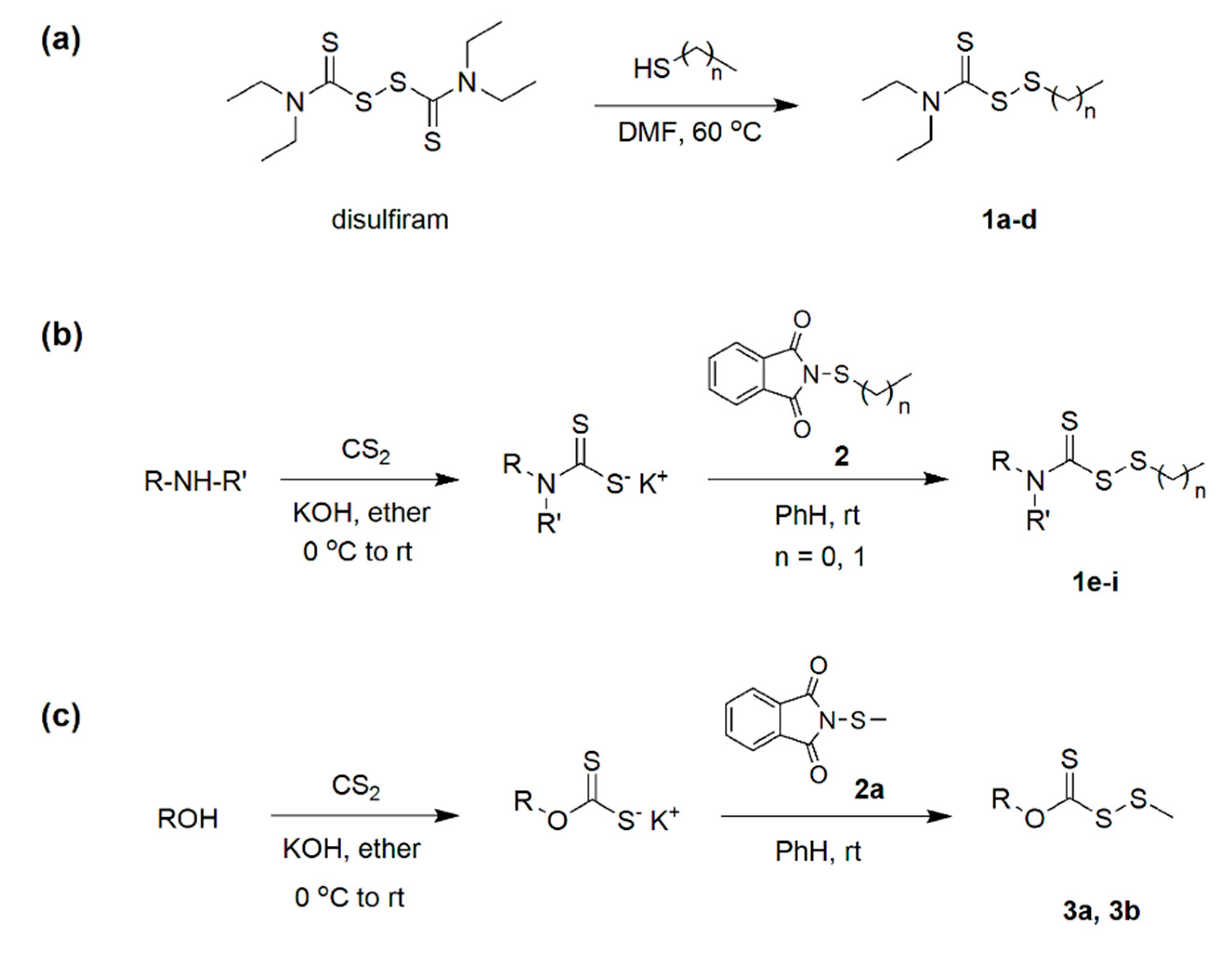
The strategies employed to synthesize the (dithioperoxo)thiolates for the investigation are depicted in Figure 2. The disulfiram-based analogs 1a–d were prepared via a thiol-disulfide exchange reaction with their respective thiols in dimethylformamide (DMF) (Figure 2a). Following workup, silica gel chromatography afforded the pure products in modest yields of 22 to 31%. The carbamo(dithioperoxo)thiolates 1e–i were alternatively synthesized in higher yields (42 to 63%) from potassium dithiocarbamates derived from their respective amines and carbon disulfide (Figure 2b). The intermediates were then reacted in benzene (PhH) with either N-methylthiophthalimide or N-ethylthiophthalimide (2) that precipitated potassium phthalimide to enable convenient product separation. Beforehand, the respective N-alkylthiophthalimides were prepared from phthalimide and their sulfenyl chloride precursors obtained by reacting sulfuryl chloride and methyldisulfide or ethanethiol. Lastly, the carbano(dithioperoxo)thioate analogs 3a and 3b were obtained in yields of 46% to 64% from their respective potassium xanthates by the same method (Figure 2c).

3.2. Susceptibility Testing

The MICs were determined according to protocols set forth by CLSI reference methods M27-A4 [19] and M38-A3 [20] for yeast and filamentous fungi, respectively. The fungi were treated with the test agents in MOPS-modified RPMI 1640 medium at a two-fold dilution range of 0.5 to 16 µg/mL. Per CLSI guidelines, the MIC endpoints were 24 h for Candida species and 48 h for A. fumigatus and Cryptococcus neoformans. Table 1 compares the modal MIC values for the disulfiram-derived (dithioperoxo)thiolates 1a–d to disulfiram and standard antifungal drugs. The data shows a consistent SAR profile across the test panel of fluconazole-resistant Candida species, fluconazole-susceptible C. neoformans, and voriconazole-resistant A. fumigatus AR-0731 (L98H/TR34). Maximal antifungal activity was achieved for the S-ethyl derivative 1a followed by the S-butyl (1b), S-hexyl (1c), and S-octyl (1d) analogs. Disulfiram was comparably less active against the yeast isolates than analog 1a, while filamentous growth was observed at the 48 h endpoint for A. fumigatus at 16 µg/mL.
The investigation then proceeded to focus on the S-methyl and S-ethyl series of (dithioperoxo)thiolates. Table 2 compares the modal MICs for derivatives obtained from various potassium dithiocarbamates and xanthates. The data shows that only the N,N-dialkyl carbamo(dithioperoxo)thiolates 1e, 1f, and 1g exhibited appreciable antifungal activity (i.e., MIC < 10 µM). Moreover, neither of the carbano(dithioperoxo)thiolates (i.e., 3a, 3b) inhibited the growth of A. fumigatus at treatments of 16 µg/mL or less (i.e., ≤95 µM). The data further shows that the MIC values for the DETC-bound S-methyl (1e) and S-ethyl (1a) (dithioperoxo)thiolates were within ±1 log2 dilution of each other. The same outcome was observed for the respective dimethyldithiocarbamate (DMTC) based analogs 1f and 1g. Altogether, the susceptibility data indicated that the S-methyl analogs 1e and 1f were more effective inhibitors of A. fumigatus whilst the Candida species appeared to be equally susceptible to the S-ethyl derivatives 1a, 1g, and 1h.
Afterwards, the (dithioperoxo)thiolates exhibiting MICs of ≤10 µM were evaluated against a ten-member A. fumigatus panel comprising strains characterized for their cyp51A mutations. The results presented in Table 3 show that the S-methyl (1e, 1f) and S-ethyl (1a, 1g) analogs had a MIC range of 0.5 to 4 µg/mL. Although no correlations between the antifungal activity and the amino acid substitutions were evident, the S-methyl (dithioperoxo)thiolates 1e and 1f displayed the lowest variation in their modal MIC values between the mutant variants. We further compared the MICs with the primary metabolite of analogs 1a and 1e (i.e., DETC) that was prepared as the sodium salt by reacting 2.5 mmol each of diethylamine, carbon disulfide, and sodium hydroxide powder in 10 mL of anhydrous diethyl ether. The results in Table 3 indicate that the modal MICs of DETC•Na were at least four times higher for the A. fumigatus panel. However, a higher degree of variability in the MICs was observed for DETC•Na with up to ±2 log2 dilution difference between experiments.

3.3. Pharmacodynamic Studies

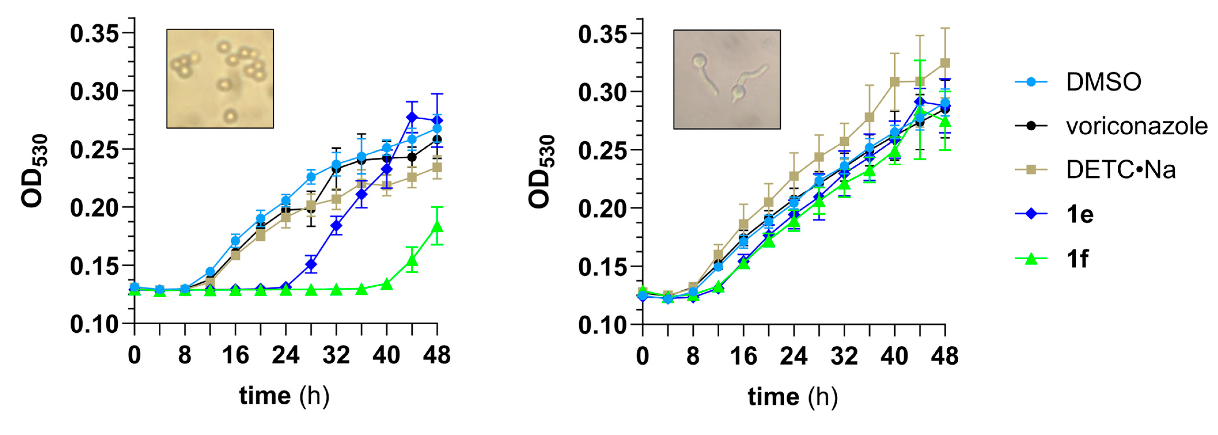
The S-methyl (dithioperoxo)thiolates 1e and 1f were selected as lead compounds to undergo further evaluation based on their optimal MIC50/MIC90 values of 2 µg/mL for the A. fumigatus mutant panel. A growth study was conducted using dormant conidia harvested from a six-day agar culture of A. fumigatus AR-1295. The results depicted in Figure 3 (left) show both analogs delayed conidial germination by at least 10 h when compared to the vehicle-treated (DMSO) control cultures. The data further indicates that DMTC analog 1f suppressed germination and hyphal formation by up to ten hours longer than the DETC analog 1e. By comparison, the ability of voriconazole and DETC•Na to impair filamentous growth was significantly lower than either of the (dithioperoxo)thiolates.
In the A. fumigatus lifecycle, the onset of conidial germination spawns isotropic swelling and emergence of germ tubes from the conidium (germlings) that continue to elongate, forming hyphae and branched hyphae called mycelium [23]. We found the same growth pattern exists in vitro for A. fumigatus AR-1295 when conidia from the six-day agar culture were incubated at 35 °C in Hanks’ balanced salt solution (HBSS) containing fetal bovine serum (FBS). The germlings obtained after 8 h incubation were isolated and assessed for sensitivity to (dithioperoxo)thiolates 1e and 1f in the study. Figure 3 (right) depicting the growth response curves indicate that the compounds impaired germling development by a similar degree. The data also indicates that voriconazole did not slow the filamentous growth of A. fumigatus, while treatment with DETC•Na resulted in a slightly elevated rate of hyphal formation.
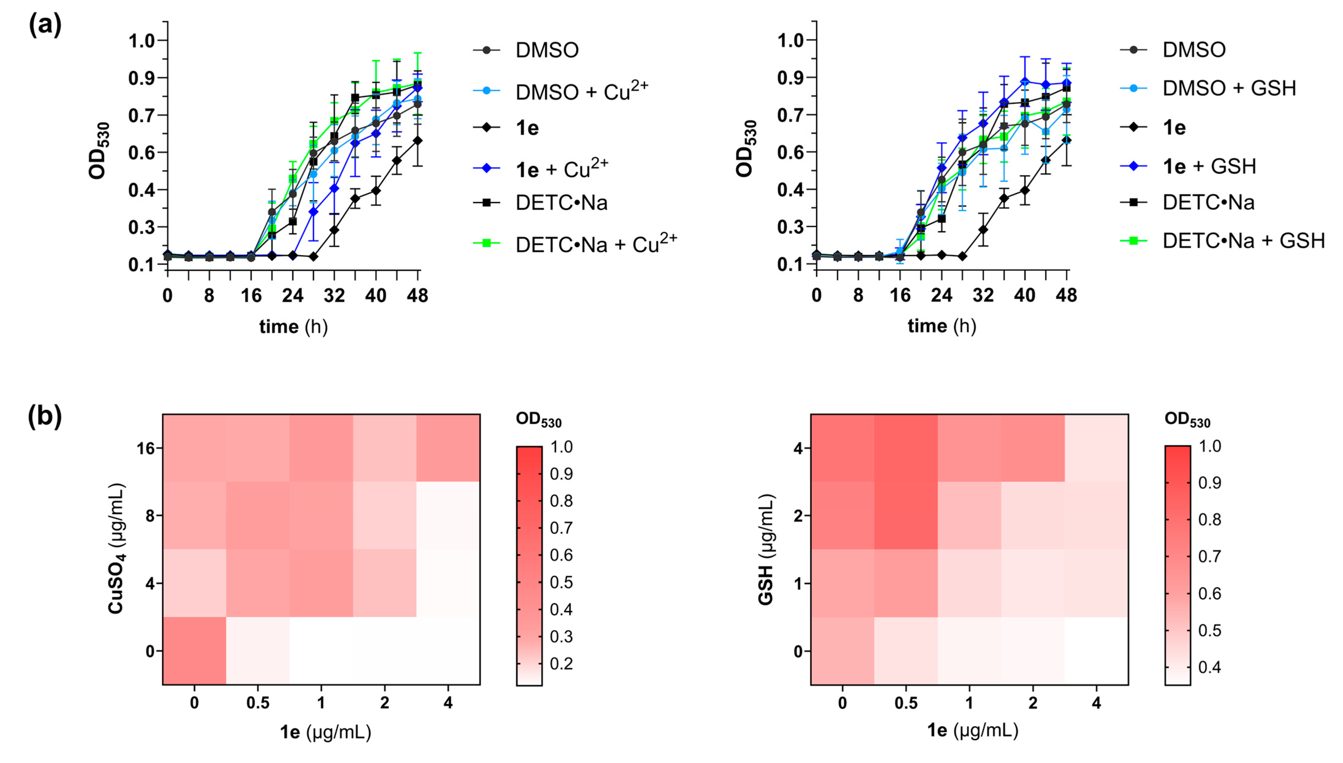
In a prior report, Shanholtzer et al. established that DETC•Na and Cu2+ had a synergistic interaction with up to a sixteen-fold reduction in the MICs for Candida glabrata and Candida auris [9]. The synergistic potential of the DETC-based analog 1e and Cu2+ was thereby assessed by differential growth curve and isobologram (checkerboard) analyses using A. fumigatus AR-1295 [21,24]. Surprisingly, both experiments revealed an antagonistic relationship between Cu2+ and the (dithioperoxo)thiolate. Figure 4a (left) shows filamentous growth was detectable 4 h earlier for conidia co-treated with 10 µM copper (II) sulfate. A heat map comparing the growth levels in the checkboard microtiter plate wells further confirmed that Cu2+ decreases the antifungal activity of analog 1e (Figure 4b, left).
As illustrated in Figure 1b, reactions between sulfhydryl-containing molecules (RSH) and (dithioperoxo)thiolate 1e by thiol-disulfide exchange generate a mixed disulfide (i.e., RS-DETC) and DETC. The most abundant thiol that fungi use to preserve intracellular redox homeostasis and cope with oxidative stress is glutathione (GSH) [25]. It was therefore believed that GSH protects A. fumigatus from thiol damage by the (dithioperoxo)thiolates. To probe for antagonism by GSH, the pharmacodynamic interaction with (dithioperoxo)thiolate 1e was examined by differential growth curve and isobologram analyses. The growth curves in Figure 4a (right) show filamentous growth was detectable 12 h earlier in the (dithioperoxo)thiolate cultures co-treated with GSH than without. The results of the checkerboard assay in Figure 4b (right) further corroborate this result of an antagonistic relationship between (dithioperoxo)thiolate 1e and GSH.

3.4. Cytotoxicity Studies

The in vitro cytotoxic activities of the lead (dithioperoxo)thiolates 1e and 1f were evaluated with two cultured human cell lines. The analogs exhibited IC50 values greater than 40 µM for human embryonic kidney cells (HEK293T) using the sulforhodamine B (SRB) colorimetric assay [22]. The compounds were likewise tested on the head and neck squamous cell carcinoma cell line MDA686TU. The data indicated that the carcinoma cells were more sensitive to the respective (dithioperoxo)thiolates with IC50 values of 22.5 ± 5.8 and 20.8 ± 2.7 µM. Additionally, cytotoxic activities were assessed using whole blood collected from a male Sprague Dawley rat. A Hemavet analyzer was used to measure the changes in blood cell counts following 2 h incubation with compound 1e. For the study, amphotericin B was enlisted as a comparator drug with known hemolytic activity [26]. The log2 fold change (log2FC) was calculated from the average measurements for the treated versus untreated groups. The log2FC and p values revealed that 100 µM of compound 1e induced a moderate decrease in erythrocyte abundance (−0.29, p 0.15) while reductions in neutrophils and lymphocytes were not observed. By comparison, only a significant decrease in erythrocytes (−0.55, p 0.02) was observed with 100 µM amphotericin B and not for the other blood cell types.

4. Discussion

The primary objective of this research was to identify lead (dithioperoxo)thiolates as antifungal agents against A. fumigatus. The MIC data supported earlier findings that antimicrobial activity was contingent on the chain length of the thioalkyl substituent. Prior antimicrobial studies revealed that C. albicans and Gram-negative bacteria (e.g., Acinetobacter baumannii) were the most susceptible to DMTC and DETC derivatives bound with an S-ethyl substituent [13,14]. Conversely, Gram-positive bacteria, including Staphylococcus aureus, were more susceptible to the S-hexyl and S-octyl analogs within those series [13,14]. Our results aligned with the prior SAR trends observed with C. albicans and further established that the thiomethyl-bound analogs maximally suppressed the growth of fungi. It was concluded that the S-methyl derivatives 1e and 1f offered the optimal combination of potency (MIC90 2 µg/mL) and lowest degree of MIC variability among the ten triazole-resistant variants in A. fumigatus panel (range: 1 to 2 µg/mL) for advancing as lead compounds in the investigation.
The ensuing pharmacodynamic studies revealed that compounds 1e and 1f inhibited the germination of dormant conidia from a triazole-resistant A. fumigatus mutant by 10 h or longer (Figure 3). The data further showed the period of impaired growth was significantly shorter with germinated conidia, which suggests the drugs may have limited therapeutic efficacy for progressing infections. Another notable observation was the antagonistic interaction of the DETC-based analog 1e with Cu2+. This result can be partly explained by the ability of DETC to chelate heavy metals such as Cu2+, which is known to be toxic to A. fumigatus in excess [27]. Consistent with this finding, the Cu2+-supplemented group treated with DETC•Na was found to have accelerated growth by the 32 h time point (Figure 4a). These data provide evidence for the protective effect of DETC•Na to heavy metals in A. fumigatus, which was investigated as a potential chelation therapy for nickel (II) poisoning [28].
Studies on the mechanisms of antifungal action of (dithioperoxo)thiolates will be a focus of future research. Previous investigations into the antibacterial mechanisms of disulfiram and the natural thiosulfinate allicin (garlic) should allow us to glean information on the antimicrobial pharmacology in fungi. The thiol-reactive compounds [29] were shown to induce an oxidative response in S. aureus that includes enrichment of the biosynthetic genes for its primary antioxidant thiol, bacillithiol [12,30]. In Escherichia coli, allicin was shown to deplete GSH and inactivate metabolic enzymes through S-allylmercapto modification of cysteine residues [31]. It is therefore projected that collateral damage stemming from oxidant accumulation and deficient GSH-mediated protein repair will be correlated to the antifungal mechanism of (dithioperoxo)thiolates.
As in the case of disulfiram, the DATC metabolite is expected to play a role in the mechanism of antifungal action. Prior differential transcriptomics and bioenergetic studies implicated DETC in the antibacterial action of disulfiram as an inhibitor of oxidative phosphorylation in S. aureus [12]. Moreover, the studies indicated that disulfiram induces feedback inhibition of central glucose catabolism by depleting coenzyme A in S. aureus [12]. It is speculated that the (dithioperoxo)thiolates will have analogous antagonistic effects on fungal cellular respiration pathways and coenzyme A. It is noteworthy that cellular respiration and protein synthesis are required to break dormancy in A. fumigatus conidia [32]. Therefore, pharmacological studies will need to consider the effects (dithioperoxo)thiolates and their DATC metabolite on conidia germination and proliferating cells.
As with other thiol-reactive disulfides [29], the pharmacokinetics and pharmacodynamics of (dithioperoxo)thiolate-based antifungals will be influenced by their stability to the GSH levels in the blood and tissues [33]. Inhalational administration may therefore be the optimal delivery route in the treatment of pulmonary aspergillosis. Their physiochemical properties further suggest that the (dithioperoxo)thiolates will have a large volume of distribution and high tissue penetration [34]. The lipophilic attribute would be advantageous for fungal infections involving lung, bone, and brain tissues. Conversely, the compounds may not be suitable treatments for fungemia if therapeutic blood levels cannot be achieved due to hepatic metabolism and a high volume of distribution. The pharmacokinetic properties of (dithioperoxo)thiolates with respect to metabolism and distribution may thereby limit the types of infections that they can be used to treat.

5. Conclusions

This research established the S-methyl (dithioperoxo)thiolates as a lead series of compounds for antifungal drug development. It was noted, however, that the MIC values of the S-ethyl analogs were generally within the range of experimental error and also showed promise as a lead series. Our future studies will focus on the DETC-based analog 1e given that the pharmacokinetic and safety parameters of disulfiram and the DETC metabolite are established [10,35].

Author Contributions

Conceptualization—T.E.L.; Investigation—S.K., K.L.J., M.E.V., A.R.M.R.A. and T.E.L.; Methodology—M.E.V., A.R.M.R.A. and T.E.L.; Data Curation—S.K., K.L.J., M.E.V., A.R.M.R.A. and T.E.L.; Formal Analysis—M.E.V., A.R.M.R.A. and T.E.L.; Resources—M.E.V., M.D.H., A.R.M.R.A. and T.E.L.; Funding Acquisition—T.E.L.; Writing—Original Draft—T.E.L.; Writing—Review and Editing—S.K., M.E.V., M.D.H., A.R.M.R.A. and T.E.L. All authors have read and agreed to the published version of the manuscript.

Funding

This work was supported by funding from the National Institutes of Health grants R15AI151970 (TEL) and R15DE032063 (ARMRA).

Institutional Review Board Statement

The animal study protocol was approved by the Institutional Animal Care and Use Committee of Marshall University (protocol #855 approved on 6 March 2025).

Informed Consent Statement

Not applicable.

Data Availability Statement

The original contributions are included in the article; further inquiries can be directed to the corresponding author.

Acknowledgments

Candida and Aspergillus isolates were acquired from the FDA-CDC Antimicrobial Resistance Isolate Bank. C. neoformans NIH-306 was obtained from BEI Resources, NIAID, NIH.

Conflicts of Interest

The authors declare no conflict of interest.

References

  1. Segal, B.H.; Walsh, T.J. Current approaches to diagnosis and treatment of invasive aspergillosis. Am. J. Respir. Crit. Care Med. 2006, 173, 707–717. [Google Scholar] [CrossRef] [PubMed]
  2. Brown, G.D.; Denning, D.W.; Gow, N.A.; Levitz, S.M.; Netea, M.G.; White, T.C. Hidden killers: Human fungal infections. Sci. Transl. Med. 2012, 4, 165rv13. [Google Scholar] [CrossRef] [PubMed]
  3. Patterson, T.F.; Thompson, G.R., 3rd; Denning, D.W.; Fishman, J.A.; Hadley, S.; Herbrecht, R.; Kontoyiannis, D.P.; Marr, K.A.; Morrison, V.A.; Nguyen, M.H.; et al. Practice Guidelines for the Diagnosis and Management of Aspergillosis: 2016 Update by the Infectious Diseases Society of America. Clin. Infect. Dis. 2016, 63, e1–e60. [Google Scholar] [CrossRef]
  4. Lewis, J.S., 2nd; Wiederhold, N.P.; Hakki, M.; Thompson, G.R., 3rd. New Perspectives on Antimicrobial Agents: Isavuconazole. Antimicrob. Agents Chemother. 2022, 66, e0017722. [Google Scholar] [CrossRef]
  5. CLSI. Voriconazole Breakpoints for Aspergillus fumigatus, 1st ed; CLSI rationale document FR01; Clinical and Laboratory Standard Institute: Wayne, PA, USA, 2024; ISBN 978-1-68440-230-4. [Google Scholar]
  6. Slavin, M.A.; Chen, Y.-C.; Cordonnier, C.; Cornely, O.A.; Cuenca-Estrella, M.; Donnelly, J.P.; Groll, A.H.; Lortholary, O.; Marty, F.M.; Nucci, M.; et al. When to Change Treatment of Acute Invasive Aspergillosis: An Expert Viewpoint. J. Antimicrob. Chemother. 2021, 77, 16–23. [Google Scholar] [CrossRef]
  7. Berger, S.; El Chazli, Y.; Babu, A.F.; Coste, A.T. Azole Resistance in Aspergillus fumigatus: A Consequence of Antifungal Use in Agriculture? Front. Microbiol. 2017, 8, 1024. [Google Scholar] [CrossRef]
  8. Hagiwara, D.; Watanabe, A.; Kamei, K.; Goldman, G.H. Epidemiological and Genomic Landscape of Azole Resistance Mechanisms in Aspergillus Fungi. Front. Microbiol. 2016, 7, 1382. [Google Scholar] [CrossRef]
  9. Shanholtzer, C.N.; Rice, C.; Watson, K.; Carreon, H.; Long, T.E. Effect of copper on the antifungal activity of disulfiram (Antabuse®) in fluconazole-resistant Candida strains. Med. Mycol. 2022, 60, myac016. [Google Scholar] [CrossRef]
  10. Custodio, M.M.; Sparks, J.; Long, T.E. Disulfiram: A Repurposed Drug in Preclinical and Clinical Development for the Treatment of Infectious Diseases. Antiinfect. Agents 2022, 20, e040122199856. [Google Scholar] [CrossRef] [PubMed]
  11. Frazier, K.R.; Moore, J.A.; Long, T.E. Antibacterial activity of disulfiram and its metabolites. J. Appl. Microbiol. 2019, 126, 79–86. [Google Scholar] [CrossRef]
  12. Long, T.E.; Naidu, S.T.; Hissom, E.G.; Meka, Y.; Chavva, H.; Brown, K.C.; Valentine, M.E.; Fan, J.; Denvir, J.; Primerano, D.A.; et al. Disulfiram induces redox imbalance and perturbations in central glucose catabolism and metal homeostasis to inhibit the growth of Staphylococcus aureus. Sci. Rep. 2025, 15, 15658. [Google Scholar] [CrossRef]
  13. Lewis, A.D.; Riedel, T.M.; Kesler, M.B.A.; Varney, M.E.; Long, T.E. Pharmacological evaluation of disulfiram analogs as antimicrobial agents and their application as inhibitors of fosB-mediated fosfomycin resistance. J. Antibiot. 2022, 75, 146–154. [Google Scholar] [CrossRef] [PubMed]
  14. Sheppard, J.G.; Frazier, K.R.; Saralkar, P.; Hossain, M.F.; Geldenhuys, W.J.; Long, T.E. Disulfiram-based disulfides as narrow-spectrum antibacterial agents. Bioorg. Med. Chem. Lett. 2018, 28, 1298–1302. [Google Scholar] [CrossRef] [PubMed]
  15. Sheppard, J.G.; Long, T.E. Allicin-inspired thiolated fluoroquinolones as antibacterials against ESKAPE pathogens. Bioorg. Med. Chem. Lett. 2016, 26, 5545–5549. [Google Scholar] [CrossRef]
  16. Heldreth, B.; Long, T.E.; Jang, S.; Reddy, G.S.K.; Turos, E.; Dickey, S.; Lim, D.V. N-Thiolated β-lactam antibacterials: Effects of the N-organothio substituent on anti-MRSA activity. Bioorg. Med. Chem. 2006, 14, 3775–3784. [Google Scholar] [CrossRef]
  17. Satyanarayana, A.N.V.; Pattanayak, P.; Nayar, P.P.; Chatterjee, T. Iodine-mediated, chalcogen–chalcogen bond formation in water: Green synthesis of carbamo(dithioperoxo)thioates, carbamo(selenothioperoxo)thioates, carbono(dithioperoxo)thioates, and carbono(selenothioperoxo)thioates. Green Chem. 2025, 27, 1054–1061. [Google Scholar] [CrossRef]
  18. Schroll, A.L.; Eastep, S.J.; Barany, G. Synthesis and characterization of methoxy(thiocarbonyl)sulfenyl chloride. J. Org. Chem. 1990, 55, 1475–1479. [Google Scholar] [CrossRef]
  19. CLSI. Reference Method for Broth Dilution Antifungal Susceptibility Testing of Yeasts, 4th ed.; CLSI standard M27; Clinical and Laboratory Standard Institute: Wayne, PA, USA, 2017. [Google Scholar]
  20. CLSI. Reference Method for Broth Dilution Antifungal Susceptibility Testing of Filamentous Fungi, 3rd ed.; CLSI standard M38; Clinical and Laboratory Standard Institute: Wayne, PA, USA, 2017. [Google Scholar]
  21. Moody, J.A. Synergism testing: Broth microdilution checkboard and broth macrodilution methods. In Clinical Procedures Handbook; Isenberg, H.D., Ed.; ASM Press: Washington, DC, USA, 1992; pp. 5.18.1–5.18.28. [Google Scholar]
  22. Adeluola, A.A.; Bosomtwe, N.; Long, T.E.; Amin, A.R.M.R. Context-dependent activation of p53 target genes and induction of apoptosis by actinomycin D in aerodigestive tract cancers. Apoptosis 2022, 27, 342–353. [Google Scholar] [CrossRef]
  23. Dagenais, T.R.; Keller, N.P. Pathogenesis of Aspergillus fumigatus in Invasive Aspergillosis. Clin. Microbiol. Rev. 2009, 22, 447–465. [Google Scholar] [CrossRef] [PubMed]
  24. Chavva, H.; Meka, Y.; Long, T.E. Antimicrobial pharmacodynamics of vancomycin and disulfiram (Antabuse®) in Staphylococcus aureus. Front. Microbiol. 2023, 13, 1092257. [Google Scholar] [CrossRef]
  25. Sato, I.; Shimizu, M.; Hoshino, T.; Takaya, N. The glutathione system of Aspergillus nidulans involves a fungus-specific glutathione S-transferase. J. Biol. Chem. 2009, 284, 8042–8053. [Google Scholar] [CrossRef]
  26. Yu, Y.; Chen, P.; Gao, M.; Lan, W.; Sun, S.; Ma, Z.; Sultani, R.; Cui, Y.; Umar, M.N.; Khan, S.W.; et al. Amphotericin B Tamed by Salicylic Acid. ACS Omega 2022, 7, 14690–14696. [Google Scholar] [CrossRef]
  27. Handelman, M.; Meir, Z.; Shadkchan, Y.; Kandil, A.A.; Amano, O.; Mariscal, M.; López-Berges, M.S.; Osherov, N.; Mitchell, A.P. Evolution of the pathogenic mold Aspergillus fumigatus on high copper levels identifies novel resistance genes. mSphere 2024, 9, e0025324. [Google Scholar] [CrossRef] [PubMed]
  28. Baselt, R.C.; Sunderman, F.W., Jr.; Mitchell, J.; Horak, E. Comparisons of antidotal efficacy of sodium diethyldithiocarbamate, D-penicillamine and triethylenetetramine upon acute toxicity of nickel carbonyl in rats. Res. Commun. Chem. Pathol. Pharmacol. 1977, 18, 677–688. [Google Scholar] [PubMed]
  29. Blume, L.; Long, T.E.; Turos, E. Applications and Opportunities in Using Disulfides, Thiosulfinates, and Thiosulfonates as Antibacterials. Int. J. Mol. Sci. 2023, 24, 8659. [Google Scholar] [CrossRef] [PubMed]
  30. Loi, V.V.; Huyen, N.T.T.; Busche, T.; Tung, Q.N.; Gruhlke, M.C.H.; Kalinowski, J.; Bernhardt, J.; Slusarenko, A.J.; Antelmann, H. Staphylococcus aureus responds to allicin by global S-thioallylation—Role of the Brx/BSH/YpdA pathway and the disulfide reductase MerA to overcome allicin stress. Free Radic. Biol. Med. 2019, 139, 55–69. [Google Scholar] [CrossRef]
  31. Müller, A.; Eller, J.; Albrecht, F.; Prochnow, P.; Kuhlmann, K.; Bandow, J.E.; Slusarenko, A.J.; Leichert, L.I. Allicin Induces Thiol Stress in Bacteria through S-Allylmercapto Modification of Protein Cysteines. J. Biol. Chem. 2016, 291, 11477–11490. [Google Scholar] [CrossRef]
  32. Baltussen, T.J.H.; Zoll, J.; Verweij, P.E.; Melchers, W.J.G. Molecular Mechanisms of Conidial Germination in Aspergillus spp. Microbiol. Mol. Biol. Rev. 2019, 84, e00049-19. [Google Scholar]
  33. Wu, G.; Fang, Y.Z.; Yang, S.; Lupton, J.R.; Turner, N.D. Glutathione metabolism and its implications for health. J. Nutr. 2004, 134, 489–492. [Google Scholar] [CrossRef]
  34. Liu, X.; Testa, B.; Fahr, A. Lipophilicity and its relationship with passive drug permeation. Pharm. Res. 2011, 28, 962–977. [Google Scholar] [CrossRef]
  35. Johansson, B. A review of the pharmacokinetics and pharmacodynamics of disulfiram and its metabolites. Acta Psychiatr. Scand. Suppl. 1992, 369, 15–26. [Google Scholar] [CrossRef] [PubMed]
Figure 1. Thiol-disulfide exchange reactions of thiols (RSH) with disulfiram and (dithioperoxo)thiolates. (a) Reactions between disulfiram and RSH generates N,N-diethyldithiocarbamate (DETC) that can form M[DETC]n complexes with metal ions (Mn+). (b) Reactions between (dithioperoxo)thiolates and RSH generates N,N-dialkyldithiocarbamates (DATCs) that can form M[DATC]n complexes with Mn+. (c) Chemical structures of M[DATC]n complexes containing divalent and trivalent metal ions.
Pathogens 14 00878 g001
Figure 2. Synthesis of (dithioperoxo)thiolates. (a) Disulfiram-derived analogs 1a–d were prepared from their respective mercaptans by a thiol-disulfide exchange reaction; (b) Carbamo(dithioperoxo)thiolates 1e–i were prepared from their respective amines and N-alkylthiophthalimides (2); (c) Carbano(dithioperoxo)thiolates 3a and 3b were prepared from their respective alcohols and N-alkylthiophthalimides.
Pathogens 14 00878 g002
Figure 3. Growth response curves for a 1 to 5 × 10⁵ CFU/mL inoculum of A. fumigatus AR-1295 consisting of dormant conidia (left) and primarily swollen conidia with emerging germ tubes (right) that were treated with 2 µg/mL of test agents in MOPS-modified RPMI 1640. Image magnification: 40×.
Pathogens 14 00878 g003
Figure 4. Pharmacodynamic studies on (dithioperoxo)thiolate 1e revealed antagonistic interactions with Cu2+ and glutathione (GSH). (a) Differential growth curve analysis showed that A. fumigatus AR-1295 cultures containing 20 µM copper (II) sulfate or GSH were less susceptible to the (dithioperoxo)thiolate. (b) Heat map representing the median growth levels in the wells of checkerboard microtiter plates illustrates the antagonistic relationships of (dithioperoxo)thiolate 1e with Cu2+ and GSH.
Pathogens 14 00878 g004
Table 1. Antifungal activity of diethylcarbamo(dithioperoxo)thioate derived from disulfiram.
CompdnSpecies a|Modal MIC: µg/mL (µM)
C. albicans
AR-0761 b
C. glabrata
AR-1104 b
C. krusei
AR-0397 b
C. tropicalis
AR-1098 b
C. auris
AR-1104 b
C. neoformans
NIH-306 c
A. fumigatus
AR-0731 c
1a10.5 (2)1 (5)0.5 (2)1 (5)2 (10)1 (5)2 (10)
1b31 (2)2 (8)2 (8)2 (8)4 (17) 1 (4)4 (17)
1c51 (4)2 (8)2 (8)2 (8)4 (15)1 (4)8 (30)
1d72 (7)2 (7)4 (17)16 (55)16 (55)2 (7)>32 (>109)
disulfiram 1 (3)8 (27)1 (3)8 (27)2 (7)8 (27)>16 (>54)
fluconazole >16 (>52)>16 (>52)>16 (>52)>16 (>52)>16 (>52)8 (23)>16 (>52)
voriconazole >16 (>45)4 (12)2 (6)8 (23)1 (3)≤0.5 (≤1)4 (12)
amphotericin B 1 (1)0.5 (0.5)1 (1)1 (1)1 (1)0.5 (0.5)1 (1)
a Species: Candida albicans, Candida glabrata, Candida krusei, Candida tropicalis, Candida auris, Cryptococcus neoformans, Aspergillus fumigatus. b MIC endpoint: 24 h c MIC endpoint: 48 h.
Table 2. Antifungal activity of (dithioperoxo)thiolates derived from potassium dithiocarbamates and xanthates.
CompdRR’nSpecies|Modal MIC: µg/mL (µM)
C. albicans
AR-0761 a
C. glabrata
AR-1104 a
C. krusei
AR-0397 a
C. tropicalis
AR-1098 a
C. auris
AR-1104 a
C. neoformans
NIH-306 b
A. fumigatus
AR-0731 b
1eEtEt01 (5)0.5 (3)1 (5)0.5 (3)1 (5)2 (10)2 (10)
1fMeMe01 (6)2 (12)2 (12)2 (12)2 (12)2 (12)1 (6)
1gMeMe10.5 (3)1 (6)1 (6)1 (6)1 (6)1 (6)1 (6)
1hMeBn11 (4)0.5 (2)0.5 (2)2 (8)2 (8)0.5 (2)4 (16)
1iMeMeO01 (6)1 (5.5)1 (6)2 (11)1 (6)4 (22)4 (22)
3aMe 016 (104)8 (52)16 (104)16 (104)8 (52)8 (52)>16 (>104)
3bEt 016 (95)8 (48)16 (95)16 (95)8 (48)8 (48)>16 (>95)
a MIC endpoint: 24 h b MIC endpoint: 48 h.
Table 3. Susceptibility of characterized isolates of voriconazole-resistant A. fumigatus.
StrainCyp51A MutationCompd|Modal MIC: µg/mL (µM)
1a1e1f1gDETC•Na aVoriconazole
AR-0732F495I, L98H, S297T, TR342 (10)2 (10)1 (6)2 (11)>16 (>71)2 (6)
AR-0733L98H, TR342 (10)2 (10)1 (6)1 (6)>16 (>71)4 (12)
AR-0734L98H, TR344 (19)2 (10)1 (6)4 (22)>16 (>71)4 (12)
AR-0735F495I, L98H, S297T, TR342 (10)2 (10)1 (6)2 (11)>16 (>71)2 (6)
AR-1283M220V2 (10)2 (10)2 (12)1 (6)>16 (>71)1 (3)
AR-1292M220K2 (10)2 (10)2 (12)1 (6)>16 (>71)2 (6)
AR-1293G54R1 (5)1 (5)2 (12)1 (6)>16 (>71)2 (6)
AR-1294T289A, TR46, Y1211 (5)1 (5)2 (12)2 (11)≥16 (≥71)>16 (>45)
AR-1295T289A, TR46, Y1214 (19)2 (10)2 (12)4 (22)≥16 (≥71)>16 (>45)
AR-1296T289A, TR46, Y1211 (5)1 (5)2 (12)4 (22)>16 (>71)>16 (>45)
a sodium diethyldithiocarbamate.
Disclaimer/Publisher’s Note: The statements, opinions and data contained in all publications are solely those of the individual author(s) and contributor(s) and not of MDPI and/or the editor(s). MDPI and/or the editor(s) disclaim responsibility for any injury to people or property resulting from any ideas, methods, instructions or products referred to in the content.