Article

Genomic Insights into the Pathogenicity of Hypervirulent Aeromonas hydrophila Strain D4 Isolated from Diseased Blunt Snout Bream with the Epidemic Sequence Type 251 Clones

1
College of Fisheries, Key Lab of Freshwater Animal Breeding, Ministry of Agriculture and Rural Affairs/Key Lab of Agricultural Animal Genetics, Breeding and Reproduction of Ministry of Education/Engineering Research Center of Green Development for Conventional Aquatic Biological Industry in the Yangtze River Economic Belt, Ministry of Education, Huazhong Agricultural University, Wuhan 430070, China
2
State Key Laboratory of Agricultural Microbiology, Huazhong Agricultural University, Wuhan 430070, China
*
Author to whom correspondence should be addressed.
Pathogens 2025, 14(6), 570; https://doi.org/10.3390/pathogens14060570
Submission received: 6 May 2025 / Revised: 29 May 2025 / Accepted: 4 June 2025 / Published: 6 June 2025

Abstract

:
Aeromonas hydrophila ST251 is a crucial pathogen responsible for the outbreaks of Motile Aeromonas Septicemia (MAS) in global aquaculture. To elucidate the genetic basis underlying its hypervirulence, we investigated strain D4, an ST251 isolate recovered from diseased blunt snout bream. Phenotypic assays revealed that, compared to the environmental strain ATCC 7966T, D4 exhibited enhanced motility, hemolytic activity, and protease production. Average nucleotide identity (ANI) analysis demonstrated that D4 clustered within a distinct ST251 clade, with ANI values ≥ 99.74%. Comparative genomic analysis of D4, nine additional ST251 strains, and ATCC 7966T identified multiple unique genomic islands in ST251 strains, including pathways for myo-inositol and L-fucose utilization and a pseudaminic acid biosynthesis gene cluster. These genetic elements are associated with nutrient acquisition and flagellar assembly, potentially enhancing colonization and environmental adaptability. In addition, distinct plasmids and prophages in ST251 strains may contribute to host adaptation and virulence by regulating stress responses and virulence-associated genes. These findings offer new insights into the molecular mechanisms driving the pathogenicity and adaptability of hypervirulent A. hydrophila ST251 strains.

1. Introduction

Aquaculture is one of the fastest-growing food production sectors worldwide, playing a vital role in meeting the increasing global demand for animal protein. The aquaculture industry contributes significantly to economic development and food security. With the expansion of farming scale and diversification of cultured species, aquaculture production continues to increase; however, it also faces challenges such as disease control and environmental pollution [1].
Aeromonas hydrophila is a Gram-negative opportunistic pathogen ubiquitously distributed in diverse aquatic environments. It is capable of infecting a broad range of hosts, including fish, amphibians, reptiles, and mammals [2,3]. In recent years, outbreaks of Motile Aeromonas Septicemia (MAS) in fish, caused by sequence type (ST) 251 A. hydrophila, have emerged as a significant challenge for the aquaculture industries in China and the United States [4,5]. These ST251 strains exhibit not only strong virulence but also notable epizootic potential. Comparative genomic analyses have revealed that epidemic isolates from diseased fish in Asia and the United States share highly similar genomes, suggesting a common origin and global dissemination through waterborne transmission or the international trade of aquatic animals [6].
The pathogenic mechanism of A. hydrophila is complex, involving a variety of virulence factors. In previous research, a number of virulence factors, including secretion systems, motility and adhesins, toxins, enzymes, quorum systems, iron acquisition, and antibiotic resistance, have been identified [7]. However, the detailed pathogenesis of A. hydrophila is still unclear.
Studying the whole genome of A. hydrophila can provide valuable insights into its pathogenic mechanism. We hypothesized that unique genetic features in ST251 strains contribute to their enhanced virulence and environmental adaptability. In our previous work, we sequenced the genome of hypervirulent ST251 strain D4, which was isolated from diseased blunt snout bream (Megalobrama amblycephala) [8]. Building on this, the present study aims to (1) characterize the virulence phenotypes of strain D4 in comparison with the environmental isolate ATCC 7966T and (2) perform a comparative genomic analysis of D4 with other ST251 strains and environmental strains. Our findings reveal distinct genomic features, including specific metabolic pathways and virulence-associated gene clusters, plasmids, and prophages, that may underpin the enhanced pathogenicity and host adaptation of ST251 strains.

2. Materials and Methods

2.1. Strains and Genomes

In this study, the virulence phenotype of strain D4 was characterized. To investigate the genetic determinants of hypervirulence in strain D4 and other A. hydrophila ST251 isolates, comparative genomic analyses were performed on nine epidemic strains from China and the United States, along with the strain ATCC 7966T (ST1).
A. hydrophila strain D4 was isolated from the liver of one of three moribund blunt snout breams (Megalobrama amblycephala: a body length of 15–18 cm and a weight of 90–110 g), collected during a MAS outbreak at a fish farm in Wuhan, China. The genomes of A. hydrophila strain D4 was sequenced and submitted to GenBank with accession numbers CP013965-CP013969 [8].
Ten A. hydrophila strains, namely D4, JBN2301, ZYAH72, NJ-35, J-1, GYK1, ML09-119, AL09-71, pc104A and ATCC 7966T, were used in this study to perform the comparative genomic analyses (Table 1). The 19 genomes (A. hydrophila: YL17, 23-C-23, WCX23, AHNIH1, AL06-06, AH10, MX16A, WCHAH045096, GSH82, KN-Mc-1R2, ZYAH75, and 4AK4; Aeromonas dhakensis: KN-Mc-6U21; Aeromonas sobria: CECT 4245; Aeromonas veronii: B565; Aeromonas salmonicida subsp. Salmonicida: A449; Aeromonas caviae: 429865; Aeromonas media: WS; and Aeromonas rivipollensis: KN-Mc-11N1) used to perform the average nucleotide identity (ANI) analysis were deposited in GenBank with the following accession numbers: NZ_CP007518, NZ_CP038465, NZ_CP038463, NZ_CP016380, NZ_CP010947, NZ_CP011100, NZ_CP018201, NZ_CP028568.2, NZ_AP019193, NZ_CP027804, NZ_CP016990, NZ_CP006579, NZ_CP023141, NZ_CDBW01000006, NC_015424, NC_009348, NZ_LIIX01000004, NZ_CP007567, and NZ_CP027856, respectively.

2.2. Phenotypic Identification of Strain D4

2.2.1. Swimming and Swarming Motility Assays

Motility assays were conducted in swimming and swarming media. The 0.3% agar LB swim medium consisted of 10 g of tryptone, 5 g of yeast extract, 10 g of NaCl, and 3 g of agar in 1 L of deionized water. The 0.5% agar LB swarm medium contained 10 g of tryptone, 5 g of yeast extract, 10 g of NaCl, and 5 g of agar in 1 L of deionized water. A 5 µL aliquot of D4 and ATCC 7966T cultures at an OD600 of 0.3 was placed in the center of each plate. The plates were covered and allowed to stand for 5 min. Then, the plates were incubated at 28 °C for 24 h. The diameters of bacterial swimming and swarming zones were measured.

2.2.2. Hemolytic and Protease Activity Assays

Hemolytic activity: Cultures of A. hydrophila D4 and ATCC 7966T, which were grown in the LB medium at 28 °C with shaking at 180 rpm for 18 h, were treated with trypsin (final concentration: 0.05%) at 28 °C for 1 h. The number of hemolytic units per milliliter of cell filtrate per 1 × 10⁸ colony-forming units (CFU) was determined and reported. The culture supernatants were mixed with rabbit red blood cells (2%) in a 96-well plate at a volume ratio of 1:3. The plate was incubated at 37 °C for 1 h and subsequently at 4 °C overnight. Hemolytic activity was evaluated by measuring the absorbance of the mixture supernatants at 540 nm.
Protease activity: An aliquot (200 μL) of overnight culture filtrates from A. hydrophila D4 and ATCC 7966T was added to disposable 5 mL snap-cap tubes. Each tube contained 800 μL of DPBS and 5 mg of the Hide azure powder substrate. The tubes were incubated in a shaker incubator at 37 °C for 3 h. As the proteinase in the culture filtrates catalyzed the substrate, a blue color was released. The intensity of the blue color was quantified at an optical density of 595 nm (OD595). The proteinase activity was calculated as the amount per milliliter of culture filtrate per 1 × 10⁸ CFU. The substrate incubated with the LB medium alone served as a negative control.

2.2.3. Biofilm Formation Assay

Ten microliters of overnight-grown bacterial cultures (1 × 10⁸ CFU/mL) of A. hydrophila D4 and ATCC 7966T was transferred into a well containing 190 μL of the LB medium in a 96-well plate and incubated under static conditions at 28 °C for 48 h. The medium was carefully aspirated, and the well was then washed three times with deionized water to remove any non-adherent cells. The biofilm was stained with a 1% crystal violet solution for 15 min. Subsequently, the excess stain was thoroughly rinsed off with deionized water. The dye bound to the biofilm structure was extracted with pure ethanol, and the absorbance of the resulting solution at 570 nm was measured.
Iron utilization: Briefly, siderophores produced by the bacterial strains were analyzed based on the chrome azurol-S (CAS) analytical method. To prepare the inoculum, bacterial strains were first grown in the sterilized LB medium at 28 °C for 18 h on a rotary shaker set at 180 rpm. Then, the cells were harvested by centrifugation at 5 000 rpm for 10 min. The supernatant was mixed with the CAS Assay Solution. A reference sample was prepared by mixing the CAS Assay Solution with the non-inoculated LB medium. Absorbances at 630 nm were determined, and values were compared with those of the reference. The OD value at 570 nm in the experimental group was recorded as ODe and the negative control group as ODc. We adopted the well-established evaluation criteria for biofilm formation ability from the work by Davey and O’toole [9]: non-adhesive when ODc ≤ Ode, weakly adhesive when ODc < ODe ≤ 2ODc, moderately adhesive when 2ODc < ODe ≤ 4ODc, and strongly adhesive when ODe > 4ODc.

2.2.4. Lethal Dose 50 Assay

Single colonies of each bacterial strain were inoculated into LB broth. The resulting overnight cultures were then diluted to a ratio of 1:100 in fresh LB broth. The inoculated cultures were incubated at 28 °C with shaking at 180 rpm until they reached the logarithmic growth phase, which was determined by measuring the optical density at OD600 until they reached 0.6. The harvested cells were washed twice with phosphate-buffered saline (PBS) and then resuspended in PBS to achieve appropriate concentrations.
For each isolate, five groups, each consisting of ten fish, fish were intraperitoneally injected with 0.02 mL of serially ten-fold diluted bacterial suspensions. The suspensions had concentrations ranging from 1 × 10⁴ to 1 × 10⁸ CFU. Another group of ten zebrafish (serving as the control group) was intraperitoneally injected with 0.02 mL of sterile PBS only.
The zebrafish were placed in separate tanks, with one tank dedicated to each bacterial infection group. The tanks were aerated and had a non-circulating water system, and the water temperature was maintained at 28 °C. The zebrafish were observed for one week post-infection. Water was occasionally added to account for evaporation, and any dead fish were removed from the tanks during the course of the experiments. For each determination, three independent experiments were conducted. Survival data were analyzed by the method of Reed and Muench for the calculation of LD50 values [10].

2.3. Statistical Analyses

All experiments were replicated at least three times. In all experiments, the data are presented as the mean ± standard deviation. Results were analyzed using one-way analysis of variance (ANOVA) followed by a post hoc Tukey’s test with the Statistical Package for the Social Sciences (SPSS) 26.0 software.

2.4. Comparative Genomics Analysis

2.4.1. ANI Analysis and General Features of Ten A. hydrophila Genomes

To further determine the classification of strain D4, the average nucleotide homology (ANI) among 29 Aeromonas spp. genomes was analyzed using the built-in ANIb program JSpecies (version 1.2.1).

2.4.2. Genomic Collinearity and Rearrangement Analysis

To explore the genomic structure and collinearity of 10 strains of A. hydrophila ST251, the linear structure of 10 genomes was compared by progressive MAUVE (version 2.3.1), with the genome of strain D4 serving as the reference genome.

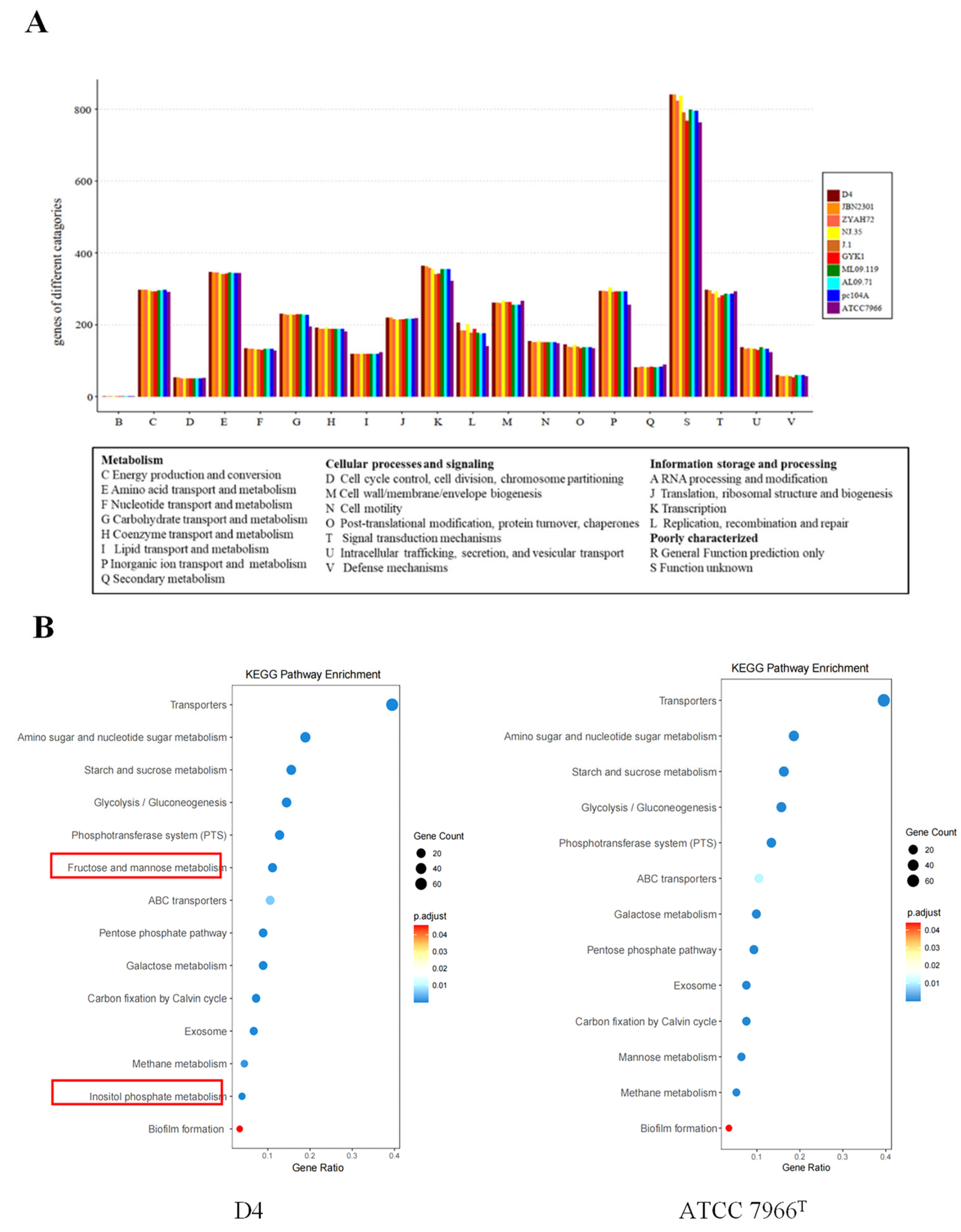
2.4.3. Functional Annotation and Pathway Analysis

To classify the functions encoded in the 10 A. hydrophila genomes, BLASTp was used to align the amino acid sequences against the database of Clusters of Orthologous Groups (http://www.ncbi.nlm.nih.gov/COG/ (accessed in 2024)). The A. hydrophila genomes were submitted to the KEGG pathway database (http://www.genome.jp/kegg/pathway.html (accessed in 2024)) for annotation.

2.5. Virulence-Related Factors Analysis

2.5.1. Virulence Gene Conservation and Absence Analysis

To comprehensively identify and analyze the virulence genes present in 10 A. hydrophila strains, BLASTp was applied. The amino acid sequences of the proteins encoded by the genomes of these 10 strains were aligned against the Virulence Factor Database (VFDB) to annotate the virulence genes.

2.5.2. Secondary Metabolite Gene Cluster Analysis

To explore the potential production of secondary metabolites by 10 A. hydrophila strains, the prediction of their secondary metabolite gene clusters was performed using the antiSMASH 5.0 online tool with default parameters (https://antismash.secondarymetabolites.org/ (accessed in 2024)) [11].

2.5.3. Prophage Content and Functional Analysis

To investigate the prophage elements harbored within 10 A. hydrophila strains, the prediction of prophages in these strains was conducted using the PHASTER online tool with default parameters (http://phaster.ca/ (accessed in 2025)).

2.5.4. Genomic Islands Analysis

To identify potential genomic islands that may contribute to the unique characteristics of 10 A. hydrophila strains, the prediction of genomic islands in these 10 strains was performed using IslandViewer 4 with default parameters (http://pathogenomics.sfu.ca/islandviewer (accessed in 2025)).

2.5.5. Plasmid Content and Functional Analysis

The collinearity comparison between pAhD4-1 (accession number: CP013966.1) and pHX3 (accession number: CP040718.1) was drawn using Easyfig (version 2.2.5) by inputting their GenBank files, performing BLAST alignment, and configuring parameters to analyze homologous regions and genetic distribution differences between the two plasmids.

3. Results

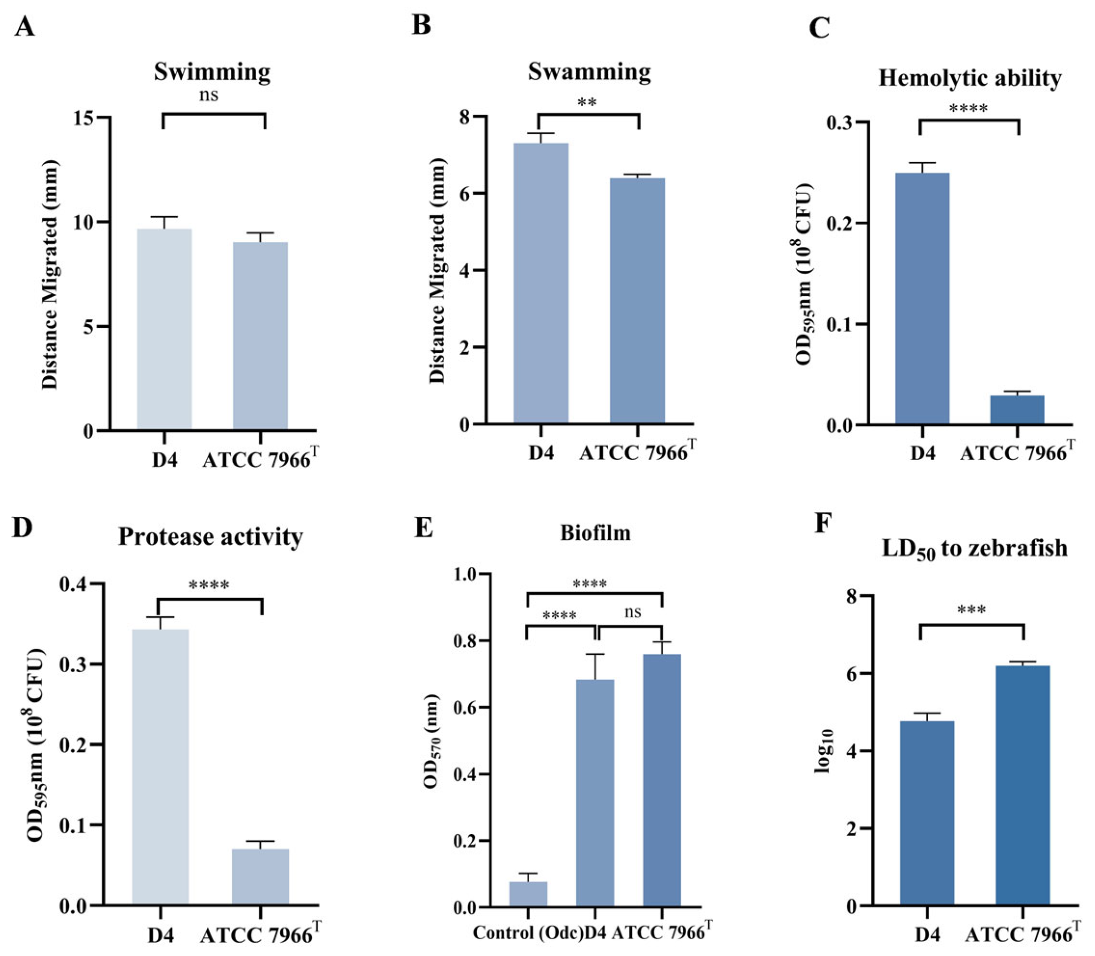
3.1. Phenotypic Characterization of Strain D4

3.1.1. Swimming and Swarming Motility

Flagella are complex surface organelles that enable bacteria to move towards favorable environments and contribute to the virulence of pathogenic bacteria via adhesion and biofilm formation on host surfaces. However, only a limited number of bacteria have dual flagella systems: a polar flagellum for swimming and lateral flagella for swarming [12,13]. To detect the difference in motility phenotypes between the epidemic ST251 strain D4 and the environmental strain ATCC 7966T, the 0.3% agar LB medium was used to assess the swimming ability of bacteria, and the 0.5% agar LB medium was employed to evaluate the swarming movement of bacteria. From the results of the swimming motility assay (Figure 1A), there was no significant difference in the migrated distance of colonies between D4 and ATCC 7966T (p > 0.05). However, in the swarming motility assay, the migrated distance of colonies of ATCC 7966T was significantly shorter than that of D4 (p < 0.01) (Figure 1B).

3.1.2. Hemolytic and Protease Activity

A. hydrophila can produce a variety of extracellular products, such as toxins (mainly including aerolysin, cytotoxic enterotoxin, and hemolysin) and extracellular proteases, most of which are associated with bacterial pathogenicity [14,15,16]. The hemolytic and protease activities of the epidemic strain D4 and the environmental strain ATCC 7966T were compared using the methods described above. As shown in Figure 1C,D, the hemolytic and protease activities of the extracellular products of strain D4 were significantly higher than those of ATCC 7966T (p < 0.0001).

3.1.3. Biofilm

Biofilm formation ability was evaluated using the 96-well plate method. The biofilm-forming abilities of the epidemic strain D4 and environmental strain ATCC 7966T are shown in Figure 1E. No significant difference was observed in biofilm formation between the two strains, but both displayed significantly higher biofilm formation than the control (p < 0.0001). Specifically, the ODe values of both strains were greater than 4ODc, indicating that both the epidemic strain D4 and the environmental strain ATCC 7966T have strong biofilm-forming capabilities.

3.1.4. Virulence Assessment via LD50 Determination

To evaluate the virulence of A. hydrophila strains D4 and ATCC 7966T, the median lethal dose (LD50) in zebrafish (Danio rerio) was determined. As shown in Figure 1F, the LD50 values for D4 and ATCC 7966T were 4.59 × 10⁴ CFU/fish and 1.65 × 10⁶ CFU/fish, respectively. According to the virulence classification criteria established by Rodríguez et al. [17], strain D4 was categorized as virulent (LD50 < 1.0 × 10⁶ CFU/fish), whereas the environmental isolate ATCC 7966T was classified as avirulent (LD50 > 1.0 × 10⁶ CFU/fish).

3.2. Comparative Genomics Analysis

3.2.1. ANI Analysis and General Features of Ten A. hydrophila Genomes

To further determine the classification of strain D4, ANI was calculated among 29 Aeromonas spp. genomes deposited in GenBank (Figure 2). As previously established, species-level classification requires ANI ≥ 95% [18]. Strain D4 clustered closely with A. hydrophila reference genomes, yielding ANI values >95%, confirming its classification as A. hydrophila. Conversely, strains YL17 and 4AK4 showed ANI values ≤ 92.88% and ≤ 86.04% with A. hydrophila genomes, respectively, suggesting potential misclassification. Subsequent analysis revealed that YL17 shared 97.1% ANI with A. dhakensis KN-Mc-6U21 and 4AK4 shared 95.5% ANI with A. rivipollensis KN-Mc-11N1, supporting their reclassification into these species. Notably, A. hydrophila strains D4, ZYAH72, NJ-35, J-1, GYK1, JBN2301, ML09-119, AL09-7, and pc104A displayed higher ANI values compared to other A. hydrophila genomes, clustering together into a distinct clade with ANI ≥ 99.74% and all belonging to ST251.
A whole-genome overview is shown in Table 1. Among the ten strains studied, D4 (this study), ZYAH72 [19], NJ-35 [20], J-1 [21], GYK1 [22], and JBN2301 [23] were the epidemic strains collected in China; ML09-119 [24], AL09-71 [25], and pc104A [26] were the epidemic strains isolated in the United States; and ATCC 7966T [27] was an environmental strain. There were four and three plasmids in the D4 and JBN2301 genomes, respectively, and no plasmid was found in other genomes. All ten strains harbored a single circular chromosome, with sizes ranging from 4.74 Mb (ATCC 7966T) to 5.28 Mb (NJ-35). Notably, the genome of the environmental isolate ATCC 7966T was significantly smaller than those of the epidemic strains. The genome G + C content was conserved (60.50–61.51%), but coding sequences (CDSs) varied from 4151 (ATCC 7966T) to 4569 (D4). All strains encoded 31 rRNAs, while tRNA, ncRNA, and pseudogene counts differed significantly. As shown in Figure 3A, comparative analysis revealed extensive CDS deletions in ATCC 7966T relative to ST251 strains, suggesting reduced functional complexity in the environmental isolate.

3.2.2. Genomic Collinearity and Rearrangement Analysis

To investigate the genomic structure and collinearity among ten A. hydrophila strains, progressive MAUVE alignment was performed using strain D4 as a reference genome (Figure 3B). Notably, no genome rearrangements were detected among the six Chinese epidemic strains (D4, ZYAH72, NJ-35, J-1, GYK1, and JBN2301) or within the three American epidemic strains (ML09-119, AL09-71, and pc104A). However, significant rearrangements were observed between the Chinese and American epidemic lineages, as well as between the environmental isolate ATCC 7966T and all epidemic strains.
The majority of genomic rearrangements were characterized by large-scale inversions of chromosomal segments. Previous studies have highlighted that such inversions play critical roles in adaptive evolution and ecological divergence [28]. The observed inversions between the Chinese and American epidemic clades may reflect geographically specific environmental adaptations, potentially contributing to their distinct epidemiological profiles.

3.2.3. Functional Annotation and Pathway Analysis

To identify functional determinants underlying virulence diversity in epidemic strains, ten A. hydrophila genomes were annotated against the Clusters of Orthologous Groups (COG) database. As shown in Figure 4A, the environmental isolate ATCC 7966T harbored significantly fewer genes in COG categories G (carbohydrate transport and metabolism), K (transcription), L (replication/recombination/repair), P (inorganic ion transport), and S (function unknown) compared to the nine epidemic ST251 strains. This observation suggests that genes in these COG categories may contribute to the enhanced virulence of epidemic strains.
KEGG pathway analysis was performed on genes categorized under COG G, K, L, and P. Notably, genes involved in the myo-inositol and L-fucose metabolic pathways (COG G) were uniquely present in ST251 strains, as shown in Figure 4B (red box). The enrichment of myo-inositol and L-fucose metabolism in ST251 strains may enhance host colonization by providing energy sources during infection [29].
Similarly, ST251-specific genes in COG L encoded transposases and helicases, including rapA, which encodes an ATP-dependent helicase. Previous studies have linked rapA homologs in Escherichia coli to biofilm formation, multidrug resistance, and stress adaptation [30]. The exclusive presence of rapA in ST251 strains suggests its potential role as a virulence determinant.

3.3. Virulence-Related Factors Analysis

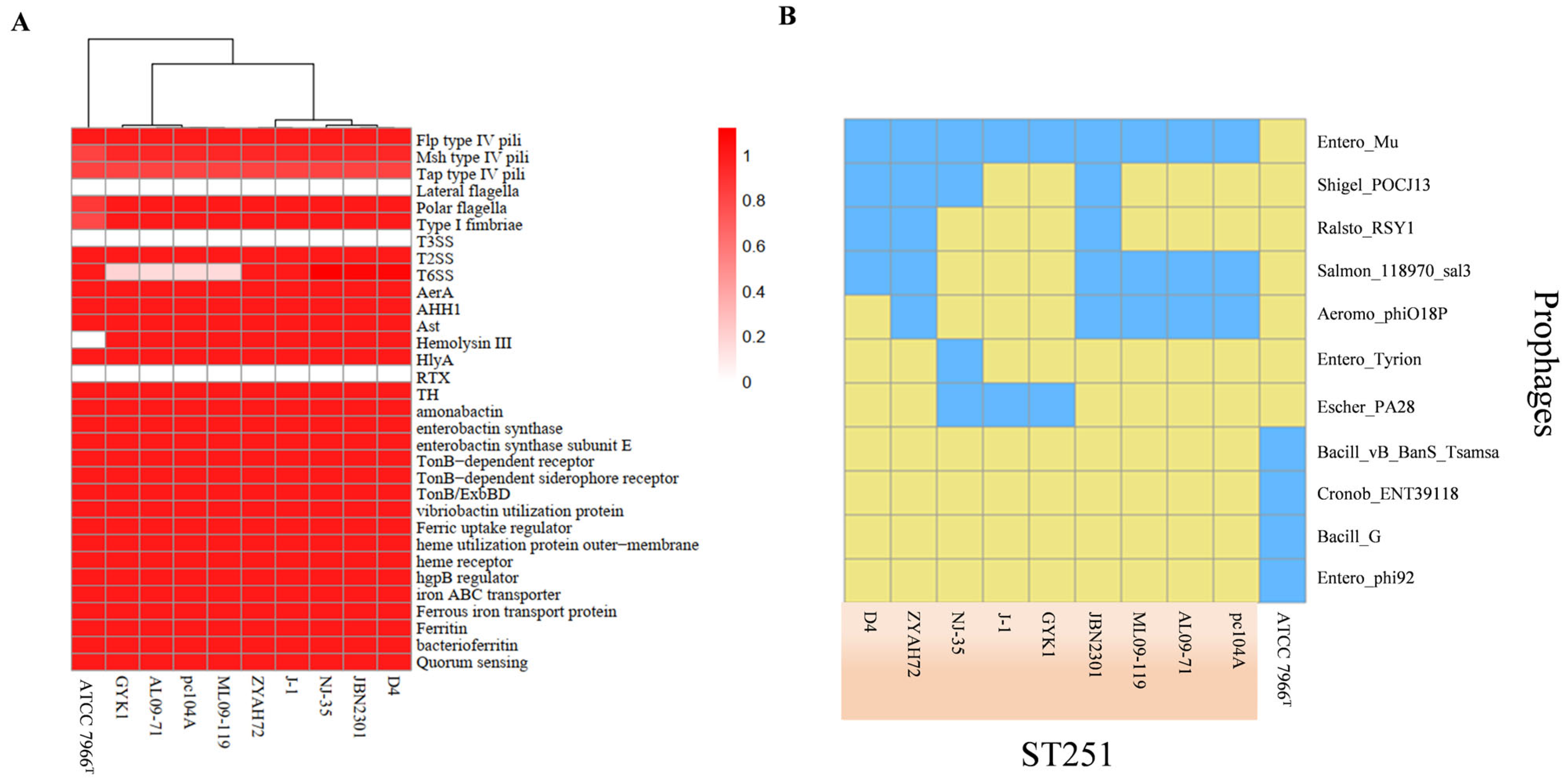
3.3.1. Virulence Gene Conservation and Absence Analysis

Previous studies have demonstrated that virulence factors are conserved among closely related bacterial species [31], and our study also revealed that nearly all the virulence genes from the VFDB were conserved across the ten A. hydrophila strains, including strain D4. However, the type III secretion system (T3SS), the repeat in toxin (RTX) system, and the lateral flagella system were absent not only in the environmental strain ATCC 7966T but also in the nine epidemic ST251 strains (Figure 5A). Additionally, the type VI secretion system (T6SS) was not present in three American strains (ML09-119, AL09-71, and pc104A) and one Chinese strain, GYK1. Intriguingly, although the T6SS was lacking in these four strains, the effector proteins (VgrG and Hcp) were detected. The above analyses suggest that the T3SS, the T6SS, the RTX system, and the lateral flagella system may not be essential virulence factors for the ST251 strain of A. hydrophila.

3.3.2. Secondary Metabolite Gene Cluster Analysis

Secondary metabolites are synthesized during specific growth phases that are dependent on primary metabolic pathways and typically play non-essential roles in microbial survival and reproduction [32]. As shown in Table 2, no differences were observed in the types of secondary metabolite gene clusters between epidemic ST251 strains and the environmental isolate ATCC 7966T. All strains harbored four conserved clusters: arylpolyene, bacteriocin, non-ribosomal peptide synthetase (NRPS), and hserlactone. Notably, ten A. hydrophila strains shared Clusters 1–4, while Cluster 5 was uniquely present in strain NJ-35.
Cluster 3 displayed 100% similarity to the amonabactin P750 biosynthesis cluster in A. hydrophila ATCC 7966T, which is associated with siderophore production [27]. Cluster 1, encoding an arylpolyene synthase, shared 61% homology with reported arylpolyene clusters in Xenorhabdus doucetiae. Arylpolyenes are widespread Gram-negative bacterial secondary metabolites with roles in oxidative stress resistance [33]. The conservation of the arylpolyene and siderophore clusters across all strains suggests their roles in fundamental survival processes, potentially contributing to ecological fitness rather than strain-specific virulence.
Clusters 2 and 4 lacked significant homology to previously characterized clusters, warranting further functional investigation.

3.3.3. Prophage Content and Functional Analysis

Prophages can enhance the pathogenicity of host bacteria by carrying virulence-related genes and facilitating their horizontal gene transfer [34]. Using the PHASTER online tool, four prophages were identified in strain D4’s genome. The number of prophages in nine ST251 strains and the environmental isolate ATCC 7966T ranged from two to five. As shown in Figure 5B, eleven distinct prophage types were detected across all strains. Notably, prophages present in ST251 strains were absent in ATCC 7966T. The prophage profiles were consistent among the American strains, while significant variations were observed in the Chinese strains.
The Entero_Mu prophage was conserved in all ST251 strains and encoded a regulatory protein (AhyD4_15485) homologous to PtrR from Pseudomonas aeruginosa, which has been linked to pathogenesis and antimicrobial resistance [35]. This suggests that PtrR-like proteins in ST251 may contribute to virulence, though experimental validation is needed. Additionally, the Entero_Mu prophage harbored genes encoding the M and S subunits of a type I restriction–modification system and secretion activator proteins (AhyD4_15495 and AhyD4_15500, respectively, which are putative virulence determinants in A. hydrophila [36]. The conservation of Entero_Mu in ST251 strains and its association with virulence-related genes suggest a role in horizontal gene transfer-mediated virulence evolution.

3.3.4. Genomic Island Analysis

Genomic islands (GIs) are segments of genomic DNA acquired through horizontal gene transfer and play a crucial role in bacterial environmental adaptation [37]. In this study, IslandViewer 4 was used to predict GIs in the genomes of ten A. hydrophila strains.
The number of GIs in the epidemic strains was significantly higher than that in the environmental strain ATCC 7966T. Moreover, most of the GIs present in the epidemic strains were absent in ATCC 7966T. All nine epidemic strains contain 14 GIs, among which GI 11, 18, 21, and 22 are functional GIs (Figure 3A).
The GI 11 region harbors a myo-inositol utilization cluster (Figure 6A). Myo-inositol can serve as a carbon source to enhance the colonization ability of bacteria in the host, thus affecting the virulence of bacteria [38]. This cluster encodes three ribose transport proteins (RbsABC) responsible for myo-inositol transport and eight proteins (IolARDG_2G_1CEB) involved in myo-inositol catabolism (Figure 6A) [39,40]. The myo-inositol utilization cluster in GI 11 encodes all the enzymes necessary for myo-inositol utilization, except 2-deoxy-5-keto-D-gluconic acid 6-phosphate aldolase, which is required for the degradation of myo-inositol to acetyl-CoA. Nevertheless, the D4 genome can encode homologs of 2-deoxy-5-keto-D-gluconic acid 6-phosphate aldolase, such as tagatose-bisphosphate aldolase (AhyD4_19035) and class II fructose-bisphosphate aldolase (AhyD4_19150).
GI 18 encodes all the enzymes required for pseudaminic acid biosynthesis, except PseH (Figure 6B) [41]. Pseudaminic acid can enhance the adhesion and motility of bacteria by participating in the modification of the bacterial surface and the glycosylation of flagellin proteins, thereby improving their colonization, survival, and immune evasion efficiency within the host [42]. A search of the D4 genome identified some genes predicted to encode homologs of PseH, such as GNAT family N-acetyltransferase (AhyD4_20800). Additionally, GI 18 encodes five proteins (FliS, FliD, and three flagellins) associated with flagellar assembly.
GI 21 encodes eight proteins involved in part of the L-fucose metabolic pathway and a peptide ABC transport system (OppABCD) (Figure 6C). Pathogens can utilize L-fucose as a carbon source and make use of its metabolic intermediates to supply energy or synthesize virulence factors such as the O antigen in lipopolysaccharide (LPS) [43,44]. The complete L-fucose metabolic pathway involves eight proteins encoded by the fucRPIKAUO operon. Typically, a regulator (FucR), a permease (FucP), a fucose mutarotase (FucU), an isomerase (FucI), a kinase (FucK), an aldolase (FucA), and a reductase (FucO) are required to produce 1,2-propanediol and dihydroxyacetone phosphate, which enter the glycolytic pathway [17]. GI 21 encodes FucK, FucA, FucU, and FucO, while the three genes fucR, fucP, and fucI were found upstream of GI 21.
GI 22, the largest among the nine epidemic-associated unique GIs with 41 ORFs, comprises hypothetical proteins, a DNA helicase, a DNA repair protein, a transcriptional regulator, an iron complex transport system, and a transposase.

3.3.5. Two-Component Regulatory System Profiling

Two-component regulatory systems (TCSs) are conserved bacterial signaling networks critical for environmental adaptation [45]. As shown in Table 3, the TCSs were highly conserved among ST251 strains, including D4. Notable exceptions were observed in the environmental isolate ATCC 7966T, which lacked the phosphoglycerate transport regulator PgtC and the vancomycin resistance sensor VanS. Conversely, the magnesium-citrate transport regulator CitT was uniquely present in ATCC 7966T.
KEGG annotation identified 61 TCSs in ST251 strains, including virulence-associated systems ArlS/ArlR and Ihk/Irr [46]. The conservation of ArlS/ArlR and Ihk/Irr in ST251 may underlie their hypervirulence by modulating biofilm formation and stress responses [47,48].

3.3.6. Plasmid Content and Functional Analysis

Plasmids are self-replicating genetic elements that confer accessory traits to microorganisms, including toxin production, stress tolerance, and anabolic pathways [49]. As shown in Table 1, among nine ST251 strains and the environmental isolate ATCC 7966T, only D4 and JBN2301 harbored plasmids (four and three plasmids, respectively). The genomic features of these seven plasmids are detailed in Table 4.
Notably, three small plasmids in D4 (pAhD4-2, pAhD4-3, and pAhD4-4) shared 99–100% similarity with those in JBN2301 (pAhJBN2301-1, pAhJBN2301-2, and pAhJBN2301-3). The largest plasmid, pAhD4-1, displayed 81% similarity to pHX3 from A. veronii strain HX3. pAhD4-1 encoded 164 genes, including putative virulence and resistance determinants such as the fluoroquinolone resistance protein, the antitoxin MazE, the antitoxin HipB, and conjugal transfer pilus assembly proteins (Figure 7; homologous to type IV secretion system components) [8]. The presence of type IV secretion system homologs in pAhD4-1 suggests potential roles in interbacterial competition or host colonization. The three small plasmids contained 6–11 CDSs, with most annotated as hypothetical proteins.

4. Discussion

In recent years, MAS has emerged as a significant threat to global aquaculture, causing substantial economic losses in China, the United States, and other regions [4]. A. hydrophila ST251 has been identified as a hypervirulent clonal group associated with MAS outbreaks in both China and the Southeastern United States [5]. Here, we characterized the virulence phenotype and complete genome of A. hydrophila D4, which was isolated from diseased M. amblycephala and belonging to ST251, and performed comparative genomic analyses with nine ST251 strains and the environmental isolate ATCC 7966T (ST1).

4.1. Phenotypic and Genomic Correlates of Virulence

Phenotypic characterization revealed obvious differences between D4 and ATCC 7966T. D4 exhibited stronger swarming motility, which aligns with it having intact flagellar assembly genes (e.g., fliS, fliD, and three flagellin genes in GI 18) critical for motility [42]. Strain ATCC 7966T lacks these genes, consistent with its reduced motility.
Additionally, D4 showed significantly higher hemolytic activity and protease production, likely stemming from plasmid-encoded type IV secretion system homologs (e.g., conjugal transfer pilus proteins in pAhD4-1) that facilitate the secretion of virulence factors [8].

4.2. Biofilm Formation and Functional Redundancy

Contrary to expectations, both D4 and ATCC 7966T formed robust biofilms with no statistically significant difference (p > 0.05; Figure 1E). This unexpected finding suggests that despite genomic differences, both strains employ distinct strategies to achieve comparable biofilm capacities. D4’s biofilm formation capabilities may rely on ST251-specific ABC transporters and the L-fucose metabolic pathway, which facilitate adhesion to host tissues [50]. Additionally, the PtrR-like regulator encoded by the prophage Entero_Mu in D4 may upregulate biofilm-associated genes, as observed in P. aeruginosa [35]. In contrast, ATCC 7966T lacks these pathways but harbors CitT, a magnesium citrate transport regulator unique to ATCC 7966T. This suggests that ATCC 7966T may utilize alternative carbon sources (e.g., citrate) to form biofilms, aligning with its adaptation to aquatic or other environments. The comparable biofilm capacities of D4 and ATCC 7966T highlight functional redundancy in biofilm formation mechanisms. While D4 relies on host-derived nutrients (e.g., L-fucose), ATCC 7966T may utilize environmental resources (e.g., citrate), indicating their distinct ecological niches. This divergence in metabolic strategies likely contributes to their differential virulence phenotypes despite similar biofilm formation abilities.

4.3. Genomic Insights into ST251 Pathogenesis

Comparative genomic analysis revealed that the environmental isolate ATCC 7966T harbored extensive CDS deletions compared to ST251 strains, suggesting reduced functional complexity (Figure 3A). The ANI among nine ST251 A. hydrophila strains ranged from 99.74% to 100%, which is significantly higher than the values observed in other A. hydrophila lineages (96.24–96.97%) (Figure 2). Phylogenetic analysis confirmed close genetic relationships within the ST251 clade, with strains from the same geographic region forming even tighter clusters. Genome collinearity analysis further supported this finding (Figure 3B).
A. hydrophila is known to harbor diverse virulence determinants, including secretion systems and flagellar apparatus, which are often recognized as critical virulence determinants [7]. Notably, VFDB annotation revealed no significant differences in core virulence gene repertoires among ten A. hydrophila genomes, except for T6SS. Strikingly, T3SS and lateral flagella were absent in all ten strains, including ST251 isolates (Figure 5A). This suggests that these systems are non-essential for ST251 pathogenesis.
To identify unique virulence determinants in ST251, COG and KEGG functional annotations were performed (Figure 4A). The analysis revealed that ST251 genomes are enriched in genes encoding transposases, integrases, ABC transporters, and enzymes involved in the myo-inositol and L-fucose metabolic pathways (Figure 4B). These findings imply that ST251 may rely on metabolic adaptation rather than classical virulence systems to establish infection.

4.4. Mobile Genetic Elements and Adaptive Evolution

Plasmids and prophages in ST251 strains may contribute to their virulence, resistance, and adaptation to the environment [51,52]. D4’s plasmid pAhD4-1 encodes type IV secretion system homologs and fluoroquinolone resistance determinants, suggesting roles in horizontal gene transfer and antimicrobial resistance. The conserved prophage Entero_Mu in ST251 (Figure 5B), which is absent in ATCC 7966T, encodes a PtrR-like regulator (AhyD4_15485) linked to P. aeruginosa pathogenesis [35]. This protein may enhance ST251’s ability to colonize host tissues by modulating stress responses.
GI analysis identified 26 GIs (4–54 kb) in A. hydrophila D4 (Figure 3A). Fourteen GIs were considered as ST251-specific virulence-associated islands, and they were absent in the environmental isolate ATCC 7966T but conserved in ST251 strains. Notably, two of these GIs harbored genes encoding the myo-inositol and L-fucose metabolic pathways, while another contained a pseudaminic acid biosynthesis cluster.

4.5. Metabolic Adaptation as a Pathogenic Strategy

Bacterial metabolic adaptability is critical for survival and proliferation in diverse environments. Studies in pathogenic Salmonella, Vibrio, and Helicobacter have demonstrated that metabolic genes are as essential as classical virulence factors during infection [53]. ST251 strains encode two distinct metabolic pathways (myo-inositol and L-fucose) and a pseudaminic acid biosynthesis cluster, which may collectively contribute to their hypervirulence.
Myo-inositol, abundant in the fish liver and intestine, may serve as a carbon source during infection [29]. D4 encodes a complete myo-inositol utilization cluster (GI 11), including iolR, which regulates aerolysin expression [40]. This suggests a nutrient-dependent regulatory axis where myo-inositol availability could enhance virulence factor production, conferring a competitive advantage in host tissues.
L-fucose, a terminal residue of intestinal mucins, may serve as both an adhesion target and an energy source [54,55]. Fucosylated glycans facilitate bacterial colonization by interacting with host lectins [56], potentially promoting ST251 persistence in mucosal environments.
Notably, ST251 strains uniquely encode pseudaminic acid biosynthesis genes (GI 18). This non-canonical sugar modifies flagellin, enabling functional flagellar assembly and motility [42]. Since pseudaminic acid is restricted to pathogenic bacteria, this pathway likely contributes to ST251’s enhanced motility and host colonization capacity.
Collectively, these metabolic pathways and biosynthesis systems represent novel virulence determinants in ST251. By exploiting host-derived nutrients (myo-inositol and L-fucose) and optimizing motility via pseudaminic acid, ST251 may outcompete environmental isolates and establish infection. This metabolic plasticity, which is absent in ATCC 7966T, likely underlies the hypervirulence of the ST251 clonal group.

4.6. Limitations and Future Research Directions

This study provides initial insights into the pathogenicity and environmental adaptability of the ST251 epidemic strain; however, further investigations are needed to validate and expand upon the current findings:
(1)
Constructing deletion or overexpression mutants of key virulence or regulatory genes (e.g., pse genes, PtrR, IolR), combined with in vitro and in vivo infection models, will help clarify their roles in motility, biofilm formation, and virulence expression.
(2)
Transcriptomic analyses under various environmental stress conditions (e.g., high temperature and salinity and nutrient limitation) and natural host infection models (e.g., blunt snout bream) could provide mechanistic insights into transcriptional regulation and virulence adaptation in complex environments. Integrating histopathology and host immune gene expression will further improve ecological relevance.
(3)
Expanding the strain collection to include more ST251 isolates from diverse geographic regions and host sources, along with host response analyses (e.g., host transcriptomics, inflammatory markers, and immune cell activation), will facilitate a systems-level understanding of host–pathogen interactions and their impact on infection outcomes.
Future research integrating genome editing, multi-omics approaches, and ecologically relevant infection models will be crucial for elucidating the molecular mechanisms underlying ST251 virulence evolution and environmental adaptation, ultimately supporting the development of effective control strategies for aquaculture-associated outbreaks.

5. Conclusions

This study provides valuable insights into the genomic and phenotypic features of A. hydrophila ST251, a hypervirulent lineage responsible for widespread outbreaks of MAS in aquaculture. Phenotypic analyses revealed that, compared to the environmental isolate ATCC 7966T, strain D4 exhibited significantly enhanced motility, hemolytic activity, and protease activity, as well as increased virulence in the zebrafish infection model, suggesting greater pathogenic potential.
Comparative genomic analysis of D4, nine additional ST251 strains, and ATCC 7966T showed that ST251 strains form a distinct phylogenetic clade with an ANI score above 99.74%. Their genome sizes and the number of CDSs were generally larger than those of ATCC 7966T. Although the G + C content remained consistent, extensive genome rearrangements were observed among ST251 strains from China and the United States, as well as between ST251 and environmental strains.
Notably, ST251-specific genomic islands encode pathways for inositol and L-fucose utilization and a pseudaminic acid biosynthesis gene cluster, which may enhance metabolic versatility and host colonization by enabling nutrient acquisition and flagellar glycosylation. In addition, although typical virulence factors such as T3SS, T6SS, RTX clusters, and lateral flagella are commonly present in ST251 strains, their absence or variation in some isolates suggests that these factors may not be the core determinants of the heightened virulence of this lineage. Moreover, the types of secondary metabolite gene clusters showed no significant differences between ST251 and environmental strains, implying that these clusters are unlikely to be major drivers of virulence enhancement.
Prophage analysis revealed that ST251 strains universally harbor a conserved Entero_Mu prophage encoding a PtrR-like transcriptional regulator that is homologous to proteins involved in antimicrobial resistance and virulence in P. aeruginosa. Additionally, conserved two-component regulatory systems such as ArlS/ArlR and Ihk/Irr were identified, which may contribute to increased environmental adaptability and pathogenicity by enhancing stress responses and biofilm formation. The detection of type IV secretion system homologs on plasmid pAhD4-1 further suggests possible roles in interbacterial competition and host colonization.
In summary, these findings provide novel insights into the molecular mechanisms underlying the pathogenicity and environmental adaptability of hypervirulent A. hydrophila ST251 strains.

Author Contributions

Conceptualization, Y.L.; methodology, L.X., X.K. and Z.W.; software, L.X. and Z.X.; validation, L.X., X.K., Z.W. and Z.X.; formal analysis, L.X. and Y.L.; investigation, L.X. and X.K.; resources, L.X., X.K., Z.W. and Z.X.; data curation, L.X., X.K. and Z.W.; writing—original draft preparation, L.X. and X.K.; writing—review and editing, L.X. and Y.L.; visualization, L.X.; supervision, Y.L.; project administration, Y.L. All authors have read and agreed to the published version of the manuscript.

Funding

This research was funded by the National Key Research and Development Plan Program under Grant No. 2022YFD2400604 and the Fundamental Research Funds for the Central Universities, grant number X2662024SCPY001.

Institutional Review Board Statement

Not applicable.

Informed Consent Statement

Not applicable.

Data Availability Statement

The original contributions presented in this study are included in the article. Further inquiries can be directed to the corresponding authors.

Conflicts of Interest

The authors declare no conflicts of interest.

Abbreviations

The following abbreviations are used in this manuscript:
ANIAverage nucleotide identity
A. hydrophilaAeromonas hydrophila
CASChrome azurol-S
CDSsCoding sequences
MASMotile Aeromonas Septicemia
PBSPhosphate-buffered saline
T3SSType III secretion system
T6SSType VI secretion system
ST251Sequence type 251

References

  1. Little, D.C.; MacKenzie, S. Grand challenges for global aquaculture. Front. Aquac. 2023, 2, 1232936. [Google Scholar] [CrossRef]
  2. Michael Janda, J.; Sharon, L.A. The genus Aeromonas: Taxonomy, pathogenicity, and infection. Clin. Microbiol. Rev. 2010, 23, 35–73. [Google Scholar] [CrossRef] [PubMed]
  3. Bartie, K.L.; Ngô, T.P.H.; Bekaert, M.; Hoang Oanh, D.T.; Hoare, R.; Adams, A.; Desbois, A.P. Aeromonas hydrophila ST251 and Aeromonas dhakensis are major emerging pathogens of striped catfish in vietnam. Front. Microbiol. 2023, 13, 1067235. [Google Scholar] [CrossRef] [PubMed]
  4. Zhang, X.; Yang, W.; Wu, H.; Gong, X.; Li, A. Multilocus sequence typing revealed a clonal lineage of Aeromonas hydrophila caused Motile Aeromonas Septicemia outbreaks in pond-cultured cyprinid fish in an epidemic area in central china. Aquaculture 2014, 432, 1–6. [Google Scholar] [CrossRef]
  5. Rasmussen-Ivey, C.R.; Hossain, M.J.; Odom, S.E.; Terhune, J.S.; Hemstreet, W.G.; Shoemaker, C.A.; Zhang, D.; Xu, D.-H.; Griffin, M.J.; Liu, Y.-J.; et al. Classification of a hypervirulent Aeromonas hydrophila pathotype responsible for epidemic outbreaks in warm-water fishes. Front. Microbiol. 2016, 7, 1615. [Google Scholar] [CrossRef]
  6. Hossain, M.J.; Sun, D.; McGarey, D.J.; Wrenn, S.; Alexander, L.M.; Martino, M.E.; Xing, Y.; Terhune, J.S.; Liles, M.R. An Asian origin of virulent Aeromonas Hydrophila responsible for disease epidemics in United States-farmed catfish. mBio 2014, 5, e00848-14. [Google Scholar] [CrossRef]
  7. Rasmussen-Ivey, C.R.; Figueras, M.J.; McGarey, D.; Liles, M.R. Virulence factors of Aeromonas hydrophila: In the wake of reclassification. Front. Microbiol. 2016, 7, 1337. [Google Scholar] [CrossRef]
  8. Zhu, L.; Zheng, J.-S.; Wang, W.-M.; Luo, Y. Complete genome sequence of highly virulent Aeromonas hydrophila Strain D4, isolated from a diseased blunt-snout bream in China. Microbiol. Resour. Announc. 2019, 8, e01035-18. [Google Scholar] [CrossRef]
  9. Davey, M.E.; O’toole, G.A. Microbial biofilms: From ecology to molecular genetics. Microbiol. Mol. Biol. Rev. 2000, 64, 847–867. [Google Scholar] [CrossRef]
  10. Saganuwan, A. A modified arithmetical method of reed and muench for determination of a relatively ideal median lethal dose (LD50). Afr. J. Pharm. Pharmacol. 2011, 5, 1543–1546. [Google Scholar] [CrossRef]
  11. Medema, M.H.; Blin, K.; Cimermancic, P.; de Jager, V.; Zakrzewski, P.; Fischbach, M.A.; Weber, T.; Takano, E.; Breitling, R. antiSMASH: Rapid identification, annotation and analysis of secondary metabolite biosynthesis gene clusters in bacterial and fungal genome sequences. Nucleic Acids Res. 2011, 39, W339–W346. [Google Scholar] [CrossRef] [PubMed]
  12. McCarter, L.L. Dual flagellar systems enable motility under different circumstances. J. Mol. Microbiol. Biotechnol. 2004, 7, 18–29. [Google Scholar] [CrossRef] [PubMed]
  13. Chaban, B.; Hughes, H.V.; Beeby, M. The flagellum in bacterial pathogens: For motility and a whole lot more. Semin. Cell Dev. Biol. 2015, 46, 91–103. [Google Scholar] [CrossRef] [PubMed]
  14. Wang, G.; Clark, C.G.; Liu, C.; Pucknell, C.; Munro, C.K.; Kruk, T.M.A.C.; Caldeira, R.; Woodward, D.L.; Rodgers, F.G. Detection and characterization of the hemolysin genes in Aeromonas hydrophila and Aeromonas sobria by multiplex PCR. J. Clin. Microbiol. 2003, 41, 1048–1054. [Google Scholar] [CrossRef]
  15. Goswami, R.; Ghosh, D.; Saha, D.R.; Padhy, P.K.; Mazumder, S. Effect of acute and chronic arsenic exposure on growth, structure and virulence of Aeromonas hydrophila isolated from fish. Microb. Pathog. 2011, 50, 63–69. [Google Scholar] [CrossRef]
  16. Abreu, R.E.F.; Magalhães, T.C.; Souza, R.C.; Oliveira, S.T.; Ibelli, A.M.; Demarqui, F.N.; Gouveia, J.J.; Costa, M.M.; Gouveia, G.V. Environmental factors on virulence of Aeromonas hydrophila. Aquacult. Int. 2018, 26, 495–507. [Google Scholar] [CrossRef]
  17. Rodríguez, I.; Novoa, B.; Figueras, A. Immune response of zebrafish (Danio Rerio) against a newly isolated bacterial pathogen Aeromonas Hydrophila. Fish Shellfish Immunol. 2008, 25, 239–249. [Google Scholar] [CrossRef]
  18. Rodriguez-R, L.M.; Conrad, R.E.; Viver, T.; Feistel, D.J.; Lindner, B.G.; Venter, S.N.; Orellana, L.H.; Amann, R.; Rossello-Mora, R.; Konstantinidis, K.T. An ani gap within bacterial species that advances the definitions of intra-species units. mBio 2024, 15, e0269623. [Google Scholar] [CrossRef]
  19. Xie, Q.; Mei, W.; Ye, X.; Zhou, P.; Islam, M.S.; Elbassiony, K.R.A.; Yuan, C.; Li, J.; Zhou, Y. The two-component regulatory system CpxA/R is required for the pathogenesis of Aeromonas hydrophila. FEMS Microbiol. Lett. 2018, 365, fny218. [Google Scholar] [CrossRef]
  20. Pang, M.; Jiang, J.; Xie, X.; Wu, Y.; Dong, Y.; Kwok, A.H.Y.; Zhang, W.; Yao, H.; Lu, C.; Leung, F.C.; et al. Novel insights into the pathogenicity of epidemic Aeromonas hydrophila ST251 clones from comparative genomics. Sci. Rep. 2015, 5, 9833. [Google Scholar] [CrossRef]
  21. Bi, Z.X.; Liu, Y.J.; Lu, C.P. Contribution of AhyR to virulence of Aeromonas hydrophila J-1. Res. Vet Sci. 2007, 83, 150–156. [Google Scholar] [CrossRef] [PubMed]
  22. Hou-jun, P. Identification, virulence, hemolytic activity of GYK1, a Strain of pathogenic Aeromonas hydrophila isolated from mandarinfish. J. Shanghai Fish. Univ. 2004, 13, e29. [Google Scholar]
  23. Yang, W.; Li, N.; Li, M.; Zhang, D.; An, G. Complete genome sequence of fish pathogen Aeromonas hydrophila JBN2301. Genome Announc. 2016, 4, e01615-15. [Google Scholar] [CrossRef] [PubMed]
  24. Tekedar, H.C.; Waldbieser, G.C.; Karsi, A.; Liles, M.R.; Griffin, M.J.; Vamenta, S.; Sonstegard, T.; Hossain, M.; Schroeder, S.G.; Khoo, L.; et al. Complete genome sequence of a channel catfish epidemic isolate, Aeromonas hydrophila Strain ML09-119. Genome Announc. 2013, 1, e00755-15. [Google Scholar] [CrossRef] [PubMed]
  25. Pridgeon, J.W.; Zhang, D.; Zhang, L. Complete genome sequence of the highly virulent Aeromonas hydrophila AL09-71 isolated from diseased channel catfish in West Alabama. Genome Announc. 2014, 2, 10–1128. [Google Scholar] [CrossRef]
  26. Pridgeon, J.W.; Zhang, D.; Zhang, L. Complete genome sequence of a moderately virulent Aeromonas hydrophila Strain, pc104A, isolated from soil of a catfish pond in West Alabama. Genome Announc. 2014, 2, e00554-14. [Google Scholar] [CrossRef] [PubMed]
  27. Seshadri, R.; Joseph, S.W.; Chopra, A.K.; Sha, J.; Shaw, J.; Graf, J.; Haft, D.; Wu, M.; Ren, Q.; Rosovitz, M.J.; et al. Genome sequence of Aeromonas hydrophila ATCC 7966T: Jack of all trades. J. Bacteriol. 2006, 188, 8272–8282. [Google Scholar] [CrossRef]
  28. Fricke, W.F.; Mammel, M.K.; McDermott, P.F.; Tartera, C.; White, D.G.; Leclerc, J.E.; Ravel, J.; Cebula, T.A. Comparative genomics of 28 Salmonella Enterica isolates: Evidence for CRISPR-mediated adaptive sublineage evolution. J. Bacteriol. 2011, 193, 3556–3568. [Google Scholar] [CrossRef]
  29. Awan, F.; Dong, Y.; Liu, J.; Wang, N.; Mushtaq, M.H.; Lu, C.; Liu, Y. Comparative genome analysis provides deep insights into Aeromonas hydrophila taxonomy and virulence-related factors. BMC Genomics 2018, 19, 712. [Google Scholar] [CrossRef]
  30. Pietras, Z.; Hardwick, S.W.; Swiezewski, S.; Luisi, B.F. Potential regulatory interactions of Escherichia Coli RraA Protein with DEAD-Box helicases. J. Biol. Chem. 2013, 288, 31919–31929. [Google Scholar] [CrossRef]
  31. Xu, T.; Rasmussen-Ivey, C.R.; Moen, F.S.; Fernández-Bravo, A.; Lamy, B.; Beaz-Hidalgo, R.; Khan, C.D.; Castro Escarpulli, G.; Yasin, I.S.M.; Figueras, M.J.; et al. A global survey of hypervirulent Aeromonas hydrophila (vAh) identified vah strains in the lower Mekong River Basin and diverse opportunistic pathogens from farmed fish and other environmental sources. Microbiol. Spectr. 2023, 11, e0370522. [Google Scholar] [CrossRef] [PubMed]
  32. Li, S.; Wang, P.; Yuan, W.; Su, Z.; Bullard, S.H. Endocidal regulation of secondary metabolites in the producing organisms. Sci. Rep. 2016, 6, 29315. [Google Scholar] [CrossRef]
  33. Lian, L.; Sun, L.; Zhao, X.; Chen, L.; Zhang, B.; Liu, Y.; Lin, X. Analysis of pathogenicity factors in the highly virulent Aeromonas hydrophila strain LP-2. Aquaculture 2025, 598, 741982. [Google Scholar] [CrossRef]
  34. Huang, J.; Dai, X.; Wu, Z.; Hu, X.; Sun, J.; Tang, Y.; Zhang, W.; Han, P.; Zhao, J.; Liu, G.; et al. Conjugative transfer of streptococcal prophages harboring antibiotic resistance and virulence genes. ISME J. 2023, 17, 1467–1481. [Google Scholar] [CrossRef] [PubMed]
  35. Sun, Z.; Shi, J.; Liu, C.; Jin, Y.; Li, K.; Chen, R.; Jin, S.; Wu, W. PrtR homeostasis contributes to Pseudomonas aeruginosa pathogenesis and resistance against ciprofloxacin. Infect. Immun. 2014, 82, 1638–1647. [Google Scholar] [CrossRef]
  36. Mehershahi, K.S.; Chen, S.L. DNA methylation by three Type I restriction modification systems of Escherichia Coli does not influence gene regulation of the host bacterium. Nucleic Acids Res. 2021, 49, 7375–7388. [Google Scholar] [CrossRef]
  37. Juhas, M.; van der Meer, J.R.; Gaillard, M.; Harding, R.M.; Hood, D.W.; Crook, D.W. Genomic islands: Tools of bacterial horizontal gene transfer and evolution. FEMS Microbiol. Rev. 2009, 33, 376–393. [Google Scholar] [CrossRef]
  38. Manske, C.; Schell, U.; Hilbi, H. Metabolism of myo-inositol by Legionella pneumophila promotes infection of amoebae and macrophages. Appl. Environ. Microbiol. 2016, 82, 5000–5014. [Google Scholar] [CrossRef]
  39. Krings, E.; Krumbach, K.; Bathe, B.; Kelle, R.; Wendisch, V.F.; Sahm, H.; Eggeling, L. Characterization of myo-inositol utilization by Corynebacterium glutamicum: The stimulon, identification of transporters, and influence on L-lysine formation. J. Bacteriol. 2006, 188, 8054–8061. [Google Scholar] [CrossRef]
  40. Dong, Y.; Li, S.; Zhao, D.; Liu, J.; Ma, S.; Geng, J.; Lu, C.; Liu, Y. IolR, a negative regulator of the myo-inositol metabolic pathway, inhibits cell autoaggregation and biofilm formation by downregulating rpma in Aeromonas hydrophila. NPJ Biofilms Microbiomes 2020, 6, 22. [Google Scholar] [CrossRef]
  41. da Silva Filho, A.C.; Marchaukoski, J.N.; Raittz, R.T.; De Pierri, C.R.; de Jesus Soares Machado, D.; Fadel-Picheth, C.M.T.; Picheth, G. Prediction and analysis in silico of genomic islands in Aeromonas hydrophila. Front. Microbiol. 2021, 12, 769380. [Google Scholar] [CrossRef] [PubMed]
  42. Salah Ud-Din, A.I.M.; Roujeinikova, A. Flagellin glycosylation with pseudaminic acid in Campylobacter and Helicobacter: Prospects for development of novel therapeutics. Cell. Mol. Life Sci. 2018, 75, 1163–1178. [Google Scholar] [CrossRef] [PubMed]
  43. Stahl, M.; Friis, L.M.; Nothaft, H.; Liu, X.; Li, J.; Szymanski, C.M.; Stintzi, A. L-fucose utilization provides campylobacter jejuni with a competitive advantage. Proc. Natl. Acad. Sci. USA 2011, 108, 7194–7199. [Google Scholar] [CrossRef] [PubMed]
  44. Zou, Y.; Li, C.; Brunzelle, J.S.; Nair, S.K. Molecular basis for substrate selectivity and specificity by an LPS biosynthetic enzyme. Biochemistry 2007, 46, 4294–4304. [Google Scholar] [CrossRef]
  45. Hirakawa, H.; Kurushima, J.; Hashimoto, Y.; Tomita, H. Progress overview of bacterial two-component regulatory systems as potential targets for antimicrobial chemotherapy. Antibiotics 2020, 9, 635. [Google Scholar] [CrossRef] [PubMed]
  46. Beier, D.; Gross, R. Regulation of bacterial virulence by two-component systems. Curr. Opin. Microbiol. 2006, 9, 143–152. [Google Scholar] [CrossRef]
  47. Han, H.; Liu, C.; Wang, Q.; Xuan, C.; Zheng, B.; Tang, J.; Yan, J.; Zhang, J.; Li, M.; Cheng, H.; et al. The two-component system Ihk/Irr contributes to the virulence of streptococcus suis serotype 2 strain 05ZYH33 through alteration of the bacterial cell metabolism. Microbiology 2012, 158, 1852–1866. [Google Scholar] [CrossRef]
  48. Crosby, H.A.; Tiwari, N.; Kwiecinski, J.M.; Xu, Z.; Dykstra, A.; Jenul, C.; Fuentes, E.J.; Horswill, A.R. The Staphylococcus aureus ArlRS two-component system regulates virulence factor expression through MgrA. Mol. Microbiol. 2020, 113, 103–122. [Google Scholar] [CrossRef]
  49. Actis, L.A.; Tolmasky, M.E.; Crosa, J.H. Bacterial plasmids: Replication of extrachromosomal genetic elements encoding resistance to antimicrobial compounds. Front. Biosci. 1999, 4, D43–D62. [Google Scholar] [CrossRef]
  50. Zhao, Y.-L.; Zhou, Y.-H.; Chen, J.-Q.; Huang, Q.-Y.; Han, Q.; Liu, B.; Cheng, G.-D.; Li, Y.-H. Quantitative proteomic analysis of Sub-MIC erythromycin inhibiting biofilm formation of S. suis in vitro. J. Proteom. 2015, 116, 1–14. [Google Scholar] [CrossRef]
  51. Brüssow, H.; Canchaya, C.; Hardt, W.-D. Phages and the evolution of bacterial pathogens: From genomic rearrangements to lysogenic conversion. Microbiol. Mol. Biol. Rev. 2004, 68, 560–602. [Google Scholar] [CrossRef] [PubMed]
  52. Dougherty, P.E.; Nielsen, T.K.; Riber, L.; Lading, H.H.; Forero-Junco, L.M.; Kot, W.; Raaijmakers, J.M.; Hansen, L.H. Widespread and largely unknown prophage activity, diversity, and function in two genera of wheat phyllosphere bacteria. ISME J. 2023, 17, 2415–2425. [Google Scholar] [CrossRef] [PubMed]
  53. Chen, Y.-J.; Leung, P.M.; Wood, J.L.; Bay, S.K.; Hugenholtz, P.; Kessler, A.J.; Shelley, G.; Waite, D.W.; Franks, A.E.; Cook, P.L.M.; et al. Metabolic flexibility allows bacterial habitat generalists to become dominant in a frequently disturbed ecosystem. ISME J. 2021, 15, 2986–3004. [Google Scholar] [CrossRef] [PubMed]
  54. Zhang, N.-Z.; Zhao, L.-F.; Zhang, Q.; Fang, H.; Song, W.-L.; Li, W.-Z.; Ge, Y.-S.; Gao, P. Core fucosylation and its roles in gastrointestinal glycoimmunology. World J. Gastrointest. Oncol. 2023, 15, 1119–1134. [Google Scholar] [CrossRef]
  55. Corfield, A.P.; Carroll, D.; Myerscough, N.; Probert, C.S. Mucins in the gastrointestinal tract in health and disease. Front. Biosci. 2001, 6, D1321–D1357. [Google Scholar] [CrossRef]
  56. Kononova, S.; Litvinova, E.; Vakhitov, T.; Skalinskaya, M.; Sitkin, S. Acceptive immunity: The role of fucosylated glycans in human host–microbiome interactions. Int. J. Mol. Sci. 2021, 22, 3854. [Google Scholar] [CrossRef]
Figure 1. Phenotypic characterization of strain D4. (A) Swimming motility of A. hydrophila strains D4 and ATCC 7966T. (B) Swarming motility of A. hydrophila strains D4 and ATCC 7966T. (C) Hemolytic activity in the culture supernatants of A. hydrophila strains D4 and ATCC 7966T. (D) Protease activity in the culture supernatants of A. hydrophila strains D4 and ATCC 7966T. (E) Biofilm formation of A. hydrophila strains D4 and ATCC 7966T. (F) Virulence of A. hydrophila assessed in zebrafish. Three independent experiments were conducted, and the data presented are the arithmetic means ± standard deviations. ** indicates a significant difference (p < 0.01); *** indicates a significant difference (p < 0.001); **** indicates a significant difference (p < 0.0001).
Pathogens 14 00570 g001
Figure 2. Average nucleotide identity of 29 strains of Aeromonas.
Pathogens 14 00570 g002
Figure 3. Whole-genome analysis of A. hydrophila. (A) Circular representation of ten A. hydrophila genome. The arrow indicates the genomic island. Red square: integrated prediction methods; blue square: IslandPath-DIMOB prediction method; orange square: SIGI-HMM prediction method. (B) Collinear analysis result of nine A. hydrophila strains and the environmental isolate ATCC 7966T.
Pathogens 14 00570 g003
Figure 4. Functional annotation and pathway analysis. (A) COG functional categories among ten A. hydrophila genomes. (B) KEGG pathway enrichment of COG category G genes in D4 and ATCC 7966T. The red box indicates that this pathway is present in strain D4 but absent in ATCC 7966T.
Pathogens 14 00570 g004
Figure 5. Virulence-related factors analysis. (A) Distribution of virulence factors among the A. hydrophila strains. (B) Type of prophages present across ten A. hydrophila strains. The blue color represents the presence of the prophage, and the yellow color represents the absence of the prophage, Orange represents the strains of A. hydrophila ST251.
Pathogens 14 00570 g005
Figure 6. Some gene clusters in GI 11, GI 18, and GI 21. (A) The pink arrow indicates the myo-inositol utilization cluster harbored in the GI 11 region. (B) The red and orange colors indicate the pseudaminic acid biosynthesis and flagellar assembly gene cluster harbored in the GI 18 region. (C) the blue and yellow colors indicate the L-fucose metabolic pathway and a peptide ABC transport system harbored in the GI 21 region. The gray arrow indicates the genes surrounding the gene cluster.
Pathogens 14 00570 g006
Figure 7. Collinear comparison of the plasmid maps between pAhD4-1 and pHX3.
Pathogens 14 00570 g007
Table 1. General features of A. hydrophila genomes.
D4JBN2301ZYAH72NJ-35J-1GYK1ML09-119AL09-71pc104AATCC 7966T
Accession No.CP013965CP013178CP016989CP006870CP006883CP016392CP005966CP007566CP007576CP000462
Date of isolation2012200920152010198920012009200920101901
LocationWuhan,
China
Wuhan,
China
Wuhan,
China
Nanjing,
China
Nanjing,
China
Guangzhou,
China
Mississippi State
USA
West
Alabama
USA
West
Alabama
USA
USA
Host/sourceDiseased
Fish (Megalobrama amblycephala)
Diseased Fish (Carassius auratus)Diseased Fish (Carassius auratus)Diseased Fish (Carassius auratus)Diseased Fish (Carassius auratus)Diseased Fish (Siniperca chuatsi)Diseased Fish (Ictalurus punctatus)Diseased Fish (Ictalurus punctatus)Environment (Soil of a Catfish Pond)Food
(Fishy milk)
Genome size (bp)5,100,5205,127,3625,159,1825,279,6445,000,8144,951,7655,024,5005,023,8615,023,8294,744,448
G + C Content (%)60.8060.7860.7060.5060.9060.8060.8060.8060.8061.51
CDS4569443843974526426842194446429743004151
rRNAs31313131313131313131
tRNAs117129123102110114112111111126
ncRNAs7172287225
Pseudo Genes504759555143102514931
Plasmid43--------
Table 2. Gene clusters of secondary metabolites of ten A. hydrophila strains.
Cluster IDCluster TypeMost Similar Known ClusterSimilarity (%)D4ZYAH72NJ-35J1GYK1JBN2301ML09-119AL09-71Pc104AATCC 7966T
Cluster1ArylpolyeneAryl polyenes, other61%
Cluster2Bacteriocin
Cluster3NRPSAmonabactin P 750, nrps100%
Cluster4Hserlactone
Cluster5Bacteriocin
The blue cells indicate that the strain harbors the corresponding gene cluster, while the gray cells indicate its absence.
Table 3. Comparison of TCSs from ten A. hydrophila strains.
StrainsTCS Family/Number
OmpR FamilyNarL FamilyNtrC FamilyChemotaxis FamilyCellcycle FamilyLuxR FamilyLux FamilyLytTR FamilyCitB FamilySporulation Family
D469222222764695
ZYAH7269222222764695
NJ-3569222222764695
J-169222222764695
GYK169222222764695
JNB230169222222764695
ML09-11969222222764695
AL09-7169222222764695
pc104A69222222764695
ATCC 7966T682221227646105
Table 4. General feature of A. hydrophila D4 and JBN2301.
PlasmidPlasmid Size (bp)GC Content %Numbers of CDSsMost Similar PlasmidSimilarity %
pAhD4-1156,08650.12164pHX381%
pAhD4-2631856.25%11pAhJBN2301-1100%
pAhD4-3616354.286pAhJBN2301-299%
pAhD4-4604551.50%9pAhJNB2301-3100%
pAhJBN2301-1631856.25%11pAhD4-2100%
pAhJBN2301-2616254.28%6pAhD4-399%
pAhJBN2301-3604551.50%9pAhD4-4100%
Disclaimer/Publisher’s Note: The statements, opinions and data contained in all publications are solely those of the individual author(s) and contributor(s) and not of MDPI and/or the editor(s). MDPI and/or the editor(s) disclaim responsibility for any injury to people or property resulting from any ideas, methods, instructions or products referred to in the content.

