Review

An Introduction to the Influence of Nutritional Factors on the Pathogenesis of Opportunist Fungal Pathogens in Humans

Western Sydney University, School of Science, Campbelltown, NSW 2560, Australia
*
Author to whom correspondence should be addressed.
Pathogens 2025, 14(4), 335; https://doi.org/10.3390/pathogens14040335
Submission received: 17 January 2025 / Revised: 27 March 2025 / Accepted: 27 March 2025 / Published: 31 March 2025
(This article belongs to the Section Fungal Pathogens)

Abstract

:
Fungi such as Aspergillus fumigatus, Candida albicans, and Cryptococcus neoformans are opportunistic pathogens in humans. They usually infect individuals whose immune system is compromised due to either a primary infection, e.g., HIV/AIDS, or as part of treatment for another condition, e.g., stem cell or solid organ transplant. In hosts with a weakened immune system, these fungi can cause life-threatening infections. Unlike true pathogens, opportunistic pathogens do not have specific mechanisms to overcome a healthy host, requiring a different approach to understand how they cause infection. The ability of fungi to adapt to various environmental conditions, including the human host, is critical for virulence. In humans, micronutrient metals, such as iron, are sequestered to reduce serum concentrations, which helps to inhibit microbial growth. Other human tissues may increase metal concentrations to toxic levels to prevent infection by pathogens. The ability of fungi to acquire or detoxify nutrients, such as iron or copper, from the host is essential for the establishment of infection. In this review, the role of fungal nutrition will be discussed in relation to opportunistic fungal pathogens. It will focus on the acquisition of micronutrients, e.g., iron, copper, and zinc, and how this enables these fungi to circumvent host nutritional immunity.

1. Introduction

This short review will act as an introduction to the connections between nutrition and pathogenesis in opportunistic fungal pathogens in humans. An estimated 6.5 million people per year are affected by life-threatening fungal diseases, with the majority caused by opportunist fungal pathogens [1]. The annual incidence of invasive aspergillosis is >1.5 million cases [1]: >1 million for invasive candidiasis and Candida septicaemia [1], and >0.5 million for cryptococcosis in individuals with HIV/AIDS [2]. The ubiquity of these fungi in our surrounding environment and an increasing population of susceptible individuals make these fungal infections a serious global health threat.
Fungi are globally distributed and found in most environments, where the majority play an important role as decomposers. Fungi are organoheterotrophs, and cannot synthesise sugars as plants do. They must obtain nutrients from the external environment, principally through the degradation of organic matter [3]. The close proximity between fungi and other organisms can lead to commensalistic, parasitic, or symbiotic interactions. The best example of fungal symbiosis is mycorrhizas, where fungi receive carbohydrates from the plant in exchange for nutrients such as phosphates that the fungi obtain from organic sources in the external environment [4]. Some soil-dwelling fungi can exploit living animals for nutrients, e.g., the nematode-trapping fungus Arthobotrys oligospora [5]. This fungus forms rings that constrict around soil nematodes, allowing for the immobilisation and consumption of the nematode. Pochonia chlamydoporia can infect the eggs of invertebrates and plant-parasitic nematodes to provide a source of nutrients [6]. Many of these invertebrate pathogens can exploit multiple ecological niches [7]. Fungi can also exploit other fungi for nutrients, including those that infect cultivated mushrooms and cause green mould diseases (Trichomderma spp.) or brown spot (Gliocladium roseum) [8]. In humans, fungi are dependent on the host as a source of nutrition, being commensalistic or parasitic. The yeast Candida albicans can be found in the oral cavity or intestines as a commensal microbe but can also cause potentially life-threating invasive mycosis [9]. Here, we describe the mechanisms that fungi use to obtain nutrients in the environment and how these are important in pathogenesis and overcoming nutritional immunity in the human host.

2. Fungal Pathogens

Fungi can be primary pathogens of plants, animals, and even other fungi, causing disease in healthy hosts. Many plant pathogens cause significant losses in agriculture, e.g., the leaf rust of wheat caused by Puccinia triticina [10]. There are fungal pathogens of animals, e.g., white-nose syndrome in bats caused by Pseudogymnoascus destructans [11] and chytridiomycosis in amphibians caused by Batrachochytrium dendrobatidis [12]. These diseases threaten entire animal populations.
A limited number of fungi are primary pathogens in humans, with the most common being those that cause superficial infections, usually referred to as dermatophytes. These typically belong to the genera Trichophyton, Epidermophyton, or Microsporum [13]. These fungi are adapted to the breakdown of keratin, which allows them to exploit the surface of humans as an ecological niche. Although they may cause superficial inflammation, they are restricted to the upper skin layers and do not usually cause serious infection [14]. More serious infections include Paracoccidioidomycosis and Coccidioidomycosis, caused by fungi in the genera Paracoccidioides and Coccidioides. These fungi are endemic to Central and South America, causing pneumonia in some cases along with cutaneous lesions [15].

2.1. Classification of Fungal Infections

In medical practice, fungal infections are classified by the site of infection and the causative agent. In terms of virulence, fungi are classified as either primary pathogens or as opportunistic pathogens. Primary pathogens cause disease in healthy hosts, whereas opportunist pathogens are generally considered to require the host’s immune system to be suppressed either by an existing condition or by medical intervention [16]. However, this is not a strict definition as there are increasing reports of opportunistic mycoses in immunocompetent individuals (discussed further in Section 2.2).
Classification by site of infection recognises superficial, subcutaneous, and invasive infections. Superficial infections are restricted to the surface of the skin or mucosa, with no penetration into the underlying tissues. These are caused by dermatophyte fungi such as Trichophyton spp. that infect the upper keratinised layers of skin and Trichosporon beigleii, which infects hair [17]. Subcutaneous infections are often caused by the traumatic implantation of spores into the subdermal layers of the skin and underlying tissues. These are often found in individuals who walk barefoot or handle materials that carry infectious spores. These are often present as masses in the subcutaneous tissues that may present with abscesses or as ulcers and can cause tissue destruction. Subcutaneous infections include chromoblastomycosis, caused by Cladophialophora carrionii; mycetoma, caused by Madurella mycetomatis; and sporotrichosis, caused by Sporothrix schenckii [18,19]. Invasive fungal infections often start in the lungs. The spores are usually inhaled and then disseminated from the lungs through the circulatory system, which can lead to the infection of internal organs [20]. These can be caused by primary pathogens such as Paracoccidioides brasiliensis or by opportunist organisms such as Aspergillus fumigatus. Fungi are morphologically divided into yeasts and moulds; yeasts are unicellular, whereas moulds are multicellular and filamentous. Amongst moulds, there are dermatophytes, which cause superficial infections of skin, nail, and hair, and non-dermatophytes, such as Aspergillus spp., that cause superficial, subcutaneous, and invasive infections [16,19,20].
Infections caused by fungi are also named after the causative agent, e.g., Aspergillus spp. cause aspergillosis, Candida spp. cause candidiasis, and Mucor spp. or members of the Mucorales cause mucormycosis [20]. Infections caused by less common moulds are termed hyalohyphomycosis (translucent) or phaeohyphomycosis (caused by melanised fungi), referring to their appearance in histopathology samples. Hyalophyomycosis is caused by fungi including Penicillium spp. and entomopathogens such as Paecilomyces spp., whereas phaeohyphomycosis is caused by fungi such as Exophiala spp. and Cladosporium spp. [21]. These terms were created from the inability to identify the species using methods where identification can only be made based on fungal morphology. Advances in diagnostic methods such as the use of DNA sequencing and mass spectrometry are increasingly allowing genus- and species-level identification [22].
Some fungi can cause a spectrum of disease in humans. For example, Aspergillus spp. can cause infections that can be invasive (invasive aspergillosis, IA) or allergic (allergic bronchopulmonary aspergillosis, ABPA) [23,24]. Aspergilloma refers to the formation of fungal balls in the lung, usually within pre-existing cavities caused by tuberculosis [23]. Aspergillus spp. can also cause cutaneous infection, sinusitis, and ocular infections [24].
The names of fungi are increasingly changing due to advances in taxonomy. Mycologists seek to define the taxonomy of fungi based on genetic evidence, which leads to reclassifications of genera and species [25]. This can cause problems in medicine as it leads to changes in the names of common pathogens, e.g., Candida glabrata was reclassified to Nakaseomyces glabrata, and Penicillium marneffei was reclassified to Talaromyces marneffei [25]. This has the capacity to cause confusion and has led to debate between mycologists and their colleagues in medicine. Lists of reclassifications of medically important fungi are often published to help scientists and clinicians remain updated with the taxonomy of fungi that cause infection [26,27].

2.2. Infections Caused by Opportunist Fungal Pathogens in Immunocompetent Hosts

The most common fungal infections in an immunocompetent host are superficial/cutaneous infections caused by dermatophytes. These are rarely life threatening, but can be a source of morbidity if they are left untreated [28]. There have been cases of dermatophyte infections progressing into the subcutaneous tissues, but this mostly occurred in individuals who were immunocompromised or had a co-morbidity that predisposed them to invasive infection, e.g., diabetes. The overall mortality for individuals with invasive dermatophyte infections was 7.9% [29].
Allergic conditions can occur in individuals who live or work in areas where soils or decomposing plant matter are disturbed; the disturbance releases large numbers of spores that become airborne and can be inhaled by unprotected individuals. Exposure to such conditions and the inhalation of large numbers of spores can lead to farmer’s lung (caused by A. fumigatus, Alternaria spp.), asthma with fungal sensitisation (caused by Aspergillus spp. and Penicillium spp.), and allergic rhinosinusitis (caused by Aspergillus spp., Alternaria spp., and Cladosporium spp.) [21].
Although relatively rare, there is an increasing incidence of severe fungal infections in immunocompetent individuals. IA has been reported in gardeners; in these cases, the gardener was exposed to large amounts of conidia at a sufficient inoculum density to overwhelm the pulmonary immune system [30,31].
The Vancouver Island outbreak of Cryptococcus gattii led to 218 cases of cryptococcosis in the period from 1999 to 2007, with many of the cases classified as occurring in immunocompetent individuals [32]. Subsequent studies in murine models indicated that C. gattii can inhibit the generation of superoxide and neutrophil migration [33]. This contrasts with infection by C. neoformans, which does not have the same ability to inhibit the immune function of neutrophils and usually causes infection in immunocompromised individuals [34].

2.3. Opportunist Fungal Pathogens

Opportunist pathogens are those that are present in the environment or are a part of the host microbiota, on the skin or mucosal surfaces, that can cause human infection under specific circumstances. An increasing number of fungi that typically act as decomposers in the environment have also been observed to be opportunistic pathogens, capable of causing severe fungal infections [35]. The major requirement for the development of these infections is for the human host to be immunosuppressed or immunocompromised. The most common causes of immunosuppression leading to fungal infections are stem-cell and solid-organ transplants [36]. Immunocompromised individuals typically include those with advanced AIDS or other primary infections such as COVID-19 [37], or tuberculosis [38]. These infections are usually characterised by higher mortality than other fungal infections, but are not as widespread. The major causes of opportunist fungal infections in humans include yeasts (Cryptococcus neoformans), moulds (Aspergillus spp., fungi of the order Mucorales), and tropical-dimorphic fungi (Candida spp., Histoplasma capsulatum, Blastomyces dermatidis, and Talaromyces marneffei) [39,40].

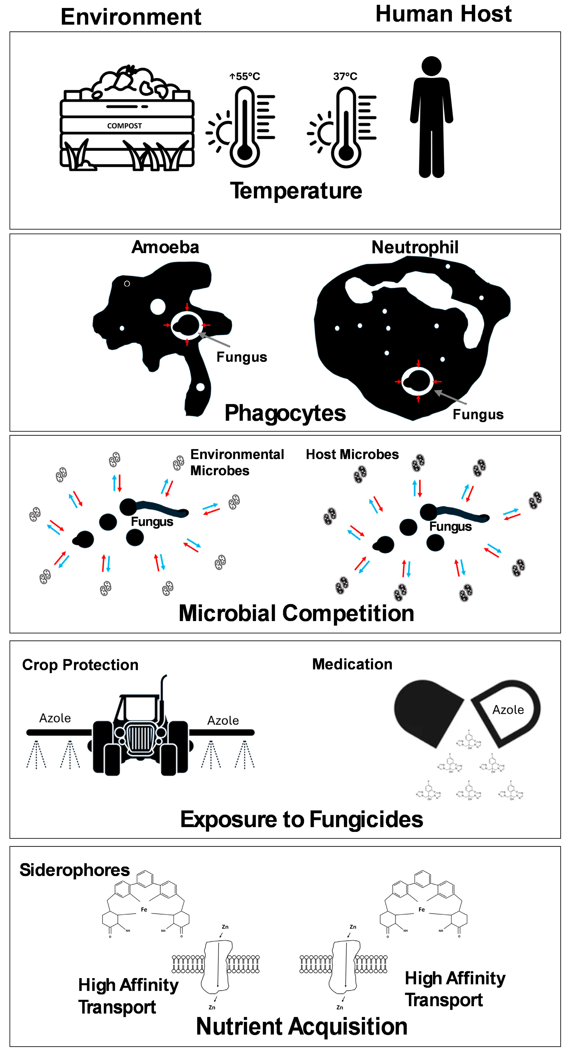
3. Virulence in Opportunist Pathogens

Unlike obligate pathogenic microbes, the majority of opportunistic pathogenic fungi, except those causing tropical mycoses and dermatophytes, do not exhibit the properties of infectious agents. Infections caused by opportunist pathogens are often governed by the immune status of the host. In the immunocompromised, the adaptive immune system is usually disrupted, leaving the innate immune system as the barrier to infection. Characteristics that enable survival in the environment can also be beneficial in adapting to the challenges presented by the host’s innate immune system, which includes body temperature, skin and mucosal surfaces, host microbiota, and macrophages (Figure 1). Using A. fumigatus as an example, this organism is often found in organic compost. Compost can reach relatively high temperatures (up to 55 °C); therefore, the fungus has developed thermotolerance, enabling it to survive at temperatures found in the human body (Figure 1) [41]. The fungus has adaptations to withstand predation by soil-dwelling phagocytic protozoa, which has resulted in adaptations that help the fungus to survive encounters with human phagocytic immune cells (Figure 1) [42]. A. fumigatus can survive within macrophages and emerge post-phagocytosis, and this is potentially a mechanism that allows the fungus to migrate from the site of initial infection to other host tissues [43]. Furthermore, the soil/compost environment imposes reduced levels of oxygen, creating hypoxic stress, to which the fungus has become adapted. This condition is also found within the human body where the internal organs have reduced oxygen and high CO2 levels, and in the lungs, there are regions of localised hypoxia that can be caused by inflammation or disease processes [44]. Adaptation to hypoxia has been linked to virulence in both A. fumigatus [44,45] and C. neoformans [46]. Fungi show metabolic adaptability with regards to glucose utilisation where fungi can be described as Crabtree-positive or Crabtree-negative. The Crabtree-positive fungi, usually yeasts, downregulate the conversion of pyruvate to acetyl-CoA when there is abundant glucose and switch to fermentation even in the presence of oxygen. The Crabtree-positive fungi upregulate the conversion of pyruvate to acetyl-CoA in order to drive more substrate through the tricarboxylic acid cycle, enabling high yields of ATP [47]. Typically, fungi may switch to ethanolic fermentation during hypoxia to reduce the requirement for oxygen [45]. This is necessary for survival, but it has been observed that hypoxic conditions have very little effect on antifungal sensitivity [48]. The widespread use of azole fungicides in agriculture has provided fungi, within these environments, the opportunity to experience the selective pressure required to develop azole resistance, which could result in fungi that cause human infection (Figure 1) [49]. The emergence of azole resistance in clinical isolates of A. fumigatus represents a significant challenge in the treatment of human aspergillosis [50]. A. fumigatus can produce secondary metabolites—many of which have antibiotic effects—that are inhibitory to competing microbes or predators, including toxins such as fumagillin and gliotoxin (Figure 1) [51,52]. Finally, the fungus must acquire nutrients from the environment and compete with other organisms for nutritional resources. For example, in low iron (Fe) environments, fungal and bacterial pathogens produce scavenging molecules called siderophores to facilitate the acquisition of Fe from the external environment (Figure 1) [53]. In humans, the resident microbiome found on mucosal surfaces competes with pathogens for resources. This provides an additional form of immunity to infection [54]. However, this protection is not present within the internal organs or bloodstream.
The overall virulence of fungal pathogens has recently been reviewed, giving a broader overview of pathogenesis that is complementary to the current review [55].

4. Nutritional Immunity

There is a form of nutritional immunity where essential micronutrients within the human are bound by host factors to reduce their availability to extremely low levels, limiting their availability to pathogens. In the case of Fe, it is mostly bound up in enzymes or to Fe-binding molecules such as ferritin or haemoglobin [56]. This greatly reduces the amount of free Fe available for uptake by pathogens. Fungal siderophores are effective within infected hosts, enabling pathogens to become established (Figure 1). As a result, the ability to acquire nutrients within the host can be considered a virulence factor. The ability to tolerate or overcome starvation within the host is an essential element of pathogenesis. However, nutritional immunity can also take the form of the increased concentration of essential metals, such as copper (Cu), where increased Cu concentrations are associated with the response to infection [57].

5. Nutrition and Pathogenesis

The goal of an opportunist pathogen is not to cause disease, but to colonise an environment, extract resources for growth and reproduction, and then disseminate to a new environment. Fungi acquire nutrients through the release of extracellular enzymes that act on their surrounding environment to break down complex substrates and release nutrients [58]. Increased nutrient acquisition can also be achieved through the production of secondary metabolites by the fungus, thereby inhibiting the growth of competing microbes in the environment [52]. The process of extracting nutrients coupled with the release of toxic secondary metabolites can be damaging to host tissues, leading to a disease state involving inflamed or necrotic lesions within tissue and organs [59].
Some of the enzymes released by fungal pathogens are beneficial in overcoming elements of the host’s innate immune system. The enzyme urease, produced by C. neoformans and A. fumigatus, can modulate the pH of phagolysosomes, preventing their acidification [60,61]. This step is important for the activation of lytic enzymes and their subsequent antimicrobial activity within the immune cell. Once disrupted, the pathogen can survive destruction and exit the immune cell to establish infection.

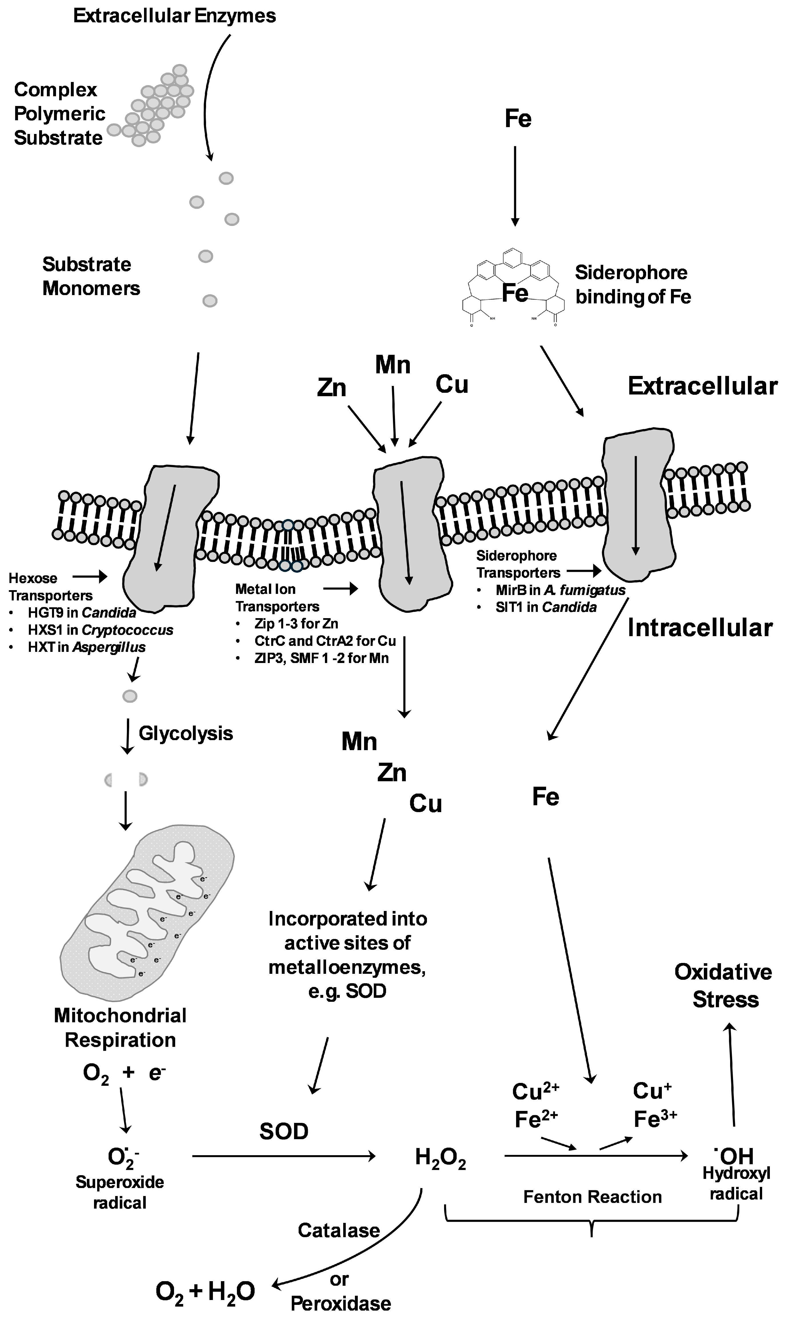
6. Macronutrient Acquisition

The ability of opportunistic fungal pathogens to obtain nutrients is a prerequisite to survival within the host. The major nutrients required, known as macronutrients, include carbon, nitrogen, and phosphorus. However, within the host, nutrient availability varies greatly; the skin is relatively inhospitable to many organisms [62], and even though nutrients are plentiful in the gut, nutrient acquisition can be inhibited by competition with the resident microbiome [63]. Carbohydrate metabolism has been extensively studied in the model yeast Saccharomyces cerevisiae and, more recently, in C. albicans [64,65,66]. When available, glucose is a simple and preferred source of carbon, with excess glucose stored as glycogen or trehalose. Fortunately, for the human host, many anatomical niches restrict glucose availability and, thus, the ability of opportunistic fungi to cause invasive infection [67]. However, this is not always the case, as evidenced by patients with diabetes in whom yeast infections are more common than in non-diabetic populations due to the reduced control of glucose levels [68,69]. For opportunists to become pathogenic, alternative carbon sources including the utilisation of molecules such as N-acetylglucosamine (GlcNAc) and carboxylic acids (lactate) are required, while proteins can double as a source of nitrogen and carbon [70]. Metabolic adaptability/plasticity is a key driver of infection, providing opportunistic fungi with a fitness advantage and allowing them to cause deep-seated infections, meaning that they should be viewed in the same way as other virulence factors. Furthermore, the central role that glycolysis plays in carbon and energy accumulation is of fundamental importance to survival and is responsible for producing the sugar precursors of the polysaccharides mannan and chitin that are vital for cell wall synthesis. It is therefore unsurprising that targeting carbohydrate metabolism represents an opportunity for therapeutic intervention—specifically, fungal glycogen metabolism. Several disruption studies have demonstrated the importance of glycogen metabolism. The disruption of genes such as glycogen synthase (gsy1∆/∆) resulted in long-term survival defects in C. albicans, impacting virulence under starvation conditions and in vivo during vaginal colonisation [64]. The same authors found that a global glycogen catabolism mutant (gph1 ∆/∆ [involved in accumulation], sga1∆/∆ [involved in utilisation]) was less virulent during invasive infection, but not lethal, suggesting that alternative carbon sources, e.g., trehalose, were consumed in vivo. A study by Kammer et al. (2020) highlighted the varying survival strategies of the four most common Candida species causing bloodstream infections [71]. Their analysis, using human blood, revealed rapid and significant transcriptional changes. These included the downregulation of glycolytic enzymes, the stable upregulation of the alternative glyoxylate cycle, and fermentative energy production. An interesting observation, from a therapeutic perspective, was that each Candida species approached the utilisation of alternative carbohydrates differently. The authors suggested that there was no universal fungal virulence strategy among the Candida species tested, and that pathogenesis could not be attributed to the regulation of individual genes but was likely more the culmination of several overlapping and redundant responses [71].
While access to these nutrients usually requires the digestion of complex substrates by fungi, this is mediated via the secretion of extracellular protease enzymes [72]. An important family of proteases includes alkaline serine proteases. These play a crucial role in the pathogenesis of invertebrate fungal pathogens, e.g., Pr1 produced by the insect pathogen Metarhizium anisopliae [73] and VCP1 from the nematode egg pathogen P. chlamydosporia [6]. Proteinase K, produced by the fungus Parengyodontium album [74], is a broad-spectrum protease used extensively for the digestion of cell walls, membranes, and tissues in nucleic acid isolation protocols. Its commercial use gives a clue to the role of these proteases in overcoming cellular barriers to infection and the digestion of tissue.
In human infections, A. fumigatus produces a serine protease, Alp1, that has a role in digesting the extracellular matrix of the pulmonary tissues [75] and in the development of fungal asthma [76]. An analysis of Alp1 deletion strains indicated that it is not required for virulence because of the upregulation of other proteases to replace its function [77]. It is not the only major serine protease that plays a role in pathogenesis: dipeptidylpeptidases (DPPs) produced by A. fumigatus have also been implicated in the binding and digestion of collagen and cytokines, which disrupts immune responses [78]. The DPP proteases are found in spore diffusate, indicating a role in the establishment of growth [79], and induce cytokine production in a murine model [80]. Proteases are important for other opportunistic fungal pathogens. An aspartic protease (Pep1p) was identified in C. neoformans that has a role in virulence. It was found that susceptible mice that produced antibodies against Pep1p have improved outcomes [81]. The secreted aspartic proteases of Candida spp. are important for several aspects of virulence from biofilm formation to epithelial cell invasion [82,83]. The release of broad-spectrum proteases by fungi in host tissues can also disrupt immune responses by digesting elements of the complement system [84] and eliciting inflammatory responses [85].
Phosphate is the third macronutrient for which environmental fungi have evolved acquisition strategies. In particular, mycorrhiza facilitate phosphate acquisition by plants and this is a central reason for the formation of mycorrhizal associations. The plant can signal phosphate starvation to the fungus, leading to increased phosphate uptake [86]. The main mechanism of phosphate uptake is the PHO system of genes, which was initially characterised in S. cerevisiae [87]. PHO-type systems have been identified in Cryptococcus [88], Candida [89], and A. fumigatus [90]. In opportunist pathogens, the PHO-type system becomes necessary for the acquisition of phosphates from host tissues. Phosphate uptake has been associated with virulence in Candida [91,92] and Cryptococcus [93], with these studies showing the PHO system being highly upregulated in virulence models or through mutational studies. PHO-regulated genes were also identified as upregulated during A. fumigatus interaction with dendritic cells [94], as well as evidence of the critical nature of PHO during C. neoformans interactions with macrophages [95]. Together, these studies indicate an important relationship between phosphate acquisition and pathogenesis.

7. Micronutrient Acquisition

Overcoming the aforementioned nutritional immunity is essential for the establishment of infection. This is achieved by sequestering metal micronutrients or, in some cases, through resistance to toxic levels of Cu. Metals are essential for basic cellular processes, including metabolism, growth, and replication. They play important roles as enzyme cofactors and assist in stress resistance [96]. Studies in several organisms using mutational analysis and infection models have revealed how essential these metals are for virulence.

7.1. Iron Acquisition

Fe is a well-studied nutrient associated with fungal pathogenesis. Fe availability is tightly regulated within the host to prevent infection. Fungal pathogens use several mechanisms to overcome this. One response to Fe deficiency is to remodel metabolism to reduce the need for Fe. In S. cerevisiae, the most obvious change is a shift from oxidative phosphorylation in the mitochondria, requiring Fe in the cytochromes of the electron transport chain, to fermentation, which is regulated by the mRNA-binding protein Cth2 [97,98]. Cth2 also decreases the expression of other Fe-consuming metabolic pathways such as amino acid biosynthesis and the tricarboxylic acid cycle. These metabolic pathways are also repressed by a Cth2 homolog in C. albicans during Fe deprivation [99,100].
In pathogens, three major mechanisms are important for Fe uptake: the production of siderophores to chelate Fe, the heme uptake pathway, and the reductive Fe assimilation system. Pathogenic fungi actively try to scavenge Fe from their growth environment using siderophores. In A. fumigatus, it was found that two extracellular siderophores, triacetylfusarinine C and desferriferricrocin, were necessary for virulence; this was achieved through the deletion of the L-ornithine-N5-monooxygenase (SidA) gene that catalyses the first step in siderophore biosynthesis [101]. In C. albicans, a transporter for Fe from siderophores was identified as important for the invasion of epithelial cells, indicating the importance of siderophore-scavenged Fe in pathogenesis [102]. Unlike A. fumigatus, C. albicans can cause septicaemia [103] and, in this environment, C. albicans uses other Fe sources [102]. In the bloodstream, C. albicans can lyse erythrocytes and has cell surface receptors for haemoglobin, which is taken up and digested to provide a source of Fe [104,105,106]. C. neoformans has been observed to use haemoglobin as a source of Fe with uptake through endocytosis, via the Chc1 protein, located putatively in the trans-Golgi network or endocytic vesicles [107]. The deletion of the chc1 gene resulted in the inability to incorporate Fe from hemin or haemoglobin. There was some indication that chc1 and other genes engaged in clathrin-mediated endocytosis have relevance to virulence because the deletion mutants lost the ability to grow at 37 °C and displayed reduced capsule size, and there was a reduction in melanin production; these traits have been corelated with virulence [107,108]. Within the oral cavity, epithelial cells store Fe as ferritin, and hyphal C. albicans can use this as a source of Fe during pathogenesis. While the mechanism of extracting Fe from the ferritin protein is not known, it is proposed that the binding of ferritin occurs via the surface protein Als3, with subsequent acidification and uptake of the released Fe via the reductive Fe system [109]. A similar system of host Fe acquisition has been proposed for C. neoformans. Macrophages that have phagocytosed C. neoformans show increased concentrations of intracellular ferritin, and an ability to extract Fe from ferritin would contribute to the survival of the fungus in the phagolysosome [110,111]. Identified in C. albicans but conserved among pathogenic fungi are the ferric reductase FRE1 and metal transporter SMF11 [112]. One major transcriptional regulator responsible for the response to Fe limitation in pathogens such as A. fumigatus and C. neoformans is HapX [113,114]. HapX leads to the upregulation of siderophore biosynthesis, and its deletion can result in virulence attenuation [113]. In yeast, the Fe response is regulated by Aft1 and Aft2 transcription factors [115].
An interesting case underscoring the importance of Fe in fungal pathogenesis was the increased incidence of Mucormycosis in patients receiving the Fe-chelating drug deferoxamine [116]. The Rhizopus spp. that caused the infection, which do not typically grow in human serum, showed enhanced growth when deferoxamine bound with Fe was present. It was subsequently demonstrated that the drug was acting as an Fe carrier for the fungus [117]. Other therapeutic Fe-chelating agents developed and incapable of being used as siderophores by Mucorales fungi have been shown to be protective in animal infection models [118].

7.2. Zinc Acquisition

Like Fe, limiting zinc (Zn) in the host can be an important defence mechanism against pathogens (nutritional immunity). Zn is required by all organisms as an important part of structural and catalytic proteins, with approximately 10% of the proteome requiring Zn [119]. Zinc-finger domain-containing proteins are the most abundant proteins displaying a range of biological functions, including protein degradation, signal transduction, and transcriptional regulation, with the structure of these proteins being stabilised by the Zn ions [120]. During infection, macrophages display decreased concentrations of Zn as a protective mechanism [121], and fungal virulence factors associated with overcoming Zn nutritional immunity in macrophages likely evolved from interactions with phagocytic amoebas such as Acanthamoeba castellanii [122]. Zn also plays an important role in the development of biofilms in the respiratory pathogen H. capsulatum [123].
A range of transporters are important in Zn homeostasis [119]. In C. gattii, Zap1P, which is highly conserved across the fungal kingdom, regulates the expression of three transporters, Zip1, Zip2, and Zip3 [124], with Zip 1 and 2 being important for Zn transport and virulence in Cryptococcus infection models [125]. The deletion of Zap1 in C. gattii led to an increase in reactive oxygen species (ROS) accumulation within the cells, causing increased oxidative stress [125]. The Zip3 gene is not involved in Zn homeostasis, although it is also important in ROS metabolism and stress response [126]. A second regulatory protein associated with Zn starvation, Zrg1, has been characterised in C. gattii and is conserved among Cryptococcus spp. [127]. Zrg1 regulates the expression of Zap1P and Zip1 and has also been found to regulate autophagic processes. Autophagy allows C. gattii to degrade host Zn-binding proteins, allowing the Zn to be redistributed to essential fungal Zn-binding proteins [127].
The genome of A. fumigatus encodes eight transporter genes associated with Zn uptake and regulation. Three of these transporters, ZrfA, ZrfB, and ZrfC, are regulated by the Zn homeostasis transcription factor ZafA [128] and have been examined [129,130]. ZrfA and ZrfB are important in Zn transport under acidic conditions [129,130]. ZrfC assists in the transport of Zn via the 310 amino acid protein Aspf2, an extracellular Zn scavenger, which acts in the same way as a siderophore [129,131,132]. Unlike siderophores, which are typically small molecules, the Aspf2 Zn scavenger (zincophore) is an extracellular protein. The Zn transporter zrfC was also expressed in the presence of the Zn/Mn-chelating calprotectin, released by neutrophils, as another mechanism by which Zn acquisition occurs in the face of the host immune response [133]. This distinction is noteworthy, as the bioavailability of Zn atoms in the interstitial spaces of host tissues is estimated to be six to seven magnitudes lower than in host cells [128].
C. albicans utilises orthologs of the A. fumigatus zincophore and transporter [134]. The Zn scavenger Pra1 is a protein identified in the secretome of C. albicans. Pra1 bound to Zn then interacts with the Zn transporter Zrt1 [134]. Similarly, candidalysin produced by C. albicans is important for Zn acquisition within the host [135]. Candidalysin is produced during the infection of epithelial cells, causing toxicity to the host and enabling the penetration of host cells where candidalysin sequesters Zn.

Zinc in Transcription Factors

Transcription factors can act on several genes, allowing adaptation to environmental signals, and Zn plays an essential role in the functioning of transcription factors [136]. Zn-finger transcription factors are common amongst fungi and control processes from filamentous growth in C. albicans by CZF1 [137] to protease expression in A. fumigatus through PrtT [138]. In opportunistic pathogenic fungi, there are >300 Zn-finger transcription factors in A. fumigatus, >100 in C. albicans, and >100 in C. gattii and C. neoformans [139]. In A. fumigatus, PacC is linked to the response to environmental pH and is also linked to host tissue invasion [140]. The PacC protein is a Zn-finger transcription factor that binds Zn atoms, allowing further interaction with DNA to regulate gene expression [141]. In C. neoformans, zinc-finger-containing transcription factors STE12α and STE12a are associated with the mating strains MATα and MATa, respectively. These STE12 transcription factors are important for virulence in C. neoformans, regulating capsule, phospholipase, and superoxide dismutase gene expression, all important factors in virulence [142,143]. Fe homeostasis is also regulated by the Zn-finger transcription factor Cir1 in C. neoformans [144]. These activities further reinforce the importance of Zn acquisition to pathogenic fungi.

7.3. Copper Acquisition

As with other metal micronutrients, Cu is an important enzyme cofactor facilitating mitochondrial electron transfer reactions, as well as the biosynthesis of melanin [145]. In A. fumigatus, Cu uptake is mediated by the transcription factor MAC1, which regulates four Cu transporters [146,147]. The transporters CtrC and CtrA2 are crucial, working synergistically under the conditions of Cu scarcity, and the deletion of mac1 leads to reduced pathogenicity in murine infection [146]. There is also overlap between the regulation of Zn and Cu homeostasis, with the Zn transcriptional regulator ZafA being able to affect Cu metabolism through the upregulation of CtrC and CtrA2 under the conditions of Zn starvation [148]. In C. albicans, in addition to the transporter CtrI, the ferric reductase Fre7 aids in uptake by reducing Cu, while the manganese (Mn) transporter SMF12 may also play a role in Cu transport [149]. MAC1 is also essential for Cu homeostasis and virulence in C. albicans, with deletion mutants displaying reduced infection in murine models and deficient mitochondrial respiration [150]. The C. albicans macI deletion mutant was also hypersensitive to ergosterol inhibitor fungicides, and was rescued by the addition of Cu to the media [149]. These deletion mutants have demonstrated that MAC1 is integral for host cell adhesion, although Cu supplementation to the media did not reverse the effect, indicating that MAC1 regulates other genes associated with adhesion not directly associated with Cu homeostasis [149]. MAC1 has a similar role in H. capsulatum, regulating the Cu transporter gene CTR3 and other genes important for the infection of phagocytes, with the deletion of mac1 decreasing intracellular macrophage proliferation [151]. Once transported across the membrane to the cytosol of the fungal pathogen, free Cu must be bound to prevent uncontrolled Fenton reactions (Figure 2) and non-specific binding to proteins. In the yeasts S. cerevisiae, N. glabrata, and C. neoformans, cysteine-rich metallothioneins are important in binding free Cu. Metallochaperones and intracellular carrier proteins are also important in transporting Cu to cuproenzymes and mitochondrial proteins or to mediate Cu storage [152].

Adaptation to High Copper Concentrations

Overcoming Cu limitation is important for fungal infection; however, high concentrations of Cu are also an important antimicrobial defence used by phagocytic cells such as macrophages [153,154]. Cu has been employed as an antimicrobial surface cleaning agent in healthcare settings where it is used on door handles to reduce fomite transmission [155]. Host Cu concentrations within serum and phagocytic cells have been demonstrated to rise during infection [57,154]. Therefore, understanding the response of fungal pathogens to increased Cu levels is of importance.
In addition to its role in Cu limitation, MAC1 is also important for survival under high Cu concentrations, where it regulates the expression of Cu efflux pumps as well as playing a role in the oxidative stress response (including the expression of catalase and superoxide dismutase (SOD)) in H. capsulatum [151]. Both adaptations are necessary for its survival within the host and phagolysosome of phagocytic cells. In A. fumigatus, MAC1 is important in regulating the efflux of Cu [146], while another transcription factor, AceA, is also capable of activating Cu efflux and resistance to Cu [156]. C. neoformans initiates infection in the lung, where Cu concentrations are high, before moving to the brain where Cu is limited, highlighting the need to understand the response to both high and low Cu concentrations. Within the host, C. neoformans possesses Cu-detoxification (CMT) genes that encode Cu-binding metallothioneins that are induced in the presence of increased Cu concentrations [157,158]. The deletion of CMT genes in C. neoformans leads to reduced virulence in a murine infection model [158]. This highlights the importance of understanding Cu homeostasis within the host and fungal pathogen during different stages of infection.

7.4. Manganese Acquisition

While a significant number of studies have investigated Fe, Cu, and Zn in pathogenesis and nutritional immunity, less is known about Mn. Like Fe, Mn is an important enzyme cofactor. Mn is also important for SOD activity, a vital response enabling pathogenic fungi to counter the oxidative stress produced as a defence mechanism by the host immune cells as well as during aerobic growth because of the Fenton reaction (Figure 2) [159]. In a murine model of infection, it was found that the deletion of Mn SOD led to a complete loss of virulence in C. neoformans [160]. Several catalytic enzymes also require Mn as an essential cofactor, with one of the most important being glycosyltransferases, specifically mannosyltransferases, which are important in processing proteins in the Golgi [161]. In C. albicans, a null mutant of Pmr1p, which transports Cu and Mn to the Golgi for transfer to glycosyltransferases, highlighted the importance of Mn-containing glycosyltransferases in maintaining the cell wall and virulence in C. albicans. In C. gattii, the Mn transporter ZIP3 is proposed to be localised to the membrane of the Golgi [126]. The deletion of ZIP3 led to imbalanced ROS metabolism, the secretion of the virulence factor GXM, and impaired virulence in a Galleria mellonella infection model, which may be the result of reduced cell wall melanisation.
As with other metal nutrients, Mn transporters are key for the survival of fungal pathogens under Mn starvation induced by the host. Interestingly, a study by Wildeman et al. (2023), which employed a mouse model of disseminated candidiasis, with the kidney as the primary target tissue, demonstrated the dramatic loss of total kidney Mn in response to infection, further highlighting the importance of the host’s restriction of this key transition metal [162]. Key to countering this during the invasion of host cells, C. albicans employs two natural resistance-associated macrophage protein (NRAMP) transporters, SMF12 and SMF13. The disruption of either gene, individually or in combination (smf12∆/∆ and smf1∆/∆ mutations), results in a dramatic (10–80-fold) reduction in cellular Mn. Furthermore, deletion mutants had impaired mitochondrial Mn-Sod2 and cytosolic Mn-Sod3 activities, defective hyphal formation, and attenuated virulence in a mouse model of disseminated candidiasis [162,163]. As mentioned previously, the Mn transporter SMF12 is also important in Cu starvation [149]. RNA-seq revealed that Mn starvation in C. albicans induced metabolic genes and Fe transporters, while the genes for Mn-containing mannosyltransferases and cell wall integrity proteins were downregulated [163]. In these Mn-starved cells, intracellular Fe levels were also reduced, a common phenomenon in other organisms. In addition, the virulence factors required for C. albicans to initiate hyphal growth and the production of candidalysin were induced [163]. Interestingly, Mn-replete conditions also moderately increased sensitivity to antifungals. The chelation of Mn by neutrophils, the predominant cell type in corneal ulcers, was mediated by the protein calprotectin, which caused the inhibition of A. fumigatus hyphal growth [164]. There is overlap in the uptake of Fe and Mn, with the transcription factor SreA playing an important role in the regulation of Fe uptake in A. fumigatus [165], as well as being involved in Mn uptake and tolerance [166].

8. Conclusions

The ability to acquire nutrients from the host is essential for the establishment of fungal infections. The limitation of any single macronutrient or metal micronutrient (Fe, Zn, Mn, and Cu) can impact viability and prevent infection, while some human tissues can use metal toxicity as an alternative resistance mechanism. Although pathogens can usually overcome nutritional immunity, the development of agents that can disrupt specific mechanisms of nutrient acquisition or homeostasis within the pathogen could help to prevent fungal infection. Given the limited classes of antifungal drugs available [167], metal ion homeostasis in pathogenic fungi could become a novel target for future antifungal drug development [168,169]. The importance of nutrition in fungal pathogenesis also highlights the necessity of understanding the basic biology of opportunist fungal pathogens in their natural habitats to explain their response to the infected host. Further study of fungal biology, including reproduction and metabolism, has the potential to reveal new avenues for the development of therapeutics or the removal of these pathogens from healthcare settings.

Author Contributions

T.J.: conception, literature search, and writing of the review. C.M.S.: conception, writing, and editing of the review. M.C.M.: conception, writing, and editing of the review. C.O.M.: conception, literature search, writing, and editing of the review. All authors have read and agreed to the published version of the manuscript.

Funding

This research received no external funding.

Institutional Review Board Statement

Not applicable to this narrative review.

Informed Consent Statement

Not applicable to this narrative review.

Data Availability Statement

No new data was generated for this narrative review.

Acknowledgments

We acknowledge Western Sydney University Graduate Research School for supporting Timothy Jong during his Master of Research programme.

Conflicts of Interest

The authors declare no conflicts of interest.

References

  1. Denning, D.W. Global incidence and mortality of severe fungal disease. Lancet Infect. Dis. 2024, 24, e428–e438. [Google Scholar] [CrossRef] [PubMed]
  2. Park, B.J.; Wannemuehler, K.A.; Marston, B.J.; Govender, N.; Pappas, P.G.; Chiller, T.M. Estimation of the current global burden of cryptococcal meningitis among persons living with HIV/AIDS. AIDS 2009, 23, 525–530. [Google Scholar] [CrossRef]
  3. Liu, S.; Garcia-Palacios, P.; Tedersoo, L.; Guirado, E.; van der Heijden, M.G.A.; Wagg, C.; Chen, D.; Wang, Q.; Wang, J.; Singh, B.K.; et al. Phylotype diversity within soil fungal functional groups drives ecosystem stability. Nat. Ecol. Evol. 2022, 6, 900–909. [Google Scholar] [CrossRef]
  4. Smith, S.E.; Jakobsen, I.; Gronlund, M.; Smith, F.A. Roles of arbuscular mycorrhizas in plant phosphorus nutrition: Interactions between pathways of phosphorus uptake in arbuscular mycorrhizal roots have important implications for understanding and manipulating plant phosphorus acquisition. Plant Physiol. 2011, 156, 1050–1057. [Google Scholar] [CrossRef] [PubMed]
  5. Huang, J.; Zheng, X.; Tian, M.; Zhang, K. Ammonia and Nematode Ascaroside Are Synergistic in Trap Formation in Arthrobotrys oligospora. Pathogens 2023, 12, 1114. [Google Scholar] [CrossRef]
  6. Segers, R.; Butt, T.M.; Kerry, B.R.; Peberdy, J.F. The nematophagous fungus Verticillium chlamydosporium produces a chymoelastase-like protease which hydrolyses host nematode proteins in situ. Microbiology 1994, 140 Pt 10, 2715–2723. [Google Scholar] [CrossRef] [PubMed]
  7. St Leger, R.J.; Wang, J.B. Metarhizium: Jack of all trades, master of many. Open Biol. 2020, 10, 200307. [Google Scholar] [CrossRef]
  8. Kim, S.W.; Kim, S.; Lee, H.J.; Park, J.W.; Ro, H.S. Isolation of Fungal Pathogens to an Edible Mushroom, Pleurotus eryngii, and Development of Specific ITS Primers. Mycobiology 2013, 41, 252–255. [Google Scholar] [CrossRef]
  9. Kumamoto, C.A.; Gresnigt, M.S.; Hube, B. The gut, the bad and the harmless: Candida albicans as a commensal and opportunistic pathogen in the intestine. Curr. Opin. Microbiol. 2020, 56, 7–15. [Google Scholar] [CrossRef]
  10. Chai, Y.; Pardey, P.G.; Hurley, T.M.; Senay, S.D.; Beddow, J.M. A Probabilistic Bio-Economic Assessment of the Global Consequences of Wheat Leaf Rust. Phytopathology 2020, 110, 1886–1896. [Google Scholar] [CrossRef]
  11. Frick, W.F.; Pollock, J.F.; Hicks, A.C.; Langwig, K.E.; Reynolds, D.S.; Turner, G.G.; Butchkoski, C.M.; Kunz, T.H. An emerging disease causes regional population collapse of a common North American bat species. Science 2010, 329, 679–682. [Google Scholar] [CrossRef] [PubMed]
  12. Olson, D.H.; Aanensen, D.M.; Ronnenberg, K.L.; Powell, C.I.; Walker, S.F.; Bielby, J.; Garner, T.W.; Weaver, G.; Bd Mapping, G.; Fisher, M.C. Mapping the global emergence of Batrachochytrium dendrobatidis, the amphibian chytrid fungus. PLoS ONE 2013, 8, e56802. [Google Scholar] [CrossRef]
  13. Havlickova, B.; Czaika, V.A.; Friedrich, M. Epidemiological trends in skin mycoses worldwide. Mycoses 2008, 51 (Suppl. S4), 2–15. [Google Scholar] [CrossRef] [PubMed]
  14. Deng, R.; Wang, X.; Li, R. Dermatophyte infection: From fungal pathogenicity to host immune responses. Front. Immunol. 2023, 14, 1285887. [Google Scholar] [CrossRef]
  15. Bonifaz, A.; Vazquez-Gonzalez, D.; Perusquia-Ortiz, A.M. Endemic systemic mycoses: Coccidioidomycosis, histoplasmosis, paracoccidioidomycosis and blastomycosis. J. Dtsch. Dermatol. Ges. 2011, 9, 705–714. [Google Scholar] [CrossRef] [PubMed]
  16. de Hoog, S.; Tang, C.; Zhou, X.; Jacomel, B.; Lustosa, B.; Song, Y.; Kandemir, H.; Ahmed, S.A.; Zhou, S.; Belmonte-Lopes, R.; et al. Fungal primary and opportunistic pathogens: An ecological perspective. FEMS Microbiol. Rev. 2024, 48, fuae022. [Google Scholar] [CrossRef]
  17. Schwartz, R.A. Superficial fungal infections. Lancet 2004, 364, 1173–1182. [Google Scholar] [CrossRef]
  18. La Hoz, R.M.; Baddley, J.W. Subcutaneous Fungal Infections. Curr. Infect. Dis. Rep. 2012, 14, 530–539. [Google Scholar] [CrossRef]
  19. Thompson, G.R., 3rd; Le, T.; Chindamporn, A.; Kauffman, C.A.; Alastruey-Izquierdo, A.; Ampel, N.M.; Andes, D.R.; Armstrong-James, D.; Ayanlowo, O.; Baddley, J.W.; et al. Global guideline for the diagnosis and management of the endemic mycoses: An initiative of the European Confederation of Medical Mycology in cooperation with the International Society for Human and Animal Mycology. Lancet Infect. Dis. 2021, 21, e364–e374. [Google Scholar] [CrossRef]
  20. Rautemaa-Richardson, R.; Richardson, M.D. Systemic fungal infections. Medicine 2017, 45, 757–762. [Google Scholar]
  21. Oliveira, M.; Oliveira, D.; Lisboa, C.; Boechat, J.L.; Delgado, L. Clinical Manifestations of Human Exposure to Fungi. J. Fungi 2023, 9, 381. [Google Scholar] [CrossRef] [PubMed]
  22. Stack, C.M.; Morton, C.O. Diagnostics for Fungal Infections in Solid Organ Transplants (SOT). Curr. Fungal Infect. R. 2021, 15, 127–135. [Google Scholar] [CrossRef]
  23. Hope, W.W.; Walsh, T.J.; Denning, D.W. The invasive and saprophytic syndromes due to Aspergillus spp. Med. Mycol. 2005, 43 (Suppl. S1), S207–S238. [Google Scholar] [CrossRef]
  24. Shahhosseini, N.; Khabiri, A.; Bagheri, F. The spectrum of diseases caused by Aspergillus fumigatus. Iran. J. Clin. Infect. Dis. 2011, 6, 136–141. [Google Scholar]
  25. Kidd, S.E.; Abdolrasouli, A.; Hagen, F. Fungal Nomenclature: Managing Change is the Name of the Game. Open Forum Infect. Dis. 2023, 10, ofac559. [Google Scholar] [CrossRef]
  26. Borman, A.M.; Johnson, E.M. Name Changes for Fungi of Medical Importance, 2018 to 2019. J. Clin. Microbiol. 2021, 59, 10-1128. [Google Scholar] [CrossRef]
  27. Borman, A.M.; Johnson, E.M. Name Changes for Fungi of Medical Importance, 2020 to 2021. J. Clin. Microbiol. 2023, 61, e0033022. [Google Scholar] [CrossRef]
  28. Kruithoff, C.; Gamal, A.; McCormick, T.S.; Ghannoum, M.A. Dermatophyte Infections Worldwide: Increase in Incidence and Associated Antifungal Resistance. Life 2023, 14, 1. [Google Scholar] [CrossRef]
  29. Wang, R.; Huang, C.; Zhang, Y.; Li, R. Invasive dermatophyte infection: A systematic review. Mycoses 2021, 64, 340–348. [Google Scholar] [CrossRef]
  30. Batard, E.; Renaudin, K.; Morin, O.; Desjars, P.; Germaud, P. Fatal acute granulomatous pulmonary aspergillosis in a healthy subject after inhalation of vegetal dust. Eur. J. Clin. Microbiol. Infect. Dis. 2003, 22, 357–359. [Google Scholar] [CrossRef]
  31. Zuk, J.A.; King, D.; Zakhour, H.D.; Delaney, J.C. Locally invasive pulmonary aspergillosis occurring in a gardener: An occupational hazard? Thorax 1989, 44, 678–679. [Google Scholar] [CrossRef] [PubMed]
  32. Galanis, E.; Macdougall, L.; Kidd, S.; Morshed, M.; British Columbia Cryptococcus gattii Working, G. Epidemiology of Cryptococcus gattii, British Columbia, Canada, 1999–2007. Emerg. Infect. Dis. 2010, 16, 251–257. [Google Scholar] [CrossRef]
  33. Bartlett, K.H.; Cheng, P.Y.; Duncan, C.; Galanis, E.; Hoang, L.; Kidd, S.; Lee, M.K.; Lester, S.; MacDougall, L.; Mak, S.; et al. A decade of experience: Cryptococcus gattii in British Columbia. Mycopathologia 2012, 173, 311–319. [Google Scholar] [CrossRef]
  34. Perfect, J.R.; Casadevall, A. Cryptococcosis. Infect. Dis. Clin. N. Am. 2002, 16, 837–874. [Google Scholar] [CrossRef]
  35. Ascioglu, S.; Rex, J.H.; de Pauw, B.; Bennett, J.E.; Bille, J.; Crokaert, F.; Denning, D.W.; Donnelly, J.P.; Edwards, J.E.; Erjavec, Z.; et al. Defining opportunistic invasive fungal infections in immunocompromised patients with cancer and hematopoietic stem cell transplants: An international consensus. Clin. Infect. Dis. 2002, 34, 7–14. [Google Scholar] [CrossRef]
  36. Hope, W.; Natarajan, P.; Goodwin, L. Invasive fungal infections. Clin. Med. 2013, 13, 507–510. [Google Scholar] [CrossRef] [PubMed]
  37. Hoenigl, M. Invasive Fungal Disease Complicating Coronavirus Disease 2019: When It Rains, It Spores. Clin. Infect. Dis. 2021, 73, e1645–e1648. [Google Scholar] [CrossRef]
  38. Yan, H.; Guo, L.; Pang, Y.; Liu, F.; Liu, T.; Gao, M. Clinical characteristics and predictive model of pulmonary tuberculosis patients with pulmonary fungal coinfection. BMC Pulm. Med. 2023, 23, 56. [Google Scholar] [CrossRef]
  39. Badiee, P.; Hashemizadeh, Z. Opportunistic invasive fungal infections: Diagnosis & clinical management. Indian. J. Med. Res. 2014, 139, 195–204. [Google Scholar]
  40. Gnat, S.; Lagowski, D.; Nowakiewicz, A.; Dylag, M. A global view on fungal infections in humans and animals: Opportunistic infections and microsporidioses. J. Appl. Microbiol. 2021, 131, 2095–2113. [Google Scholar] [CrossRef]
  41. Beffa, T.; Staib, F.; Lott Fischer, J.; Lyon, P.F.; Gumowski, P.; Marfenina, O.E.; Dunoyer-Geindre, S.; Georgen, F.; Roch-Susuki, R.; Gallaz, L.; et al. Mycological control and surveillance of biological waste and compost. Med. Mycol. 1998, 36 (Suppl. S1), 137–145. [Google Scholar] [PubMed]
  42. Novohradska, S.; Ferling, I.; Hillmann, F. Exploring Virulence Determinants of Filamentous Fungal Pathogens through Interactions with Soil Amoebae. Front. Cell Infect. Microbiol. 2017, 7, 497. [Google Scholar] [CrossRef]
  43. Slesiona, S.; Gressler, M.; Mihlan, M.; Zaehle, C.; Schaller, M.; Barz, D.; Hube, B.; Jacobsen, I.D.; Brock, M. Persistence versus escape: Aspergillus terreus and Aspergillus fumigatus employ different strategies during interactions with macrophages. PLoS ONE 2012, 7, e31223. [Google Scholar] [CrossRef]
  44. Willger, S.D.; Puttikamonkul, S.; Kim, K.H.; Burritt, J.B.; Grahl, N.; Metzler, L.J.; Barbuch, R.; Bard, M.; Lawrence, C.B.; Cramer, R.A., Jr. A sterol-regulatory element binding protein is required for cell polarity, hypoxia adaptation, azole drug resistance, and virulence in Aspergillus fumigatus. PLoS Pathog. 2008, 4, e1000200. [Google Scholar] [CrossRef]
  45. Grahl, N.; Puttikamonkul, S.; Macdonald, J.M.; Gamcsik, M.P.; Ngo, L.Y.; Hohl, T.M.; Cramer, R.A. In vivo hypoxia and a fungal alcohol dehydrogenase influence the pathogenesis of invasive pulmonary aspergillosis. PLoS Pathog. 2011, 7, e1002145. [Google Scholar] [CrossRef]
  46. Chun, C.D.; Liu, O.W.; Madhani, H.D. A link between virulence and homeostatic responses to hypoxia during infection by the human fungal pathogen Cryptococcus neoformans. PLoS Pathog. 2007, 3, e22. [Google Scholar] [CrossRef]
  47. Fleck, C.B.; Schobel, F.; Brock, M. Nutrient acquisition by pathogenic fungi: Nutrient availability, pathway regulation, and differences in substrate utilization. Int. J. Med. Microbiol. 2011, 301, 400–407. [Google Scholar] [CrossRef]
  48. Warn, P.A.; Sharp, A.; Guinea, J.; Denning, D.W. Effect of hypoxic conditions on in vitro susceptibility testing of amphotericin B, itraconazole and micafungin against Aspergillus and Candida. J. Antimicrob. Chemother. 2004, 53, 743–749. [Google Scholar] [CrossRef]
  49. Ren, J.; Jin, X.; Zhang, Q.; Zheng, Y.; Lin, D.; Yu, Y. Fungicides induced triazole-resistance in Aspergillus fumigatus associated with mutations of TR46/Y121F/T289A and its appearance in agricultural fields. J. Hazard. Mater. 2017, 326, 54–60. [Google Scholar] [CrossRef]
  50. Verweij, P.E.; Snelders, E.; Kema, G.H.; Mellado, E.; Melchers, W.J. Azole resistance in Aspergillus fumigatus: A side-effect of environmental fungicide use? Lancet Infect. Dis. 2009, 9, 789–795. [Google Scholar] [CrossRef]
  51. Stevanovic, J.; Stanimirovic, Z.; Radakovic, M.; Stojic, V. In vitro evaluation of the clastogenicity of fumagillin. Environ. Mol. Mutagen. 2008, 49, 594–601. [Google Scholar] [CrossRef] [PubMed]
  52. Svahn, K.S.; Goransson, U.; Chryssanthou, E.; Olsen, B.; Sjolin, J.; Stromstedt, A.A. Induction of gliotoxin secretion in Aspergillus fumigatus by bacteria-associated molecules. PLoS ONE 2014, 9, e93685. [Google Scholar] [CrossRef]
  53. Haas, H.; Eisendle, M.; Turgeon, B.G. Siderophores in fungal physiology and virulence. Annu. Rev. Phytopathol. 2008, 46, 149–187. [Google Scholar] [CrossRef] [PubMed]
  54. Chow, E.W.L.; Pang, L.M.; Wang, Y. Impact of the host microbiota on fungal infections: New possibilities for intervention? Adv. Drug Deliv. Rev. 2023, 198, 114896. [Google Scholar] [CrossRef] [PubMed]
  55. Brown, G.D.; Ballou, E.R.; Bates, S.; Bignell, E.M.; Borman, A.M.; Brand, A.C.; Brown, A.J.P.; Coelho, C.; Cook, P.C.; Farrer, R.A.; et al. The pathobiology of human fungal infections. Nat. Rev. Microbiol. 2024, 22, 687–704. [Google Scholar] [CrossRef]
  56. Hood, M.I.; Skaar, E.P. Nutritional immunity: Transition metals at the pathogen-host interface. Nat. Rev. Microbiol. 2012, 10, 525–537. [Google Scholar] [CrossRef]
  57. Mandarano, A.H.; McGargill, M.A. The critical role of copper homeostasis during the immune response. J. Immunol. 2023, 210, 148.113. [Google Scholar] [CrossRef]
  58. Richards, T.A.; Talbot, N.J. Osmotrophy. Curr. Biol. 2018, 28, R1179–R1180. [Google Scholar] [CrossRef]
  59. Stergiopoulou, T.; Meletiadis, J.; Roilides, E.; Kleiner, D.E.; Schaufele, R.; Roden, M.; Harrington, S.; Dad, L.; Segal, B.; Walsh, T.J. Host-dependent patterns of tissue injury in invasive pulmonary aspergillosis. Am. J. Clin. Pathol. 2007, 127, 349–355. [Google Scholar] [CrossRef]
  60. Fu, M.S.; Coelho, C.; De Leon-Rodriguez, C.M.; Rossi, D.C.P.; Camacho, E.; Jung, E.H.; Kulkarni, M.; Casadevall, A. Cryptococcus neoformans urease affects the outcome of intracellular pathogenesis by modulating phagolysosomal pH. PLoS Pathog. 2018, 14, e1007144. [Google Scholar] [CrossRef]
  61. Xiong, Z.; Zhang, N.; Xu, L.; Deng, Z.; Limwachiranon, J.; Guo, Y.; Han, Y.; Yang, W.; Scharf, D.H. Urease of Aspergillus fumigatus Is Required for Survival in Macrophages and Virulence. Microbiol. Spectr. 2023, 11, e0350822. [Google Scholar] [CrossRef]
  62. Scott, J.A.; Untereiner, W.A. Determination of keratin degradation by fungi using keratin azure. Med. Mycol. 2004, 42, 239–246. [Google Scholar] [CrossRef] [PubMed]
  63. Kruger, W.; Vielreicher, S.; Kapitan, M.; Jacobsen, I.D.; Niemiec, M.J. Fungal-Bacterial Interactions in Health and Disease. Pathogens 2019, 8, 70. [Google Scholar] [CrossRef] [PubMed]
  64. Miao, J.; Regan, J.; Cai, C.; Palmer, G.E.; Williams, D.L.; Kruppa, M.D.; Peters, B.M. Glycogen Metabolism in Candida albicans Impacts Fitness and Virulence during Vulvovaginal and Invasive Candidiasis. mBio 2023, 14, e0004623. [Google Scholar] [CrossRef] [PubMed]
  65. Ries, L.N.A.; Beattie, S.; Cramer, R.A.; Goldman, G.H. Overview of carbon and nitrogen catabolite metabolism in the virulence of human pathogenic fungi. Mol. Microbiol. 2018, 107, 277–297. [Google Scholar] [CrossRef]
  66. Wilson, W.A.; Roach, P.J.; Montero, M.; Baroja-Fernandez, E.; Munoz, F.J.; Eydallin, G.; Viale, A.M.; Pozueta-Romero, J. Regulation of glycogen metabolism in yeast and bacteria. FEMS Microbiol. Rev. 2010, 34, 952–985. [Google Scholar] [CrossRef]
  67. Man, A.; Ciurea, C.N.; Pasaroiu, D.; Savin, A.I.; Toma, F.; Sular, F.; Santacroce, L.; Mare, A. New perspectives on the nutritional factors influencing growth rate of Candida albicans in diabetics. An in vitro study. Mem. Inst. Oswaldo Cruz 2017, 112, 587–592. [Google Scholar] [CrossRef]
  68. Balan, P.; Gogineni, S.B.; Kumari, N.S.; Shetty, V.; Rangare, A.L.; Castelino, R.L.; Areekat, K.F. Candida Carriage Rate and Growth Characteristics of Saliva in Diabetes Mellitus Patients: A Case-Control Study. J. Dent. Res. Dent. Clin. Dent. Prospect 2015, 9, 274–279. [Google Scholar] [CrossRef]
  69. Dornelas Figueira, L.M.; Ricomini Filho, A.P.; da Silva, W.J.; Del Be, L.C.A.A.; Ruiz, K.G.S. Glucose effect on Candida albicans biofilm during tissue invasion. Arch. Oral. Biol. 2020, 117, 104728. [Google Scholar] [CrossRef]
  70. Ene, I.V.; Brunke, S.; Brown, A.J.; Hube, B. Metabolism in fungal pathogenesis. Cold Spring Harb. Perspect. Med. 2014, 4, a019695. [Google Scholar] [CrossRef]
  71. Kammer, P.; McNamara, S.; Wolf, T.; Conrad, T.; Allert, S.; Gerwien, F.; Hunniger, K.; Kurzai, O.; Guthke, R.; Hube, B.; et al. Survival Strategies of Pathogenic Candida Species in Human Blood Show Independent and Specific Adaptations. mBio 2020, 11, 10-1128. [Google Scholar] [CrossRef]
  72. Ortiz-Ramirez, J.A.; Cuellar-Cruz, M.; Villagomez-Castro, J.C.; Lopez-Romero, E. Fungal Glycosidases in Sporothrix Species and Candida albicans. J. Fungi 2023, 9, 919. [Google Scholar] [CrossRef]
  73. Wang, C.; Typas, M.A.; Butt, T.M. Detection and characterisation of pr1 virulent gene deficiencies in the insect pathogenic fungus Metarhizium anisopliae. FEMS Microbiol. Lett. 2002, 213, 251–255. [Google Scholar] [CrossRef] [PubMed]
  74. Jany, K.D.; Mayer, B. Proteinase K from Tritirachium album limber. I. Molecular mass and sequence around the active site serine residue. Biol. Chem. Hoppe Seyler 1985, 366, 485–492. [Google Scholar] [CrossRef] [PubMed]
  75. Tomee, J.F.; Wierenga, A.T.; Hiemstra, P.S.; Kauffman, H.K. Proteases from Aspergillus fumigatus induce release of proinflammatory cytokines and cell detachment in airway epithelial cell lines. J. Infect. Dis. 1997, 176, 300–303. [Google Scholar] [CrossRef]
  76. Basu, T.; Seyedmousavi, S.; Sugui, J.A.; Balenga, N.; Zhao, M.; Kwon Chung, K.J.; Biardel, S.; Laviolette, M.; Druey, K.M. Aspergillus fumigatus alkaline protease 1 (Alp1/Asp f13) in the airways correlates with asthma severity. J. Allergy Clin. Immunol. 2018, 141, 423–425. [Google Scholar] [CrossRef]
  77. Bergmann, A.; Hartmann, T.; Cairns, T.; Bignell, E.M.; Krappmann, S. A regulator of Aspergillus fumigatus extracellular proteolytic activity is dispensable for virulence. Infect. Immun. 2009, 77, 4041–4050. [Google Scholar] [CrossRef]
  78. Beauvais, A.; Monod, M.; Debeaupuis, J.P.; Diaquin, M.; Kobayashi, H.; Latge, J.P. Biochemical and antigenic characterization of a new dipeptidyl-peptidase isolated from Aspergillus fumigatus. J. Biol. Chem. 1997, 272, 6238–6244. [Google Scholar] [CrossRef]
  79. Portaels, J.; Van Crombrugge, E.; Van Den Broeck, W.; Lagrou, K.; Laval, K.; Nauwynck, H. Aspergillus Fumigatus Spore Proteases Alter the Respiratory Mucosa Architecture and Facilitate Equine Herpesvirus 1 Infection. Viruses 2024, 16, 1208. [Google Scholar] [CrossRef]
  80. Bozza, S.; Clavaud, C.; Giovannini, G.; Fontaine, T.; Beauvais, A.; Sarfati, J.; D’Angelo, C.; Perruccio, K.; Bonifazi, P.; Zagarella, S.; et al. Immune sensing of Aspergillus fumigatus proteins, glycolipids, and polysaccharides and the impact on Th immunity and vaccination. J. Immunol. 2009, 183, 2407–2414. [Google Scholar] [CrossRef]
  81. Vernel-Pauillac, F.; Laurent-Winter, C.; Fiette, L.; Janbon, G.; Aimanianda, V.; Dromer, F. Cryptococcus neoformans infections: Aspartyl protease potential to improve outcome in susceptible hosts. mBio 2024, 15, e0273324. [Google Scholar] [CrossRef]
  82. Naglik, J.R.; Challacombe, S.J.; Hube, B. Candida albicans secreted aspartyl proteinases in virulence and pathogenesis. Microbiol. Mol. Biol. Rev. 2003, 67, 400–428. [Google Scholar] [CrossRef] [PubMed]
  83. Naglik, J.R.; Rodgers, C.A.; Shirlaw, P.J.; Dobbie, J.L.; Fernandes-Naglik, L.L.; Greenspan, D.; Agabian, N.; Challacombe, S.J. Differential expression of Candida albicans secreted aspartyl proteinase and phospholipase B genes in humans correlates with active oral and vaginal infections. J. Infect. Dis. 2003, 188, 469–479. [Google Scholar] [CrossRef]
  84. Behnsen, J.; Lessing, F.; Schindler, S.; Wartenberg, D.; Jacobsen, I.D.; Thoen, M.; Zipfel, P.F.; Brakhage, A.A. Secreted Aspergillus fumigatus protease Alp1 degrades human complement proteins C3, C4, and C5. Infect. Immun. 2010, 78, 3585–3594. [Google Scholar] [CrossRef] [PubMed]
  85. Redes, J.L.; Basu, T.; Ram-Mohan, S.; Ghosh, C.C.; Chan, E.C.; Sek, A.C.; Zhao, M.; Krishnan, R.; Rosenberg, H.F.; Druey, K.M. Aspergillus fumigatus-Secreted Alkaline Protease 1 Mediates Airways Hyperresponsiveness in Severe Asthma. Immunohorizons 2019, 3, 368–377. [Google Scholar] [CrossRef]
  86. Muller, L.M.; Harrison, M.J. Phytohormones, miRNAs, and peptide signals integrate plant phosphorus status with arbuscular mycorrhizal symbiosis. Curr. Opin. Plant Biol. 2019, 50, 132–139. [Google Scholar] [CrossRef]
  87. Secco, D.; Wang, C.; Shou, H.; Whelan, J. Phosphate homeostasis in the yeast Saccharomyces cerevisiae, the key role of the SPX domain-containing proteins. FEBS Lett. 2012, 586, 289–295. [Google Scholar] [CrossRef]
  88. Toh-e, A.; Ohkusu, M.; Li, H.M.; Shimizu, K.; Takahashi-Nakaguchi, A.; Gonoi, T.; Kawamoto, S.; Kanesaki, Y.; Yoshikawa, H.; Nishizawa, M. Identification of genes involved in the phosphate metabolism in Cryptococcus neoformans. Fungal Genet. Biol. 2015, 80, 19–30. [Google Scholar] [CrossRef]
  89. Ikeh, M.A.; Kastora, S.L.; Day, A.M.; Herrero-de-Dios, C.M.; Tarrant, E.; Waldron, K.J.; Banks, A.P.; Bain, J.M.; Lydall, D.; Veal, E.A.; et al. Pho4 mediates phosphate acquisition in Candida albicans and is vital for stress resistance and metal homeostasis. Mol. Biol. Cell 2016, 27, 2784–2801. [Google Scholar] [CrossRef]
  90. de Gouvea, P.F.; Soriani, F.M.; Malavazi, I.; Savoldi, M.; Goldman, M.H.; Loss, O.; Bignell, E.; da Silva Ferreira, M.E.; Goldman, G.H. Functional characterization of the Aspergillus fumigatusPHO80 homologue. Fungal Genet. Biol. 2008, 45, 1135–1146. [Google Scholar] [CrossRef]
  91. Thewes, S.; Kretschmar, M.; Park, H.; Schaller, M.; Filler, S.G.; Hube, B. In vivo and ex vivo comparative transcriptional profiling of invasive and non-invasive Candida albicans isolates identifies genes associated with tissue invasion. Mol. Microbiol. 2007, 63, 1606–1628. [Google Scholar] [CrossRef] [PubMed]
  92. Urrialde, V.; Prieto, D.; Pla, J.; Alonso-Monge, R. The Candida albicans Pho4 Transcription Factor Mediates Susceptibility to Stress and Influences Fitness in a Mouse Commensalism Model. Front. Microbiol. 2016, 7, 1062. [Google Scholar] [CrossRef]
  93. Lev, S.; Kaufman-Francis, K.; Desmarini, D.; Juillard, P.G.; Li, C.; Stifter, S.A.; Feng, C.G.; Sorrell, T.C.; Grau, G.E.; Bahn, Y.S.; et al. Pho4 Is Essential for Dissemination of Cryptococcus neoformans to the Host Brain by Promoting Phosphate Uptake and Growth at Alkaline pH. mSphere 2017, 2, 10-1128. [Google Scholar] [CrossRef]
  94. Morton, C.O.; Varga, J.J.; Hornbach, A.; Mezger, M.; Sennefelder, H.; Kneitz, S.; Kurzai, O.; Krappmann, S.; Einsele, H.; Nierman, W.C.; et al. The temporal dynamics of differential gene expression in Aspergillus fumigatus interacting with human immature dendritic cells in vitro. PLoS ONE 2011, 6, e16016. [Google Scholar] [CrossRef]
  95. Fan, W.; Kraus, P.R.; Boily, M.J.; Heitman, J. Cryptococcus neoformans gene expression during murine macrophage infection. Eukaryot. Cell 2005, 4, 1420–1433. [Google Scholar] [CrossRef]
  96. Weiss, G.; Carver, P.L. Role of divalent metals in infectious disease susceptibility and outcome. Clin. Microbiol. Infect. 2018, 24, 16–23. [Google Scholar] [CrossRef] [PubMed]
  97. Philpott, C.C.; Leidgens, S.; Frey, A.G. Metabolic remodeling in iron-deficient fungi. Biochim. Biophys. Acta 2012, 1823, 1509–1520. [Google Scholar] [CrossRef]
  98. Ramos-Alonso, L.; Romero, A.M.; Martinez-Pastor, M.T.; Puig, S. Iron Regulatory Mechanisms in Saccharomyces cerevisiae. Front. Microbiol. 2020, 11, 582830. [Google Scholar] [CrossRef]
  99. Duval, C.; Macabiou, C.; Garcia, C.; Lesuisse, E.; Camadro, J.M.; Auchere, F. The adaptive response to iron involves changes in energetic strategies in the pathogen Candida albicans. Microbiologyopen 2020, 9, e970. [Google Scholar] [CrossRef]
  100. Wells, M.L.; Washington, O.L.; Hicks, S.N.; Nobile, C.J.; Hartooni, N.; Wilson, G.M.; Zucconi, B.E.; Huang, W.; Li, L.; Fargo, D.C.; et al. Post-transcriptional regulation of transcript abundance by a conserved member of the tristetraprolin family in Candida albicans. Mol. Microbiol. 2015, 95, 1036–1053. [Google Scholar] [CrossRef]
  101. Schrettl, M.; Bignell, E.; Kragl, C.; Joechl, C.; Rogers, T.; Arst, H.N., Jr.; Haynes, K.; Haas, H. Siderophore biosynthesis but not reductive iron assimilation is essential for Aspergillus fumigatus virulence. J. Exp. Med. 2004, 200, 1213–1219. [Google Scholar] [CrossRef] [PubMed]
  102. Heymann, P.; Gerads, M.; Schaller, M.; Dromer, F.; Winkelmann, G.; Ernst, J.F. The siderophore iron transporter of Candida albicans (Sit1p/Arn1p) mediates uptake of ferrichrome-type siderophores and is required for epithelial invasion. Infect. Immun. 2002, 70, 5246–5255. [Google Scholar] [CrossRef]
  103. Duggan, S.; Leonhardt, I.; Hunniger, K.; Kurzai, O. Host response to Candida albicans bloodstream infection and sepsis. Virulence 2015, 6, 316–326. [Google Scholar] [CrossRef]
  104. Pendrak, M.L.; Chao, M.P.; Yan, S.S.; Roberts, D.D. Heme oxygenase in Candida albicans is regulated by hemoglobin and is necessary for metabolism of exogenous heme and hemoglobin to alpha-biliverdin. J. Biol. Chem. 2004, 279, 3426–3433. [Google Scholar] [CrossRef]
  105. Weissman, Z.; Kornitzer, D. A family of Candida cell surface haem-binding proteins involved in haemin and haemoglobin-iron utilization. Mol. Microbiol. 2004, 53, 1209–1220. [Google Scholar] [CrossRef]
  106. Weissman, Z.; Shemer, R.; Conibear, E.; Kornitzer, D. An endocytic mechanism for haemoglobin-iron acquisition in Candida albicans. Mol. Microbiol. 2008, 69, 201–217. [Google Scholar] [CrossRef] [PubMed]
  107. Bairwa, G.; Caza, M.; Horianopoulos, L.; Hu, G.; Kronstad, J. Role of clathrin-mediated endocytosis in the use of heme and hemoglobin by the fungal pathogen Cryptococcus neoformans. Cell Microbiol. 2019, 21, e12961. [Google Scholar] [CrossRef]
  108. Xue, P.; Sanchez-Leon, E.; Damoo, D.; Hu, G.; Jung, W.H.; Kronstad, J.W. Heme sensing and trafficking in fungi. Fungal Biol. Rev. 2023, 43, 100286. [Google Scholar] [CrossRef]
  109. Almeida, R.S.; Brunke, S.; Albrecht, A.; Thewes, S.; Laue, M.; Edwards, J.E.; Filler, S.G.; Hube, B. the hyphal-associated adhesin and invasin Als3 of Candida albicans mediates iron acquisition from host ferritin. PLoS Pathog. 2008, 4, e1000217. [Google Scholar] [CrossRef]
  110. Jung, W.H.; Kronstad, J.W. Iron and fungal pathogenesis: A case study with Cryptococcus neoformans. Cell Microbiol. 2008, 10, 277–284. [Google Scholar] [CrossRef]
  111. Song, M.; Thak, E.J.; Kang, H.A.; Kronstad, J.W.; Jung, W.H. Cryptococcus neoformans can utilize ferritin as an iron source. Med. Mycol. 2022, 60, myac056. [Google Scholar] [CrossRef] [PubMed]
  112. Patel, N.K.; David, M.S.; Yang, S.; Garg, R.; Zhao, H.; Cormack, B.P.; Culotta, V.C. Converging Roles of the Metal Transporter SMF11 and the Ferric Reductase FRE1 in Iron Homeostasis of Candida albicans. Mol. Microbiol. 2024, 122, 879–895. [Google Scholar] [CrossRef] [PubMed]
  113. Schrettl, M.; Beckmann, N.; Varga, J.; Heinekamp, T.; Jacobsen, I.D.; Jochl, C.; Moussa, T.A.; Wang, S.; Gsaller, F.; Blatzer, M.; et al. HapX-mediated adaption to iron starvation is crucial for virulence of Aspergillus fumigatus. PLoS Pathog. 2010, 6, e1001124. [Google Scholar] [CrossRef]
  114. Jung, W.H.; Saikia, S.; Hu, G.; Wang, J.; Fung, C.K.; D’Souza, C.; White, R.; Kronstad, J.W. HapX positively and negatively regulates the transcriptional response to iron deprivation in Cryptococcus neoformans. PLoS Pathog. 2010, 6, e1001209. [Google Scholar] [CrossRef] [PubMed]
  115. Rutherford, J.C.; Ojeda, L.; Balk, J.; Muhlenhoff, U.; Lill, R.; Winge, D.R. Activation of the iron regulon by the yeast Aft1/Aft2 transcription factors depends on mitochondrial but not cytosolic iron-sulfur protein biogenesis. J. Biol. Chem. 2005, 280, 10135–10140. [Google Scholar] [CrossRef]
  116. Windus, D.W.; Stokes, T.J.; Julian, B.A.; Fenves, A.Z. Fatal Rhizopus infections in hemodialysis patients receiving deferoxamine. Ann. Intern. Med. 1987, 107, 678–680. [Google Scholar] [CrossRef]
  117. Boelaert, J.R.; de Locht, M.; Van Cutsem, J.; Kerrels, V.; Cantinieaux, B.; Verdonck, A.; Van Landuyt, H.W.; Schneider, Y.J. Mucormycosis during deferoxamine therapy is a siderophore-mediated infection. In vitro and in vivo animal studies. J. Clin. Investig. 1993, 91, 1979–1986. [Google Scholar] [CrossRef]
  118. Ibrahim, A.S.; Gebermariam, T.; Fu, Y.; Lin, L.; Husseiny, M.I.; French, S.W.; Schwartz, J.; Skory, C.D.; Edwards, J.E., Jr.; Spellberg, B.J. The iron chelator deferasirox protects mice from mucormycosis through iron starvation. J. Clin. Investig. 2007, 117, 2649–2657. [Google Scholar] [CrossRef]
  119. Wilson, D.; Deepe, G.S., Jr. The intersection of host and fungus through the zinc lens. Curr. Opin. Microbiol. 2019, 52, 35–40. [Google Scholar] [CrossRef]
  120. Li, Y.H.; Liu, T.B. Zinc Finger Proteins in the Human Fungal Pathogen Cryptococcus neoformans. Int. J. Mol. Sci. 2020, 21, 1361. [Google Scholar] [CrossRef]
  121. Subramanian Vignesh, K.; Landero Figueroa, J.A.; Porollo, A.; Caruso, J.A.; Deepe, G.S., Jr. Granulocyte macrophage-colony stimulating factor induced Zn sequestration enhances macrophage superoxide and limits intracellular pathogen survival. Immunity 2013, 39, 697–710. [Google Scholar] [CrossRef] [PubMed]
  122. Joao, M.E.D.; Tavanti, A.G.; Vargas, A.N.; Kmetzsch, L.; Staats, C.C. The influence of amoeba metal homeostasis on antifungal activity against Cryptococcus gattii. Genet. Mol. Biol. 2024, 47, e20230320. [Google Scholar] [CrossRef] [PubMed]
  123. Pires, A.; Carvalho, A.R.; Vaso, C.O.; Mendes-Giannini, M.J.S.; Singulani, J.L.; Fusco-Almeida, A.M. Influence of Zinc on Histoplasma capsulatum Planktonic and Biofilm Cells. J. Fungi 2024, 10, 361. [Google Scholar] [CrossRef]
  124. Schneider, R.O.; Diehl, C.; Dos Santos, F.M.; Piffer, A.C.; Garcia, A.W.A.; Kulmann, M.I.R.; Schrank, A.; Kmetzsch, L.; Vainstein, M.H.; Staats, C.C. Effects of zinc transporters on Cryptococcus gattii virulence. Sci. Rep. 2015, 5, 10104. [Google Scholar] [CrossRef]
  125. Schneider Rde, O.; Fogaca Nde, S.; Kmetzsch, L.; Schrank, A.; Vainstein, M.H.; Staats, C.C. Zap1 regulates zinc homeostasis and modulates virulence in Cryptococcus gattii. PLoS ONE 2012, 7, e43773. [Google Scholar] [CrossRef]
  126. Garcia, A.W.A.; Kinskovski, U.P.; Diehl, C.; Reuwsaat, J.C.V.; Motta de Souza, H.; Pinto, H.B.; Trentin, D.D.S.; de Oliveira, H.C.; Rodrigues, M.L.; Becker, E.M.; et al. Participation of Zip3, a ZIP domain-containing protein, in stress response and virulence in Cryptococcus gattii. Fungal Genet. Biol. 2020, 144, 103438. [Google Scholar] [CrossRef]
  127. Diehl, C.; Garcia, A.W.A.; Kinskovski, U.P.; Sbaraini, N.; Schneider, R.O.; Ferrareze, P.A.G.; Gerber, A.L.; de Vasconcelos, A.T.R.; Kmetzsch, L.; Vainstein, M.H.; et al. Zrg1, a cryptococcal protein associated with regulation of growth in nutrient deprivation conditions. Genomics 2021, 113, 805–814. [Google Scholar] [CrossRef]
  128. Vicentefranqueira, R.; Amich, J.; Marin, L.; Sanchez, C.I.; Leal, F.; Calera, J.A. The Transcription Factor ZafA Regulates the Homeostatic and Adaptive Response to Zinc Starvation in Aspergillus fumigatus. Genes 2018, 9, 318. [Google Scholar] [CrossRef]
  129. Amich, J.; Vicentefranqueira, R.; Leal, F.; Calera, J.A. Aspergillus fumigatus survival in alkaline and extreme zinc-limiting environments relies on the induction of a zinc homeostasis system encoded by the zrfC and aspf2 genes. Eukaryot. Cell 2010, 9, 424–437. [Google Scholar] [CrossRef]
  130. Vicentefranqueira, R.; Moreno, M.A.; Leal, F.; Calera, J.A. The zrfA and zrfB genes of Aspergillus fumigatus encode the zinc transporter proteins of a zinc uptake system induced in an acid, zinc-depleted environment. Eukaryot. Cell 2005, 4, 837–848. [Google Scholar] [CrossRef]
  131. Garstka, K.; Hecel, A.; Kozlowski, H.; Rowinska-Zyrek, M. Specific Zn(II)-binding site in the C-terminus of Aspf2, a zincophore from Aspergillus fumigatus. Metallomics 2022, 14, mfac042. [Google Scholar] [CrossRef]
  132. Segurado, M.; Lopez-Aragon, R.; Calera, J.A.; Fernandez-Abalos, J.M.; Leal, F. Zinc-regulated biosynthesis of immunodominant antigens from Aspergillus spp. Infect. Immun. 1999, 67, 2377–2382. [Google Scholar] [CrossRef] [PubMed]
  133. Amich, J.; Vicentefranqueira, R.; Mellado, E.; Ruiz-Carmuega, A.; Leal, F.; Calera, J.A. The ZrfC alkaline zinc transporter is required for Aspergillus fumigatus virulence and its growth in the presence of the Zn/Mn-chelating protein calprotectin. Cell Microbiol. 2014, 16, 548–564. [Google Scholar] [CrossRef]
  134. Citiulo, F.; Jacobsen, I.D.; Miramon, P.; Schild, L.; Brunke, S.; Zipfel, P.; Brock, M.; Hube, B.; Wilson, D. Candida albicans scavenges host zinc via Pra1 during endothelial invasion. PLoS Pathog. 2012, 8, e1002777. [Google Scholar] [CrossRef] [PubMed]
  135. Sprague, J.L.; Schille, T.B.; Allert, S.; Trumper, V.; Lier, A.; Grossmann, P.; Priest, E.L.; Tsavou, A.; Panagiotou, G.; Naglik, J.R.; et al. Candida albicans translocation through the intestinal epithelial barrier is promoted by fungal zinc acquisition and limited by NFkappaB-mediated barrier protection. PLoS Pathog. 2024, 20, e1012031. [Google Scholar] [CrossRef]
  136. Shelest, E. Transcription factors in fungi. FEMS Microbiol. Lett. 2008, 286, 145–151. [Google Scholar] [CrossRef] [PubMed]
  137. Brown, D.H., Jr.; Giusani, A.D.; Chen, X.; Kumamoto, C.A. Filamentous growth of Candida albicans in response to physical environmental cues and its regulation by the unique CZF1 gene. Mol. Microbiol. 1999, 34, 651–662. [Google Scholar] [CrossRef]
  138. Sharon, H.; Hagag, S.; Osherov, N. Transcription factor PrtT controls expression of multiple secreted proteases in the human pathogenic mold Aspergillus fumigatus. Infect. Immun. 2009, 77, 4051–4060. [Google Scholar] [CrossRef]
  139. Staats, C.C.; Kmetzsch, L.; Schrank, A.; Vainstein, M.H. Fungal zinc metabolism and its connections to virulence. Front. Cell Infect. Microbiol. 2013, 3, 65. [Google Scholar] [CrossRef]
  140. Bertuzzi, M.; Schrettl, M.; Alcazar-Fuoli, L.; Cairns, T.C.; Munoz, A.; Walker, L.A.; Herbst, S.; Safari, M.; Cheverton, A.M.; Chen, D.; et al. The pH-responsive PacC transcription factor of Aspergillus fumigatus governs epithelial entry and tissue invasion during pulmonary aspergillosis. PLoS Pathog. 2014, 10, e1004413. [Google Scholar] [CrossRef]
  141. Tilburn, J.; Sarkar, S.; Widdick, D.A.; Espeso, E.A.; Orejas, M.; Mungroo, J.; Penalva, M.A.; Arst, H.N., Jr. The Aspergillus PacC zinc finger transcription factor mediates regulation of both acid- and alkaline-expressed genes by ambient pH. EMBO J. 1995, 14, 779–790. [Google Scholar] [CrossRef]
  142. Chang, Y.C.; Penoyer, L.A.; Kwon-Chung, K.J. The second STE12 homologue of Cryptococcus neoformans is MATa-specific and plays an important role in virulence. Proc. Natl. Acad. Sci. USA 2001, 98, 3258–3263. [Google Scholar] [CrossRef] [PubMed]
  143. Chang, Y.C.; Wright, L.C.; Tscharke, R.L.; Sorrell, T.C.; Wilson, C.F.; Kwon-Chung, K.J. Regulatory roles for the homeodomain and C2H2 zinc finger regions of Cryptococcus neoformans Ste12alphap. Mol. Microbiol. 2004, 53, 1385–1396. [Google Scholar] [CrossRef]
  144. Jung, W.H.; Sham, A.; White, R.; Kronstad, J.W. Iron regulation of the major virulence factors in the AIDS-associated pathogen Cryptococcus neoformans. PLoS Biol. 2006, 4, e410. [Google Scholar] [CrossRef]
  145. Garcia-Santamarina, S.; Thiele, D.J. Copper at the Fungal Pathogen-Host Axis. J. Biol. Chem. 2015, 290, 18945–18953. [Google Scholar] [CrossRef]
  146. Cai, Z.; Du, W.; Zeng, Q.; Long, N.; Dai, C.; Lu, L. Cu-sensing transcription factor Mac1 coordinates with the Ctr transporter family to regulate Cu acquisition and virulence in Aspergillus fumigatus. Fungal Genet. Biol. 2017, 107, 31–43. [Google Scholar] [CrossRef] [PubMed]
  147. Park, Y.S.; Kang, S.; Seo, H.; Yun, C.W. A copper transcription factor, AfMac1, regulates both iron and copper homeostasis in the opportunistic fungal pathogen Aspergillus fumigatus. Biochem. J. 2018, 475, 2831–2845. [Google Scholar] [CrossRef] [PubMed]
  148. Kang, S.; Seo, H.; Moon, H.S.; Kwon, J.H.; Park, Y.S.; Yun, C.W. The Role of Zinc in Copper Homeostasis of Aspergillus fumigatus. Int. J. Mol. Sci. 2020, 21, 7665. [Google Scholar] [CrossRef]
  149. Khemiri, I.; Tebbji, F.; Sellam, A. Transcriptome Analysis Uncovers a Link Between Copper Metabolism, and Both Fungal Fitness and Antifungal Sensitivity in the Opportunistic Yeast Candida albicans. Front. Microbiol. 2020, 11, 935. [Google Scholar] [CrossRef]
  150. Culbertson, E.M.; Bruno, V.M.; Cormack, B.P.; Culotta, V.C. Expanded role of the Cu-sensing transcription factor Mac1p in Candida albicans. Mol. Microbiol. 2020, 114, 1006–1018. [Google Scholar] [CrossRef]
  151. Ray, S.C.; Rappleye, C.A. Mac1-Dependent Copper Sensing Promotes Histoplasma Adaptation to the Phagosome during Adaptive Immunity. mBio 2022, 13, e0377321. [Google Scholar] [CrossRef]
  152. Smith, A.D.; Logeman, B.L.; Thiele, D.J. Copper Acquisition and Utilization in Fungi. Annu. Rev. Microbiol. 2017, 71, 597–623. [Google Scholar] [CrossRef] [PubMed]
  153. White, C.; Lee, J.; Kambe, T.; Fritsche, K.; Petris, M.J. A role for the ATP7A copper-transporting ATPase in macrophage bactericidal activity. J. Biol. Chem. 2009, 284, 33949–33956. [Google Scholar] [CrossRef]
  154. Flemming, A. Copper boosts pro-inflammatory state of macrophages. Nat. Rev. Immunol. 2023, 23, 344. [Google Scholar] [CrossRef] [PubMed]
  155. Casey, A.L.; Adams, D.; Karpanen, T.J.; Lambert, P.A.; Cookson, B.D.; Nightingale, P.; Miruszenko, L.; Shillam, R.; Christian, P.; Elliott, T.S. Role of copper in reducing hospital environment contamination. J. Hosp. Infect. 2010, 74, 72–77. [Google Scholar] [CrossRef] [PubMed]
  156. Handelman, M.; Meir, Z.; Shadkchan, Y.; Abo Kandil, A.; Amano, O.; Mariscal, M.; Lopez-Berges, M.S.; Osherov, N. Evolution of the pathogenic mold Aspergillus fumigatus on high copper levels identifies novel resistance genes. mSphere 2024, 9, e0025324. [Google Scholar] [CrossRef]
  157. Sun, T.S.; Ju, X.; Gao, H.L.; Wang, T.; Thiele, D.J.; Li, J.Y.; Wang, Z.Y.; Ding, C. Reciprocal functions of Cryptococcus neoformans copper homeostasis machinery during pulmonary infection and meningoencephalitis. Nat. Commun. 2014, 5, 5550. [Google Scholar] [CrossRef]
  158. Ding, C.; Festa, R.A.; Chen, Y.L.; Espart, A.; Palacios, O.; Espin, J.; Capdevila, M.; Atrian, S.; Heitman, J.; Thiele, D.J. Cryptococcus neoformans copper detoxification machinery is critical for fungal virulence. Cell Host Microbe 2013, 13, 265–276. [Google Scholar] [CrossRef]
  159. Frealle, E.; Noel, C.; Viscogliosi, E.; Camus, D.; Dei-Cas, E.; Delhaes, L. Manganese superoxide dismutase in pathogenic fungi: An issue with pathophysiological and phylogenetic involvements. FEMS Immunol. Med. Microbiol. 2005, 45, 411–422. [Google Scholar] [CrossRef]
  160. Giles, S.S.; Batinic-Haberle, I.; Perfect, J.R.; Cox, G.M. Cryptococcus neoformans mitochondrial superoxide dismutase: An essential link between antioxidant function and high-temperature growth. Eukaryot. Cell 2005, 4, 46–54. [Google Scholar] [CrossRef]
  161. Bates, S.; MacCallum, D.M.; Bertram, G.; Munro, C.A.; Hughes, H.B.; Buurman, E.T.; Brown, A.J.; Odds, F.C.; Gow, N.A. Candida albicans Pmr1p, a secretory pathway P-type Ca2+/Mn2+-ATPase, is required for glycosylation and virulence. J. Biol. Chem. 2005, 280, 23408–23415. [Google Scholar] [CrossRef] [PubMed]
  162. Wildeman, A.S.; Patel, N.K.; Cormack, B.P.; Culotta, V.C. The role of manganese in morphogenesis and pathogenesis of the opportunistic fungal pathogen Candida albicans. PLoS Pathog. 2023, 19, e1011478. [Google Scholar] [CrossRef] [PubMed]
  163. Henry, M.; Khemiri, I.; Tebbji, F.; Abu-Helu, R.; Vincent, A.T.; Sellam, A. Manganese homeostasis modulates fungal virulence and stress tolerance in Candida albicans. mSphere 2024, 9, e0080423. [Google Scholar] [CrossRef] [PubMed]
  164. Clark, H.L.; Jhingran, A.; Sun, Y.; Vareechon, C.; de Jesus Carrion, S.; Skaar, E.P.; Chazin, W.J.; Calera, J.A.; Hohl, T.M.; Pearlman, E. Zinc and Manganese Chelation by Neutrophil S100A8/A9 (Calprotectin) Limits Extracellular Aspergillus fumigatus Hyphal Growth and Corneal Infection. J. Immunol. 2016, 196, 336–344. [Google Scholar] [CrossRef]
  165. Schrettl, M.; Kim, H.S.; Eisendle, M.; Kragl, C.; Nierman, W.C.; Heinekamp, T.; Werner, E.R.; Jacobsen, I.; Illmer, P.; Yi, H.; et al. SreA-mediated iron regulation in Aspergillus fumigatus. Mol. Microbiol. 2008, 70, 27–43. [Google Scholar] [CrossRef]
  166. Zhai, P.; Du, W.; Long, N.; Lu, L. A GATA-type transcription factor SreA affects manganese susceptibility by regulating the expression of iron uptake-related genes. Fungal Genet. Biol. 2022, 163, 103731. [Google Scholar] [CrossRef]
  167. Chen, S.C.; Sorrell, T.C. Antifungal agents. Med. J. Aust. 2007, 187, 404–409. [Google Scholar] [CrossRef]
  168. Zhai, P.; Chai, Y.; Lu, L. Fungal Zinc Homeostasis and Its Potential as an Antifungal Target: A Focus on the Human Pathogen Aspergillus fumigatus. Microorganisms 2022, 10, 2469. [Google Scholar] [CrossRef]
  169. Hunsaker, E.W.; Franz, K.J. Copper potentiates azole antifungal activity in a way that does not involve complex formation. Dalton Trans. 2019, 48, 9654–9662. [Google Scholar] [CrossRef]
Figure 1. Summary of the challenges faced by opportunist pathogenic fungi in the environment and in the human host. The red and blue arrows in the microbial competition section indicate interactions between the fungal pathogen and environmental/host microbes.
Pathogens 14 00335 g001
Figure 2. Relationship between metal micronutrients and oxidative stress. Metal micronutrients and macronutrients, such as glucose, must be acquired from the environment and then imported into the cell. Respiration leads to the production of superoxide radicals, and these are detoxified by the enzyme superoxide dismutase (SOD), which requires Mn, Cu, or Zn as co-factors. Production of hydrogen peroxide by this reaction can react with excess Fe or Cu, leading to the Fenton reaction, which produces hydroxyl radicals leading to oxidative stress.
Pathogens 14 00335 g002
Disclaimer/Publisher’s Note: The statements, opinions and data contained in all publications are solely those of the individual author(s) and contributor(s) and not of MDPI and/or the editor(s). MDPI and/or the editor(s) disclaim responsibility for any injury to people or property resulting from any ideas, methods, instructions or products referred to in the content.

