
Determination and Characterization of (Novel) Circulating Strains of Brucella sp. Within the National Bovine Brucellosis Control Program in Ecuador

 , , ,
, , ,  and
and
Abstract
1. Introduction
2. Materials and Methods
2.1. Sampling Protocol
2.2. Bacteriological Culture
2.2.1. Sample Preparation
2.2.2. Morphological and Biochemical Characterization
2.3. DNA Extraction
2.4. PCR Assays
2.5. Statistical Analysis
3. Results
3.1. Morphological and Biochemical Characterization
3.2. PCR Identification
4. Discussion
5. Conclusions
Author Contributions
Funding
Institutional Review Board Statement
Informed Consent Statement
Data Availability Statement
Acknowledgments
Conflicts of Interest
Appendix A

| Sample ID | Province | Year of Sampling | Farrell Medium | CITA Medium | PCR Brucella abortus | Bruce-Ladder |
|---|---|---|---|---|---|---|
| M1 | Pichincha | 2024 | NI | + | + | B. abortus |
| M2 | Pichincha | 2024 | NI | + | + | B. abortus |
| M3 | Pichincha | 2024 | NI | + | + | B. abortus |
| M4 | Pichincha | 2024 | NI | + | + | B. abortus |
| M5 | Tungurahua | 2024 | + | + | + | B. abortus |
| M6 | Tungurahua | 2024 | + | + | + | B. abortus |
| M7 | Tungurahua | 2024 | + | + | + | B. abortus |
| M8 | Azuay | 2023 | NI | NI | NI | NI |
| M9 | Azuay | 2023 | NI | NI | NI | NI |
| M10 | Azuay | 2023 | + | + | + | B. abortus |
| M11 | Cotopaxi | 2023 | NI | NI | NI | NI |
| M12 | Cotopaxi | 2023 | NI | NI | NI | NI |
| M13 | Cotopaxi | 2023 | NI | NI | NI | NI |
| M14 | Cotopaxi | 2023 | NI | NI | NI | NI |
| M15 | Cotopaxi | 2023 | NI | + | + | B. abortus |
| M16 | Cotopaxi | 2023 | + | + | + | B. abortus |
| M17 | Cotopaxi | 2023 | NI | + | + | B. abortus |
| M18 | Cotopaxi | 2023 | NI | + | + | B. abortus |
| M19 | Cotopaxi | 2023 | NI | + | + | B. abortus |
| M20 | Cotopaxi | 2023 | NI | + | + | B. abortus |
| M21 | Cotopaxi | 2023 | NI | + | + | B. abortus |
| M22 | Cotopaxi | 2023 | NI | + | + | B. abortus |
| M23 | Cotopaxi | 2023 | NI | + | + | B. abortus |
| M24 | Cotopaxi | 2023 | NI | + | + | B. abortus |
| M25 | Cotopaxi | 2023 | NI | + | + | B. abortus |
| M26 | Cotopaxi | 2023 | NI | + | + | B. abortus |
| M27 | Cotopaxi | 2023 | NI | + | + | B. abortus |
| M28 | Cotopaxi | 2023 | NI | + | + | B. abortus |
| M29 | Cotopaxi | 2023 | + | + | + | B. abortus |
| M30 | Cotopaxi | 2023 | NI | + | + | B. abortus |
| M31 | Cotopaxi | 2023 | NI | + | + | B. abortus |
| M32 | Cotopaxi | 2023 | NI | + | + | B. abortus |
| M33 | Cotopaxi | 2023 | NI | + | + | B. abortus |
| M34 | Santo Domingo | 2022 | + | + | + | B. abortus |
| M35 | Santo Domingo | 2022 | NI | NI | NI | NI |
| M36 | Santo Domingo | 2022 | + | NI | + | B. abortus |
| M37 | Santo Domingo | 2022 | + | + | + | B. abortus |
| M38 | Santo Domingo | 2022 | NI | NI | NI | NI |
| M39 | Santo Domingo | 2022 | NI | NI | NI | NI |
| M40 | Santo Domingo | 2022 | + | + | + | B. abortus |
| M41 | Morona Santiago | 2023 | NI | NI | NI | NI |
| M42 | Cotopaxi | 2022 | NI | + | + | B. abortus |
| M43 | Cotopaxi | 2023 | + | NI | + | B. abortus |
| M44 | Pichincha | 2023 | + | + | + | B. abortus |
| M45 | Pichincha | 2023 | NI | NI | NI | NI |
| M46 | Pichincha | 2024 | NI | NI | NI | NI |
| M47 | Pichincha | 2023 | NI | NI | NI | NI |
| M48 | Pichincha | 2023 | NI | NI | NI | NI |
| M49 | Morona Santiago | 2023 | NI | NI | NI | NI |
| M50 | Cotopaxi | 2023 | + | + | + | B. abortus |
| Primers | Sequence | Amplicon Size | DNA Target | Source of Genetic Differences |
|---|---|---|---|---|
| BMEI0998f | ATC CTA TTG CCC CGA TAA GG | 1682 | Glucosyltransferase, wboA gene | Insertion of IS711 in BMEI0998 in B. abortus RB51 and deletion of 15,079 bp in BMEI0993-BMEI1012 in B. ovis |
| BMEI0997r | GCT TCG CAT TTT CAC TGT AGC | |||
| BMEI0535f | GCG CAT TCT TCG GTT ATG AA | 450 (1320) a | Immunodominant antigen, bp26 gene | Insertion of IS711 in BMEI0535-BMEI0536 in Brucella strains isolated from marine mammals |
| BMEI0536r | CGC AGG CGA AAA CAG CTA TAA | |||
| BMEII0843f | TTT ACA CAG GCA ATC CAG CA | 1071 | Outer membrane protein, gene omp31 | Deletion of 25,061 bp in BMEII826-BMEII0850 in B. abortus |
| BMEII0844r | GCG TCC AGT TGT TGT TGA TG | |||
| BMEI1436f | ACG CAG ACG ACC TTC GGT AT | 794 | Polysaccharide deacetylase | Deletion of 976 bp in BMEI1435 in B. canis |
| BMEI1435r | TTT ATC CAT CGC CCT GTC AC | |||
| BMEII0428f | GCC GCT ATT ATG TGG ACT GG | 587 | Erythritol catabolism, eryC gene | Deletion of 702 bp in BMEII0427-BMEII0428 in B. abortus S19 |
| BMEII0428r | AAT GAC TTC ACG GTC GTT CG | |||
| BR0953f | GGA ACA CTA CGC CAC CTT GT | 272 | ABC transporter-binding protein | Deletion of 2653 bp in BR0951-BR0955 in B. melitensis and B. abortus |
| BR0953r | GAT GGA GCA AAC GCT GAA G | |||
| BMEI0752f | CAG GCA AAC CCT CAG AAG C | 218 | Ribosomal protein S12, rpsL gene | Point mutation in BMEI0752 in B. melitensis Rev.1 |
| BMEI0752r | GAT GTG GTA ACG CAC ACC AA |
| Sample ID | Sample | Province | Year of Sampling | Farrell Medium | CITA Medium | PCR Brucella abortus | Bruce-Ladder |
|---|---|---|---|---|---|---|---|
| G1 | Retromammary node | Morona Santiago | 2023 | + | + | + | B. abortus |
| G2 | Retromammary node | Azuay | 2023 | NI | + | + | B. abortus |
| G3 | Retromammary node | Azuay | 2023 | + | + | + | B. abortus |
| G4 | Retromammary node | Azuay | 2023 | + | NI | + | B. abortus |
| G5 | Retromammary node | Cotopaxi | 2023 | + | + | + | B. abortus |
| G6 | Retromammary node | Cotopaxi | 2023 | + | NI | + | B. abortus |
| G7 | Retromammary node | Cotopaxi | 2023 | NI | + | + | B. abortus |
| G8 | Retromammary node | Cotopaxi | 2023 | NI | NI | NI | - |
| G9 | Retromammary node | Cotopaxi | 2023 | NI | + | + | B. abortus |
| G10 | Retromammary node | Cotopaxi | 2023 | NI | NI | NI | - |
| G11 | Retromammary node | Santo Domingo | 2022 | + | NI | + | B. abortus |
| G12 | Retromammary node | Santo Domingo | 2022 | + | + | + | B. abortus |
| G13 | Retromammary node | Santo Domingo | 2022 | + | + | + | B. abortus |
| G14 | Retromammary node | Santo Domingo | 2022 | + | + | + | B. abortus |
| G15 | Retromammary node | Morona Santiago | 2023 | + | + | + | B. abortus |
| G16 | Retromammary node | Morona Santiago | 2023 | NI | NI | NI | - |
| G17 | Retromammary node | Morona Santiago | 2023 | NI | + | + | B. abortus |
| G18 | Retromammary node | Morona Santiago | 2023 | NI | NI | NI | - |
| G19 | Retromammary node | Cotopaxi | 2023 | NI | + | + | B. abortus |
| G20 | Retromammary node | Cotopaxi | 2023 | + | NI | + | B. abortus |
| G21 | Retromammary node | Cotopaxi | 2023 | NI | + | + | B. abortus |
| G22 | Retromammary node | Azuay | 2023 | NI | NI | NI | - |
| G23 | Retromammary node | Azuay | 2023 | NI | + | + | B. abortus |
| G24 | Retromammary node | Cotopaxi | 2023 | NI | + | + | B abortus |
| G25 | Retromammary node | Pichincha | 2024 | NI | NI | NI | - |
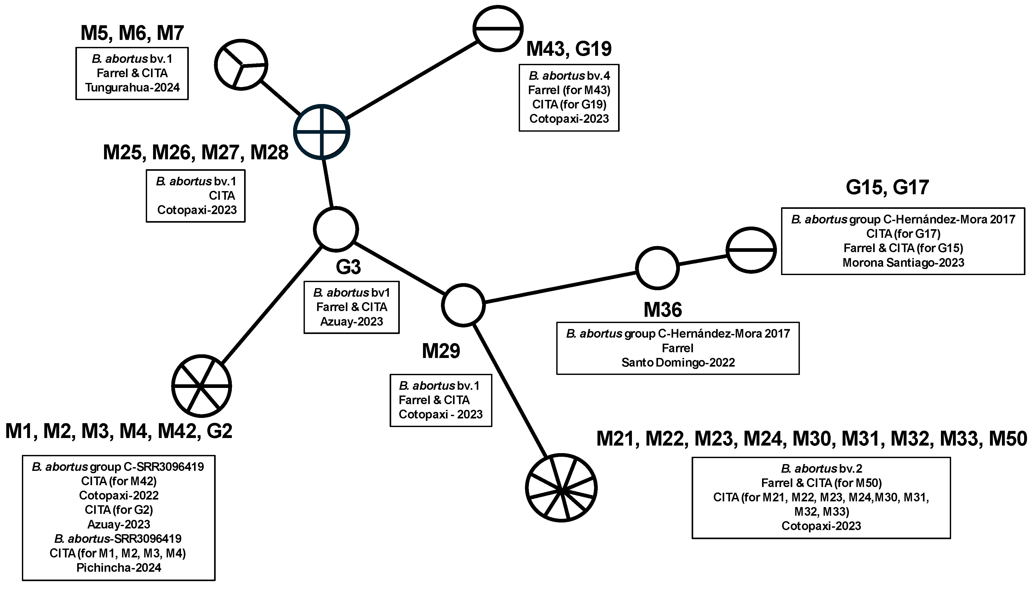
| ID Number | Province | Date of sample | Host | Bruce Number | Biovar of B. abortus | |||||||||||||||
|---|---|---|---|---|---|---|---|---|---|---|---|---|---|---|---|---|---|---|---|---|
| 06 | 08 | 11 | 12 | 42 | 43 | 45 | 55 | 18 | 19 | 21 | 04 | 07 | 09 | 16 | 30 | |||||
| M1 | Pichincha | 2024 | Cattle | 4 | 5 | 3 | 12 | 2 | 2 | 3 | 3 | 6 | 43 | 8 | 3 | 6 | 3 | 5 | 6 | B. abortus-groupC-SRR3096419 |
| M2 | Pichincha | 2024 | Cattle | 4 | 5 | 3 | 12 | 2 | 2 | 3 | 3 | 6 | 43 | 8 | 3 | 6 | 3 | 5 | 6 | B. abortus-groupC-SRR3096419 |
| M3 | Pichincha | 2024 | Cattle | 4 | 5 | 3 | 12 | 2 | 2 | 3 | 3 | 6 | 43 | 8 | 3 | 6 | 3 | 5 | 6 | B. abortus-groupC-SRR3096419 |
| M4 | Pichincha | 2024 | Cattle | 4 | 5 | 3 | 12 | 2 | 2 | 3 | 3 | 6 | 43 | 8 | 3 | 6 | 3 | 5 | 6 | B. abortus-groupC-SRR3096419 |
| M5 | Tungurahua | 2024 | Cattle | 4 | 5 | 4 | 12 | 2 | 2 | 3 | 3 | 6 | 43 | 8 | 4 | 4 | 3 | 4 | 7 | B.abortus bv1 |
| M6 | Tungurahua | 2024 | Cattle | 4 | 5 | 4 | 12 | 2 | 2 | 3 | 3 | 6 | 43 | 8 | 4 | 4 | 3 | 4 | 7 | B.abortus bv1 |
| M7 | Tungurahua | 2024 | Cattle | 4 | 5 | 4 | 12 | 2 | 2 | 3 | 3 | 6 | 43 | 8 | 4 | 4 | 3 | 4 | 7 | B.abortus bv1 |
| M21 | Cotopaxi | 2023 | Cattle | 4 | 5 | 4 | 12 | 2 | 2 | 3 | 3 | 6 | 45 | 7 | 3 | 5 | 3 | 4 | 5 | B.abortus bv2 |
| M22 | Cotopaxi | 2023 | Cattle | 4 | 5 | 4 | 12 | 2 | 2 | 3 | 3 | 6 | 45 | 7 | 3 | 5 | 3 | 4 | 5 | B.abortus bv2 |
| M23 | Cotopaxi | 2023 | Cattle | 4 | 5 | 4 | 12 | 2 | 2 | 3 | 3 | 6 | 45 | 7 | 3 | 5 | 3 | 4 | 5 | B.abortus bv2 |
| M24 | Cotopaxi | 2023 | Cattle | 4 | 5 | 4 | 12 | 2 | 2 | 3 | 3 | 6 | 45 | 7 | 3 | 5 | 3 | 4 | 5 | B.abortus bv2 |
| M25 | Cotopaxi | 2023 | Cattle | 4 | 5 | 4 | 12 | 2 | 2 | 3 | 3 | 6 | 43 | 8 | 4 | 4 | 3 | 4 | 6 | B.abortus bv1 |
| M26 | Cotopaxi | 2023 | Cattle | 4 | 5 | 4 | 12 | 2 | 2 | 3 | 3 | 6 | 43 | 8 | 4 | 4 | 3 | 4 | 6 | B.abortus bv1 |
| M27 | Cotopaxi | 2023 | Cattle | 4 | 5 | 4 | 12 | 2 | 2 | 3 | 3 | 6 | 43 | 8 | 4 | 4 | 3 | 4 | 6 | B.abortus bv1 |
| M28 | Cotopaxi | 2023 | Cattle | 4 | 5 | 4 | 12 | 2 | 2 | 3 | 3 | 6 | 43 | 8 | 4 | 4 | 3 | 4 | 6 | B.abortus bv1 |
| M29 | Cotopaxi | 2023 | Cattle | 4 | 5 | 4 | 12 | 2 | 2 | 3 | 3 | 6 | 43 | 8 | 3 | 6 | 3 | 4 | 5 | B.abortus bv1 |
| M30 | Cotopaxi | 2023 | Cattle | 4 | 5 | 4 | 12 | 2 | 2 | 3 | 3 | 6 | 45 | 7 | 3 | 5 | 3 | 4 | 5 | B.abortus bv2 |
| M31 | Cotopaxi | 2023 | Cattle | 4 | 5 | 4 | 12 | 2 | 2 | 3 | 3 | 6 | 45 | 7 | 3 | 5 | 3 | 4 | 5 | B.abortus bv2 |
| M32 | Cotopaxi | 2023 | Cattle | 4 | 5 | 4 | 12 | 2 | 2 | 3 | 3 | 6 | 45 | 7 | 3 | 5 | 3 | 4 | 5 | B.abortus bv2 |
| M33 | Cotopaxi | 2023 | Cattle | 4 | 5 | 4 | 12 | 2 | 2 | 3 | 3 | 6 | 45 | 7 | 3 | 5 | 3 | 4 | 5 | B.abortus bv2 |
| M36 | Santo Domingo | 2022 | Cattle | 4 | 5 | 3 | 12 | 2 | 2 | 3 | 3 | 6 | 43 | 8 | 3 | 5 | 3 | 3 | 5 | B.abortus group C (Hernández-Mora et al., 2017 [26]) |
| M42 | Cotopaxi | 2022 | Cattle | 4 | 5 | 3 | 12 | 2 | 2 | 3 | 3 | 6 | 43 | 8 | 3 | 6 | 3 | 5 | 6 | B.abortus group C-SRR3096419 |
| M43 | Cotopaxi | 2023 | Cattle | 4 | 5 | 4 | 12 | 1 | 2 | 3 | 3 | 6 | 45 | 8 | 1 | 4 | 3 | 4 | 6 | B.abortus bv4 |
| M50 | Cotopaxi | 2023 | Cattle | 4 | 5 | 4 | 12 | 2 | 2 | 3 | 3 | 6 | 45 | 7 | 3 | 5 | 3 | 4 | 5 | B.abortus bv2 |
| G2 | Azuay | 2023 | Cattle | 4 | 5 | 3 | 12 | 2 | 2 | 3 | 3 | 6 | 43 | 8 | 3 | 6 | 3 | 5 | 6 | B.abortus group C-SRR3096419 |
| G3 | Azuay | 2023 | Cattle | 4 | 5 | 4 | 12 | 2 | 2 | 3 | 3 | 6 | 43 | 8 | 3 | 4 | 3 | 4 | 6 | B.abortus bv1 |
| G15 | Morona Santiago | 2023 | Cattle | 4 | 5 | 3 | 12 | 2 | 3 | 3 | 3 | 6 | 43 | 8 | 3 | 5 | 3 | 3 | 5 | B.abortus group C (Hernández-Mora et al., 2017 [26]) |
| G17 | Morona Santiago | 2023 | Cattle | 4 | 5 | 3 | 12 | 2 | 3 | 3 | 3 | 6 | 43 | 8 | 3 | 5 | 3 | 3 | 5 | B.abortus group C (Hernández-Mora et al., 2017 [26]) |
| G19 | Cotopaxi | 2023 | Cattle | 4 | 5 | 4 | 12 | 1 | 2 | 3 | 3 | 6 | 45 | 8 | 1 | 4 | 3 | 4 | 6 | B.abortus bv4 |
References
- Corbel, M.J. Brucellosis: An overview. Emerg. Infect. Dis. 1997, 3, 213–221. [Google Scholar] [CrossRef] [PubMed]
- WOAH. Capítulo 3.1.4. Brucelosis (Infección por B. abortus, B. melitensis Y B. suis); WOAH—Organizacion Mundial de Sanidad Animal: Paris, France, 2022; Available online: https://www.woah.org/fileadmin/Home/eng/Health_standards/tahm/3.01.04_BRUCELLOSIS.pdf (accessed on 10 July 2024).
- Saegerman, C.; Berkvens, D.; Godfroid, J.; Walravens, K. Bovine brucellosis. En Bovine brucellosis (pp. 959–988). Tec et Doc, France. 2010. Available online: https://orbi.uliege.be/handle/2268/59477 (accessed on 18 July 2024).
- Moreno, E.; Middlebrook, E.A.; Altamirano-Silva, P.; Al Dahouk, S.; Araj, G.F.; Arce-Gorvel, V.; Arenas-Gamboa, Á.; Ariza, J.; Barquero-Calvo, E.; Battelli, G.; et al. If You’re Not Confused, You’re Not Paying Attention: Ochrobactrum Is Not Brucella. J. Clin. Microbiol. 2023, 61, e0043823. [Google Scholar] [CrossRef] [PubMed]
- Bricker, B.J.; Halling, S.M. Differentiation of Brucella abortus bv. 1, 2, and 4, Brucella melitensis, Brucella ovis, and Brucella suis bv. 1 by PCR. J. Clin. Microbiol. 1994, 32, 2660–2666. [Google Scholar] [CrossRef] [PubMed]
- Hanot Mambres, D.; Boarbi, S.; Michel, P.; Bouker, N.; Escobar-Calle, L.; Desqueper, D.; Fancello, T.; Van Esbroeck, M.; Godfroid, J.; Fretin, D.; et al. Imported human brucellosis in Belgium: Bio and molecular typing of bacterial isolates, 1996–2015. PLoS ONE 2017, 12, e0174756. [Google Scholar] [CrossRef]
- Le Flèche, P.; Jacques, I.; Grayon, M.; Al Dahouk, S.; Bouchon, P.; Denoeud, F.; Nöckler, K.; Neubauer, H.; Guilloteau, L.A.; Vergnaud, G. Evaluation and selection of tandem repeat loci for a Brucella MLVA typing assay. BMC Microbiol. 2006, 6, 9. [Google Scholar] [CrossRef]
- Moreno, E.; Moriyón, I. The Genus Brucella. In The Prokaryotes: Volume 5: Proteobacteria: Alpha and Beta Subclasses; Dworkin, M., Falkow, S., Rosenberg, E., Schleifer, K.H., Stackebrandt, E., Eds.; Springer: Berlin/Heidelberg, Germany, 2006; pp. 315–456. [Google Scholar] [CrossRef]
- Minharro, S.; Mol, J.P.S.; Dorneles, E.M.S.; Pauletti, R.B.; Neubauer, H.; Melzer, F.; Poester, F.P.; Dasso, M.G.; Pinheiro, E.S.; Filho, P.M.S.; et al. Biotyping and Genotyping (MLVA16) of Brucella abortus Isolated from Cattle in Brazil, 1977 to 2008. PLOS ONE 2013, 8, e81152. [Google Scholar] [CrossRef] [PubMed]
- Whatmore, A.M. Current understanding of the genetic diversity of Brucella, an expanding genus of zoonotic pathogens. Infect. Genet. Evol. J. Mol. Epidemiol. Evol. Genet. Infect. Dis. 2009, 9, 1168–1184. [Google Scholar] [CrossRef]
- Godfroid, J.; Scholz, H.C.; Barbier, T.; Nicolas, C.; Wattiau, P.; Fretin, D.; Whatmore, A.M.; Cloeckaert, A.; Blasco, J.M.; Moriyon, I.; et al. Brucellosis at the animal/ecosystem/human interface at the beginning of the 21st century. Prev. Vet. Med. 2011, 102, 2. [Google Scholar] [CrossRef] [PubMed]
- Nielsen, K.; Yu, W.L. Serological diagnosis of brucellosis. Prilozi 2010, 31, 65–89. [Google Scholar] [PubMed]
- López-Goñi, I.; García-Yoldi, D.; Marín, C.M.; de Miguel, M.J.; Muñoz, P.M.; Blasco, J.M.; Jacques, I.; Grayon, M.; Cloeckaert, A.; Ferreira, A.C.; et al. Evaluation of a multiplex PCR assay (Bruce-ladder) for molecular typing of all Brucella species, including the vaccine strains. J. Clin. Microbiol. 2008, 46, 3484–3487. [Google Scholar] [CrossRef]
- Bonilla-Aldana, D.K.; Trejos-Mendoza, A.E.; Pérez-Vargas, S.; Rivera-Casas, E.; Muñoz-Lara, F.; Zambrano, L.I.; Arteaga-Livias, K.; Ulloque-Badaracco, J.R.; Alarcon-Braga, E.A.; Hernandez-Bustamante, E.A.; et al. A systematic review and meta-analysis of bovine brucellosis seroprevalence in Latin America and the Caribbean. New Microbes New Infect. 2023, 54, 101168. [Google Scholar] [CrossRef]
- Eras, R.; Lalangui, M. The Agricultural Sector in Ecuador: Descriptive analysis of the impact on sustainability by COVID-19. South Fla. J. Dev. 2021, 2, 4105–4122. [Google Scholar] [CrossRef]
- Garrido-Haro, A.; Barrionuevo-Samaniego, M.; Moreno-Caballeros, P.; Burbano-Enriquez, A.; Sánchez-Vázquez, M.J.; Pompei, J.; Humblet, M.-F.; Ron-Román, J.; Saegerman, C. Seroprevalence and Risk Factors Related to Bovine Brucellosis in Continental Ecuador. Pathogens 2023, 12, 1134. [Google Scholar] [CrossRef] [PubMed]
- Ron-Román, J.; Ron-Garrido, L.; Abatih, E.; Celi-Erazo, M.; Vizcaíno-Ordóñez, L.; Calva-Pacheco, J.; González-Andrade, P.; Berkvens, D.; Benítez-Ortíz, W.; Brandt, J.; et al. Human brucellosis in northwest Ecuador: Typifying Brucella spp., seroprevalence, and associated risk factors. Vector Borne Zoonotic Dis. 2014, 14, 124–133. [Google Scholar] [CrossRef]
- Ron-Román, J.; Saegerman, C.; Minda-Aluisa, E.; Benítez-Ortíz, W.; Brandt, J.; Douce, R.Y. First report of orchitis in man caused by Brucella abortus biovar 1 in Ecuador. Am. J. Trop. Med. Hyg. 2012, 87, 3. [Google Scholar] [CrossRef] [PubMed]
- De Miguel, M.J.; Marín, C.M.; Muñoz, P.M.; Dieste, L.; Grilló, M.J.; Blasco, J.M. Development of a selective culture medium for primary isolation of the main Brucella species. J. Clin. Microbiol. 2011, 49, 1458–1463. [Google Scholar] [CrossRef] [PubMed]
- Farrell, I.D. The Development of a New Selective Medium for the Isolation of Brucella abortus from Contaminated Sources. Res. Vet. Sci. 1974, 16, 280–286. [Google Scholar] [CrossRef]
- Alton, G.G.; Jones, L.M.; Pietz, D.E. Las Tecnicas de Laboratorio en la Brucelosis, 2nd ed.; Food and Agriculture Organization of the United Nations: Rome, Italy; World Health Organization (WHO): Geneva, Switzerland, 1988; Available online: https://apps.who.int/iris/bitstream/handle/10665/38787/924340055X-spa.pdf?sequence=1 (accessed on 11 August 2024).
- Al Dahouk, S.; Flèche, P.L.; Nöckler, K.; Jacques, I.; Grayon, M.; Scholz, H.C.; Tomaso, H.; Vergnaud, G.; Neubauer, H. Evaluation of Brucella MLVA typing for human brucellosis. J. Microbiol. Methods 2007, 69, 137–145. [Google Scholar] [CrossRef] [PubMed]
- NCBI. WGS of Brucella abortus: Sample B11-0148—SRA—NCBI. 2024. Available online: https://www.ncbi.nlm.nih.gov/sra/SRR3096419 (accessed on 18 August 2024).
- Corbel, M.J.; Food and Agriculture Organization of the United Nations; World Health Organization; World Organisation for Animal Health. Brucellosis in Humans and Animals. WHO/CDS/EPR/2006.7. 2006. Available online: https://iris.who.int/handle/10665/43597 (accessed on 14 August 2024).
- Lucero, N.E.; Ayala, S.M.; Escobar, G.I.; Jacob, N.R. Brucella isolated in humans and animals in Latin America from 1968 to 2006. Epidemiol. Infect. 2008, 136, 4. [Google Scholar] [CrossRef]
- Hernández-Mora, G.; Ruiz-Villalobos, N.; Bonilla-Montoya, R.; Romero-Zúniga, J.-J.; Jiménez-Arias, J.; González-Barrientos, R.; Barquero-Calvo, E.; Chacón-Díaz, C.; Rojas, N.; Chaves-Olarte, E.; et al. Epidemiology of bovine brucellosis in Costa Rica: Lessons learned from failures in the control of the disease. PLOS ONE 2017, 12, e0182380. [Google Scholar] [CrossRef] [PubMed]
- Godfroid, J.; Nielsen, K.; Saegerman, C. Diagnosis of brucellosis in livestock and wildlife. Croat. Med. J. 2010, 51, 4. [Google Scholar] [CrossRef]
- Stack, J.A.; Harrison, M.; Perrett, L.L. Evaluation of a selective medium for Brucella isolation using natamycin. J. Appl. Microbiol. 2002, 92, 724–728. [Google Scholar] [CrossRef] [PubMed]
- Marín, C.M.; Alabart, J.L.; Blasco, J.M. Effect of antibiotics contained in two Brucella selective media on growth of Brucella abortus, B. melitensis, and B. ovis. J. Clin. Microbiol. 1996, 34, 426–428. [Google Scholar] [CrossRef]
- Hornsby, R.L.; Jensen, A.E.; Olsen, S.C.; Thoen, C.O.Y. Selective media for isolation of Brucella abortus strain RB51. Vet. Microbiol. 2000, 73, 51–60. [Google Scholar] [CrossRef] [PubMed]
- Acha, P.N.; Szyfres, B. Zoonoses and Communicable Diseases Common to Man and Animals. 1: Bacterioses and Mycoses, 3rd ed.; Pan American Health Organization: Washington, DC, USA, 2003. [Google Scholar]
- Samartino, L.E. Brucellosis in Argentina. Vet. Microbiol. 2002, 90, 71–80. [Google Scholar] [CrossRef] [PubMed]
- Hollender, D.; Conde, S.B.; Salustio, E.; Samartino, L.E. Detección de un complejo clonal con el genotipo de Brucella abortus biovariedad 2 como fundador en aislamientos de B. abortus de Argentina. Rev. Argent. Microbiol. 2013, 45, 229–239. [Google Scholar] [CrossRef]
- Torres Higuera, L.D.; Jiménez Velásquez, S.D.C.; Rodríguez Bautista, J.L.; Patiño Burbano, R.E. Identification of Brucella abortus biovar 4 of bovine origin in Colombia. Rev. Argent. Microbiol. 2019, 51, 221–228. [Google Scholar] [CrossRef]
- Rossetti, C.A.; Arenas-Gamboa, A.M.; Maurizio, E. Caprine brucellosis: A historically neglected disease with significant impact on public health. PLoS Neglected Trop. Dis. 2017, 11, e0005692. [Google Scholar] [CrossRef] [PubMed]
- Megid, J.; Albert, D.; Fagliari, J.J.; Paes, A.C.; Listoni, F.P.; Pinto, M.R.A.; Ribeiro, M.G.; Thiébaud, M.; Ueno, T.; Garin-Bastuji, B. Isolation of Brucella abortus from cattle and water buffalo in Brazil. Vet Rec. 2005, 156, 147–148. [Google Scholar] [CrossRef]
- Poester, F.P.; Picão Gonçalves, V.S.; Pereira Lage, A. Brucellosis in Brazil. Vet Microbiol. 2002, 90, 55–62. [Google Scholar] [CrossRef]
- AGROCALIDAD. Dirección de Certificación Zoosanitaria. 2024. Available online: https://www.agrocalidad.gob.ec/direccion-de-certificacion-zoosanitaria/ (accessed on 5 July 2024).
- Moreno, E.; Cloeckaert, A.; Moriyón, I. Brucella evolution and taxonomy. Vet. Microbiol. 2002, 90, 209–227. [Google Scholar] [CrossRef]
- Godfroid, J.; Al Dahouk, S.; Pappas, G.; Roth, F.; Matope, G.; Muma, J.; Marcotty, T.; Pfeiffer, D.; Skjerve, E. A «One Health» surveillance and control of brucellosis in developing countries: Moving away from improvisation. Comp. Immunol. Microbiol. Infect. Dis. 2013, 36, 241–248. [Google Scholar] [CrossRef] [PubMed]
- Ocholi, R.A.; Kwaga, J.K.P.; Ajogi, I.; Bale, J.O.O. Phenotypic characterization of Brucella strains isolated from livestock in Nigeria. Vet. Microbiol. 2004, 103, 47–53. [Google Scholar] [CrossRef] [PubMed]
- MSP. Ministerio de Salud Pública, Dirección Nacional de Vigilancia Epidemiológica, Enfermedades Zoonóticas. 2025. Available online: https://www.salud.gob.ec/enfermedades-zoonoticas/ (accessed on 3 January 2025).

| Sample Type | Province | Number of Samples | Medium Farrel | Medium Cita | PCR Brucella abortus |
|---|---|---|---|---|---|
| Milk | Pichincha | 9 | 1 | 5 | 5 |
| Milk | Tungurahua | 3 | 3 | 3 | 3 |
| Milk | Azuay | 3 | 1 | 1 | 1 |
| Milk | Cotopaxi | 26 | 4 | 21 | 22 |
| Milk | Santo Domingo | 7 | 4 | 3 | 4 |
| Retromammary lymph node | Morona Santiago | 5 | 2 | 3 | 3 |
| Retromammary lymph node | Azuay | 5 | 2 | 3 | 4 |
| Retromammary lymph node | Cotopaxi | 10 | 3 | 6 | 8 |
| Retromammary lymph node | Santo Domingo | 4 | 4 | 3 | 4 |
| Total | 6 | 75 | 24 | 48 | 54 |
| Biovar/Group | Isolated | Genotypes | Herds |
|---|---|---|---|
| B. abortus biovar 1 | 9 | 4 | 3 |
| B. abortus biovar 2 | 9 | 1 | 3 |
| B. abortus group C-SRR309419 | 6 | 1 | 3 |
| B. abortus C-Hernández-Mora 2017 | 3 | 2 | 3 |
| B. abortus biovar 4 | 2 | 1 | 2 |
Disclaimer/Publisher’s Note: The statements, opinions and data contained in all publications are solely those of the individual author(s) and contributor(s) and not of MDPI and/or the editor(s). MDPI and/or the editor(s) disclaim responsibility for any injury to people or property resulting from any ideas, methods, instructions or products referred to in the content. |
© 2025 by the authors. Licensee MDPI, Basel, Switzerland. This article is an open access article distributed under the terms and conditions of the Creative Commons Attribution (CC BY) license (https://creativecommons.org/licenses/by/4.0/).
Share and Cite
Garrido-Haro, A.D.; Falconí, M.; Moreno-Caballeros, P.; Elena-Rovalino, M.; Rosero-Mayanquer, H.; Yugcha-Díaz, M.; Fretin, D.; Wielick, C.; Saegerman, C.; Ron-Román, J. Determination and Characterization of (Novel) Circulating Strains of Brucella sp. Within the National Bovine Brucellosis Control Program in Ecuador. Pathogens 2025, 14, 158. https://doi.org/10.3390/pathogens14020158
Garrido-Haro AD, Falconí M, Moreno-Caballeros P, Elena-Rovalino M, Rosero-Mayanquer H, Yugcha-Díaz M, Fretin D, Wielick C, Saegerman C, Ron-Román J. Determination and Characterization of (Novel) Circulating Strains of Brucella sp. Within the National Bovine Brucellosis Control Program in Ecuador. Pathogens. 2025; 14(2):158. https://doi.org/10.3390/pathogens14020158
Chicago/Turabian StyleGarrido-Haro, Ana Dolores, Merci Falconí, Paola Moreno-Caballeros, María Elena-Rovalino, Hugo Rosero-Mayanquer, Michelle Yugcha-Díaz, David Fretin, Constance Wielick, Claude Saegerman, and Jorge Ron-Román. 2025. "Determination and Characterization of (Novel) Circulating Strains of Brucella sp. Within the National Bovine Brucellosis Control Program in Ecuador" Pathogens 14, no. 2: 158. https://doi.org/10.3390/pathogens14020158
APA StyleGarrido-Haro, A. D., Falconí, M., Moreno-Caballeros, P., Elena-Rovalino, M., Rosero-Mayanquer, H., Yugcha-Díaz, M., Fretin, D., Wielick, C., Saegerman, C., & Ron-Román, J. (2025). Determination and Characterization of (Novel) Circulating Strains of Brucella sp. Within the National Bovine Brucellosis Control Program in Ecuador. Pathogens, 14(2), 158. https://doi.org/10.3390/pathogens14020158