Article

The Global Outbreak of M. chimaera Infection Following Cardiac Surgery: Another Piece of the Puzzle

1
Department of Public Health and Paediatrics, University of Turin, 10124 Torino, TO, Italy
2
Department of Life Sciences and Systems Biology, University of Turin, 10124 Torino, TO, Italy
*
Author to whom correspondence should be addressed.
Pathogens 2025, 14(10), 964; https://doi.org/10.3390/pathogens14100964
Submission received: 13 August 2025 / Revised: 16 September 2025 / Accepted: 18 September 2025 / Published: 24 September 2025
(This article belongs to the Section Emerging Pathogens)

Abstract

Invasive cases of Mycobacterium chimaera have been found in Europe, and beyond, and have been associated with the use of heater–cooler units necessary to regulate the temperature of blood in extracorporeal circulation during cardiac surgery, mostly due to contamination of patients by aerosol coming from the water in the tanks of the devices. An outbreak of five cases of M. chimaera infection associated with heater–cooler units Stockert 3T (LivaNova) was also identified in the Piedmont region (Italy). M. chimaera strains isolated from 10 Stockert 3T heater–cooler units used in 3 regional cardiac surgery operating rooms were subjected to whole-genome sequencing. The results were analysed according to van Ingen’s criteria. M. chimaera was isolated from 59% of heater–cooler units Stockert 3T (LivaNova) monitored. By whole-genome sequencing analysis performed on M. chimaera strains obtained from 10 heater–cooler units, four strains were classified as subgroup 1.1 or 1.8, which are the two subgroups associated with the HCU-related outbreak worldwide. Our data and their comparisons with global heater–cooler unit isolates provide further evidence of the hypothesis that contamination occurs at the production site of LivaNova following exposure to M. chimaera-contaminated factory plumbing systems.

1. Introduction

Non-tuberculous mycobacteria (NTMs) are widely present in water [1,2,3], soil [3], dust, and other natural environments. Potting soil, hospital water, and even shower heads can be rich in NTMs that can form difficult-to-eliminate biofilms [4]. Potable water in residential buildings and healthcare settings is a common source of exposure to NTMs. Contaminated water can spread through decorative fountains and water features, hydrotherapy equipment such as jetted therapy baths, ice machines, intravenous infusions or intramuscular or intradermal injections, medical equipment such as respiratory machines and bronchoscopes, as well as heater–cooler devices, shower heads, and sink faucets [5].
Large healthcare facilities present particular challenges due to their complex water systems, which can lead to water stagnation and the proliferation of microorganisms [6], including NTMs [2,7].
Human exposure to NTMs is common, but outbreaks of NTM infections are fairly infrequent, suggesting that NTMs have low-to-moderate pathogenicity and that host risk factors play an integral role in susceptibility to NTM disease [8]. Individuals with immunodeficiencies or underlying conditions are susceptible to NTM infections. NTM outbreaks typically occur in healthcare settings when procedures expose patients to contaminated water. Pulmonary infections are the most common clinical manifestation of NTM disease. Extrapulmonary infection surveillance can help detect healthcare-associated infections (HAIs) and outbreaks more quickly [5].
Mycobacterium chimaera, first identified in 2004, is an emerging NTM causing both pulmonary and extrapulmonary infections. Invasive M. chimaera infections have been reported in Europe [9,10,11,12,13], and beyond [14,15], and have been linked to the use of heater–cooler units (HCUs) used to regulate blood temperature during extracorporeal circulation in cardiac surgery, mostly due to contamination of patients by aerosol coming from the water in the tanks of the devices [10,12]. The first identified infection case linked to these devices dates back to 2012 [10,16], although through retrospective investigations, it was possible to also recognize cases that occurred previously [9,17]. The incubation period after exposure to M. chimaera is long, with a median of 17 months (range 3–72 months). Signs and symptoms include fatigue, fever, weight loss, prosthetic valve endocarditis, prosthetic valve infection, sternotomy wound infection, mediastinitis, and manifestations of disseminated infection (sepsis)—including embolic and immunological manifestations (e.g., splenomegaly, arthritis, osteomyelitis, spondylodiscitis, bone marrow involvement with cytopenia, chorioretinitis, pulmonary involvement, hepatitis, nephritis, myocarditis, central nervous system manifestations, and septic abscesses) [18]. There is no established therapy, and the mortality rate is approximately 50%.
The extent of the global epidemic is currently not known with precision. In order to elucidate the origin of the HCU colonization and understand the transmission dynamics of M. chimaera within clinical and environmental settings, van Ingen and coworkers [19] performed a comprehensive phylogenetic analysis using whole-genome sequencing (WGS) on 250 M. chimaera isolates collected from HCUs and patients, as well as from water systems at the HCU manufacturing site. Van Ingen extracted a set of variants specific for groups of isolates defined by the phylogenetic analysis, i.e., phylogenetic single nucleotide polymorphisms (SNPs) that are characteristic of isolate groups, subgroups, or branches. He grouped isolates together based on a maximum distance to the nearest group member of 1000 base substitutions for group attribution or 10 base substitutions for subgroup attribution; for each isolate, the mean allele frequency was calculated for each group-specific set of SNP alleles, setting a threshold of at least 5% for detection of a strain from that group.
In Italy, a death in a northern region during autumn 2018 prompted a retrospective analysis to identify all subjects infected with M. chimaera, as indicated by the Ministry of Health. As of 10 October 2019, 36 confirmed cases and 2 possible cases of invasive M. chimaera infection (according to the ECDC case definition) [20] had been reported to the Ministry of Health. The ECDC case definition for HCU-related invasive M. chimaera infection is based on both clinical and exposure criteria [20]. Confirmed case: a patient who meets the clinical and exposure criteria and has M. chimaera isolated by culture and identified by Sanger sequencing in a significant biological sample. Probable case: a patient who meets the clinical and exposure criteria and has M. chimaera identified by direct PCR and Sanger sequencing in a significant biological sample, or a Mycobacterium avium complex strain isolated by culture or by direct PCR from a significant biological sample, or histopathological detection of nongaseous granuloma and foamy/swollen macrophages with the presence of acid-fast bacilli in cardiac or vascular tissue or in a sternotomy wound specimen.
In Italy, the mortality rate for M. chimaera remains very high, at 58.3% among confirmed cases reported up to the October 2019 date [21].
In the Piedmont region (north-west Italy), clinical and environmental surveillance activity (identification of contaminated devices) indicated by the Ministry of Health led to retrospective identification of five confirmed infection cases, one of which underwent surgery in a facility in the Lombardy region. These infections were acquired in the operating room during cardiac surgery from 2009 until 2013. The mean time between surgery and diagnosis was 7.2 years (range 5–9 years).
Since 2017, our microbiological laboratory has operated as a regional reference service for microbiological surveillance of all HCUs and Extra Corporeal Membrane Oxygenation (ECMO) machines [22] located in seven cardiac surgery facilities across the Piedmont region. Our investigations have highlighted NTMs and M. chimaera in a considerable number of water samples from HCUs [23,24].
Here, we report the results of whole-genome sequencing (WGS) analysis of M. chimaera strains isolated from Stockert 3T HCUs used in regional cardiac surgery operating rooms, according to the method described by van Ingen.

2. Materials and Methods

During 2020–2024, we analysed (by NTM culture) 100 water samples from 22 Stockert 3T HCUs (LivaNova, Sorin Group Deutschland GmbH, Norderstedt, Germany) located in five regional facilities. Mycobacterial cultures were performed according to our internal method [24] on 100 mL water samples using Elite agar. All M. chimaera confirmed colonies were subcultured on 7H11 medium for 10–14 days at 37 °C to obtain one to three loopfuls of cells for pelleting and DNA extraction, yielding approximately 50–100 μL in volume [25]. Below are the details of the laboratory procedures.

2.1. Sample Collection

This study collected a total of 100 water samples from five regional health facilities. Facility A and Facility C are two hospitals, located in the western and southwestern parts of the Piedmont region, respectively, with approximately 450–600 beds. Facilities B, D, and E are three private clinics, situated in the western, southwestern, and northwestern parts of the region. These clinics are highly specialized centres with around 100 beds each. The facilities serve distinct patient populations and operate independently, with no exchange of equipment or resources between them.
Water samples were collected in sterile 1 L plastic bottles containing 20 mg of sodium thiosulphate. Samples were immediately sent to the laboratory or, if not possible, stored at a controlled temperature (5 ± 3 °C for 12–15 h). All analyses were performed within 24 h of collection.

2.2. Culture

NTM isolation was performed using the following procedure: 100 mL of each water sample was filtered through a mixed cellulose esters filter, and the filter was directly added to a Petri dish containing NTM Elite agar (0.47% Middlebrook 7H9 base, 0.5% glycerol, 1.3% agar, 0.4% yeast extract, 0.2% glucose, 0.5% bovine serum albumin, 0.0056% oleic acid, and 0.0494% of a mix of selective antibiotics and antifungals) (bioMérieux, Marcy-l’Étoile, France). Subsequently, the NTM Elite agar plates were incubated at 30 °C in a sealed plastic bag to prevent dehydration. Starting from the seventh day, cultures were examined weekly for seven weeks. After realizing that mycobacteria from environmental samples needed a longer time to grow, we opted to extend the incubation time from 4 weeks—as advised by the manufacturer—to 8 weeks. The results from the NTM Elite agar analyses were reported as CFU/100 mL [24].

2.3. Identification

The NTM colonies that grew on the Elite agar plates appeared to be small to medium in size and often took several days to weeks to become visible. Most of the colonies had a dry, rough surface (as M. chelonae and M. fortuitum), although some species showed smooth and shiny colonies (as M. avium complex and M. kansasii). Many of the NTM colonies were non-pigmented (white or cream-coloured appearance, as M. avium and M. marinum), but some produced pigmented colonies—either photochromogenic (pigmented upon exposure to light, as M. fortuitum and M. smegmatis) or scotochromogenic (pigmented in darkness, as M. gordonae, M. paragordonae, and M. kansasii).
In particular, colonies of M. chimaera grew to be small to moderate in size and had a smooth, cream or beige appearance [26]. Initially, they had a shiny surface, which became rough with prolonged incubation. Colonies grew slowly and usually took weeks to develop on solid soils. Over time, they sometimes showed a slightly rounded or convex shape and had a characteristic macroscopic appearance with slightly pigmented yellow colonies.
All colonies growing on the NTM Elite agar plates were confirmed as acid-fast bacilli by Kinyoun stain and were tested as non-tuberculous mycobacteria by Real-Time PCR (Anyplex plus MTB/NTM MDR-TB Real-Time Detection, V2.0; Seegene Technologies, Inc., Seoul, Republic of Korea) [27]. Colonies were further identified as M. chimaera by Real-Time PCR using the On-Demand Advanced DNA Kit for M. chimaera Detection by Genesig (Primer Design Ltd., Southampton, UK) [23].

2.4. Whole-Genome Sequencing

Mycobacteria cells were collected from 7H11 medium, gently resuspended in a balanced salt solution (PAGE), and heat-killed at 80 °C for 20 min. After removal of the supernatant, the enriched cell pellet was subjected to immediate DNA extraction or stored frozen (e.g., at −20 °C) until extraction could begin.
Cell pellets were resuspended in 100 μL lysis buffer (15% sucrose, 0.05 M Tris-Cl, pH 8.0, 0.05 M EDTA, pH 8.0) and briefly vortexed. A quantity of 25 μL of lysozyme was added (100 mg/mL in lysis buffer, and samples were incubated for 2 h at 37 °C under gentle agitation (500 rpm). Following the lysozyme step, 3.1 μL of 20 mg/mL proteinase K and 100 μL of 10% SDS were added. The samples were then incubated for 10 min at 65 °C and subsequently stored frozen. [25].
Genomic DNA was purified from bacterial lysate by phenol/chloroform extraction followed by ethanol precipitation. A quantity of 100 ng of DNA was fragmented on the Bioruptor Pico instrument (Diagenode SA, Ougrée, Belgium) using the following settings: 30 s ON/30 s OFF pulses, easy mode, for 3 cycles. Sonicated DNA was concentrated by ethanol precipitation.
Libraries were prepared from fragmented DNA using the Watchmaker DNA Library Prep Kit (Watchmaker Genomics, Boulder, CO, USA) following the manufacturer’s instructions and sequenced on the Illumina MiSeq platform generating 2 × 150 paired-end reads.
Raw reads were quality controlled and trimmed with fastp (version 0.23.4). Variants were called with snippy (version 4.6.0, https://github.com/tseemann/snippy (accessed on 21 March 2025) against the reference genome of M. chimaera strain DSM 44623 (GenBank accession number CP015278.1) and then compared with M. chimaera group-specific SNP alleles defined by van Ingen et al. [19] (Supplementary Table S2).
[https://ars.els-cdn.com/content/image/1-s2.0-S1473309917303249-mmc1.pdf (accessed on 21 March 2025)] using bcftools (version 1.20) [28].
Sustainable data analysis was performed using Snakemake [29]. The group and subgroup classification of strains was carried out according to the method described by van Ingen and colleagues [19].

3. Results

NTMs were found in 36 water samples from 18 HCUs and M. chimaera was found in 13/22 (59%) HCUs, representing four cardiac surgeries in our region (Figure 1).
Overall, among the analysed isolates, four could be classified as 1.1 or 1.8, which are the two subgroups associated with the worldwide HCU-related outbreak. These four strains were isolated from four HCUs operating in two regional cardiac surgeries in which four patients underwent valve replacement surgery (between the years 2009 and 2013) and developed M. chimaera infections.
Moreover, in our study: one strain belonged to group 1 branch 2, and three strains belonged to group 2 (Table 1).

4. Discussion

Invasive M. chimaera infections have been reported in Europe [8,9,10,11], and beyond, and have been linked to the use of heater–cooler units (HCUs) mostly due to contamination of patients by aerosol coming from the water in the tanks of the devices [9,11].
Van Ingen and coworkers [21] conducted a phylogenetic analysis based on whole-genome sequencing (WGS) of 250 M. chimaera isolates from HCUs and patients, as well as from water systems at the HCU manufacturing site in order to understand the origin of the HCU colonization. This analysis revealed two major M. chimaera groups, with isolates related to cardiac surgery patients mainly classified into subgroup 1.1. Additionally, the WGS analysis demonstrated a high level of genetic similarity between isolates from the HCU factory and those from surgery-related patients, supporting the hypothesis that contamination originated from a single point source during HCU production.
In this study, the M. chimaera that has been assigned to subgroups 1.1 and 1.8 associated with the worldwide outbreak [10,19,30,31,32,33,34] linked to Stockert 3T HCUs were detected in four HCUs used in two regional cardiac surgery operating rooms. The literature reported that subgroups 1.1 and 1.8 were isolated from the LivaNova production site and from the majority of the analysed HCUs [33].
Our data and their comparisons with global HCU-associated isolates provide further evidence for the hypothesis that contamination occurs at the LivaNova production site, following exposure to M. chimaera-contaminated factory plumbing systems.
We also isolated one strain belonging to group 1 branch 2 (notably, this strain had also been found in one patient in Australia) [19] and three strains belonging to group 2.
The group 2 strains were found in three HCUs; however, these strains had never been encountered in patients who underwent open-heart surgery: we hypothesize that the strains other than those highlighted worldwide are strains that have colonized the HCUs starting from the local populations of waterborne M. chimaera in the plumbing systems of each facility. The remaining two strains were un-grouped because they did not exhibit any distinguishing SNP markers (Table 1).
This study has some limitations: (1) we did not store or analyse M. chimaera strains from all 13 M. chimaera-positive HCUs; (2) we analysed only one strain of M. chimaera for each HCU monitored. Most likely, the analysis of more strains would have given us the possibility to highlight a greater number of HCUs linked to the global outbreak; (3) we were not able to link each patient to the actual HCU adopted during cardiothoracic surgery because device serial numbers were not recorded before the international alert.
To date, no cases of postoperative invasive infections linked to Maquet devices have been reported; however, our microbiological results emphasize the need for surveillance.
In fact, surveillance of the Maquet devices adopted in our regional facilities has revealed a similar problem: widespread contamination with strains of Mycobacterium gordonae and Mycobacterium paragordonae despite repeated disinfections of the water tanks [35].
In our setting, the evidence of M. chimaera transmission in the cardiac surgery theatre has prompted the adoption of the best prevention practices (follow the HCU manufacturer’s instructions, microbiological surveillance of HCU water supplies, and clinical surveillance of patients undergoing cardiac surgery) in all healthcare facilities.
Following our findings, some facilities, in order to ensure patient safety, decided to move the LivaNova HCUs outside of the operating room, while others replaced their LivaNova HCUs with Maquet HCU40 devices (Maquet, Getinge Group, Rastatt, Germany).
The next effort will be to evaluate whether contamination of Maquet devices originates from a common source, like the LivaNova case, or from each facility’s individual water supply.

5. Conclusions

The outbreak in our region spanned several years (2009–2013) and affected only four patients.
Whole-genome sequencing revealed that our outbreak is part of the global M. chimaera outbreak. The high degree of similarity between our M. chimaera strains and those isolated from LivaNova HCUs worldwide supports the hypothesis of a single common source of infection and suggests that contamination occurred during manufacturing at the production site in Germany.

Author Contributions

S.D.: conceptualization, supervision, writing—review and editing; G.M.: formal analysis; M.G.: formal analysis, writing—review and editing; F.A.: formal analysis; I.M.: data curation; C.M.Z.: resources. All authors have read and agreed to the published version of the manuscript.

Funding

This research was funded by EU funding within the MUR PNRR Extended Partnership initiative on Emerging Infectious Diseases Project no. PE00000007, INF-ACT.

Institutional Review Board Statement

This study did not involve human or animal experiments; therefore, ethical review was not required.

Informed Consent Statement

Not applicable.

Data Availability Statement

The original contributions presented in this study are included in the article. Further inquiries can be directed to the corresponding author.

Conflicts of Interest

The authors declare no conflicts of interest.

References

  1. Falkinham, J.O. Environmental Sources of Nontuberculous Mycobacteria. Clin. Chest Med. 2015, 36, 35–41. [Google Scholar] [CrossRef] [PubMed]
  2. Falkinham, J.O.; Hilborn, E.D.; Arduino, M.J.; Pruden, A.; Edwards, M.A. Epidemiology and Ecology of Opportunistic Premise Plumbing Pathogens: Legionella Pneumophila, Mycobacterium Avium, and Pseudomonas Aeruginosa. Environ. Health Perspect. 2015, 123, 749–758. [Google Scholar] [CrossRef] [PubMed]
  3. Walsh, C.M.; Gebert, M.J.; Delgado-Baquerizo, M.; Maestre, F.T.; Fierer, N. A Global Survey of Mycobacterial Diversity in Soil. Appl. Environ. Microbiol. 2019, 85, e01180-19. [Google Scholar] [CrossRef]
  4. Gebert, M.J.; Delgado-Baquerizo, M.; Oliverio, A.M.; Webster, T.M.; Nichols, L.M.; Honda, J.R.; Chan, E.D.; Adjemian, J.; Dunn, R.R.; Fierer, N. Ecological Analyses of Mycobacteria in Showerhead Biofilms and Their Relevance to Human Health. mBio 2018, 9, e01614-18. [Google Scholar] [CrossRef] [PubMed]
  5. CDC Clinical Overview of Nontuberculous Mycobacteria (NTM). Available online: https://www.cdc.gov/nontuberculous-mycobacteria/hcp/clinical-overview/index.html (accessed on 1 September 2025).
  6. U.S. Environmental Protection Agency. Legionella in the Indoor Environment. Available online: https://www.epa.gov/indoor-air-quality-iaq/legionella-indoor-environment (accessed on 1 September 2025).
  7. Li, T. A Systematic Review of Waterborne Infections from Nontuberculous Mycobacteria in Health Care Facility Water Systems. Int. J. Environ. Health 2017, 220, 611–620. [Google Scholar] [CrossRef]
  8. Honda, J.R.; Alper, S.; Bai, X.; Chan, E.D. Acquired and Genetic Host Susceptibility Factors and Microbial Pathogenic Factors That Predispose to Nontuberculous Mycobacterial Infections. Curr. Opin. Immunol. 2018, 54, 66–73. [Google Scholar] [CrossRef]
  9. Achermann, Y.; Rössle, M.; Hoffmann, M.; Deggim, V.; Kuster, S.; Zimmermann, D.R.; Bloemberg, G.; Hombach, M.; Hasse, B. Prosthetic Valve Endocarditis and Bloodstream Infection Due to Mycobacterium Chimaera. J. Clin. Microbiol. 2013, 51, 1769–1773. [Google Scholar] [CrossRef]
  10. Sax, H.; Bloemberg, G.; Hasse, B.; Sommerstein, R.; Kohler, P.; Achermann, Y.; Rössle, M.; Falk, V.; Kuster, S.P.; Böttger, E.C.; et al. Prolonged Outbreak of Mycobacterium chimaera Infection After Open-Chest Heart Surgery. Clin. Infect. Dis. 2015, 61, 67–75. [Google Scholar] [CrossRef]
  11. Haller, S.; Höller, C.; Jacobshagen, A.; Hamouda, O.; Abu Sin, M.; Monnet, D.L.; Plachouras, D.; Eckmanns, T. Contamination during Production of Heater-Cooler Units by Mycobacterium chimaera Potential Cause for Invasive Cardiovascular Infections: Results of an Outbreak Investigation in Germany, April 2015 to February 2016. Euro Surveill. 2016, 21, 1–6. [Google Scholar] [CrossRef]
  12. Sommerstein, R.; Rüegg, C.; Kohler, P.; Bloemberg, G.; Kuster, S.P.; Sax, H. Transmission of Mycobacterium chimaera from Heater-Cooler Units during Cardiac Surgery despite an Ultraclean Air Ventilation System. Emerg. Infect. Dis. 2016, 22, 1008–1013. [Google Scholar] [CrossRef]
  13. Chand, M.; Lamagni, T.; Kranzer, K.; Hedge, J.; Moore, G.; Parks, S.; Collins, S.; Del Ojo Elias, C.; Ahmed, N.; Brown, T.; et al. Insidious Risk of Severe Mycobacterium chimaera Infection in Cardiac Surgery Patients. Clin. Infect. Dis 2017, 64, 335–342. [Google Scholar] [CrossRef] [PubMed]
  14. Perkins, K.M. Mycobacterium chimaera Contamination of Heater-Cooler Devices Used in Cardiac Surgery—United States. MMWR Morb. Mortal. Wkly. Rep. 2016, 65, 1117–1118. [Google Scholar] [CrossRef] [PubMed]
  15. Xu, K.; Finn, L.E.; Geist, R.L.; Prestel, C.; Moulton-Meissner, H.; Kim, M.; Stacey, B.; McAllister, G.A.; Gable, P.; Kamali, T.; et al. Mycobacterium chimaera Infections among Cardiothoracic Surgery Patients Associated with Heater-Cooler Devices-Kansas and California, 2019. Infect. Control Hosp. Epidemiol. 2021, 43, 1333–1338. [Google Scholar] [CrossRef] [PubMed]
  16. Kohler, P.; Kuster, S.P.; Bloemberg, G.; Schulthess, B.; Frank, M.; Tanner, F.C.; Rössle, M.; Böni, C.; Falk, V.; Wilhelm, M.J.; et al. Healthcare-Associated Prosthetic Heart Valve, Aortic Vascular Graft, and Disseminated Mycobacterium chimaera Infections Subsequent to Open Heart Surgery. Eur. Heart J. 2015, 36, 2745–2753. [Google Scholar] [CrossRef]
  17. Bush, L.M.; Paturi, A.; Chaparro-Rojas, F.; Perez, M.T. Mycobacterial Prosthetic Valve Endocarditis. Curr. Infect. Dis. Rep. 2010, 12, 257–265. [Google Scholar] [CrossRef]
  18. Hasse, B.; Hannan, M.M.; Keller, P.M.; Maurer, F.P.; Sommerstein, R.; Mertz, D.; Wagner, D.; Fernández-Hidalgo, N.; Nomura, J.; Manfrin, V.; et al. International Society of Cardiovascular Infectious Diseases Guidelines for the Diagnosis, Treatment and Prevention of Disseminated Mycobacterium chimaera Infection Following Cardiac Surgery with Cardiopulmonary Bypass. J. Hosp. Infect. 2020, 104, 214–235. [Google Scholar] [CrossRef]
  19. van Ingen, J.; Kohl, T.A.; Kranzer, K.; Hasse, B.; Keller, P.M.; Katarzyna Szafrańska, A.; Hillemann, D.; Chand, M.; Schreiber, P.W.; Sommerstein, R.; et al. Global Outbreak of Severe Mycobacterium chimaera Disease after Cardiac Surgery: A Molecular Epidemiological Study. Lancet Infect. Dis. 2017, 17, 1033–1041. [Google Scholar] [CrossRef]
  20. ECDC. EU Protocol for Case Detection, Laboratory Diagnosis and Environmental Testing of Mycobacterium chimaera infections Potentially Associated with Heater-Cooler Units: Case Definition and Environmental Testing Methodology; ECDC: Stockholm, Sweden, 2015.
  21. Ministero della Salute. Indicazioni Operative Riguardanti Gli Aspetti Di Laboratorio in Merito Ai Casi Di Infezione Da Mycobacterium chimaera in Italia e Aggiornamento Delle Informazioni Disponibili; Ministero della Salute: Rome, Italy, 2019.
  22. Ditommaso, S.; Giacomuzzi, M.; Memoli, G.; Garlasco, J.; Zotti, C.M. Persistence of Legionella in Routinely Disinfected Heater-Cooler Units and Heater Units Assessed by Propidium Monoazide QPCR. Pathogens 2020, 9, 978. [Google Scholar] [CrossRef]
  23. Ditommaso, S.; Giacomuzzi, M.; Memoli, G.; Zotti, C.M. Real-Time PCR, the Best Approaches for Rapid Testing for Mycobacterium chimaera Detection in Heater Cooler Units and Extracorporeal Membrane Oxygenation. Perfusion 2021, 36, 626–629. [Google Scholar] [CrossRef]
  24. Ditommaso, S.; Giacomuzzi, M.; Memoli, G.; Garlasco, J.; Curtoni, A.; Iannaccone, M.; Zotti, C.M. A New Culture Method for the Detection of Non-Tuberculous Mycobacteria in Water Samples from Heater-Cooler Units and Extracorporeal Membrane Oxygenation Machines. Int. J. Environ. Res. Public Health 2022, 19, 10645. [Google Scholar] [CrossRef]
  25. Epperson, L.E.; Strong, M. A Scalable, Efficient, and Safe Method to Prepare High Quality DNA from Mycobacteria and Other Challenging Cells. J. Clin. Tuberc. Other Mycobact. Dis. 2020, 19, 100150. [Google Scholar] [CrossRef] [PubMed]
  26. Giacomuzzi, M.; Memoli, G.; Garlasco, J.; Zotti, C.M.; Ditommaso, S. Image Dataset of Environmental Non-Tuberculous Mycobacteria Isolated from Heater Cooler Units Water by the New NTM Elite Agar. Data Brief 2025, 59, 111358. [Google Scholar] [CrossRef] [PubMed]
  27. Ditommaso, S.; Giacomuzzi, M.; Memoli, G.; Cavallo, R.; Curtoni, A.; Avolio, M.; Silvestre, C.; Zotti, C.M. Reduction of Turnaround Time for Non-Tuberculous Mycobacteria Detection in Heater-Cooler Units by Propidium Monoazide-Real-Time Polymerase Chain Reaction. J. Hosp. Infect. 2019, 104, 365–373. [Google Scholar] [CrossRef]
  28. Danecek, P.; Bonfield, J.K.; Liddle, J.; Marshall, J.; Ohan, V.; Pollard, M.O.; Whitwham, A.; Keane, T.; McCarthy, S.A.; Davies, R.M.; et al. Twelve Years of SAMtools and BCFtools. Gigascience 2021, 10, giab008. [Google Scholar] [CrossRef]
  29. Mölder, F.; Jablonski, K.P.; Letcher, B.; Hall, M.B.; Tomkins-Tinch, C.H.; Sochat, V.; Forster, J.; Lee, S.; Twardziok, S.O.; Kanitz, A.; et al. Sustainable Data Analysis with Snakemake. F1000Research 2021, 10, 33. [Google Scholar] [CrossRef]
  30. Cannas, A.; Campanale, A.; Minella, D.; Messina, F.; Butera, O.; Nisii, C.; Mazzarelli, A.; Fontana, C.; Lispi, L.; Maraglino, F.; et al. Epidemiological and Molecular Investigation of the Heater–Cooler Unit (HCU)-Related Outbreak of Invasive Mycobacterium chimaera Infection Occurred in Italy. Microorganisms 2023, 11, 2251. [Google Scholar] [CrossRef]
  31. Hasan, N.A.; Epperson, L.E.; Lawsin, A.; Rodger, R.R.; Perkins, K.M.; Halpin, A.L.; Perry, K.A.; Moulton-Meissner, H.; Diekema, D.J.; Crist, M.B.; et al. Genomic Analysis of Cardiac Surgery–Associated Mycobacterium chimaera Infections, United States. Emerg. Infect. Dis. 2019, 25, 559–563. [Google Scholar] [CrossRef]
  32. Svensson, E.; Jensen, E.T.; Rasmussen, E.M.; Folkvardsen, D.B.; Norman, A.; Lillebaek, T. Mycobacterium chimaera in Heater-Cooler Units in Denmark Related to Isolates from the United States and United Kingdom. Emerg. Infect. Dis. 2017, 23, 507–509. [Google Scholar] [CrossRef]
  33. Schreiber, P.W.; Sax, H. Mycobacterium chimaera Infections Associated with Heater-Cooler Units in Cardiac Surgery. Curr. Opin. Infect. Dis. 2017, 30, 388–394. [Google Scholar] [CrossRef]
  34. Williamson, D.; Howden, B.; Stinear, T. Mycobacterium chimaera Spread from Heating and Cooling Units in Heart Surgery. N. Engl. J. Med. 2017, 376, 600–602. [Google Scholar] [CrossRef]
  35. Ditommaso, S.; Garlasco, J.; Memoli, G.; Curtoni, A.; Bondi, A.; Ceccarelli, A.; Giacomuzzi, M. Emergence of Mycobacterium gordonae in Heater-Cooler Units: A Five-Year Prospective Surveillance of Devices Frequently Subjected to Chloramine-T Booster Disinfection. J. Hosp. Infect. 2024, 155, 9–16. [Google Scholar] [CrossRef]
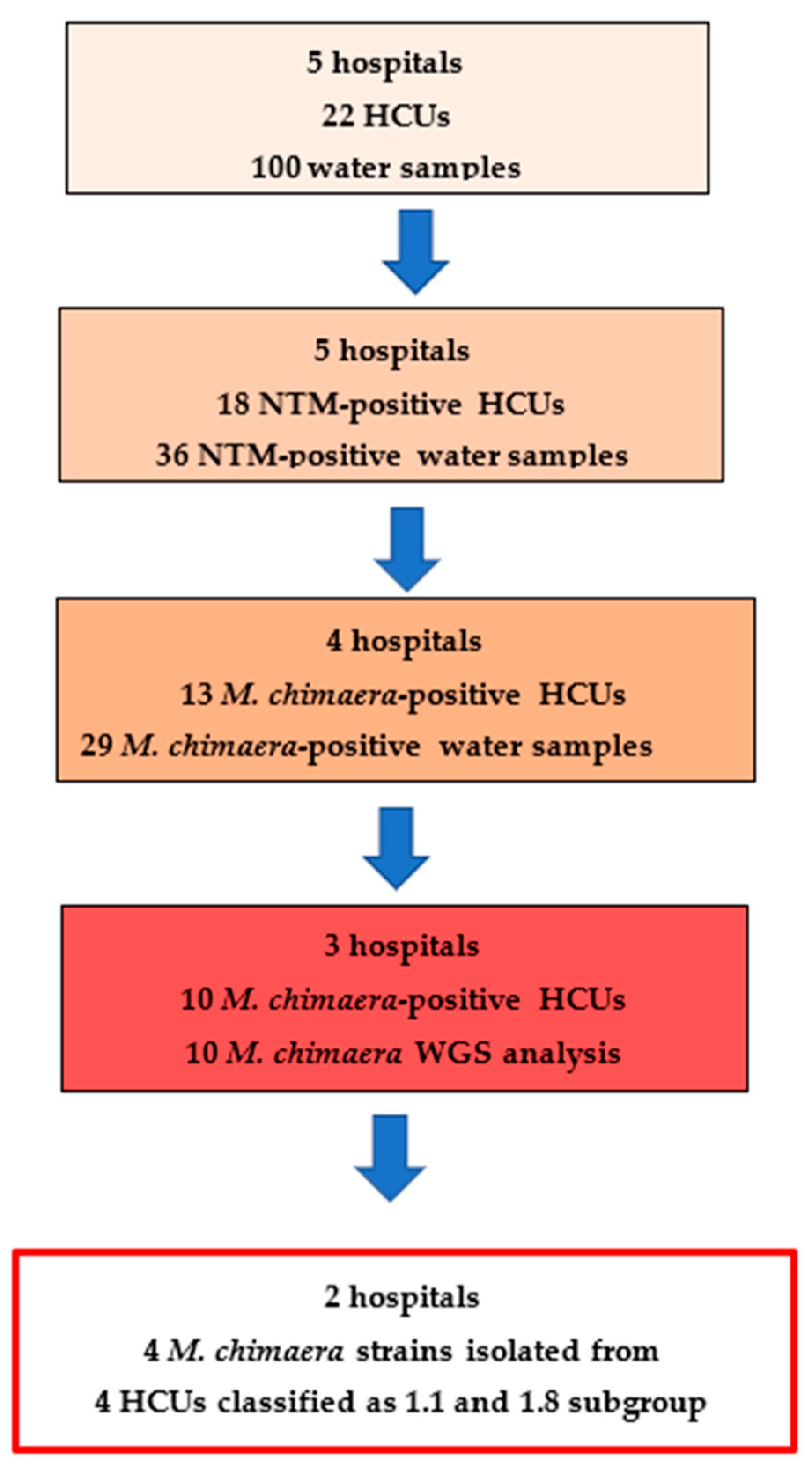
Figure 1. Prevalence of M. chimaera-positive devices in the Piedmont region and classification of sequenced strains according to van Ingen’s criteria: five years of HCU surveillance data.
Pathogens 14 00964 g001
Table 1. Informative SNPs and classification of sequenced M. chimaera isolates according to group-specific SNPs defined by van Ingen.
Sample ID
MC-1180 MC-830 MC-988 MC-1271B MC-873 MC-750 MC-791 MC-829 MC-1345 MC-872
Group/
Branch
Associated
reference SNP position
Branch_1 4977262 0000000000
Branch_2 2339764 0000000100
Branch_2 5003561 0000000100
Group_1.1 113518 0000011000
Group_1.1 209278 0000011000
Group_1.11 1419163 0000000000
Group_1.11 3132089 0000000000
Group_1.6 4050336 0000000000
Group_1.6 5033374 0000000000
Group_1.8 *~ 281696 0000000000
Group_1.8 ~ 1611282 0001100000
Group_1.8 ~ 2366314 0001100000
Group_1 *~ 2329494 1110000000
Group_1 *~ 3949608 1110000000
Group_2.1 1828053 0000000000
Group_2.1 3406341 0000000000
Group_2 ~ 3022332 1110000000
Group_2 ~ 5709901 1110000000
Sample Classification Group 2 Group 2 Group 2 Group 1.8 Group 1.8 Group 1.1 Group 1.1Branch 2Un-
grouped
Un
grouped
~ All marked SNPs are required for a specific group identification. * Reference allele is the one specific to the group. SNP: Single Nucleotide Polymorphism. MC: Mycobacterium chimaera. 1 indicates that the SNP variant is present; 0 indicates that it is absent. The full list of SNPs found in the 10 sequenced samples can be found at https://drive.google.com/file/d/1uzS3jXGIMV7_zYwdhoZZkp1QXpiH6AaZ/view?usp=sharing (accessed on 21 March 2025).
Disclaimer/Publisher’s Note: The statements, opinions and data contained in all publications are solely those of the individual author(s) and contributor(s) and not of MDPI and/or the editor(s). MDPI and/or the editor(s) disclaim responsibility for any injury to people or property resulting from any ideas, methods, instructions or products referred to in the content.