Article

Genetic and Biochemical Characterization of AXC-2 from Achromobacter ruhlandii

by
Mariana Papalia
1,2,*,
Francisco González-Espinosa
1,2,
Fátima Quiroga Castedo
1,
Gabriel Gutkind
1,2,
María Soledad Ramírez
3,
Pablo Power
1,2 and
Marcela Radice
1,2
1
Laboratorio de Resistencia Bacteriana, Instituto de Bacteriología y Virología Molecular, Facultad de Farmacia y Bioquímica, Universidad de Buenos Aires, Junín 956, Buenos Aires 1113, Argentina
2
Consejo Nacional de Investigaciones Científicas y Técnicas (CONICET), Godoy Cruz 2290, Buenos Aires C1425FQB, Argentina
3
Center for Applied Biotechnology Studies, Department of Biological Science, California State University Fullerton, Fullerton, CA 92831, USA
*
Author to whom correspondence should be addressed.
Pathogens 2024, 13(2), 115; https://doi.org/10.3390/pathogens13020115
Submission received: 7 December 2023 / Revised: 18 January 2024 / Accepted: 25 January 2024 / Published: 27 January 2024
(This article belongs to the Section Bacterial Pathogens)

Abstract

:
Achromobacter spp. are intrinsically resistant to multiple antibiotics and can also acquire resistance to those commonly used for the treatment of respiratory infections, especially in patients with cystic fibrosis. The aim of this study was to perform the genetic and biochemical characterization of AXC-2 from A. ruhlandii and to analyze all available AXC variants. Steady-state kinetic parameters were determined on a purified AXC-2 enzyme. It exhibited higher catalytic efficiencies towards amino-penicillins and older cephalosporins, while carbapenems behaved as poor substrates. Phylogenetic analysis of all blaAXC variants available in the NCBI was conducted. AXC was encoded in almost all A. ruhlandii genomes, whereas it was only found in 30% of A. xylosoxidans. AXC-1 was prevalent among A. xylosoxidans. AXC variants were clustered in two main groups, correlating with the Achromobacter species. No association could be established between the presence of blaAXC variants and a specific lineage of A. xylosoxidans; however, a proportion of AXC-1-producing isolates corresponded to ST 182 and ST 447. In conclusion, this study provides valuable insights into the genetic context and kinetic properties of AXC-2, identified in A. ruhlandii. It also provides a thorough description of all AXC variants and their association with Achromobacter species and various lineages.

1. Introduction

Achromobacter spp. are rod-shaped, aerobic, non-fermenting Gram-negative bacteria widely distributed in the environment and typically regarded as opportunistic pathogens.
The Achromobacter genus currently comprises 21 officially designated species, including A. xylosoxidans (the type species of the genus), A. denitrificans, A. insolitus, A. marplatensis, A. piechaudii, A. ruhlandii, A. spanius, A. dolens, A. insuavis, A. pulmonis, A. deleyi, A. mucicolens, A. aegrifaciens, A. animicus, A. anxifer, A. kerstersii, A. sediminum, A. agilis, A. aloeverae, A. pestifer, A. aestuarii, and various genogroups [1].
A. xylosoxidans was the sole species when the Achromobacter genus was proposed by Yabuuchi et al. in 1981 [2]. A. ruhlandii and A. piechaudii were reassigned from the Alcaligenes genus by Yabuuchi and Kawamura et al. in 1998, while A. xylosoxidans subsp. denitrificans was reclassified as A. denitrificans [3]. Subsequently, A. spanius, A. insolitus, and A. marplatensis were described by Gomila et al. in 2011 [4]. More recently, the remaining taxa were incorporated into the genus [5].
The accurate species identification of Achromobacter isolates is challenging, and unfortunately, they are often erroneously identified as A. xylosoxidans. Traditional phenotypic identification schemes frequently fail to discriminate between the various species within the Achromobacter genus. In 2013, the amplification and sequencing of the conserved gene nrdA was proposed for identification [6]. This method, along with multilocus sequence typing (MLST), is currently widely employed in research laboratories [7]. Additionally, identification can also be conducted by calculating the average nucleotide identity (ANI) in genomes. Veschetti et al. established that isolates with ANI ≥ 95% are classified within the same species, while isolates with ANI < 95% are considered to belong to different genogroups when compared to all available genomes of Achromobacter species [8]. The authors also reported a noticeable correlation with both MLST analysis and sequencing of the nrdA gene. This method proves valuable when genomes are accessible. However, sequence-based identification techniques are not practical for clinical microbiology laboratories. More recently, mass spectrometry (MALDI-TOF) has been introduced as a valuable tool in microbiology laboratories, enabling rapid bacterial identification (30 min). While this technique generally facilitates the accurate identification of A. xylosoxidans, it may yield inconclusive or even erroneous results for less common species. Currently, several research groups are actively working to enhance databases and achieve reliable species-level identification within the Achromobacter genus [9,10].
Achromobacter, mainly A. xylosoxidans, has been associated with pneumonia, bacteremia, and even meningitis, predominantly in immunocompromised patients, although cases have also been reported in immunocompetent individuals [11]. Additionally, Achromobacter species, distinct from A. xylosoxidans, have traditionally been considered environmental inhabitants and have only recently been associated with human infections. Achromobacter spp. have been isolated from the blood cultures and cerebrospinal fluid of neonatal patients (in cases of pneumonia or meningitis) and are linked to high mortality rates [12]. The majority of infections are acquired in the healthcare setting, while only one-third are in the community.
Since the year 2000, Achromobacter spp. have been recognized as emerging pathogens, especially in patients with cystic fibrosis (CF), as their prevalence in respiratory secretions has shown an upward trend [13]. The reported prevalence rate for A. xylosoxidans in cystic fibrosis centers worldwide is approximately 10%, although this rate is steadily increasing, with reports of prevalence reaching up to 29% [14,15]. This increase is likely attributed to the prolonged life expectancy of patients with CF, the selective pressures exerted by multi-antimicrobial therapy, and advancements in microbiological and molecular techniques. A. xylosoxidans is the most frequent Achromobacter species reported from the respiratory samples in these patients. Moreover, A. ruhlandii stands as the second most prevalent species in the Americas [6,16], while A. dolens and A. insuavis exhibit a higher prevalence in Europe [17,18].
Achromobacter spp. represent a serious threat to patients, as members of this genus are intrinsically resistant to multiple antibiotics, including cephalothin, cefoxitin, cefotaxime, aztreonam, and aminoglycosides [19]. Furthermore, Achromobacter species can acquire resistance to antimicrobial agents commonly employed in the treatment of respiratory infections, such as colistin, carbapenems, and fluoroquinolones. Extended-spectrum β-lactams are often considered the primary therapeutic option for treating Achromobacter infections in patients with cystic fibrosis, while carbapenems are reserved as last-resort antibiotics. Inhaled antibiotics, such as colistin or tobramycin, have been demonstrated to provide benefits as adjuvants to intravenous medications [20].
The two main intrinsic resistance mechanisms in Achromobacter species include multidrug efflux pumps, and chromosomal OXA-114-like β-lactamases [19]. Achromobacter spp. harbor two well-characterized multidrug efflux pumps. The AxyABM efflux pump is present in all publicly available Achromobacter genomes and shares common properties with the MexAB-OprM efflux pump of P. aeruginosa [21]. Several studies have demonstrated that imipenem is more effective than meropenem against Achromobacter isolates, possibly due to the overexpression of this efflux pump [22]. AxyABM plays a significant role in extruding aztreonam and cephalosporins other than cefepime and cefuroxime. The second efflux pump, AxyXY-OprZ, has a broader spectrum and is involved in extruding aminoglycosides, cefepime, carbapenems, fluoroquinolones, tetracyclines, and erythromycin [23,24,25]. AxyXY-OprZ is the primary determinant of high-level intrinsic aminoglycoside resistance in Achromobacter spp. For other antibiotics, AxyXY-OprZ appears to contribute to resistance, rather than being the primary resistance mechanism [19].
Regarding β-lactamases, A. xylososidans produces a constitutive chromosomal β-lactamase known as OXA-114, exhibiting activity against penicillin G, early cephalosporins, piperacillin, and ticarcillin [26]. However, extended-spectrum cephalosporins such as ceftazidime, cefotaxime, and cefepime are not substrates for OXA-114-like enzymes. OXA-114 demonstrates poor hydrolytic activity against imipenem. Additionally, other OXA-type enzymes were identified, proving to be ubiquitous and species-specific, such as OXA-258 in A. ruhlandii, OXA-243 in A. insuavis, and OXA-364 in A. dolens [27,28]. These enzymes efficiently hydrolyze penicillins and first-generation cephalosporins, but not third-generation cephalosporins and carbapenems.
Acquired antimicrobial resistance can occur through mutational events, alteration in gene expression, or acquisition of resistance markers encoded within mobile genetic elements. In terms of β-lactam resistance, plasmid-encoded enzymes have sporadically been reported in A. xylosoxidans, including broad-spectrum β-lactamases (BSBLs) and extended-spectrum β-lactamases (ESBLs), such as VEB-1 [29]. Concerning carbapenem resistance, the presence of class B enzymes (IMP-1, IMP-10, VIM-1, and VIM-2) has also been documented [30,31,32,33]. More recently, in 2018, a novel class A β-lactamase named AXC-1 (A. xylosoxidans carbapenemase), exhibiting slight activity against meropenem, was reported in A. xylosoxidans [34]. Kinetics parameters for this enzyme have not been described. Furthermore, in 2023, blaAMZ–1 was identified and characterized in Achromobacter mucicolens, demonstrating resistance against certain β-lactam antibiotics, including amoxicillin and cephalothin [35].
Although studies concerning A. ruhlandii antimicrobial resistance are limited, it has been reported that this species may exhibit a more resistant phenotype. A pandrug-resistant A. ruhlandii epidemic clone designated the Danish epidemic strain (DES) spread among patients with CF in 2006 in two Danish cystic fibrosis centers [36,37].
In a previous study, we characterized Achromobacter isolates recovered from respiratory samples of patients with CF [38]. Based on phenotypic and molecular techniques, A. xylosoxidans was the most frequently identified Achromobacter species in the airways of these patients, followed by A. ruhlandii as the second most prevalent. Achromobacter isolates included in this study were resistant to a wide range of antibiotics, including fluoroquinolones, aminoglycosides, and most broad-spectrum β-lactams. While carbapenems proved to be the most effective antibiotics, it is noteworthy that 3 out of 7 A. ruhlandii isolates and 1 out of 26 A. xylosoxidans isolates were resistant [38].
Considering the limited knowledge regarding A. ruhlandii and its antimicrobial resistance, efforts were focused on characterizing these isolates. Whole-genome sequencing was performed on one of the carbapenem-resistant A. ruhlandii isolates mentioned above. This particular isolate, Ar38, was recovered from the sputum of a pediatric patient in Buenos Aires in 2007 (ASM280300v1). In-depth in silico genome analysis revealed the presence of three β-lactamase coding genes, including a new blaAXC variant.
The aim of this study was to conduct genetic and biochemical characterization of this AXC variant from A. ruhlandii Ar38 and to evaluate its possible contribution to the observed resistance phenotype. Additionally, a phylogenetic analysis of all available AXC variants and their presence in different Achromobacter species was investigated.

2. Materials and Methods

2.1. Genetic and Biochemical Characterization of AXC-2 from Ar38

2.1.1. Whole-Genome Sequencing of A. ruhlandii Ar38

Whole-genome sequencing (WGS) was conducted using Illumina GAIIx, with Nextera XT libraries utilized for sample preparation. De novo assembly was performed with the SPAdes assembler v. 3.10.1 [39]. The RAST server was used to predict all open reading frames and their genetic context [40]. Sequence alignment and similarity searching to confirm predictions were accomplished using BLAST software v. 2.15.0.

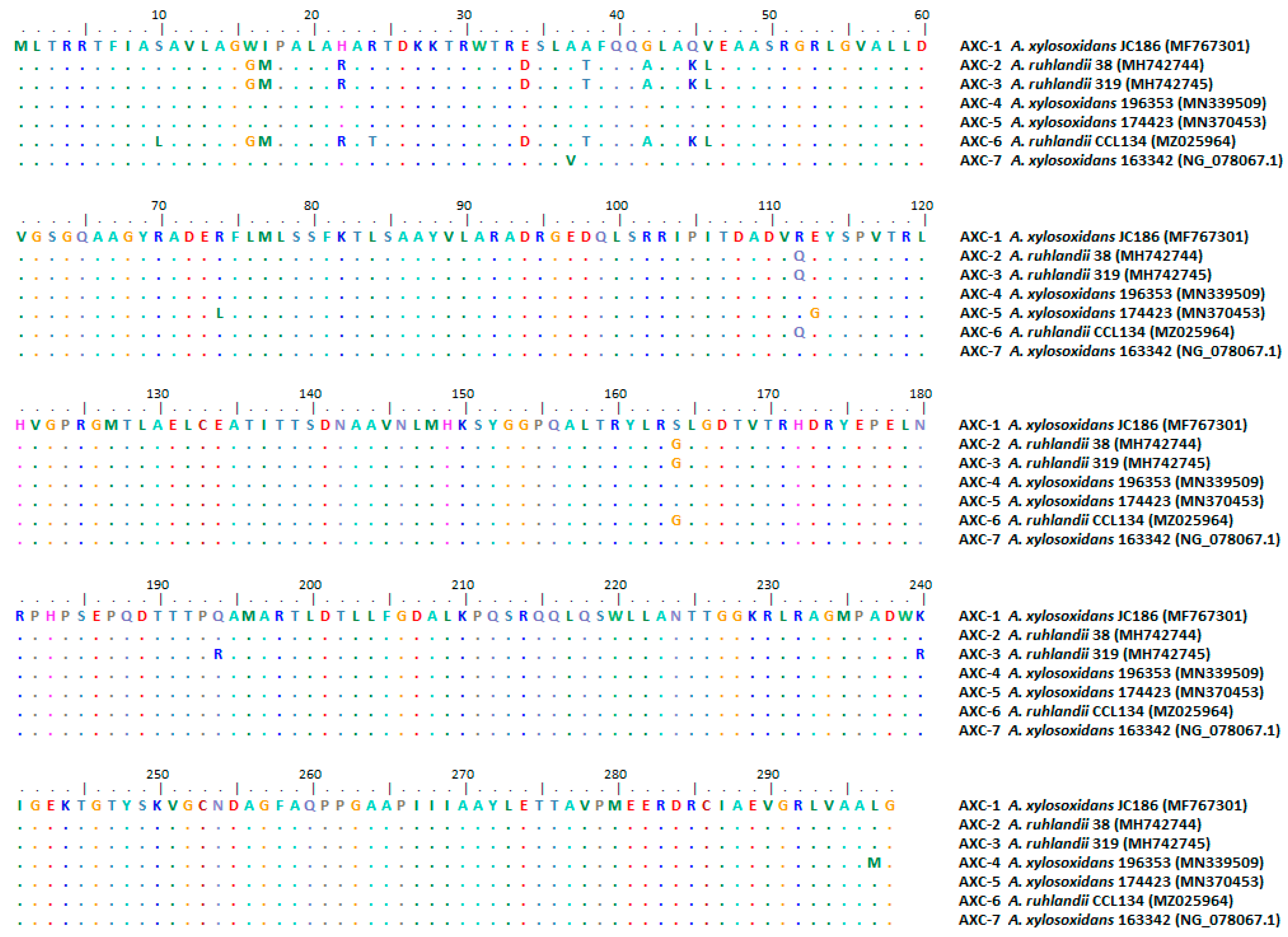
2.1.2. Comparative Analysis of AXC Sequences Deposited in the NCBI Database

Sequences of the 7 AXC variants deposited to date were downloaded. Alignment was performed using ClustalX. Analysis of the genetic context of these blaAXC was performed using RAST and BLAST tools v. 2.15.0.

2.1.3. β-Lactamase Production and Purification

Full-length blaAXC-2 was amplified by PCR using custom-designed primers AXC-F (5′ CCATATGTTGCTGACGAGAAGAAGAACCTT 3′), containing the NdeI restriction site, and AXC-R (5′ TCGAATTCCTAGCCCAATGCCGCCA 3′), containing the EcoRI restriction site. The NdeI-EcoRI amplicon was cloned into the pK19 vector and the pET-28a(+) expression vector (Novagen, Inc., Madison, WI, USA). The authenticity of the cloned fragments was confirmed by sequencing.
Escherichia coli TOP 10F’ was transformed with the pK19-AXC-2 recombinant plasmid, and the minimum inhibitory concentrations (MICs) of a representative panel of β-lactam antibiotics were determined using the broth microdilution test, according to CLSI guidelines.
A pET-AXC-2 recombinant plasmid was introduced into E. coli BL21 (DE3) (Novagen Inc., Madison, WI, USA) to overproduce the enzyme. An overnight culture of E. coli BL21(DE3)-pET-AXC-2 was diluted (1/20) in 1 L LB medium containing 30 µg/mL kanamycin (Sigma-Aldrich, Misuri, USA) and grown at 37 °C until 0.8 OD units (λ = 600 nm). IPTG (Sigma-Aldrich, Misuri, USA) was added at a final concentration of 1 mM, and culture was grown at 18 °C overnight. After centrifugation at 8000× g at 4 °C, cells were resuspended in 25 mL of 25 mM sodium phosphate buffer (pH 7.5). Cells were disrupted by sonication, and cellular debris was separated by centrifugation. The supernatant fraction of the induced culture displayed the highest specific activities when measured spectrophotometrically, with total enzymatic extracts and 100 μM cephalothin as a substrate.
Enzyme purification was carried out as follows. The supernatant fraction was dialyzed overnight at 4 °C against the same buffer used to equilibrate the ion-exchange columns (25 mM sodium phosphate buffer pH 6.5; buffer A) and clarified by centrifugation at 18,000× g for 30 min and filtration through a 0.45 μm pore membrane. Then, it was loaded onto a 5 mL Hi TrapTM SP HP column (flow rate, 1 mL/min) (GE Healthcare Life Sciences, Milwaukee, WI, USA) equilibrated with buffer A. The column was thoroughly washed to remove unbound proteins, and the β-lactamase was eluted with a linear gradient of 1 M sodium chloride. β-lactamase containing fractions were pooled, and a second purification was performed. For this purpose, an ion exchange was carried out using a 1 mL RESOURCETM S column (GE Healthcare Life Sciences, Milwaukee, WI, USA) equilibrated with buffer A. Once again, the column was washed to remove unbound proteins, and the β-lactamase was eluted with a linear gradient of 1 M sodium chloride. β-lactamase containing fractions were pooled and dialyzed overnight against 25 mM sodium phosphate buffer (pH 7.5). Purified AXC-2 was stored at −20 °C until use. Enzyme purity was over 99%, as estimated by SDS-PAGE

2.1.4. Kinetic Studies

Steady-state kinetic parameters (kcat and Km) were determined by measuring the rate of hydrolysis of different substrates, using an ultraviolet/visible (UV/Vis) spectrophotometer [41]. The reactions were performed at 25 °C in 25 mM sodium phosphate buffer (pH 7.5). The kcat and Km values were determined for a representative set of β-lactam antibiotics, including ampicillin, penicillin, cephalexin, cefoxitin, ceftazidime, ceftriaxone, cefepime, imipenem, and meropenem.

2.2. In Silico Analysis of AXC Variants from Achromobacter spp.

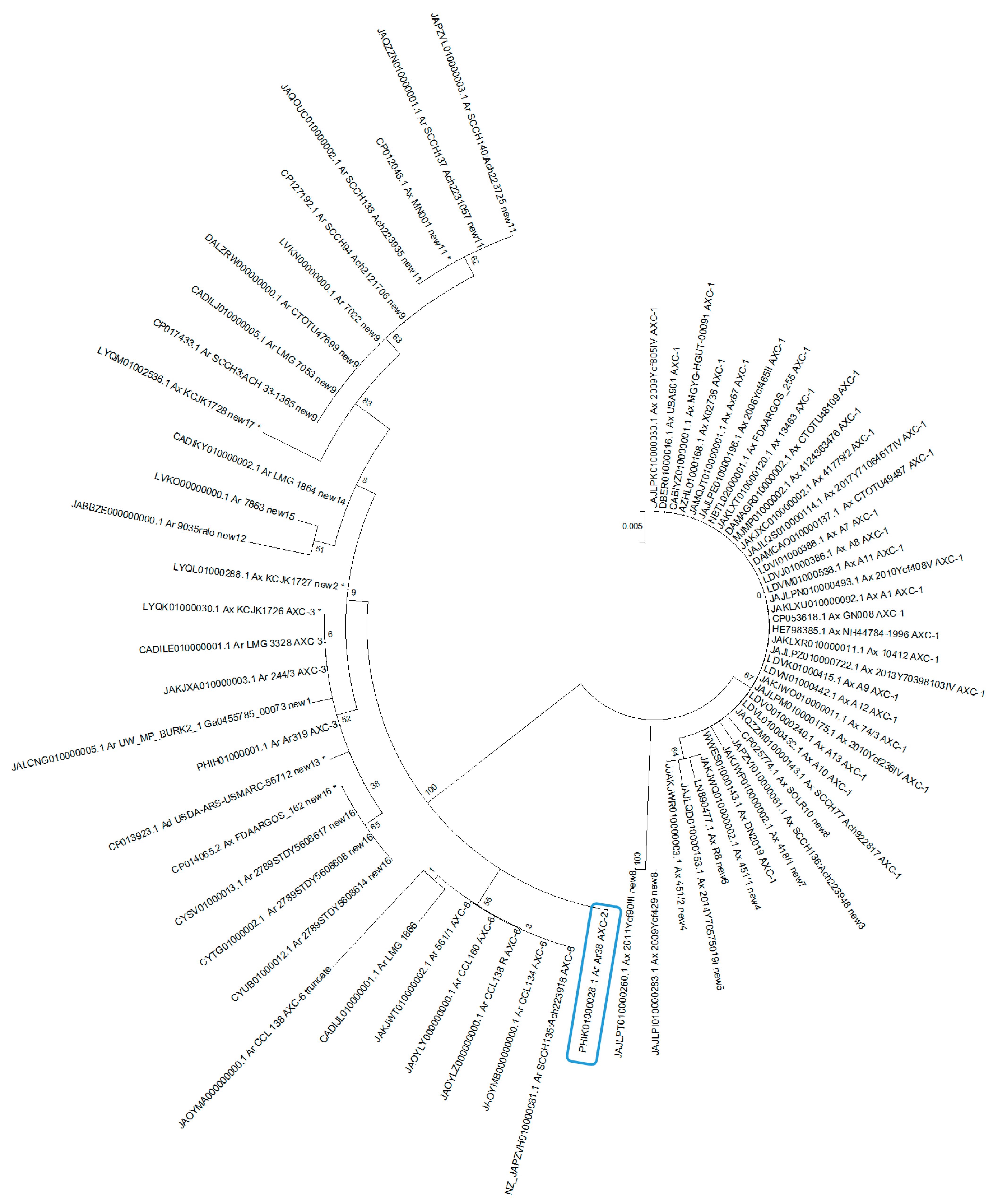
2.2.1. Phylogenetic Analysis of AXC Variants from Achromobacter Genomes Available in the Genome NCBI Database

A search for blaAXC variants was conducted using BlastN v. 2.15.0. Subsequently, all available genomes of Achromobacter species where blaAXC was detected were downloaded. Species identity was confirmed by searching for the nrdA gene and comparing alleles according to the PubMLST database (https://pubmlst.org/organisms/Achromobacter-spp. accessed on 10 November 2023). Genomes that displayed low-quality or small-length sequences were not included. The alignment of AXC variants translated from the aforementioned genomes was performed using ClustalX. A dendrogram based on maximum-likelihood estimation was conducted using MEGA 5.05 software.

2.2.2. Analysis of the Distribution of AXC in A. xylosoxidans Genomes

To highlight the distribution of AXC in A. xylosoxidans, a phylogenetic analysis was performed. PROKKA was used for genome annotation; Roary for phylogenetic analysis based on core genes, using a blastp 80% similarity threshold; and the SNPsites program for core SNP filtering. The IQ-TREE plus Microreact was employed for the construction and visualization of the phylogenetic tree.

2.2.3. Analysis of the Genetic Context of blaAXC in A. xylosoxindans and A. ruhlandii Genomes

blaAXC genetic context was investigated in selected A. xylosoxidans and A. ruhlandii genomes downloaded from the NCBI genome database. Clinker was used for the alignment and graphical representation of the genetic context.

3. Results

Three putative β-lactamase coding genes were detected by WGS and confirmed through in silico genomic analysis in Ar38. One of these open reading frames (ORFs) corresponded to blaOXA-258 (PHIK01000006.1:64095-64919), previously characterized by our group and proposed as a species-specific gene for A. ruhlandii [42]. The second ORF corresponded to blaAXC, not previously reported in A. ruhlandii, and is the main focus of this study (PHIK01000028.1:44673-45569). The third ORF encoded a putative class C β-lactamase that remains uncharacterized (PHIK01000013.1:c178058-176736).
AXC from Ar38 displayed 97% amino acid identity with AXC-1 from A. xylosoxidans, corresponding to a new variant named AXC-2, deposited in the GenBank database under the accession number MH742744. The alignment of AXC-2 and other AXC variants deposited in the NCBI database is shown in Figure 1.
The presence of the transcriptional regulator axcR, previously described by Fleurbaaij et al. [34], was observed in the closest surrounding region of blaAXC-2, displaying 90% nucleotide similarity between Ar38 and the A. xylosoxidans strain previously described. The genetic context of the blaAXC variants deposited in the NCBI was analyzed (Figure 2). It was observed that blaAXC-2, blaAXC-3, and blaAXC-6, present in A. ruhlandii, shared a common genetic environment, whereas blaAXC-1, present in A. xylosoxidans, displayed differences upstream of the gene. Unfortunately, a comparison of the genetic contexts for blaAXC-4, blaAXC-5, and blaAXC-7 could not be conducted due to the unavailability of the respective genomes. No insertion sequences or transposable elements that could indicate mobilization within the bacterial chromosome were observed.
An increase in the MIC values of ampicillin and cephalexin was observed in E. coli transformants producing AXC-2. Additionally, a slight increase in the MICs was observed for the carbapenems, although not enough to go over the breakpoints for these antibiotics (Table 1).
The purity of the AXC-2 extract was estimated to be >99% by sodium dodecyl sulfate-polyacrylamide gel electrophoresis. Kinetic constants for β-lactam antibiotics are shown in Table 2. It was observed that this enzyme exhibits higher catalytic efficiencies (kcat/ Km) towards amino-penicillins and older cephalosporins, and, to a lesser extent, benzyl-penicillin and ceftriaxone. Also, AXC-2 showed very low affinity for ceftazidime and cefoxitin, with Km values in the millimolar range and not detectable by hydrolysis. Finally, cefepime and carbapenems behaved as poor substrates, mainly due to very low turnover (kcat) values. The slight hydrolysis of carbapenems by AXC-2 was consistent with the hydrolysis determined in the crude extracts obtained from the clinical isolate. This result suggested that this β-lactamase could be expressed in the isolate, even at a low level.
The presence of blaAXC was detected in A. ruhlandii, A. xylosoxidans, and one A. denitrificans genome, but not in other species of this genus. Subsequently, all available A. ruhlandii, A. xylosoxidans, and A. denitrificans genomes were downloaded. Given the complexity associated with species identification within the genus and the potential misplacement of genomes in databases, their identity was confirmed by searching for the nrdA gene and comparing alleles according to the PubMLST database (https://pubmlst.org/organisms/Achromobacter-spp accessed on 10 November 2023). Of them, five isolates previously designated as A. xylosoxidans and one A. denitrificans corresponded to A. ruhlandii. Finally, a total of 144 A. xylosoxidans and 33 A. ruhlandii genomes were analyzed (Table S1, Supplementary Material).
Among the 33 A. ruhlandii genomes, blaAXC was detected in 31, corresponding to AXC-2, AXC-3, AXC-6, and new AXC variants (Table S1, Supplementary Material). In a thorough examination of the two A. ruhlandii genomes lacking blaAXC, and its associated regulator, a uniform genetic context was revealed between them. These genomes exhibited consistent nrdA alleles and corresponded to ST 35. Additionally, a putative transposase, InsK, was detected in association with the insertion sequence element IS150, in the surrounding region of the blaAXC location observed in other A. ruhlandii genomes. These collective findings give rise to a credible hypothesis of a gene loss event within this species.
Of the 144 A. xylosoxidans genomes, 40 were found harboring a blaAXC gene. Comparison of A. xylosoxidans genomes through core gene analysis is available at https://microreact.org/project/knWPR2m1ErkcrWDFyVyabh-arbol-axylo-con-referencias-de-araiad. (accessed on 10 November 2023) No association was observed between the presence of AXC, or even its variants, and any specific ST; however, a discernible trend indicates that a proportion of those A. xylosoxidans carrying blaAXC corresponded to ST 182 and 447.
A dendrogram of all AXC variants downloaded from Achromobacter genomes revealed that these enzymes clustered into two main groups. One group corresponded to those variants detected in A. ruhlandii, while the other clustered those from A. xylosoxidans. (Figure 3; and Table S1, Supplementary Material).
Analysis of the genetic context of blaAXC in selected A. ruhlandii and A. xylosoxidans genomes (Figure S2, Supplementary Material) exhibited an overall conservative genetic context among isolates of the same species.

4. Discussion

This study represents the first comprehensive biochemical and kinetic characterization of an AXC enzyme. AXC-1 was previously referred to as a carbapenemase (Achromobacter xylosoxidans Carbapenemases); however, no kinetic data of this enzyme or any AXC variant is available [34]. AXC-2 displayed limited hydrolytic activity towards carbapenems; thus, the concomitant presence of multiple resistance mechanisms may be responsible for the carbapenem resistance observed in A. ruhlandii Ar38. Upon analyzing the genetic context of blaAXC-2, the presence of a regulator, axcR, was detected. AxcR exhibited 94% identity with the previously reported AxcR of A. xylosoxidans [34]. Mutations in the regulator could be responsible for changes in enzyme expression. Considering the presence of a regulator, a plausible scenario similar to that observed in AmpC-type enzymes can be hypothesized [43]. In isolates harboring an inducible blaAmpC gene, its expression is determined by the regulator. In some cases, it might be stably derepressed, either partially or entirely, due to mutations in ampR, leading to the sustained production of large quantities of the enzyme. Additional experiments are essential to confirm this hypothesis in AXC.
Moreover, as mentioned by Chalhoub et al. [44], the active efflux of antibiotics in A. xylosoxidans mediated by the AxyABM, AxyXY-OprZ, or AxyEF-OprN pumps is potentially involved in antibiotic resistance. In a previous study, we characterized the AxyABM pump from A. ruhlandii Ar38, proving its active transport capability for chloramphenicol, nalidixic acid, and trimethoprim/sulfamethoxazole, while showing no efflux activity towards carbapenems [27]. Furthermore, we detected the presence of AxyXY-OprZ coding genes in A ruhlandii Ar38 genome, which has been proposed by multiple authors as a potential mediator of carbapenem efflux [23,24]. Further studies will be conducted to evaluate the carbapenem extrusion capability of this pump.
Currently, seven AXC variants have been documented in the NCBI database. However, BlastN-based analysis for blaAXC identified many new enzymatic variants. Notably, blaAXC was only detected in A. ruhlandii and A. xylosoxidans genomes, but not in any other bacterial species. An AXC enzyme was encoded in almost all available A. ruhlandii genomes, whereas it was only found in 40 out of 144 A. xylosoxidans. The AXC-1 coding gene was prevalent among the A. xylosoxidans isolates.
The phylogenetic analysis of AXC variants revealed two main clusters, indicating a correlation between AXC variants and Achromobacter species. When analyzing A. xylosoxidans genomes, no association could be established between the presence of blaAXC, or its variants, and a specific lineage. However, a noticeable pattern suggests that a proportion of AXC-1-producing A. xylosoxidans corresponded to ST 182 and 447. Interestingly, ST 182 isolates were recovered from both patients with CF and patients with non-CF in Thailand, Russia, and France, and a multidrug-resistant isolate in the United States was also found [45]. On the other hand, ST 447 was described in France in isolates recovered from patients with CF, and was associated with acquired resistance to ciprofloxacin [46].
The genetic context remained consistent among all variants observed in A. ruhlandii, with no discernible insertion sequences or transposable elements that might suggest mobilization within the bacterial chromosome. In the two A. ruhlandii genomes lacking blaAXC, its associated regulator was also absent; however, a putative transposase, InsK, was identified in the proximity of this region, probably indicative of a gene loss event. For its part, the genetic context of blaAXC in A. xylosoxidans was similar in all analyzed genomes but differed from that observed for A. ruhlandii.
In conclusion, this study provides valuable insights into the genetic context and kinetic properties of AXC-2 identified in A. ruhlandii. AXC-2 displayed limited hydrolytic activity against carbapenems, prompting the need for future studies to evaluate the role of axcR in the expression of blaAXC-2, and the contribution of the AxyXY-OprZ efflux pump. Furthermore, we provide a thorough description of all AXC variants and their association with Achromobacter species and their various lineages.
Understanding the clinical relevance of A. ruhlandii, mainly in patients with CF, and the resistance mechanisms is of utmost importance for making accurate decisions in antimicrobial treatment. A study conducted in Brazil revealed that over half (57.1%) of the patients from whom A. ruhlandii was isolated developed a chronic infection, whereas the rate was only 13.3% for those with A. xylosoxidans [16]. These observations suggest that patients colonized/infected with A. ruhlandii are at a higher risk of developing chronic infection. Accordingly, in the study reported in Denmark, concerning the DES, 13 patients were chronically infected. Moreover, this species has been implicated in causing cross-infections among patients with cystic fibrosis, even in scenarios of limited and indirect contact, raising concerns within the CF communities [47]. Additionally, it has been reported that A. ruhlandii isolates present higher resistance levels compared to other species within the genus [36,37,38]. This knowledge is critical for optimizing the management of infections and ensuring the ongoing success of antibiotic therapies in the fight against infectious diseases.

Supplementary Materials

The following supporting information can be downloaded at: https://www.mdpi.com/article/10.3390/pathogens13020115/s1, Figure S1: Alignment of AXC variants found in A. ruhlandii and A. xylosoxidans genomes; Figures S2: blaAXC genetic context investigated in selected A. xylosoxidans and A. ruhlandii genomes downloaded from NCBI genome database. Table S1: Distribution of blaAXC in A. xylosoxidans and A. ruhlandii Genomes

Author Contributions

Conceptualization, methodology, formal analysis, investigation, writing—original draft preparation, M.P.; methodology, investigation, F.G.-E.; methodology, F.Q.C.; writing—review and editing, resources, funding acquisition, G.G. and P.P.; methodology, writing—review and editing M.S.R.; writing—review and editing, resources, funding acquisition, supervision, M.R. All authors have read and agreed to the published version of the manuscript.

Funding

This work was partially supported by grants from UBACyT to M. Radice (20020190100031BA), G. Gutkind (20020170100473BA;), and P. Power (20020170100072BA). PIP to M. Radice (11220200102425CO). PICT-2021-GRFTII 00309 to M. Radice. G. Gutkind, P. Power, and M. Papalia are members of Carrera del Investigador Científico (CONICET).

Institutional Review Board Statement

Not applicable.

Informed Consent Statement

Not applicable.

Data Availability Statement

The data presented in this study are available on request to the corresponding author.

Conflicts of Interest

The authors declare no conflicts of interest.

References

  1. Parte, A.C.; Sarda Carbasse, J.; Meier-Kolthoff, J.P.; Reimer, L.C.; Goker, M. List of Prokaryotic names with Standing in Nomenclature (LPSN) moves to the DSMZ. Int. J. Syst. Evol. Microbiol. 2020, 70, 5607–5612. [Google Scholar] [CrossRef]
  2. Yabuuchi, E.; Yano, I. Achromobacter gen. nov. and Achromobacter xylosoxidans (ex Yabuuchi and Ohyama 1971) nom. rev. Int. J. Syst. Bacteriol. 1981, 31, 477–478. [Google Scholar] [CrossRef]
  3. Yabuuchi, E.; Kawamura, Y.; Kosako, Y.; Ezaki, T. Emendation of genus Achromobacter and Achromobacter xylosoxidans (Yabuuchi and Yano) and proposal of Achromobacter ruhlandii (Packer and Vishniac) comb. nov., Achromobacter piechaudii (Kiredjian et al.) comb. nov., and Achromobacter xylosoxidans subsp. denitrificans (Ruger and Tan) comb. nov. Microbiol. Immunol. 1998, 42, 429–438. [Google Scholar] [CrossRef]
  4. Gomila, M.; Tvrzova, L.; Teshim, A.; Sedlacek, I.; Gonzalez-Escalona, N.; Zdrahal, Z.; Sedo, O.; Gonzalez, J.F.; Bennasar, A.; Moore, E.R.; et al. Achromobacter marplatensis sp. nov., isolated from a pentachlorophenol-contaminated soil. Int. J. Syst. Evol. Microbiol. 2011, 61, 2231–2237. [Google Scholar] [CrossRef]
  5. Dumolin, C.; Peeters, C.; Ehsani, E.; Tahon, G.; De Canck, E.; Cnockaert, M.; Boon, N.; Vandamme, P. Achromobacter veterisilvae sp. nov., from a mixed hydrogen-oxidizing bacteria enrichment reactor for microbial protein production. Int. J. Syst. Evol. Microbiol. 2019, 70, 530–536. [Google Scholar] [CrossRef] [PubMed]
  6. Spilker, T.; Vandamme, P.; Lipuma, J.J. Identification and distribution of Achromobacter species in cystic fibrosis. J. Cyst. Fibros. 2013, 12, 298–301. [Google Scholar] [CrossRef] [PubMed]
  7. Spilker, T.; Vandamme, P.; Lipuma, J.J. A multilocus sequence typing scheme implies population structure and reveals several putative novel achromobacter species. J. Clin. Microbiol. 2012, 50, 3010–3015. [Google Scholar] [CrossRef] [PubMed]
  8. Veschetti, L.; Sandri, A.; Patuzzo, C.; Melotti, P.; Malerba, G.; Lleo, M.M. Genomic characterization of Achromobacter species isolates from chronic and occasional lung infection in cystic fibrosis patients. Microb. Genom. 2021, 7, 606. [Google Scholar] [CrossRef] [PubMed]
  9. Garrigos, T.; Dollat, M.; Magallon, A.; Chapuis, A.; Varin, V.; Bador, J.; Makki, N.; Cremet, L.; Persyn, E.; Cardot-Martin, E.; et al. Matrix-Assisted Laser Desorption Ionization-Time of Flight Mass Spectrometry for Rapid Detection of Isolates Belonging to the Epidemic Clones Achromobacter xylosoxidans ST137 and Achromobacter ruhlandii DES from Cystic Fibrosis Patients. J. Clin. Microbiol. 2021, 59, e0094621. [Google Scholar] [CrossRef] [PubMed]
  10. Papalia, M.; Figueroa-Espinosa, R.; Steffanowski, C.; Barberis, C.; Almuzara, M.; Barrios, R.; Vay, C.; Gutkind, G.; Di Conza, J.; Radice, M. Expansion and improvement of MALDI-TOF MS databases for accurate identification of Achromobacter species. J. Microbiol. Methods 2020, 172, 105889. [Google Scholar] [CrossRef]
  11. De Baets, F.; Schelstraete, P.; Van Daele, S.; Haerynck, F.; Vaneechoutte, M. Achromobacter xylosoxidans in cystic fibrosis: Prevalence and clinical relevance. J. Cyst. Fibros. 2007, 6, 75–78. [Google Scholar] [CrossRef]
  12. Turel, O.; Kavuncuoglu, S.; Hosaf, E.; Ozbek, S.; Aldemir, E.; Uygur, T.; Hatipoglu, N.; Siraneci, R. Bacteremia due to Achromobacter xylosoxidans in neonates: Clinical features and outcome. Braz. J. Infect. Dis. 2013, 17, 450–454. [Google Scholar] [CrossRef]
  13. Hansen, C.R.; Pressler, T.; Nielsen, K.G.; Jensen, P.O.; Bjarnsholt, T.; Hoiby, N. Inflammation in Achromobacter xylosoxidans infected cystic fibrosis patients. J. Cyst. Fibros. 2010, 9, 51–58. [Google Scholar] [CrossRef] [PubMed]
  14. Firmida, M.C.; Pereira, R.H.; Silva, E.A.; Marques, E.A.; Lopes, A.J. Clinical impact of Achromobacter xylosoxidans colonization/infection in patients with cystic fibrosis. Braz. J. Med. Biol. Res. 2016, 49, e5097. [Google Scholar] [CrossRef] [PubMed]
  15. Edwards, B.D.; Greysson-Wong, J.; Somayaji, R.; Waddell, B.; Whelan, F.J.; Storey, D.G.; Rabin, H.R.; Surette, M.G.; Parkins, M.D. Prevalence and Outcomes of Achromobacter Species Infections in Adults with Cystic Fibrosis: A North American Cohort Study. J. Clin. Microbiol. 2017, 55, 2074–2085. [Google Scholar] [CrossRef] [PubMed]
  16. Pereira, R.H.; Leao, R.S.; Carvalho-Assef, A.P.; Albano, R.M.; Rodrigues, E.R.; Firmida, M.C.; Folescu, T.W.; Plotkowski, M.C.; Bernardo, V.G.; Marques, E.A. Patterns of virulence factor expression and antimicrobial resistance in Achromobacter xylosoxidans and Achromobacter ruhlandii isolates from patients with cystic fibrosis. Epidemiol. Infect. 2017, 145, 600–606. [Google Scholar] [CrossRef] [PubMed]
  17. Gade, S.S.; Norskov-Lauritsen, N.; Ridderberg, W. Prevalence and species distribution of Achromobacter sp. cultured from cystic fibrosis patients attending the Aarhus centre in Denmark. J. Med. Microbiol. 2017, 66, 686–689. [Google Scholar] [CrossRef]
  18. Amoureux, L.; Bador, J.; Bounoua Zouak, F.; Chapuis, A.; de Curraize, C.; Neuwirth, C. Distribution of the species of Achromobacter in a French Cystic Fibrosis Centre and multilocus sequence typing analysis reveal the predominance of A. xylosoxidans and clonal relationships between some clinical and environmental isolates. J. Cyst. Fibros. 2016, 15, 486–494. [Google Scholar] [CrossRef] [PubMed]
  19. Isler, B.; Kidd, T.J.; Stewart, A.G.; Harris, P.; Paterson, D.L. Achromobacter Infections and Treatment Options. Antimicrob. Agents Chemother. 2020, 64, e01025-20. [Google Scholar] [CrossRef]
  20. Wang, M.; Ridderberg, W.; Hansen, C.R.; Hoiby, N.; Jensen-Fangel, S.; Olesen, H.V.; Skov, M.; Lemming, L.E.; Pressler, T.; Johansen, H.K.; et al. Early treatment with inhaled antibiotics postpones next occurrence of Achromobacter in cystic fibrosis. J. Cyst. Fibros. 2013, 12, 638–643. [Google Scholar] [CrossRef]
  21. Bador, J.; Amoureux, L.; Duez, J.M.; Drabowicz, A.; Siebor, E.; Llanes, C.; Neuwirth, C. First description of an RND-type multidrug efflux pump in Achromobacter xylosoxidans, AxyABM. Antimicrob. Agents Chemother. 2011, 55, 4912–4914. [Google Scholar] [CrossRef] [PubMed]
  22. Magallon, A.; Amoureux, L.; Garrigos, T.; Sonois, M.; Varin, V.; Neuwirth, C.; Bador, J. Role of AxyABM overexpression in acquired resistance in Achromobacter xylosoxidans. J. Antimicrob. Chemother. 2022, 77, 926–929. [Google Scholar] [CrossRef] [PubMed]
  23. Bador, J.; Neuwirth, C.; Liszczynski, P.; Mezier, M.C.; Chretiennot, M.; Grenot, E.; Chapuis, A.; de Curraize, C.; Amoureux, L. Distribution of innate efflux-mediated aminoglycoside resistance among different Achromobacter species. New Microbes New Infect. 2016, 10, 1–5. [Google Scholar] [CrossRef] [PubMed]
  24. Bador, J.; Neuwirth, C.; Grangier, N.; Muniz, M.; Germe, L.; Bonnet, J.; Pillay, V.G.; Llanes, C.; de Curraize, C.; Amoureux, L. Role of AxyZ Transcriptional Regulator in Overproduction of AxyXY-OprZ Multidrug Efflux System in Achromobacter Species Mutants Selected by Tobramycin. Antimicrob. Agents Chemother. 2017, 61, e00290-17. [Google Scholar] [CrossRef] [PubMed]
  25. Magallon, A.; Roussel, M.; Neuwirth, C.; Tetu, J.; Cheiakh, A.C.; Boulet, B.; Varin, V.; Urbain, V.; Bador, J.; Amoureux, L. Fluoroquinolone resistance in Achromobacter spp.: Substitutions in QRDRs of GyrA, GyrB, ParC and ParE and implication of the RND efflux system AxyEF-OprN. J. Antimicrob. Chemother. 2021, 76, 297–304. [Google Scholar] [CrossRef] [PubMed]
  26. Doi, Y.; Poirel, L.; Paterson, D.L.; Nordmann, P. Characterization of a naturally occurring class D beta-lactamase from Achromobacter xylosoxidans. Antimicrob. Agents Chemother. 2008, 52, 1952–1956. [Google Scholar] [CrossRef] [PubMed]
  27. Papalia, M.; Traglia, G.; Ruggiero, M.; Almuzara, M.; Vay, C.; Gutkind, G.; Ramirez, S.M.; Radice, M. Characterization of OXA-258 enzymes and AxyABM efflux pump from Achromobacter ruhlandii. J. Glob. Antimicrob. Resist. 2018, 14, 233–237. [Google Scholar] [CrossRef]
  28. Traglia, G.; Papalia, M.; Almuzara, M.; Gutkind, G.; Centron, D.; Vay, C.; Radice, M.; Ramirez, M.S. Presence of OXA-type enzymes in Achromobacter insuavis and A. dolens. Curr. Microbiol. 2014, 69, 501–506. [Google Scholar] [CrossRef]
  29. Neuwirth, C.; Freby, C.; Ogier-Desserrey, A.; Perez-Martin, S.; Houzel, A.; Pechinot, A.; Duez, J.M.; Huet, F.; Siebor, E. VEB-1 in Achromobacter xylosoxidans from cystic fibrosis patient, France. Emerg. Infect. Dis. 2006, 12, 1737–1739. [Google Scholar] [CrossRef]
  30. Chen, Z.; Fang, H.; Wang, L.; Sun, F.; Wang, Y.; Yin, Z.; Yang, H.; Yang, W.; Wang, J.; Xia, P.; et al. IMP-1 encoded by a novel Tn402-like class 1 integron in clinical Achromobacter xylosoxidans, China. Sci. Rep. 2014, 4, 7212. [Google Scholar] [CrossRef]
  31. Yamamoto, M.; Nagao, M.; Hotta, G.; Matsumura, Y.; Matsushima, A.; Ito, Y.; Takakura, S.; Ichiyama, S. Molecular characterization of IMP-type metallo-beta-lactamases among multidrug-resistant Achromobacter xylosoxidans. J. Antimicrob. Chemother. 2012, 67, 2110–2113. [Google Scholar] [CrossRef] [PubMed]
  32. Sofianou, D.; Markogiannakis, A.; Metzidie, E.; Pournaras, S.; Tsakris, A. VIM-2 metallo-beta-lactamase in Achromobacter xylosoxidans in Europe. Eur. J. Clin. Microbiol. Infect. Dis. 2005, 24, 854–855. [Google Scholar] [CrossRef] [PubMed]
  33. Di Pilato, V.; Pollini, S.; Rossolini, G.M. Characterization of plasmid pAX22, encoding VIM-1 metallo-beta-lactamase, reveals a new putative mechanism of In70 integron mobilization. J. Antimicrob. Chemother. 2014, 69, 67–71. [Google Scholar] [CrossRef]
  34. Fleurbaaij, F.; Henneman, A.A.; Corver, J.; Knetsch, C.W.; Smits, W.K.; Nauta, S.T.; Giera, M.; Dragan, I.; Kumar, N.; Lawley, T.D.; et al. Proteomic identification of Axc, a novel beta-lactamase with carbapenemase activity in a meropenem-resistant clinical isolate of Achromobacter xylosoxidans. Sci. Rep. 2018, 8, 8181. [Google Scholar] [CrossRef] [PubMed]
  35. Zhang, Y.; Zhao, J.; Zhang, G.; Lin, N.; Sha, Y.; Lu, J.; Zhu, T.; Zhang, X.; Li, Q.; Zhang, H.; et al. Identification and characterization of a novel beta-lactamase gene, bla(AMZ-1), from Achromobacter mucicolens. Front. Microbiol. 2023, 14, 1252427. [Google Scholar] [CrossRef]
  36. Ronne Hansen, C.; Pressler, T.; Hoiby, N.; Gormsen, M. Chronic infection with Achromobacter xylosoxidans in cystic fibrosis patients; a retrospective case control study. J. Cyst. Fibros. 2006, 5, 245–251. [Google Scholar] [CrossRef]
  37. Ridderberg, W.; Bendstrup, K.E.; Olesen, H.V.; Jensen-Fangel, S.; Norskov-Lauritsen, N. Marked increase in incidence of Achromobacter xylosoxidans infections caused by sporadic acquisition from the environment. J. Cyst. Fibros. 2011, 10, 466–469. [Google Scholar] [CrossRef]
  38. Papalia, M.; Steffanowski, C.; Traglia, G.; Almuzara, M.; Martina, P.; Galanternik, L.; Vay, C.; Gutkind, G.; Ramirez, M.S.; Radice, M. Diversity of Achromobacter species recovered from patients with cystic fibrosis, in Argentina. Rev. Argent. De Microbiol. 2019, 52, 13–18. [Google Scholar] [CrossRef]
  39. Bankevich, A.; Nurk, S.; Antipov, D.; Gurevich, A.A.; Dvorkin, M.; Kulikov, A.S.; Lesin, V.M.; Nikolenko, S.I.; Pham, S.; Prjibelski, A.D.; et al. SPAdes: A new genome assembly algorithm and its applications to single-cell sequencing. J. Comput. Biol. A J. Comput. Mol. Cell Biol. 2012, 19, 455–477. [Google Scholar] [CrossRef]
  40. Aziz, R.K.; Bartels, D.; Best, A.A.; DeJongh, M.; Disz, T.; Edwards, R.A.; Formsma, K.; Gerdes, S.; Glass, E.M.; Kubal, M.; et al. The RAST Server: Rapid annotations using subsystems technology. BMC Genom. 2008, 9, 75. [Google Scholar] [CrossRef]
  41. Ghiglione, B.; Rodriguez, M.M.; Brunetti, F.; Papp-Wallace, K.M.; Yoshizumi, A.; Ishii, Y.; Bonomo, R.A.; Gutkind, G.; Klinke, S.; Power, P. Structural and Biochemical Characterization of the Novel CTX-M-151 Extended-Spectrum beta-Lactamase and Its Inhibition by Avibactam. Antimicrob. Agents Chemother. 2021, 65, e01757-20. [Google Scholar] [CrossRef] [PubMed]
  42. Papalia, M.; Almuzara, M.; Cejas, D.; Traglia, G.; Ramirez, M.S.; Galanternik, L.; Vay, C.; Gutkind, G.; Radice, M. OXA-258 from Achromobacter ruhlandii: A species-specific marker. J. Clin. Microbiol. 2013, 51, 1602–1605. [Google Scholar] [CrossRef] [PubMed]
  43. Jacoby, G.A. AmpC beta-lactamases. Clin. Microbiol. Rev. 2009, 22, 161–182. [Google Scholar] [CrossRef] [PubMed]
  44. Chalhoub, H.; Kampmeier, S.; Kahl, B.C.; Van Bambeke, F. Role of Efflux in Antibiotic Resistance of Achromobacter xylosoxidans and Achromobacter insuavis Isolates from Patients with Cystic Fibrosis. Front. Microbiol. 2022, 13, 762307. [Google Scholar] [CrossRef] [PubMed]
  45. Choi, H.; Jinadatha, C.; Chatterjee, P.; Allton, Y.; Navarathna, D.H. Draft Genome Sequence of an Unusually Multidrug-Resistant Strain of Achromobacter xylosoxidans from a Blood Isolate. Microbiol. Resour. Announc. 2020, 9, e00194-20. [Google Scholar] [CrossRef]
  46. Amoureux, L.; Sauge, J.; Sarret, B.; Lhoumeau, M.; Bajard, A.; Tetu, J.; Bador, J.; Neuwirth, C. Study of 109 Achromobacter spp. isolates from 9 French CF centres reveals the circulation of a multiresistant clone of A. xylosoxidans belonging to ST 137. J. Cyst. Fibros. 2019, 18, 804–807. [Google Scholar] [CrossRef]
  47. Hansen, C.R.; Pressler, T.; Ridderberg, W.; Johansen, H.K.; Skov, M. Achromobacter species in cystic fibrosis: Cross-infection caused by indirect patient-to-patient contact. J. Cyst. Fibros. 2013, 12, 609–615. [Google Scholar] [CrossRef]
Figure 1. Alignment of AXC β-lactamases family.
Pathogens 13 00115 g001
Figure 2. Genetic context of blaAXC genes. Arrows represent the direction of transcription.
Pathogens 13 00115 g002
Figure 3. Dendrogram based on the maximum-likelihood estimation conducted using MEGA 5.05 for AXC genes. The enzyme AXC from Ar38 is highlighted in blue. * Genome analysis and organism identification by nrdA confirms that these isolates belong to the species A. ruhlandii and have been inaccurately identified.
Pathogens 13 00115 g003
Table 1. β-lactams MICs for A. ruhlandii strain Ar38, E. coli TOP10F’ harboring pK19 plasmid, E. coli TOP10F’ harboring pK19 plasmid expressing AXC-2, and E. coli TOP10F’ (reference strain). The changes in MIC due to the presence of the β-lactamase can be observed in bold, in comparison with the control.
MIC (μg/mL)
AMPPIPCEXFOXCAZCROFEPIPMMEM
E. coli TOP 10 F´81420.25≤0.125≤0.1250.1250.125
E. coli TOP 10 F´ + pK1981420.25≤0.125≤0.1250.1250.125
E. coli TOP 10 F´ + pK19 + AXC-23226420.5≤0.125≤0.1250.50.5
A. ruhlandii Ar38512165122561651264168
AMP: ampicillin, PIP: piperacillin, CEX: cephalexin, FOX: cefoxitin, CAZ: ceftazidime, CRO: ceftriaxone, FEP: cefepime, IPM: imipenem, MEM: meropenem.
Table 2. Steady-state kinetic parameters of AXC-2.
AntibioticKm (μM)kcat (s−1)kcat/Km (μM−1·s−1)
Nitrocefin202 ± 2440 ± 30.20 ± 0.04
Ampicillin344 ± 1462 ± 50.18 ± 0.02
Benzyl-penicillin1297 ± 409125 ± 280.10 ± 0.05
Cephalexin74 ± 14 16 ± 2 0.22 ± 0.07
Cefoxitin>1000NHND
Ceftazidime>1000NHND
Ceftriaxone59 ± 6 5.5 ± 0.3 0.09 ± 0.01
Cefepime872 ± 2540.29 ± 0.070.0003 ± 0.0001
Imipenem6.3 ± 0.8 0.0364 ± 0.00090.0058 ± 0.0009
Meropenem8 ± 1 0.051 ± 0.0020.007 ± 0.001
ND, not determined; NH, no hydrolysis was observed under the used experimental conditions.
Disclaimer/Publisher’s Note: The statements, opinions and data contained in all publications are solely those of the individual author(s) and contributor(s) and not of MDPI and/or the editor(s). MDPI and/or the editor(s) disclaim responsibility for any injury to people or property resulting from any ideas, methods, instructions or products referred to in the content.

