Article

Serological Survey of Leptospira spp. in Livestock and Rodents from Different Settlements in the Kilombero Wetland, Tanzania

by
Mwajabu Selemani
1,2,*,
Rhodes H. Makundi
3,
Apia W. Massawe
1 and
Abdul S. Katakweba
1
1
Institute of Pest Management, Sokoine University of Agriculture (SUA), Morogoro 67101, Tanzania
2
Department of Wildlife Management, Sokoine University of Agriculture (SUA), Morogoro 67101, Tanzania
3
The African Centre of Excellence for Innovative Rodents Pest Management and Biosensor Technology (ACE IRPM & BTD), Institute of Pest Management, Sokoine University of Agriculture (SUA), Morogoro 67101, Tanzania
*
Author to whom correspondence should be addressed.
Pathogens 2024, 13(12), 1059; https://doi.org/10.3390/pathogens13121059
Submission received: 16 October 2024 / Revised: 21 November 2024 / Accepted: 25 November 2024 / Published: 1 December 2024
(This article belongs to the Section Epidemiology of Infectious Diseases)

Abstract

:
Background: The circulation of Leptospira has been linked to various occupational activities globally. This study investigated the seroprevalence of Leptospira spp. in rodents and livestock (cattle and goats) in three settlements/villages involved in agriculture, livestock keeping, and mixed agriculture and livestock in the Kilombero district, Tanzania. Methods: Data were collected during the wet and dry seasons. A total of 179 rodents were live-captured from selected habitats. Livestock samples were collected from 80 cattle in a livestock settlement and 120 goats from both livestock and mixed agricultural–livestock settlements. The microscopic agglutination test was utilized to identify Leptospira serovars. Results: The seroprevalence of Leptospira spp. was 17.3% in rodents (21.7% in Mastomys natalensis and 3.9% in Rattus rattus) and 8.3% in livestock (13.5% in cattle and 12.6% in goats). The prevalence among rodents and livestock differed between settlements (p = 0.01). A higher prevalence was observed among rodents in the agricultural settlement relative to the other settlements. A higher prevalence of antibodies in livestock was observed in the livestock settlement compared with the mixed agricultural–livestock settlement. The Leptospira serovars Sokoine (serogroup Icterohaemorrhagiae) and Hebdomadis (serogroup Hebdomadis) were detected in both rodents and livestock. The serovars Hardjo (serogroup Sejroe) and Gripothyphosa (serogroup Gripothyphosa) were found exclusively in cattle, whereas the serovars Pomona (serogroup Pomona) and Lora (serogroup Australis) were identified in rodents. Leptospira antibodies were found to be elevated during the rainy season compared with the dry season (p = 0.05) in all settlements, with the exception of rodents in the mixed agricultural–livestock settlement. Conclusions: This study demonstrates the presence of anti-Leptospira antibodies in rodents and livestock related to occupational activities in human settlements. It further demonstrates that wild animals (rodents) and livestock are reservoirs of Leptospira and are important in the epidemiology of leptospirosis. Management and control strategies should target both rodents and livestock.

1. Introduction

Leptospirosis is an important zoonotic disease that can be transmitted from animals to humans [1]. The disease is of public health concern, especially in tropical and subtropical areas, where favorable climatic and environmental factors can facilitate the persistence and transmission of the bacteria [2]. The disease impacts both humans and animals, presenting a wide range of clinical symptoms in humans, ranging from mild, flu-like symptoms to severe conditions, such as jaundice, renal failure, and pulmonary hemorrhage, which may be fatal if left untreated [3]. Leptospirosis in livestock can result in reproductive failure, reduced milk production, and mortality, contributing to significant economic losses [4]. A good understanding of the epidemiology of leptospirosis in endemic regions is essential in order to reduce human and animal infections [5].
Wetlands are recognized as high-risk zones for leptospirosis transmission, attributed to their warm and humid conditions that promote the survival and proliferation of Leptospira [6,7]. This kind of environment also favors numerous mammalian species, particularly rodents. Rodents are particularly important in the epidemiology of Leptospirosis since they are the primary reservoirs of the bacteria, facilitating the maintenance and dissemination of the infection both within and among species [8]
The wetlands of Kilombero, Tanzania, are characterized by intensive agricultural activities, limited pastoralism, and various human occupations, creating a distinct ecological niche that facilitates frequent interactions between humans and animals [9]. This environment increases the potential for zoonotic disease transmission, in particular, leptospirosis from the abiotic environment and animals, such as rodents, to humans [10]. In the Kilombero wetlands, various occupational activities, such as irrigated rice farming, fishing, and livestock rearing, expose individuals to water and soil that may be contaminated with Leptospira [11], which can increase the risks of transmission of Leptospira within the ecosystem. Studies on the prevalence of Leptospira in animal reservoirs in Kilombero remain limited. A few studies in the past were focused on its prevalence among febrile patients [12]. There is, therefore, a lack of a clear understanding of the epidemiology of leptospirosis, which is critical knowledge necessary for developing targeted interventions and control strategies for the disease in Tanzania.
To address this gap, it is essential to assess the seroprevalence of Leptospira spp. in both livestock and rodents in Tanzania’s wetlands, considering the various occupational activities that may influence the risks of exposure. This study investigated the seroprevalence of Leptospira spp. associated with rodents as primary reservoir hosts, livestock, and human settlements (occupational activities) in the wetlands of the Kilombero Valley in the Morogoro region, Tanzania.

2. Materials and Methods

2.1. Area of Study

This study was conducted in the wetland area of the Kilombero Valley, situated in the Kilombero district in the Morogoro region, Tanzania (Figure 1). We selected human settlements based on their main occupational activities, which were considered to possibly influence the prevalence of Leptospira. The first settlement, Misufini Village, is primarily agricultural, with both rainfed and irrigated rice, with rice fields close to homesteads and no livestock. This settlement was classified as agriculture-based. The second settlement, Kipingu Village, is predominantly inhabited by livestock keepers, the main livestock being cattle and goats. Some residents also engage in the cultivation of rice and maize, mainly rainfed and, therefore, seasonal, with the farms being far from the homesteads. The settlement was classified as livestock-based. The third settlement, Sagamaganga Village, is a mixed agricultural and livestock keeping. The crops are rainfed, with farms close to homesteads, and the livestock are mainly goats. The settlement was classified as mixed farming and livestock.

2.2. Rodent Trapping and Sample Collection

Samples were collected once in the rainy season (February 2023) and once in the dry season (August 2023). Rodents were sampled in both settlements; however, livestock sampling occurred exclusively in the mixed and livestock-based settlements. Rodents were captured in houses, peridomestic surroundings, and agricultural fields using Sherman live-capture traps (Sherman® traps: 7.5 × 9.0 × 23.0 cm; HB Traps, Inc., Tallahassee, FL, USA) and locally made wire cage traps. The distribution of the traps was as follows: one Sherman and one wire cage trap were placed inside houses and in peridomestic areas, while four Sherman and four wire cage traps were set in agricultural fields. The traps were baited with a mixture of maize flour and peanut butter and were left at the trapping station for four consecutive nights. Every morning, the traps were inspected, and captured animals were removed and placed individually in a cloth bag and labeled for further processing in a field laboratory. Data on the capture locality were recorded.
In the field laboratory, the following data were recorded: species identity, morphological data (body weight, head and body length, and tail length), sex and reproductive condition (male: scrotal or abdominal testes; female: a perforated or closed vagina, and pregnant or lactating) were recorded. Blood samples (0.5–1 mL) were obtained through cardiac puncture and were centrifuged at 4000 rpm for 5 min to isolate sera and subsequently stored at −20 °C for further analysis [13].

2.3. Livestock Sample Collection

Blood samples were collected from the jugular vein of each animal using a sterilized syringe and needle. An amount of 10 mL of blood was obtained per animal and centrifuged in the field, and the resulting sera were stored at −20 °C for subsequent analysis. Samples were collected from 20 households in the livestock-based settlement. For each household, two cattle and two goats were randomly selected for blood collection (a total of 80 blood samples). In the mixed agricultural–livestock settlement, five households were randomly selected, and from each of them, blood samples were taken from four goats (a total of 20 blood samples). Sampling was performed once in the rainy season (February 2023) and once in the dry season (August 2023). A total of 200 livestock blood samples (cattle and goats) were collected from the livestock and mixed farm–livestock settlements.

2.4. Detection of Anti-Leptospira Antibodies Using Microscopic Agglutination Test (MAT)

We used the microscopic agglutination test (MAT), a gold standard for Leptospira serology and classification [14]. A panel of known Leptospira serovars of both local isolates and foreign origins as reference was used. For this study, live cultures from the Leptospira serogroup Hebdomadis (Leptospira santarosai serovar Hebdomadis), Icterohaemorrhagie (Leptospira interrogans serovar Sokoine), Sejroe (Leptospira interrogans serovar Hardjo), Grippotyphosa (Leptospira kirschneri serovar Grippotyphosa), Pomona (Leptospira interrogans serovar Pomona), Australis (Leptospira interrogans serovar Lora), and Canicola (Leptospira interrogans serovar Canicola), were used. These serovars are known to infect both rodents and livestock and are prevalent in Tanzania [15,16,17]. The serovars were inoculated into Ellinghausen and McCullough medium modified by Johnson and Harris (EMJH) and incubated at 30 °C for 5–7 days. Fully grown Leptospira cultures, with a density of 300 × 108 leptospires/mL, as determined by McFarksley, were used as reference antigens in the MAT.
For the screening, 10 μL of each sample was serially diluted in 90 μL of phosphate-buffered saline (pH 7.0) at dilutions of 1:10, 1:20, and 1:80. Afterward, 50 μL of live leptospire antigen was added to all wells containing serially diluted serum, thus increasing the dilutions to 1:20, 1:40, and 1:160. Samples that tested positive during screening were subjected to titration at various dilutions (i.e., 1:10, 1:20, 1:40, 1:80, 1:160, 1:320, 1:640, 1:1280, etc.) [18]. The positive samples were identified by observing the cut-off point of >1:160, at which 50% of Leptospira exhibited agglutination [8,19,20]. This was conducted using a dark-field microscope. Each test incorporated negative and positive controls. Phosphate-buffered saline (PBS) was used as a negative control, whereas positive controls were assigned to each unique testing serovar.

2.5. Analysis

The prevalence of anti-Leptospira antibodies in rodents and livestock was determined as the ratio of infected animals to the total number collected, categorized by the species of the rodents and livestock as well as the settlement/village categories. The locality-specific prevalences of Leptospira associated with rodents and livestock were assessed using the Fisher exact test. The seroprevalence of Leptospira serovars in rodents and livestock was calculated as the percentage of infected animals with specific serovars against the number of animals sampled. The variations across serovars in different settlements/villages were assessed using the Fisher exact test, while the seasonal effects on cattle and rodents across settlements/villages were also evaluated using the Fisher exact test. All statistical analyses were conducted using the R software, version 4.3.3.

3. Results

3.1. Rodent and Livestock Population Analysis/Demography

A total of 179 rodents were live captured in the three settlements. Of these, 46% (n = 84/179) were collected from the agriculture-based settlement, with 32% (n = 59/179) captured in February 2022 and 13% (n = 25/179) in August 2023. In the livestock settlement, 29.6% (n = 53/179) of rodents were captured, with 29% (n = 52/179) collected in February 2023 and 1 in August 2023. In the mixed agricultural–livestock settlement, 13% (n = 42/179) of rodents were captured, with 20% (n = 36/179) collected in February 2023 and 0.3% (n = 6/179) in August 2023.
The rodent captures comprised two species, of which 51% (n = 92/179) were multimammate rats (Mastomys natalensis) and 48% (n = 87/179) were black rats (Rattus rattus). The distribution of the rodent captures within settlements was 30% (n = 54/179) in agricultural fields, 52% (n = 94/179) in households, and 17% (n = 31/179) in the peridomestic environment. Most of the rodents captured were adults, at 78% (n = 141); the rest comprised juveniles, at 21% (n = 38). The percentages of female and male captures were 53.1% (n = 95/179) and 46.9% (n = 84/179), respectively.
For the livestock, a total of 200 animals were sampled, consisting of 40.0% (n = 80/200) cattle and 60.0% (n = 120/200) goats. In the livestock settlement/village, a total of 160 animals were sampled, consisting of 80 cattle and 80 goats. A total of 40 goats were sampled in the mixed agricultural–livestock settlement/village; cattle were not reared in this settlement.

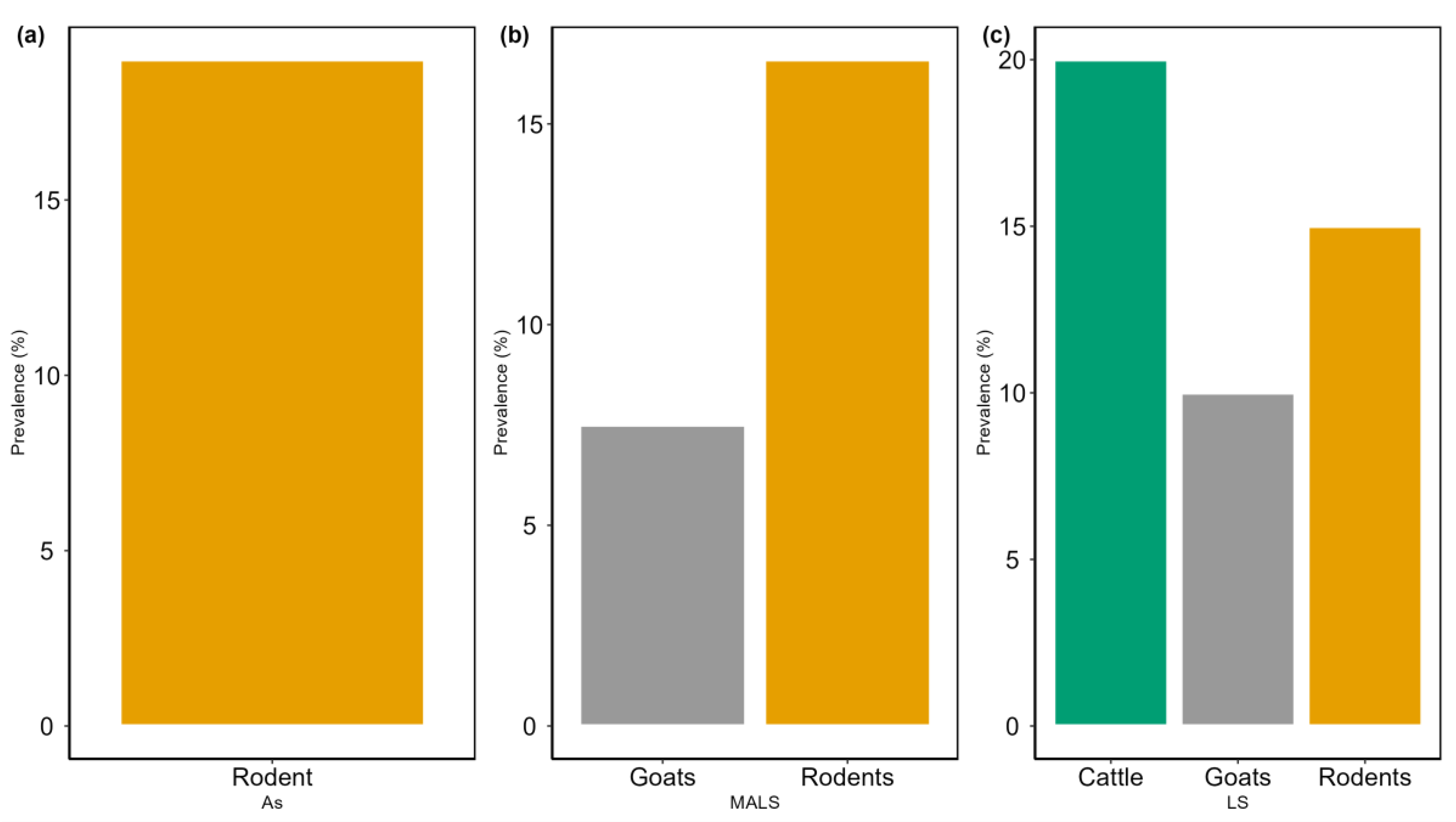
3.2. Prevalence of Leptospira Antibodies in Rodents and Livestock Across Settlements/Villages

The prevalence of Leptospira antibodies differed between rodent species and livestock, with the highest (17.3%) in rodents compared with livestock (13.5%). Within the livestock settlement, the prevalence of seropositive Leptospira was significantly higher (p = 0.001) in livestock (20.0% and 10.0% for cattle and goats, respectively) than in rodents (15.0%) (Figure 2). In the mixed agricultural–livestock settlement, the prevalence was significantly higher (p = 0.001) in rodents (16.6%) than in goats (7.5%). In the agricultural settlement, the prevalence in rodents was 19% (n = 16/84) (Figure 2).
The prevalence of Leptospira antibodies was highest in M. natalensis (21.7%; n = 20/92) relative to R. rattus (12.6%; n = 11/87). There were variations in Leptospira antibodies’ prevalence in rodents associated with the settlement type. The prevalence in rodents was significantly higher (p = 0.001) in the agricultural settlement than in the mixed agricultural–livestock and livestock settlements. There were no significant differences observed in Leptospira seropositivity between crop vegetation areas (25.9%; n = 14/54) and peridomestic areas (28.1%; n = 9/32). However, a significantly lower (p = 0.001) prevalence was observed in houses (8.6%; n = 8/92) (Table 1).

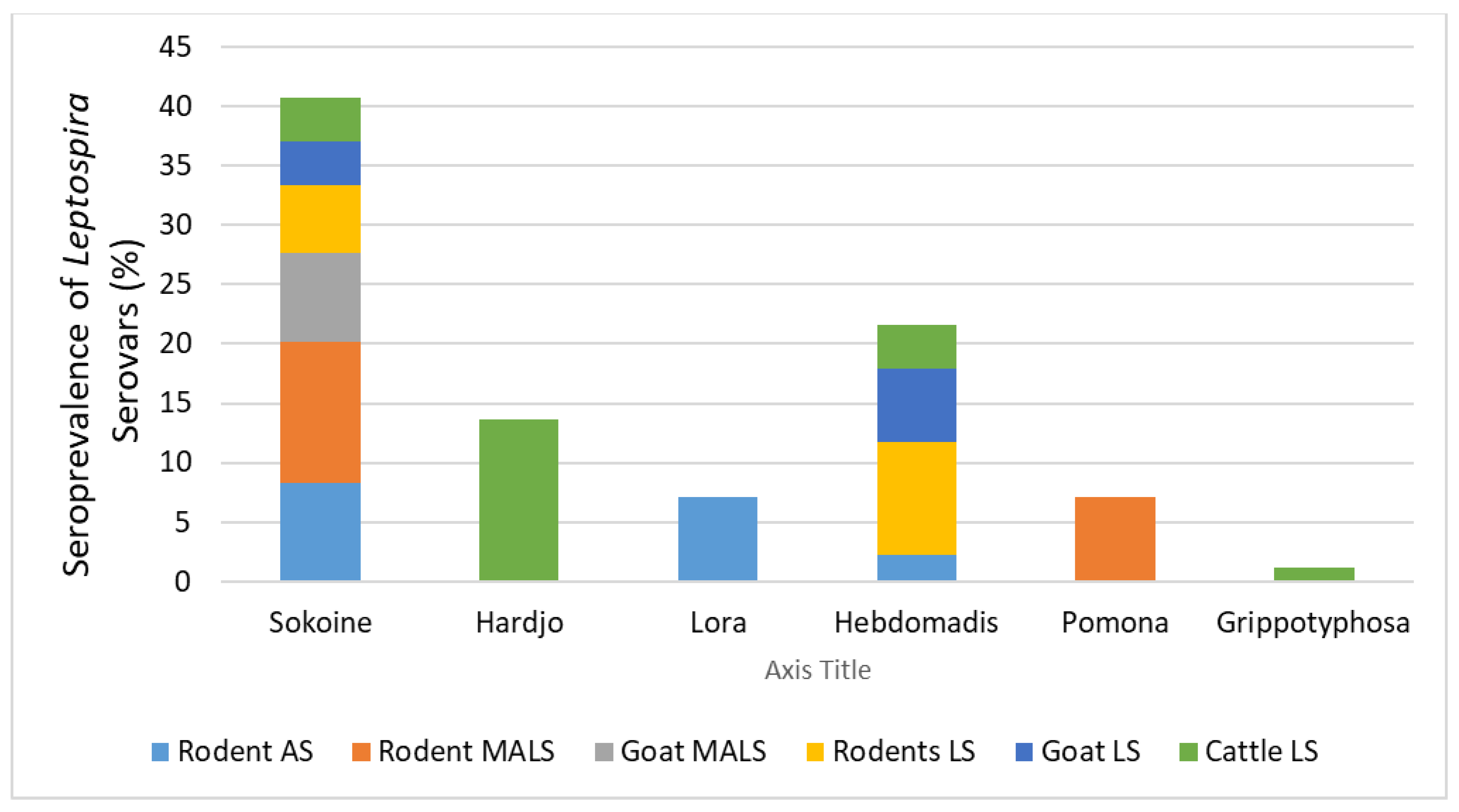
3.3. Seroprevalence of Leptospira Serovars

The seropositivity of rodents for the Leptospira serovars Sokoine (serogroup Icterohaemorrhagiae) and Hebdomadis (serogroup Hebdomadis) was 8.3% (n = 15/179) and 3.9% (n = 7/179), respectively, whereas the same serovars had a seropositivity of 4.5% (n = 9/200) and 3% (n = 6/200) in livestock. The serovars Hardjo (serogroup Sejroe) and Gripothyphosa (serogroup Gripothyphosa) were exclusively found in cattle with a seropositivity of 5.5% (n = 11/200) and 1.5% (n = 3/200), respectively. The serovars Pomona (serogroup Pomona) and Lora (serogroup Australis) were detected in rodents only with a seropositivity of 1.6% (n = 3/179) and 3.3% (n = 6/179), respectively. The serovar Canicola was not detected in either rodents or livestock (Table 2).
There were some variations in seropositivity associated with the type of settlement. Rodents in the agricultural settlement had a higher prevalence of the serovars Sokoine (8.3%; n = 7/84) and Lora (7.1%; n = 6/84) and a low prevalence of serovar Hebdomadis (2.3%; n = 2/84). In the mixed agricultural–livestock settlement, rodents were infected with the serovars Sokoine (11.9%; n = 5/42) and Pomona (7.1%; n = 3/42). In the livestock settlement, rodents were more infected with the serovars Hebdomadis (9.4%; n = 5/53) and Sokoine (5.6%; n = 3/53).
The seroprevalence of Leptospira spp. found in goats in the mixed agricultural–livestock settlement was the serovar Sokoine (7.5%; n = 3/40), whereas goats were positive for the serovars Sokoine (3.7%; n = 3/80) and Hebdomadis (6.2%; n = 5/80) in the livestock settlement. Cattle in the livestock settlement were predominantly infected with the serovars Hardjo (13.7%; n = 11/80), Hebdomadis (3.7%; n = 3/80), Gppotyphosa (1.25%; n =1/80), and Sokoine (3.7%; n = 3/80) (Figure 3).

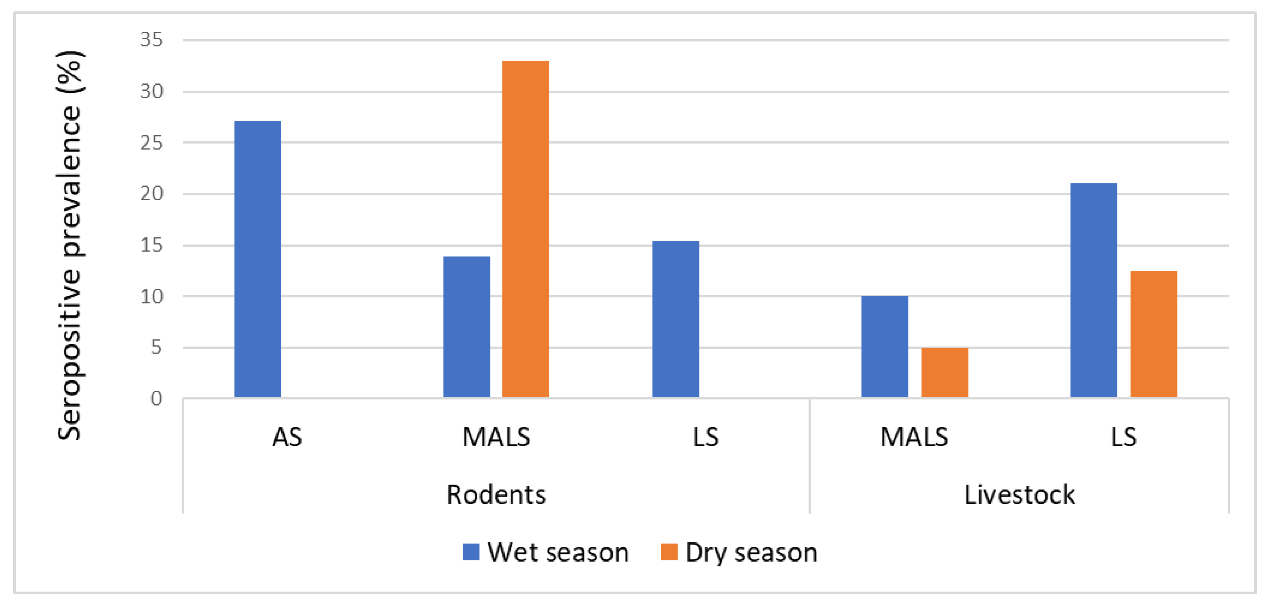
3.4. Seasonal Prevalence of Leptospira Antibodies

There were significant variations in the seropositivity of Leptospira in livestock (p < 0.5) and rodents (p < 0.05) in the three settlements associated with seasonality. In the livestock settlement, the prevalence was higher during the rainy season (18.7%; n = 15/80) relative to the dry season (12.5%; n = 10/80). In the mixed agricultural–livestock settlement, the prevalence in goats was 10% (n = 2/20) and 5% (n = 1/20) in the rainy and dry seasons, respectively (Figure 4).
For rodent species, more positive cases were identified during the rainy season in both agricultural and livestock settlements. In the mixed agricultural–livestock settlement, the prevalence of Leptospira antibodies in rodents was higher during the dry season compared with the rainy season (Figure 4).

4. Discussion

This study investigated the seroprevalence of Leptospira spp. in rodents and livestock (cattle and goats) across three settlements/villages characterized by the dominance of three different occupational activities, namely, agriculture, livestock keeping, and a combination of both.
In the livestock-based settlements where both rodents and livestock were sampled, the prevalence of seropositive Leptospira was higher in cattle than in rodents, followed by goats. This suggests livestock are important sources of infection of Leptospira in communities where livestock keeping is an occupational activity [21,22]. Primary reservoirs of Leptospira could potentially infect and discharge the bacteria into the environment from which the cattle and goats get infected when feeding. Humans probably get infected when they consume undercooked meat or milk that has not been boiled [23,24]. The higher seroprevalence of Leptospira in cattle than in goats could be attributed to feeding habits. Cattle are grazers, and, therefore, they are more likely to become infected by Leptospira from the soil [25].
The lower seroprevalence of Leptospira in rodents in livestock settlements can be attributed to the wider foraging range of livestock [26] and the increased risk of getting infected at water ponds. Rodents, in contrast, have a restricted home range determined by their habitat preferences [27].
The higher seroprevalence of Leptospira in goats in livestock settlements than in mixed agricultural–livestock settlements is probably due to different livestock management practices. In livestock settlements, goats are free-roaming and have frequent contact with other infected animals and contaminated environments, hence elevating their infection risk [28]. The increased prevalence of rodents in agricultural settlements relative to mixed agricultural–livestock and livestock settlements suggests that rodent abundance significantly influences infection dynamics [29,30]. A higher abundance of rodents in an agricultural settlement certainly increases the infection rate among rodents. An increase in the number of rodents most likely elevates the contamination of the environment and transmission between individuals [31,32].
Among rodent species, M. natalensis had a higher seroprevalence rate than R. rattus, indicating that M. natalensis may have a greater and more substantial role in the maintenance and dissemination of Leptospira serovars in the three types of settlements. M. natalensis has higher mobility than R. rattus and frequently burrows in the ground, potentially exposing it to contaminated urine or waste from soil and water [33]. R. rattus is usually more active on roofs [34], which, therefore, renders it less vulnerable to encounters with Leptospira on the ground. M. natalensis was the most common in all three settlements but was more abundant in the agricultural settlement within crop fields and peridomestic areas where there was a higher seroprevalence of Leptospira than in houses. The lower prevalence of Leptospira in houses could be due to an environment that was less favorable to infestation by rodents and the maintenance of good hygienic conditions, which do not favor contamination with Leptospira and transmission [35]. The maintenance of dry conditions in houses does not favor the survival of Leptospira and reduces the risk of human infections [35,36].
The serovars Sokoine and Hebdomadis were the most prevalent in both rodents and livestock, but they have also been documented in multiple locations and isolated from diverse animals in Tanzania, including freshwater fish [37], domestic animals, such as cattle, goats, and sheep [38,39], wild animals, [20], and humans [40]. The serovar Sokoine was found in all three settlements in rodents and livestock. The serovar Sokoine has been isolated in various regions of Tanzania [41], across multiple animal species [37,42,43], and has been reported in Kenya [44].
The serovars Hardjo and Gripotyphosa were found in cattle and showed host specificity. The serovar Hardjo is recognized as the primary Leptospira responsible for significantly impacting cattle output and serving as the principal cause of infection in humans. This serovar has been recorded in many regions of Tanzania, including the northern regions [5], the southern highlands [45], and the western regions [20]. The absence of Grippotyphosa in rodents and Lora and Pomona in livestock may be attributed to the prevailing environmental and climatic conditions during the time of our study; these serovars have been documented in previous studies involving rodents and livestock [8].
The serovar Sokoine was the most dominant in rodents, with the exception of the livestock settlement, whereas Hebdomadis was more prevalent in livestock and rodents in this settlement. This distribution pattern is probably determined by the abundance of hosts, in this case, the higher abundance of cattle. The predominance of the serovar Hebdomadis in livestock, particularly cattle, suggests that environmental and animal husbandry practices in livestock-based villages may facilitate the transmission of this serovar [5]. The coexistence of multiple serovars in animals within a settlement can be attributed to host specificity and/or infection of the same animal species by different serovars. For instance, the serovar Hardjo is unique to cattle, but other serovars may inadvertently infect cattle due to their presence in an area or being shed by other species, such as rodents [45].
The survival of Leptospira is highly influenced by climatic factors and is more prevalent in areas characterized by moderate temperatures, substantial rainfall, and elevated humidity [46]. Our study demonstrated an increased seropositivity during the rainy (wet) season relative to the dry season in both rodents and animals from agricultural and livestock settlements. Precipitation facilitates the transfer of leptospires from contaminated soil, feces, or urine into water sources accessible to livestock during the rainy season [47]. In rodents, the rainy season leads to increased water availability in agricultural fields, resulting in a moist environment that enhances Leptospira survival, while increased resources for rodents enhance reproduction, population abundance, and an increase in bacterial circulation.

5. Conclusions

This research highlights the prevalence of Leptospira antibodies in rodents and livestock in three settlements/villages characterized by different occupational activities. The high prevalence of Leptospira antibodies in rodents in agricultural settlements suggests that targeted interventions, including improved water management strategies and enhanced rodent control measures, are necessary. Mixed agricultural livestock settlements may benefit from initiatives aimed at addressing both agricultural and residential infection sources. In livestock-based settlements, while rodents are less abundant, the high prevalence of infection in cattle must be taken into account, as cattle may be the main source of infection in humans. The significant differences in infection rates across various species of animals and village types highlight the need for appropriate strategies for the prevention of Leptospira infection, which must consider local epidemiological conditions, host species, whether wild or livestock, and occupational activities that render or increase the risk of human infections.

Author Contributions

Conceptualization, M.S. and M.S.; methodology, M.S.; software, M.S.; validation, M.S. and A.S.K.; formal analysis, M.S.; investigation, M.S.; resources, A.W.M.; data curation, M.S. and A.S.K.; writing—original draft preparation, M.S.; writing—review and editing, R.H.M.; visualization, A.S.K.; supervision, R.H.M., A.S.K. and A.W.M.; project administration, M.S.; funding acquisition, A.W.M. and R.H.M. All authors have read and agreed to the published version of the manuscript.

Funding

This study was funded by the African Centre of Excellence for Innovative Rodents Pest Management and Biosensor Technology (ACE IRPM & BTD) (Grant No.: 5799/TZ), based in the Institute of Pest Management, SUA Morogoro, Tanzania and partially by the Global Challenges Research Fund (MR/T028962/1)-Developing Effective Rodent Control Strategies to Reduce Disease Risk in Ecologically and Culturally Diverse Rural Landscapes.

Institutional Review Board Statement

This study was conducted according to the guidelines of the Declaration of Helsinki and approved by the Directorate of Postgraduate Studies, Research, Technology, Transfer, and Consultancy, which is within the Sokoine University of Agriculture (SUA/DPRTC/PFC/D/2021/0002/04) and received a permit from the Office of the Reginal Commissioner, Morogoro, Tanzania (Na. AB. 175/245/01.’1’/07) and the Office of the District Commissioner, the Kilombero district, Tanzania (Na. AB. 23/367/01B/239). The animals were handled in accordance with the ARRIVE guidelines [48].

Data Availability Statement

The data supporting the reported results can be made available from the Institute of Pest Management (IPM) upon request.

Acknowledgments

We acknowledge the funders and the Sokoine University of Agriculture (SUA) for providing the necessary logistical support to complete this study. The authors would also like to thank the Institute of Pest Management staff for their assistance during field and laboratory data collection. We also thank the local authorities and farmers in the working villages for their support.

Conflicts of Interest

The authors declare no conflicts of interest.

References

  1. Adler, B.; de la Peña Moctezuma, A. Leptospira and leptospirosis. Vet. Microbiol. 2010, 140, 287–296. [Google Scholar] [CrossRef] [PubMed]
  2. Costa, F.; Hagan, J.E.; Calcagno, J.; Kane, M.; Torgerson, P.; Martinez-Silveira, M.S.; Stein, C.; Abela-Ridder, B.; Ko, A.I. Global Morbidity and Mortality of Leptospirosis: A Systematic Review. PLoS Negl. Trop. Dis. 2015, 9, e0003898. [Google Scholar] [CrossRef]
  3. Haake, D.A.; Levett, P.N. Leptospirosis in humans. Curr. Top. Microbiol. Immunol. 2015, 387, 65–97. [Google Scholar] [CrossRef]
  4. Mwachui, M.A.; Crump, L.; Hartskeerl, R.; Zinsstag, J.; Hattendorf, J. Environmental and behavioural determinants of leptospirosis transmission: A systematic review. PLoS Neglected Trop. Dis. 2015, 9, e0003843. [Google Scholar] [CrossRef]
  5. Allan, K.J.; Halliday, J.E.B.; Moseley, M.; Carter, R.W.; Ahmed, A.; Goris, M.G.A.; Hartskeerl, R.A.; Keyyu, J.; Kibona, T.; Maro, V.P.; et al. Assessment of animal hosts of pathogenic Leptospira in northern Tanzania. PLoS Negl. Trop. Dis. 2018, 12, e0006444. [Google Scholar] [CrossRef] [PubMed]
  6. Hartskeerl, R.; Collares-Pereira, M.; Ellis, W.A. Emergence, control and re-emerging leptospirosis: Dynamics of infection in the changing world. Clin. Microbiol. Infect. 2011, 17, 494–501. [Google Scholar] [CrossRef] [PubMed]
  7. Ganoza, C.A.; Matthias, M.A.; Collins-Richards, D.; Brouwer, K.C.; Cunningham, C.B.; Segura, E.R.; Gilman, R.H.; Gotuzzo, E.; Vinetz, J.M. Determining risk for severe leptospirosis by molecular analysis of environmental surface waters for pathogenic Leptospira. PLoS Med. 2006, 3, 1329–1340. [Google Scholar] [CrossRef] [PubMed]
  8. Majawa, C.A.; Lupindu, A.M.; Mhamphi, G.G.; Katakweba, A.A.S. Seroprevalence of Leptospira antibodies in rodents and shrews of Kibondo and Kakonko Districts, Kigoma region, Tanzania. Malawi J. Sci. Technol. 2023, 15, 1. [Google Scholar]
  9. Msofe, N.; Sheng, L.; Lyimo, J. Land use change trends and their driving forces in the Kilombero Valley Floodplain, Southeastern Tanzania. Sustainability 2019, 11, 505. [Google Scholar] [CrossRef]
  10. Sasaki, D.M.; Pang, L.; Minette, H.P.; Wakida, C.K.; Fujimoto, W.J.; Manea, S.J.; Kunioka, R.; Middleton, C.R. Active surveillance and risk factors for leptospirosis in Hawaii. Am. J. Trop. Med. Hyg. 1993, 48, 35–43. [Google Scholar] [CrossRef] [PubMed]
  11. Chiani, Y.T.; Jacob, P.; Mayora, G.; Aquino, D.S.; Quintana, R.D.; Mesa, L. Presence of Leptospira spp. in a Mosaic of Wetlands Used for Livestock Raising under Differing Hydroclimatic Conditions. Appl. Environ. Microbiol. 2023, 89, e0197122. [Google Scholar] [CrossRef]
  12. Hercik, C.; Cosmas, L.; Mogeni, O.D.; Wamola, N.; Kohi, W.; Houpt, E.; Liu, J.; Ochieng, C.; Onyango, C.; Fields, B.; et al. A combined syndromic approach to examine viral, bacterial, and parasitic agents among febrile patients: A pilot study in Kilombero, Tanzania. Am. J. Trop. Med. Hyg. 2018, 98, 625–632. [Google Scholar] [CrossRef] [PubMed]
  13. Mgode, G.F.; Machang’u, R.S.; Mhamphi, G.G.; Katakweba, A.; Mulungu, L.S.; Durnez, L.; Leirs, H.; Hartskeerl, R.A.; Belmain, S.R. Leptospira Serovars for Diagnosis of Leptospirosis in Humans and Animals in Africa: Common Leptospira Isolates and Reservoir Hosts. PLoS Negl. Trop. Dis. 2015, 9, e0004251. [Google Scholar] [CrossRef] [PubMed]
  14. Goris, M.G.A.; Leeflang, M.M.G.; Loden, M.; Wagenaar, J.F.P.; Klatser, P.R.; Hartskeerl, R.A.; Boer, K.R. Prospective evaluation of three rapid diagnostic tests for diagnosis of human leptospirosis. PLoS Negl. Trop. Dis. 2013, 7, e2290. [Google Scholar] [CrossRef]
  15. Motto, S.K.; Shirima, G.M.; de Clare Bronsvoort, B.M.; Cook, E.A.J. Epidemiology of leptospirosis in tanzania: A review of the current status, serogroup diversity and reservoirs. PLoS Negl. Trop. Dis. 2021, 15, e0009918. [Google Scholar] [CrossRef] [PubMed]
  16. Schoonman, L.; Swai, E.S. Risk factors associated with the seroprevalence of leptospirosis, amongst at-risk groups in and around Tanga city, Tanzania. Ann. Trop. Med. Parasitol. 2009, 103, 711–718. [Google Scholar] [CrossRef]
  17. Biggs, H.M.; Hertz, J.T.; Munishi, O.M.; Galloway, R.L.; Marks, F.; Saganda, W.; Maro, V.P.; Crump, J.A. Estimating Leptospirosis Incidence Using Hospital-Based Surveillance and a Population-Based Health Care Utilization Survey in Tanzania. PLoS Negl. Trop. Dis. 2013, 7, e2589. [Google Scholar] [CrossRef]
  18. Goris, M.G.A.; Hartskeerl, R.A. Leptospirosis serodiagnosis by the microscopic agglutination test. Curr. Protoc. Microbiol. 2014, 32, 12E.5.1–12E.5.18. [Google Scholar] [CrossRef] [PubMed]
  19. Schoonman, L.; Swai, E.S. Herd- and animal-level risk factors for bovine leptospirosis in Tanga region of Tanzania. Trop. Anim. Heal. Prod. 2010, 42, 1565–1572. [Google Scholar] [CrossRef]
  20. Assenga, J.A.; Matemba, L.E.; Muller, S.K.; Mhamphi, G.G.; Kazwala, R.R. Predominant leptospiral serogroups circulating among humans, livestock and wildlife in Katavi-Rukwa ecosystem, Tanzania. PLoS Negl. Trop. Dis. 2015, 9, e0003607. [Google Scholar] [CrossRef] [PubMed]
  21. Sunaryo, S.; Priyanto, D. Leptospirosis in rats and livestock in Bantul and Gunungkidul district, Yogyakarta, Indonesia. Vet. World 2022, 15, 1449–1455. [Google Scholar] [CrossRef] [PubMed]
  22. Hairgrove, T.B. Leptospirosis in cattle. In Proceedings of the Thirty-Seventh Annual Conference, Fort Worth, TX, USA, 23–25 September 2004; Available online: https://bovine-ojs-tamu.tdl.org/AABP/article/view/4897 (accessed on 12 October 2024).
  23. Di Azevedo, M.I.N.; Lilenbaum, W. An overview on the molecular diagnosis of animal leptospirosis. Lett. Appl. Microbiol. 2021, 72, 496–508. [Google Scholar] [CrossRef]
  24. Cook, E.A.J.; de Glanville, W.A.; Thomas, L.F.; Kariuki, S.; Bronsvoort, B.M.d.C.; Fèvre, E.M. Risk factors for leptospirosis seropositivity in slaughterhouse workers in western Kenya. Occup. Environ. Med. 2017, 74, 357–365. [Google Scholar] [CrossRef] [PubMed]
  25. Herrik, A.L.; Mogensen, N.; Svenning, J.-C.; Buitenwerf, R. Rotational grazing with cattle-free zones supports the coexistence of cattle and wild herbivores in African rangelands. J. Appl. Ecol. 2023, 60, 2154–2166. [Google Scholar] [CrossRef]
  26. Ruvuga, P.R.; Wredle, E.; Nyberg, G.; Hussein, R.A.; Masao, C.A.; Selemani, I.S.; Sangeda, A.Z.; Kronqvist, C. Evaluation of rangeland condition in miombo woodlands in eastern Tanzania in relation to season and distance from settlements. J. Environ. Manag. 2021, 290, 112635. [Google Scholar] [CrossRef] [PubMed]
  27. Ssuuna, J.; Makundi, R.H.; Chidodo, S.J.; Isabirye, M.; Mbije, N.E.; Mulungu, L.S. Spatio-temporal home range of the dominant rodent species in Mabira central forest reserve, Uganda. BMC Evol. Biol. 2023, 23, 40. [Google Scholar] [CrossRef] [PubMed]
  28. Gomard, Y.; Dellagi, K.; Goodman, S.M.; Mavingui, P.; Tortosa, P. Tracking Animal Reservoirs of Pathogenic Leptospira: The Right Test for the Right Claim. Trop. Med. Infect. Dis. 2021, 6, 205. [Google Scholar] [CrossRef]
  29. Herrera, J.P.; Wickenkamp, N.R.; Turpin, M.; Baudino, F.; Tortosa, P.; Goodman, S.M.; Soarimalala, V.; Ranaivoson, T.N.; Nunn, C.L. Effects of land use, habitat characteristics, and small mammal community composition on leptospira prevalence in northeast madagascar. PLoS Negl. Trop. Dis. 2020, 14, e0008946. [Google Scholar] [CrossRef] [PubMed]
  30. Peterson, A.C.; Ghersi, B.M.; Riegel, C.; Wunder, E.A.; Childs, J.E.; Blum, M.J. Amplification of pathogenic Leptospira infection with greater abundance and co-occurrence of rodent hosts across a counter-urbanizing landscape. Mol. Ecol. 2021, 30, 2145–2161. [Google Scholar] [CrossRef]
  31. Ricardo, T.; Jacob, P.; Chiani, Y.; Schmeling, M.F.; Cornejo, P.; Ojeda, A.A.; Teta, P.V.; Vanasco, N.B.; Previtali, M.A. Seroprevalence of leptospiral antibodies in rodents from riverside communities of Santa Fe, Argentina. PLoS Negl. Trop. Dis. 2020, 14, e0008222. [Google Scholar] [CrossRef]
  32. Rosli, M.Z.; Mohd-Taib, F.S.; Khoo, J.J.; Chee, H.Y.; Wong, Y.P.; Shafie, N.J.; Mohamed, N.Z.; AbuBakar, S.; Nor, S.M. A Multi-landscape Assessment of Leptospira Prevalence on a Diversity of Small Mammals. Ecohealth 2023, 20, 208–224. [Google Scholar] [CrossRef]
  33. Mulungu, L.S.; Mahlaba, T.A.; Massawe, A.W.; Kennis, J.; Crauwels, D.; Eiseb, S.; Monadjem, A.; Makundi, R.H.; Katakweba, A.A.S.; Leirs, H.; et al. Dietary differences of the multimammate mouse, Mastomys natalensis (Smith, 1834), across different habitats and seasons in Tanzania and Swaziland. Wildl. Res. 2011, 38, 640–646. [Google Scholar] [CrossRef]
  34. Harper, G.; Dickinson, K.J.M.; Seddon, P.J. Habitat use by three rat species (Rattus spp.) on Stewart Island/Rakiura, New Zealand. N. Z. J. Ecol. 2005, 29, 251–260. [Google Scholar]
  35. Munoz-Zanzi, C.; Mason, M.; Encina, C.; Gonzalez, M.; Berg, S. Household characteristics associated with rodent presence and Leptospira infection in rural and urban communities from Southern Chile. Am. J. Trop. Med. Hyg. 2014, 90, 497–506. [Google Scholar] [CrossRef] [PubMed]
  36. Muñoz-Zanzi, C.; Mason, M.R.; Encina, C.; Astroza, A.; Romero, A. Leptospira Contamination in Household and Environmental Water in Rural Communities in Southern Chile. Int. J. Environ. Res. Public Health 2014, 11, 6666–6680. [Google Scholar] [CrossRef] [PubMed]
  37. Mgode, G.; Mhamphi, G.; Katakweba, A.; Thomas, M. Leptospira infections in freshwater fish in Morogoro Tanzania: A hidden public health threat. Tanzan. J. Health Res. 2014, 16, 112–117. [Google Scholar] [CrossRef]
  38. Allan, K.J.; Biggs, H.M.; Halliday, J.E.B.; Kazwala, R.R.; Maro, V.P.; Cleaveland, S.; Crump, J.A. Epidemiology of leptospirosis in Africa: A systematic review of a neglected zoonosis and a paradigm for ‘One Health’ in Africa. PLoS Negl. Trop. Dis. 2015, 9, e0003899. [Google Scholar] [CrossRef]
  39. Mgode, G.F.; Japhary, M.M.; Mhamphi, G.G.; Kiwelu, I.; Athaide, I.; Machang’u, R.S. Leptospirosis in sugarcane plantation and fishing communities in Kagera northwestern Tanzania. PLOS Negl. Trop. Dis. 2019, 13, e0007225. [Google Scholar] [CrossRef] [PubMed]
  40. Maze, M.J.; Shirima, G.M.; Lukambagire, A.-H.S.; Bodenham, R.F.; Rubach, M.P.; Cash-Goldwasser, S.; Carugati, M.; Thomas, K.M.; Sakasaka, P.; Mkenda, N.; et al. Prevalence and risk factors for human leptospirosis at a hospital serving a pastoralist community, Endulen, Tanzania. PLoS Negl. Trop. Dis. 2023, 17, e0011855. [Google Scholar] [CrossRef] [PubMed]
  41. Msemwa, B.; Mirambo, M.M.; Silago, V.; Samson, J.M.; Majid, K.S.; Mhamphi, G.; Genchwere, J.; Mwakabumbe, S.S.; Mngumi, E.B.; Mgode, G.; et al. Existence of Similar Leptospira Serovars among Dog Keepers and Their Respective Dogs in Mwanza, Tanzania, the Need for a One Health Approach to Control. Pathogens 2021, 10, 609. [Google Scholar] [CrossRef]
  42. Kessy, M.J.; Machang’u, R.S.; Swai, E.S. A microbiological and serological study of leptospirosis among pigs in the Morogoro municipality, Tanzania. Trop. Anim. Health Prod. 2010, 42, 523–530. [Google Scholar] [CrossRef]
  43. Said, K.; Bakari, G.G.; Machang’u, R.; Katakweba, A.S.; Muhairwa, A.P. Seroprevalence of canine leptospirosis, in Urban and Periurban, Morogoro, Tanzania. Afr. J. Microbiol. Res. 2018, 12, 481–487. [Google Scholar] [CrossRef]
  44. Ngugi, J.N.; Fèvre, E.M.; Mgode, G.F.; Obonyo, M.; Mhamphi, G.G.; Otieno, C.A.; Cook, E.A.J. Seroprevalence and associated risk factors of leptospirosis in slaughter pigs; a neglected public health risk, western Kenya. BMC Vet. Res. 2019, 15, 403. [Google Scholar] [CrossRef] [PubMed]
  45. Motto, S.K.; Hernandez-Castro, L.E.; Shirima, G.M.; Mengele, I.J.; Bwatota, S.F.; Bronsvoort, B.M.d.C.; Lyatuu, E.T.; Komwihangilo, D.M.; Cook, E.A.J. Seroepidemiology of Leptospira serovar Hardjo and associated risk factors in smallholder dairy cattle in Tanzania. PLOS Negl. Trop. Dis. 2023, 17, e0011199. [Google Scholar] [CrossRef] [PubMed]
  46. Joshi, Y.P.; Kim, E.-H.; Cheong, H.-K. The influence of climatic factors on the development of hemorrhagic fever with renal syndrome and leptospirosis during the peak season in Korea: An ecologic study. BMC Infect. Dis. 2017, 17, 406. [Google Scholar] [CrossRef] [PubMed]
  47. Thibeaux, R.; Geroult, S.; Benezech, C.; Chabaud, S.; Soupé-Gilbert, M.-E.; Girault, D.; Bierque, E.; Goarant, C. Seeking the environmental source of Leptospirosis reveals durable bacterial viability in river soils. PLoS Negl.Trop. Dis. 2017, 11, e0005414. [Google Scholar] [CrossRef] [PubMed]
  48. Mcgrath, J.C.; Drummond, G.B.; Mclachlan, E.M.; Kilkenny, C.; Wainwright, C.L. Guidelines for reporting experiments involving animals: The ARRIVE guidelines. Br. J. Pharmacol. 2010, 160, 1573–1576. [Google Scholar] [CrossRef]
Figure 1. Map illustrating the geographical position of the study villages within the Kilombero wetland, in the Kilombero district, Tanzania. a = Misufini Village (agriculture-based settlement); b = Kipingu Village (livestock-based settlement), and c = Sagamaganga Village (mixed agricultural–livestock settlement).
Pathogens 13 01059 g001
Figure 2. Variations in the prevalence of Leptospira seropositivity in rodents and livestock in three different settlements in the Kilombero Valley, Tanzania. (As: agricultural settlement, MALS: mixed agricultural–livestock settlement, and LS: livestock settlement). (a) Rodent prevalence, (b) Goats and Rodent prevalence (c) Cattle, Goats and Rodent prevalence.
Pathogens 13 01059 g002
Figure 3. Seropositivity in relation to Leptospira serovars detected in rodents, cattle, and goats from three settlements/villages. As: agricultural settlement, MALS: mixed agricultural–livestock settlement, and LS: livestock settlement.
Pathogens 13 01059 g003
Figure 4. Seasonal differences in the prevalence of anti-Leptospira antibodies in agricultural, mixed agricultural–livestock, and livestock settlements in the Kilombero Valley, Tanzania. (As: agricultural settlement, MALS: mixed agricultural–livestock settlement, and LS: livestock settlement).
Pathogens 13 01059 g004
Table 1. The prevalence of Leptospira seropositivity in rodents (M. natalensis and R. rattus) captured in houses, peridomestic surroundings, and agricultural fields in three settlements/villages in the Kilombero Valley, Tanzania.
AgriculturalSettlement/Village Category Mixed (Agricultural–Livestock)Livestock
M. natalensis R. rattus M. natalensis R. rattus M. natalensis R. rattus
Agricultural fields18.4% (7/38)0 (0/0)25% (3/12)100% (1/1)100% (3/3)0% (0/0)
Peridomestic surroundings11.5% (3/26)50% (1/2)100% (2/2)100% (1/1)0% (0/0)100% (1/1)
In houses20% (2/10)37.5% (3/8)0% (0/1)0% (0/25)0% (0/0)8.1% (4/49)
Table 2. Leptospira serovars and their corresponding serogroups identified from livestock and rodents in the Kilombero Valley, Tanzania.
Livestock Rodents
CattleGoats M. natalensis R. rattus
Leptospira SerovarSerogroup
SokoineIcterohaemorrhagiae3.7% (n = 3/80)5% (n = 6/120)7.6% (n = 7/92)9% (n = 8/87)
HebdomadisHebdomadis3.7% (n = 3/80)4% (n = 5/120)4.3% (n = 4/92)3.4% (n=3/87)
LoraAustralis006.5% (n = 6/92)0
GripothyphosaGripothyphosa1.2% (n = 1/80)000
CanicolaCanicola0000
PomonaPomona0 3.2% (n = 3/92)0
HardjoSejroe13% (n = 11/80)000
Disclaimer/Publisher’s Note: The statements, opinions and data contained in all publications are solely those of the individual author(s) and contributor(s) and not of MDPI and/or the editor(s). MDPI and/or the editor(s) disclaim responsibility for any injury to people or property resulting from any ideas, methods, instructions or products referred to in the content.

