
Unusual Masquerading Infraorbital Mass—A Case Report of Human Ocular Dirofilariosis Found in A Ukrainian Patient in Switzerland

 ,
,  , and
, and 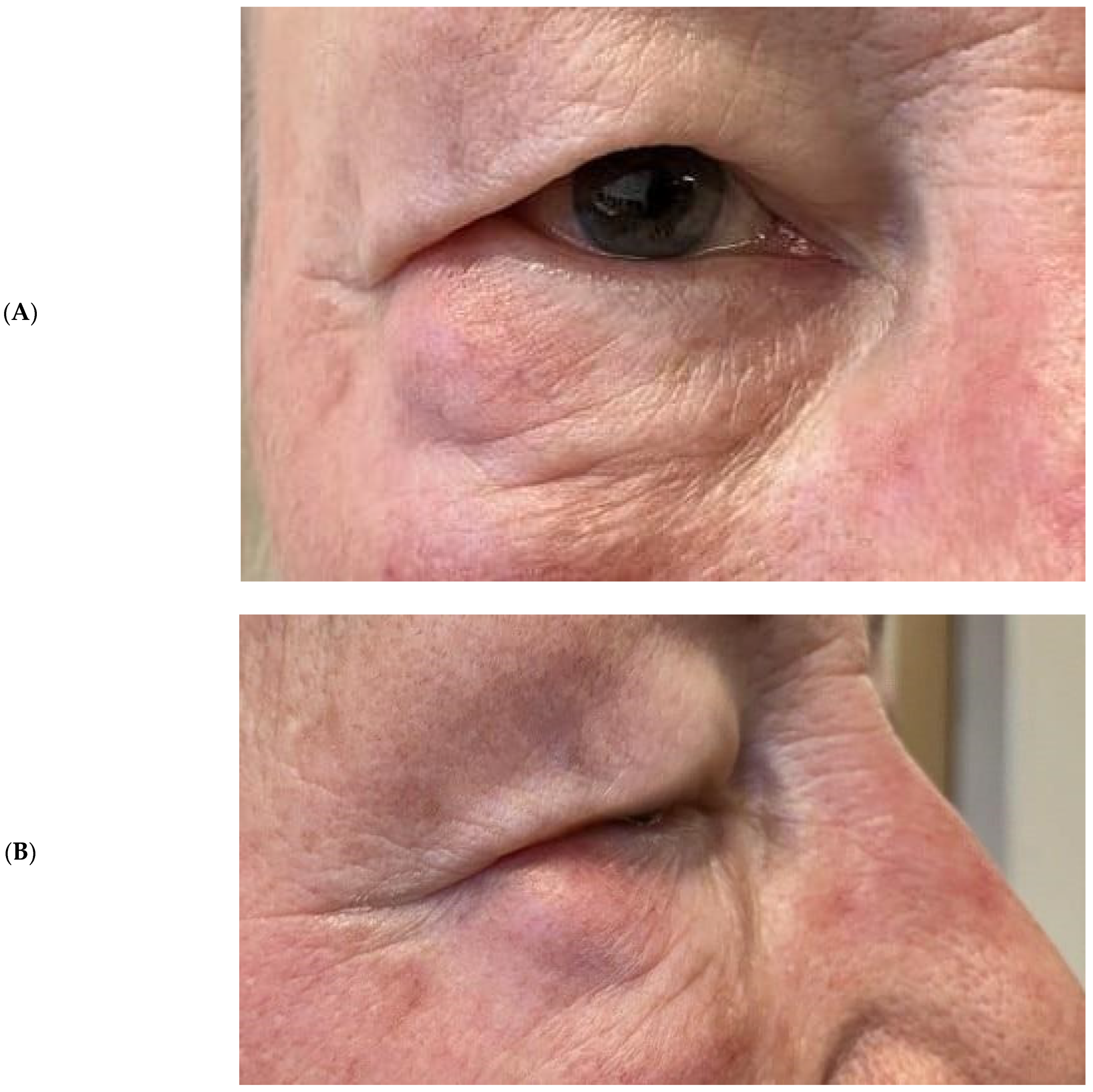
Abstract
1. Introduction
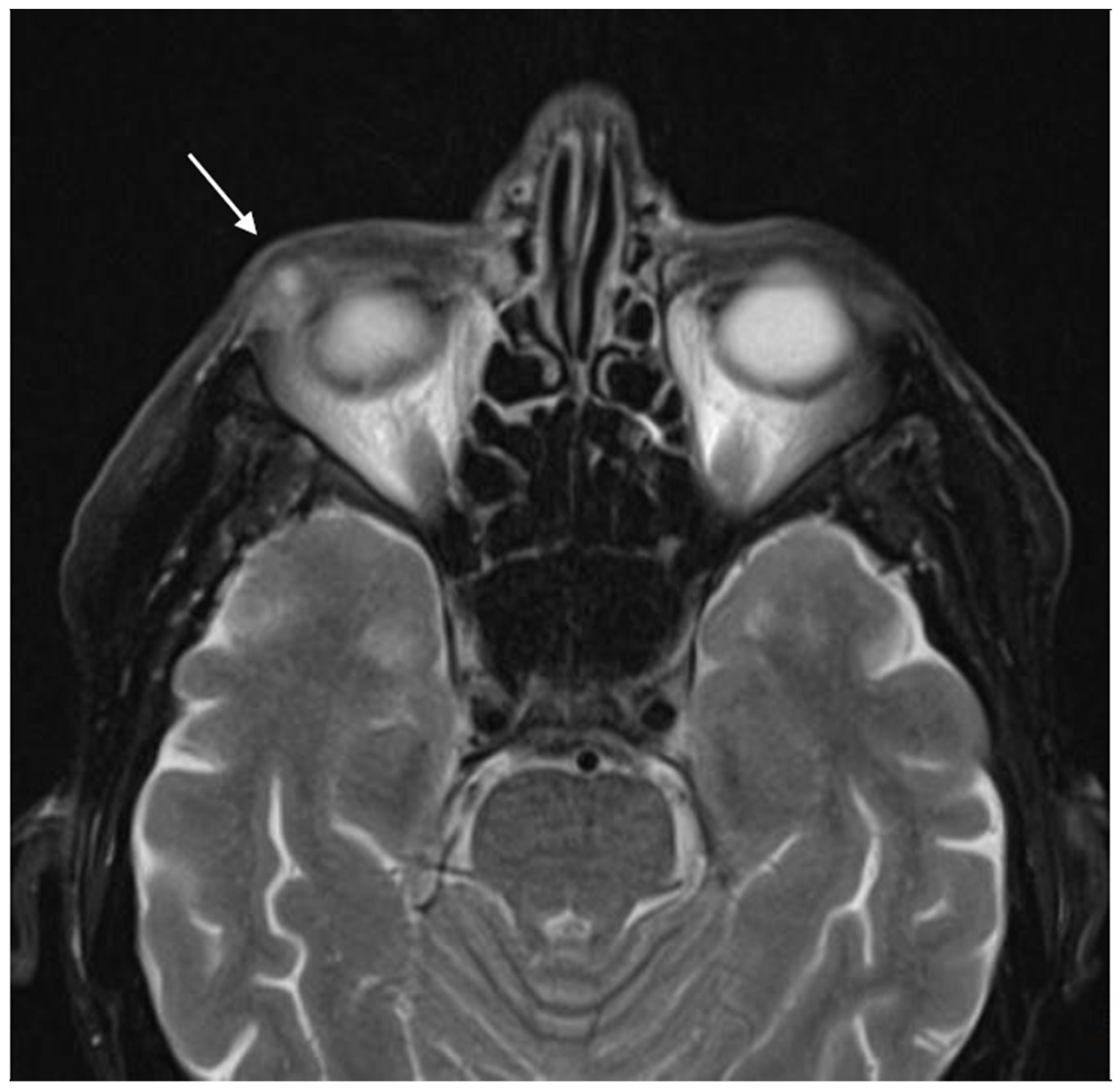
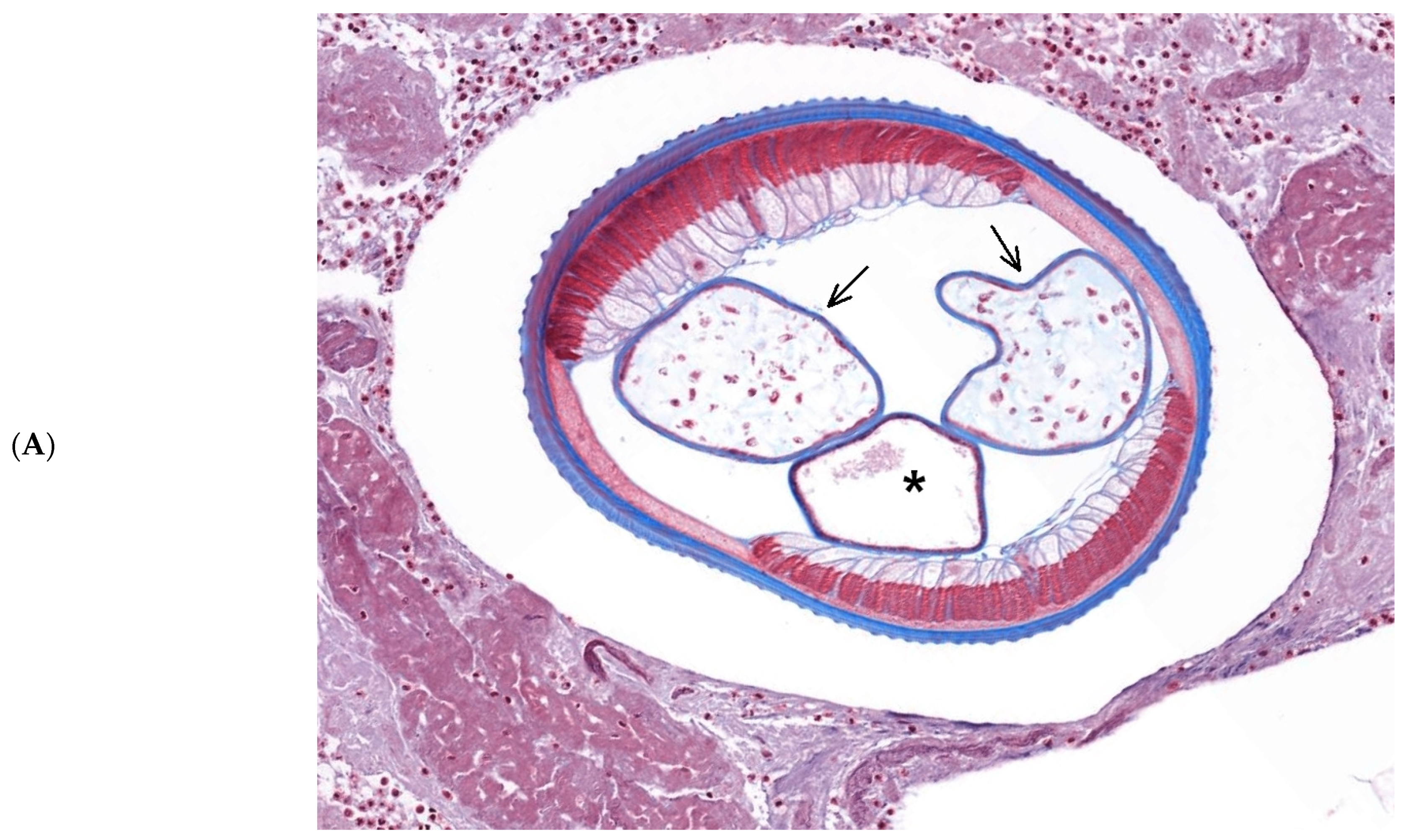
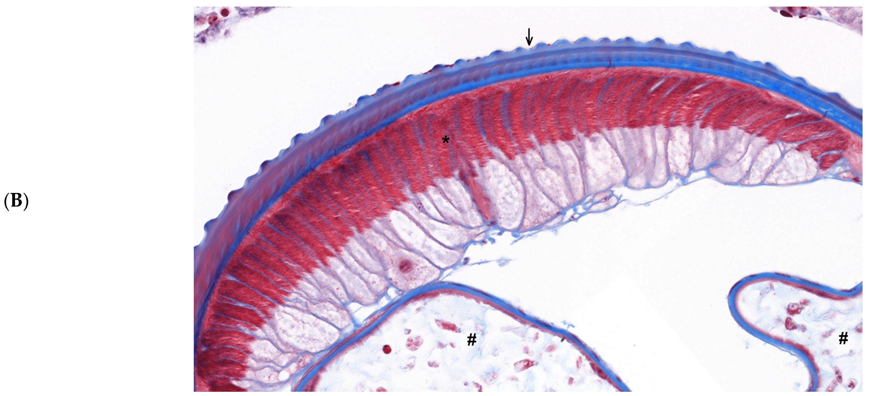
2. Case Report
3. Discussion
4. Conclusions
Author Contributions
Funding
Institutional Review Board Statement
Informed Consent Statement
Data Availability Statement
Conflicts of Interest
References
- Khan, S.N.; Sepahdari, A.R. Orbital masses: CT and MRI of common vascular lesions, benign tumors, and malignancies. Saudi J. Ophthalmol. 2012, 26, 373–383. [Google Scholar] [CrossRef] [PubMed]
- Gutiérrez-Jara, J.P.; Salazar-Viedma, M.; González, C.R.; Cancino-Faure, B. The emergence of Dirofilaria repens in a non-endemic area influenced by climate change: Dynamics of transmission using a mathematical model. Acta Tropica 2022, 226, 106230. [Google Scholar] [CrossRef] [PubMed]
- Capelli, G.; Genchi, C.; Baneth, G.; Bourdeau, P.; Brianti, E.; Cardoso, L.; Danesi, P.; Fuehrer, H.-P.; Giannelli, A.; Ionică, A.M.; et al. Recent advances on Dirofilaria repens in dogs and humans in Europe. Parasites Vectors 2018, 11, 663. [Google Scholar] [CrossRef] [PubMed]
- Rymgayłło-Jankowska, B.; Ziaja-Sołtys, M.; Flis, B.; Bogucka-Kocka, A.; Żarnowski, T. Subcutaneous Dirofilariosis of the Eyelid Brought to Poland from the Endemic Territory of Ukraine. Pathogens 2023, 12, 196. [Google Scholar] [CrossRef] [PubMed]
- Simón, F.; Diosdado, A.; Siles-Lucas, M.; Kartashev, V.; González-Miguel, J. Human dirofilariosis in the 21st century. A scoping review of clinical cases reported in the literature. Transbound. Emerg. Dis. 2022, 69, 2424–2439. [Google Scholar] [CrossRef]
- Salamatin, R.; Pavlikovska, T.M.; Sagach, O.S.; Nikolayenko, S.M.; Kornyushin, V.V.; Kharchenko, V.O.; Masny, A.; Cielecka, D.; Konieczna-Sałamatin, J.; Conn, D.B.; et al. Human dirofilariasis due to Dirofilaria repens in Ukraine, an emergent zoonosis: Epidemiological report of 1465 cases. Acta Parasitol. 2013, 58, 592–598. [Google Scholar] [CrossRef]
- Pampiglione, S.; Rivasi, F. Human dirofilariasis due to Dirofilaria (Nochtiella) repens: An update of world literature from 1995 to 2000. Parassitologia 2000, 42, 231–254. [Google Scholar]
- Petruschke, G.; Rossi, L.; Genchi, C.; Pollono, F. Canine dirofilariasis in the canton of Ticino and in the neighboring areas of northern Italy. Schweiz. Arch. Tierheilkd. 2001, 143, 141–147. [Google Scholar]
- Smitha, M.; Rajendran, V.R.; Devarajan, E.; Anitha, P.M. Case report: Orbital dirofilariasis. Indian J. Radiol. Imaging 2008, 18, 60–62. [Google Scholar]
- Strianese, D.; Martini, A.; Molfino, G.; Falabella, L.; Tranfa, F. Orbital dirofilariasis. Eur. J. Ophthalmol. 1998, 8, 258–262. [Google Scholar] [CrossRef]
- Addario, C. Su di un mematode dell’occhio umano. Ann. Ottalmol. Clin. Ocul. 1985, 14, 135–147. [Google Scholar]
- Font, R.L.; Neafie, R.C.; Pery, H.D. Subcutaneous dirofilaria of the eyelid and ocular adnexa. Report of six cases. Arch. Ophthalmol. 1980, 98, 1079–1082. [Google Scholar] [CrossRef]
- Makaveev, I.; Galev, A.; Marinova, P.; Martinov, M.; Harizanov, R.; Rainova, I.; Karsheva, M. First case of ocular dirofilariosis in Bulgaria caused by gravid female Dirofilaria repens. Ann. Parasitol. 2022, 68, 191–193. [Google Scholar]
- Bergin, D.J.; Parmley, V. Orbital neurilemoma. Arch. Ophthalmol. 1988, 106, 414–415. [Google Scholar] [CrossRef]
- Goto, H.; Mori, H.; Shirato, S.; Usui, M. Ciliary body schwannoma successfully treated by local resection. Jpn. J. Ophthalmol. 2006, 50, 543–546. [Google Scholar] [CrossRef]
- Smith, P.A.; Damato, B.E.; Ko, M.K.; Lyness, R.W. Anterior uveal neurilemmoma—A rare neoplasm simulating malignant melanoma. Br. J. Ophthalmol. 1987, 71, 34–40. [Google Scholar] [CrossRef]
- Fan, J.T.; Campbell, R.J.; Robertson, D.M. A survey of intraocular schwannoma with a case report. Can. J. Ophthalmol. 1995, 30, 37–41. [Google Scholar]
- Pakdel, F.; Ghadimi, H.; Nozarian, Z.; Amoli, F.A.; Pirmarzdashti, N.; Karimi, M.; Mehrpour, M. Orbital Dirofilariasis Masquerading as Orbital Rhabdomyosarcoma. J. Ophthalmic Vis. Res. 2022, 17, 587–591. [Google Scholar] [CrossRef]
- Otranto, D.; Dantas-Torres, F.; Cebeci, Z.; Yeniad, B.; Buyukbabani, N.; Boral, O.B.; Gustinelli, A.; Mounir, T.; Mutafchiev, Y.; Bain, O. Human ocular filariasis: Further evidence on the zoonotic role of Onchocerca lupi. Parasit Vectors. 2012, 5, 84. [Google Scholar]
- Džamić, A.M.; Čolović, I.V.; Arsić-Arsenijević, V.S.; Stepanović, S.; Boričić, I.; Džamić, Z.; Mitrović, S.M.; Rašić, D.M.; Stefanović, I.; Latković, Z.; et al. Human Dirofilaria repens infection in Serbia. J. Helminthol. 2009, 83, 129–137. [Google Scholar] [CrossRef]
- Kalogeropoulos, C.; Stefaniotou, M.; Gorgoli, K.; Papadopoulou, C.; Pappa, C.; Paschidis, C. Ocular dirofilariasis: A case series of 8 patients. Middle East Afr. J. Ophthalmol. 2014, 21, 312–316. [Google Scholar] [CrossRef] [PubMed]
- Stringfellow, G.J.; Francis, I.C.; Coroneo, M.T.; Walker, J. Orbital dirofilariasis. Clin. Exp. Ophthalmol. 2002, 30, 378–380. [Google Scholar] [CrossRef] [PubMed]
- Kartashev, V.; Afonin, A.; González-Miguel, J.; Sepúlveda, R.; Simón, L.; Morchón, R.; Simón, F. Regional warming and emerging vector-borne zoonotic dirofilariosis in the Russian Federation, Ukraine, and other post-Soviet states from 1981 to 2011 and projection by 2030. BioMed Res. Int. 2014, 2014, 858936. [Google Scholar] [PubMed]


Disclaimer/Publisher’s Note: The statements, opinions and data contained in all publications are solely those of the individual author(s) and contributor(s) and not of MDPI and/or the editor(s). MDPI and/or the editor(s) disclaim responsibility for any injury to people or property resulting from any ideas, methods, instructions or products referred to in the content. |
© 2023 by the authors. Licensee MDPI, Basel, Switzerland. This article is an open access article distributed under the terms and conditions of the Creative Commons Attribution (CC BY) license (https://creativecommons.org/licenses/by/4.0/).
Share and Cite
Menna, F.; Billia, F.; Papazoglou, A.; Tschopp, M.; Grobholz, R. Unusual Masquerading Infraorbital Mass—A Case Report of Human Ocular Dirofilariosis Found in A Ukrainian Patient in Switzerland. Pathogens 2023, 12, 982. https://doi.org/10.3390/pathogens12080982
Menna F, Billia F, Papazoglou A, Tschopp M, Grobholz R. Unusual Masquerading Infraorbital Mass—A Case Report of Human Ocular Dirofilariosis Found in A Ukrainian Patient in Switzerland. Pathogens. 2023; 12(8):982. https://doi.org/10.3390/pathogens12080982
Chicago/Turabian StyleMenna, Feliciana, Filippo Billia, Anthia Papazoglou, Markus Tschopp, and Rainer Grobholz. 2023. "Unusual Masquerading Infraorbital Mass—A Case Report of Human Ocular Dirofilariosis Found in A Ukrainian Patient in Switzerland" Pathogens 12, no. 8: 982. https://doi.org/10.3390/pathogens12080982
APA StyleMenna, F., Billia, F., Papazoglou, A., Tschopp, M., & Grobholz, R. (2023). Unusual Masquerading Infraorbital Mass—A Case Report of Human Ocular Dirofilariosis Found in A Ukrainian Patient in Switzerland. Pathogens, 12(8), 982. https://doi.org/10.3390/pathogens12080982