Article

Whole Genome Sequencing and Comparative Genomic Analysis of Chlamydia gallinacea Field Strains Isolated from Poultry in Poland

by
Kinga Zaręba-Marchewka
1,*,
Arkadiusz Bomba
2,
Sabine Scharf
3,
Krzysztof Niemczuk
1,4,
Christiane Schnee
3 and
Monika Szymańska-Czerwińska
1,4
1
Department of Cattle and Sheep Diseases, National Veterinary Research Institute, Al. Partyzantow 57, 24-100 Pulawy, Poland
2
Department of Omics Analyses, National Veterinary Research Institute, Al. Partyzantow 57, 24-100 Pulawy, Poland
3
Institute of Molecular Pathogenesis, Friedrich-Loeffler-Institut (Federal Research Institute for Animal Health), Naumburger Str. 96 a, D-07743 Jena, Germany
4
Laboratory of Serological Diagnosis, National Veterinary Research Institute, Al. Partyzantow 57, 24-100 Pulawy, Poland
*
Author to whom correspondence should be addressed.
Pathogens 2023, 12(7), 891; https://doi.org/10.3390/pathogens12070891
Submission received: 30 May 2023 / Revised: 22 June 2023 / Accepted: 27 June 2023 / Published: 29 June 2023
(This article belongs to the Section Bacterial Pathogens)

Abstract

:
Chlamydia gallinacea is an intracellular bacterium belonging to the Chlamydiaceae family. Poultry is considered to be the major reservoir of this agent, which has worldwide distribution and a particularly consistent worldwide occurrence in chicken flocks. The bacterium has been linked to respiratory disease in humans but without definitive confirmation; nevertheless, while it has not been proved to be the cause of human respiratory disease, a recent report from Italy verified its bird-to-human transmission. This aspect being significant for public health, more research is needed to gain insight into the infection biology of C. gallinacea. In this study, the genomes of eleven novel C. gallinacea field strains from different regions of Poland were analyzed comparatively. It was confirmed that C. gallinacea strains are closely related, with at least 99.46% sequence identity. They possess a conservative genome structure involving the plasticity zone with a complete cytotoxin, the type three secretion system, inclusion membrane proteins, polymorphic membrane proteins, hctA and hctB histone-like proteins, and the chlamydial protease-like activating factor exoenzyme, as well as plasmids. Genetic diversity seems to be restricted. However, some genetic loci, such as ompA and multi-locus sequence typing target genes, are diverse enough to enable high-resolution genotyping and epidemiological tracing.

1. Introduction

Chlamydiae are obligate intracellular bacteria belonging to the two genera, Chlamydia and Chlamydiifrater, in the family Chlamydiaceae [1]. The genus Chlamydia currently includes 15 characterized species (C. abortus, C. psittaci, C. avium, C. gallinacea, C. buteonis, C. caviae, C. crocodili, C. felis, C. muridarum, C. pecorum, C. pneumoniae, C. poikilotherma, C. serpentis, C. suis, and C. trachomatis) [2,3,4,5,6] and four Candidatus species (Cand. C. ibidis, Cand. C. sanzinia, Cand. C. corallus, and Cand. C. testudinis) [7,8,9,10]. Chlamydiae have worldwide distribution and occur in a wide range of hosts: humans, companion animals, livestock, including poultry, and wildlife and exotic species. The most common Chlamydia species reported in birds is C. psittaci. Species long known to infect birds, as well as recently described species, might also be involved in avian chlamydiosis. One emerging chlamydial agent is C. gallinacea, first described in 2014 by Sachse et al. [11]. Chlamydia gallinacea is most common in poultry, and its presence has been confirmed worldwide in flocks of chickens, turkeys, and guinea fowl including in birds in Poland [12,13,14]. Initially, it was thought that C. gallinacea was a commensal in the gastrointestinal tract of poultry [15]; however, subsequent studies have also proven its occurrence in cattle in China [16] and in parrots (Eolophus roseicapillus) in Australia [17]. Data on its pathogenicity in birds are limited. So far, in field studies on poultry, only asymptomatic shedding of C. gallinacea has been reported while, in experimentally infected broilers, significantly lower body weight gains [18,19] and low mortality in embryonated eggs after yolk sac inoculation with C. gallinacea have been observed [15,20].
Until recently, the zoonotic potential of this agent was unclear. Slaughterhouse workers in France developed atypical pneumonia after exposure to C. gallinacea-infected poultry, but an animal origin of the pneumonia was neither unambiguously supported nor denied [21]. However, the latest report published by Italian researchers [22] finally confirmed bird-to-human-transmission of C. gallinacea, based on testing sputum taken from farmers having contact with poultry. Infection was asymptomatic in both hosts, however, more investigations are needed to understand better the possible risk to public health and to design and take measures to protect poultry industry workers and minimize the risk of infection. This elucidation is crucial for human health because poultry is the meat eaten most in the world (132.3 million tons were consumed in 2021 worldwide) and is also important to Poland as a leader in poultry production in the European Union [23,24]. Taking into account the impact of C. gallinacea on poultry production parameters and the potential risks to human health, more investigations are needed to understand this agent more comprehensively. Molecular techniques, including whole genome sequencing (WGS), are crucial for evaluating the variability and pathogenicity of C. gallinacea. Different genetic variants of C. gallinacea occur in the world, and Poland also has a heterogenous C. gallinacea population [12,14,25]. Only four whole genome sequences of C. gallinacea strains were available in GenBank as of May 2022. Similar to other Chlamydiaceae representatives, C. gallinacea genomes are conserved, reduced, and possess the hallmark chlamydial virulence factors [20]. The purpose of this study was to evaluate the genetic diversity of eleven novel C. gallinacea field strains from different parts of Poland by comparative genomic analysis and compares the strains with reference C. gallinacea genomes and other Chlamydia spp. representatives.

2. Materials and Methods

2.1. Samples

Cloacal swabs were collected from randomly selected, apparently healthy poultry flocks in all Polish voivodeships in the course of the execution of the multiannual “Protection of animal and public health” monitoring program in the years 2019–2023. Ten random samples were collected in duplicate from each flock. Dry swabs for DNA preparation were stored at −20 °C, while swabs in Chlamydia-stabilizing medium for cultural isolation were stored at −80 °C. All swabs were collected by authorized veterinarians, following standard procedures and with the farmers’ consent. According to the Local Ethical Committee on Animal Testing at the University of Life Sciences in Lublin (Poland), formal ethical approval is not required for this kind of study. Detailed results of this monitoring study have not been published yet as this program is still ongoing.
Six flocks of Gallus gallus sampled between October 2019 and August 2020 were chosen from the positive poultry flocks and included in this survey. Each flock was located in a different voivodeship of Poland (Figure 1). Additionally, bacterial strain 15-56/1, obtained in a previous study [14], was included in this research (Figure 1).

2.2. DNA Extraction and Chlamydia Identification

Dry swabs were used for DNA extraction with a QIAamp DNA Mini Kit (Qiagen, Hilden, Germany) following the manufacturer’s instructions. In addition, an internal positive control (TaqMan Exogenous Internal Positive Control, Applied Biosystems, Foster City, CA, USA) was added to each sample according to the manufacturer’s protocol, in order that, in further PCR analysis, true negatives could be differentiated from negatives which were false because of inhibition. DNA extracts were stored at −20 °C before analysis. A Chlamydiaceae-specific real-time PCR targeting the 23S rRNA gene fragment (111 bp) [26] conserved in all members of Chlamydiaceae was applied for screening. Furthermore, a species-specific real-time PCR was conducted on all Chlamydiaceae-positive samples to identify the Chlamydia species, which was one of C. gallinacea [27], C. psittaci [28], C. avium [29], C. abortus, C. pecorum, or C. suis [30]. In each assay, a panel of controls was included: species-specific positive controls (C. gallinacea, C. psittaci, C. avium, C. abortus, C. pecorum, and C. suis) and negative equivalents using DNase-RNase free water (Qiagen). Threshold cycle (Ct) values above 36 were considered negative. All PCR analyses were conducted on a 7500 Real-Time PCR System (Applied Biosystems, Foster City, CA, USA).

2.3. Isolation and Propagation in Cell Culture

One or two cloacal swabs in stabilizing medium per flock with the highest C. gallinacea-DNA concentration(s) (Ct values 23.59–31.68) were chosen from six poultry flocks for isolation (Table 1) on Buffalo green monkey (BGM) cells. The isolation procedure was as described before [14] except that adjustments were made to the concentrations of nystatin (9 µg/mL), gentamycin (40 mg/mL), and vancomycin (25 mg/mL). A sample was considered positive when inclusions of typical chlamydial morphology appeared as bright apple-green spots upon staining with an IMAGEN Chlamydia kit (Oxoid Ltd., Basingstoke, UK) after two passages.
Additionally, strain 15-56/1 (Table 1) was propagated on BGM cell culture with UltraMDCK (Madin–Darby canine kidney) serum-free medium (Lonza, Cologne, Germany) in T25 flasks and incubated at 37 °C with 5% CO2 in a fully humidified cabinet for 48–72 h [14]. The medium was replaced after 18 h.

2.4. Preparation of Genomic DNA for Illumina and Nanopore (MinION) Sequencing

At 60–72 h post-infection, infected cells from four 25 cm2 tissue culture flasks were harvested using a scraper, frozen at −80 °C, thawed at 37 °C, and centrifuged at 25,830× g for 40 min at 4 °C. Supernatants were discarded and pellets were washed with ice-cold PBS, sonicated for 2 × 30 s to dissolve the pellets and centrifuged at 1000× g for 10 min at 4 °C to remove cellular debris. Supernatants were then centrifuged at 25,830× g for 40 min at 4 °C and pellets were washed with ice-cold PBS. The three-step centrifugation process was repeated. Genomic DNA was extracted from pellets with Qiagen 20/G Genomic-tips and a Genomic DNA buffer set according to the “Preparation of Gram-Negative and some Gram-Positive Bacterial Samples” protocol in the QIAGEN Genomic DNA Handbook.
DNA extracts were confirmed by analysis with a Nanodrop One spectrophotometer and Qubit 3.0 fluorometer (both from Thermo Fisher Scientific, Waltham, MA, USA) to confirm the quality and quantity. Capillary electrophoresis was applied to assess DNA integrity.

2.5. Illumina and Nanopore Sequencing, Assembly, and Draft Annotation

Genomic libraries were constructed using the Nextera XT DNA library preparation kit and Nextera XT index kit (Illumina, San Diego, CA, USA). Sequencing was conducted on a MiSeq sequencer (Illumina, San Diego, CA, USA) with the 2 × 300-bp paired-end protocol. For sequencing with the MinION system (Oxford Nanopore Technologies, Oxford, UK), a 1D genomic DNA sequencing protocol, in combination with an SQK-LSK109 1D ligation sequencing kit and the EXP-NBD104 and EXP-NBD114 native barcode expansion kits, were used as recommended by the manufacturer (Oxford Nanopore Technologies, Oxford, UK). Sequencing was performed in the MinION system using a FLO-MIN106D (R9.4.1) flow cell according to a standard protocol (Oxford Nanopore Technologies, Oxford, UK) and base-called with Guppy version 5.0.17. Strains 20-339/2, 20-339/3, 20-303/8, 20-291/9, 19-530/1, 19-502/5, 19-473/7, and 19-473/10 underwent this procedure. Genomic DNA of strains 20-303/10, 19-502/7, and 15-56/1 were sent for sequencing to Eurofins Genomics (Ebersberg, Germany). Illumina NovaSeq genome sequencer technology with a 2 × 150-bp paired-end protocol was applied for sequencing. The quality of the reads was checked using FastQC version 0.11.9 [31], then trimmed with fastp 0.12.4 [32], Porechop 0.2.4 (https://github.com/rrwick/Porechop accessesd on 6 April 2022) or NanoFilt 2.8.0 [33], depending on the read type. Genome sequences for the 20-339/2, 20-339/3, 20-303/8, 20-291/9, 19-530/1, 19-502/5, 19-473/7, and 19-473/10 strains were generated by combining data from both the Illumina and MinION data sets using Unicycler version 0.5.0 [34], whilst sequence reads obtained for the remaining three strains were assembled using SPAdes version 3.15.3 [35]. All genome sequences were annotated using Prokka version 1.14.5 [36]. Default parameters were used for all software programs, unless otherwise specified.

2.6. Phylogenetic and Genome Analysis

Pairwise similarity of genomes was found using JSpecies version 3.9.5 [37] with default parameters to determine the average nucleotide identity (ANIb) and the correlation of the tetra-nucleotide signatures between pairwise genomic comparisons of C. gallinacea strains and chlamydial representatives.
Whole-genome single-nucleotide polymorphism (SNP) analysis was conducted using fastq files of eleven C. gallinacea strains and four fasta files of reference sequences, which were input to Snippy version 4.4.3 (https://github.com/tseemann/snippy accessed on 31 January 2023). The type strain C. gallinacea 08-1274/3 was used as a reference genome. A phylogenic tree was built based on core SNP alignment from Snippy using RAxML-NG version 1.0.1 [38] with the substitution model GTR+G+ASC_STAM, with support values calculated from 1000 bootstraps. The phylogenetic tree was visualized with Interactive Tree of Life (ITOL) version 6.7.6 [39].
The gene sequences of a variety of important diagnostic markers and putative virulence factors such as 16S rRNA, 23S rRNA, ompA, the plasticity zone (PZ) fragments as well as plasmid sequences (reoriented using Circlator v.1.5.5 [40]) were extracted from whole genome sequences of C. gallinacea isolates and Chlamydia spp. by Geneious Pro 8.0 software (Biomatters, Auckland, New Zealand) or downloaded from GenBank. The acetyl-CoA carboxylase (accB) and einosine-5′-monophosphate dehydrogenase (guaB) genes were established as the boundaries of the plasticity zone. Where the guaB gene was missing, the queuosine precursor transporter was substituted. The fragments of genome determined by these genes were extracted from analyzed genomes.
The multi-locus sequence typing application MLST version 2.19.0 (https://github.com/tseemann/mlst accessed on 13 June 2022) was used with default settings to query the assemblies against the PubMLST Chlamydiales database. The gatA, oppA, hflX, gidA, enoA, hemN, and fumC gene sequences were extracted from C. gallinacea isolates. Seven house-keeping genes from 38 C. gallinacea and one C. avium reference strain (available in the PubMLST database) were included in the analysis.
Alignments were created by MAFFT [41] version 7.017 and ClustalW in Geneious Pro 8.0 [42]. Nucleotide and amino acid identities were calculated based on multiple sequence alignments in Geneious Pro 8.0. To construct dendrograms, IQ-TREE version 1.6.12 [43] with 1000 bootstrap replicates and the best-fit model according to the Bayesian information criterion were utilized, while the Model Finder tool of IQ-TREE v1.6.12 [44,45,46] was used to calculate the models. ITOL [39] was applied for the visualization of the phylogeny. Chlamydial plasmid arrangements and BLAST comparisons of plasticity zone fragments were visualized using EasyFig 2.1 [47].
The Virulence Factor Database (VFDB) (http://www.mgc.ac.cn/cgi-bin/VFs/genus.cgi?Genus=Chlamydia accessed on 20 January 2023) was used to predict chlamydial virulence genes including type III–secreted proteins. The output virulence factors were manually filtered based on datasets available in the VFDB “Effector delivery system” section for type three secretion system (T3SS) and the effectors thereof (http://www.mgc.ac.cn/cgi-bin/VFs/compvfs.cgi?Genus=Chlamydia&hl=VF0711#VF0711 accessed on 20 January 2023). The resulting hits were filtered for a bit score of minimum 50, because for average length proteins, a bit score of 50 is almost always significant [48].
Inclusion membrane proteins (Incs) and the histone-like proteins hctA and hctB were identified based on reference sequences representing different subtypes of Incs, hctA, and hctB genes available in the GenBank and UniProt databases. Their sequences were compared with sequences from the strains studied in this paper by BLASTp version 2.12.0 [49].
The DeepTMHMM version 1.0.18 tool (https://www.biolib.com/DTU/DeepTMHMM accessed on 23 November 2022) was used to predict chlamydial membrane proteins based on the presence of bi-lobed hydrophobic domains, because this tool currently provides the most complete and best-performing method for the prediction of the topology of both alpha-helical and beta-barrel transmembrane proteins [50]. The cut-off was set as 40 or more amino acids in the bi-lobed hydrophobic domain.
All sequences of polymorphic membrane proteins (pmps) of Chlamydia spp., available in the GenBank and UniProt databases and described by Vasilevsky [51], were used to create protein databases. All genome sequences from this study were compared with generated databases using BLASTp version 2.12.0 [49]. The resulting hits were filtered for a bit score and ‘qcovhsp’ of minimum 50 [48].

3. Results

3.1. Identification and Isolation of Chlamydia gallinacea Strains

The Chlamydiaceae 23S rRNA real-time PCR confirmed shedding in all six poultry flocks. Chlamydia gallinacea was identified in all tested poultry flocks by the species-specific real-time PCR. The presence of other Chlamydia species (C. psittaci, C. avium, C. abortus, C. pecorum, and C. suis) was excluded (Table 1). Ten novel strains were successfully isolated in total in this study: 20-339/2, 20-339/3, 20-303/8, 20-303/10, 20-291/9, 19-530/1, 19-502/5, 19-502/7, 19-473/7, and 19-473/10. The bacterial strain 15-56/1 was propagated.

3.2. Chlamydia gallinacea Genomes

Eleven strains were sequenced with second- and third-generation sequencing methods. All strains contained a ~1.1 Mb genome and a conserved plasmid of ~7.5 kb. For four strains (20-339/2, 20-339/3, 20-303/8, and 19-530/1), sequencing and assembly resulted in single circular chromosomal contigs while, for the remaining seven strains, multiple chromosomal contigs (n = 6–27) were obtained. The G+C content of the chromosomal DNA was 37.9% for all strains, whilst the G+C content of plasmidial DNA ranged from 31.6 to 31.8%. The average number of coding sequences (CDS) was about 905, which is comparable with results obtained for reference strains of C. gallinacea. The basic genomic parameters of these eleven strains in comparison with other C. gallinacea representatives are given in Supplementary Table S1. All chromosomal and plasmidial sequences, as well as the raw data obtained in this study, have been deposited in the European Nucleotide Archive (ENA) as a part of bioproject PRJEB55472 under the accession numbers shown in Supplementary Table S2.

3.3. Phylogenetic and Genomic Analysis

3.3.1. ANIb and Tetra-Nucleotide Signatures

The ANIb was calculated by BLAST and tetra-nucleotide regressions between the sequences of the eleven Polish C. gallinacea field strains, four reference strains of C. gallinacea, and other Chlamydia spp. The ANIb and tetra-nucleotide regression between all C. gallinacea sequences ranged from 99.34% to 100% and from 0.99956 to 1, well above the respective cut off values for species separation of 95% and 0.989, and thus confirming species affiliation. In contrast, the ANIb between all analyzed C. gallinacea sequences, and those of other Chlamydiaceae, ranged between 81.17% and 67.98%. Detailed results are presented in Supplementary Table S3.

3.3.2. Analysis of 16S rRNA, 23S rRNA and ompA

A concurring result was provided by 16S rRNA and 23S rRNA phylogenetic analysis, which assigned all analyzed strains to C. gallinacea with 99.9% to 100% nucleotide sequence identity at these important diagnostic marker genes.
In contrast, C. gallinacea ompA sequences presented high variability with nucleotide identity ranging from 69.23% to 100% (Supplementary Table S4). The sequences from our study grouped with various reference C. gallinacea sequences, including European strains originating from France (08-1274/3 and 09-309/2 D031), Croatia (11-1879_Q083 39/11), Slovenia (11-1649 682/10 Q007), and Poland (14-67/1, 15-43/8, 14-154/9 and 15-56/2), but also a strain from China (11-2521_X001) (Figure 2). Therefore, ompA-based phylogeny did not reflect the geographical origin of C. gallinacea strains.

3.3.3. Multi-Locus Sequence Typing

Based on the analysis of seven MLST housekeeping genes established for Chlamydiaceae [52] (http://pubmlst.org/chlamydiales/ accessed on 13 June 2022) (enoA, fumC, gatA, gidA, hemN, hlfX, and oppA), eight new sequence types (ST)—ST321–328—were determined for eleven C. gallinacea strains from Poland (Table 2), among which isolates from the same farm often showed the same genotype. The obtained sequences and all sequences of C. gallinacea representatives available in the PubMLST database (http://pubmlst.org/chlamydiales/), as well as the C. avium 10DC88 type strain, were included in the phylogenetic analysis, resulting in the dendrogram shown in Supplementary Figure S1. This illustrates the genetic diversity of the Polish isolates, which clustered in four distinct clades together with strains of diverse geographical origin.

3.3.4. SNP-Based Analysis

The eleven strains from this study and four C. gallinacea reference strains (08-1274/3, JX-1, NL F725, and NL G47) were used for whole-genome SNP analysis. A dendrogram constructed based on core SNP alignment is presented in Figure 3. The number of core SNPs between the eleven strains from this study ranged from 0 to 3613, and, between the studied strains and C. gallinacea reference strains, it ranged from 1611 to 4525. There were no or very few SNPs between three pairs of strains originating from the same flocks, which can be regarded as clonal: 20-339/2 and 20-339/3 (0 SNPs), 20-303/8 and 20-303/10 (5 SNPs), and 19-473/7 and 19-473/10 (6 SNPs). In contrast, strains 19-502/5 and 19-502/7 sampled from the same flock showed 2804 SNPs and clearly represent epidemiologically independent C. gallinacea strains (Supplementary Table S5).

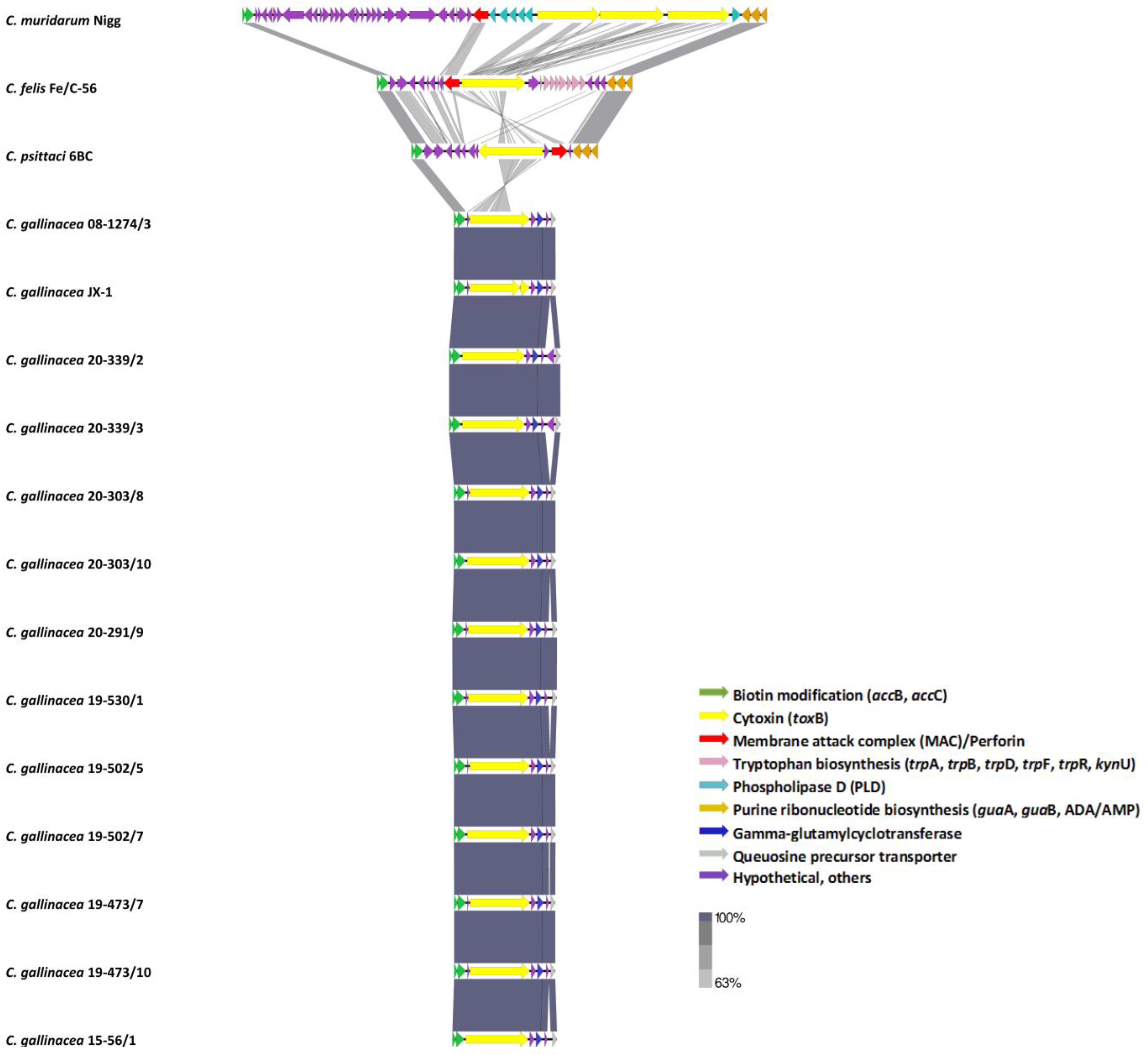
3.3.5. Plasticity Zone

The median size of the PZ of C. gallinacea field strains was 15,845 bp with nucleotide identity ranging from 90.10% to 100% (Supplementary Table S6). With respect to the PZ, the novel Polish strains were closely related to the reference strains 08-1274/3 (90.89–99.80%) and JX-1 (90.62–99.14%) of C. gallinacea. The level of nucleotide identity was significantly lower with other avian species at 48.70–48.91% for C. avium, 46.67–49.13% for Cand. C. ibidis, 30.22–32.46% for C. psittaci, and 27.57–30.10% for C. buteonis. Chlamydia gallinacea representatives demonstrated gene content reduction in their PZs in comparison to other chlamydial species such as avian C. abortus 15-70d24 (20,909 bp), C. buteonis RSHA (26,735 bp), C. psittaci 6BC (29,144 bp), and Cand. C. ibidis 10-1398/6 (31,344 bp), while the PZ of C. avium (5694 bp) was approximately three times smaller. All PZs of C. gallinacea strains started with Acetyl-CoA-carboxylase (accBC) and ended with the queuosine precursor transporter. Cytotoxin virulence factor (toxB) and gamma-glutamylcyclotransferase could be found in all C. gallinacea strain PZs, while purine and pyrimidine biosynthesis genes (guaAB-add), membrane attack complex (MAC)/perforin genes, the tryptophan operon (trpABFCDR, kynU, and prsA), and phospholipase D (PLD) were absent from this region. Interestingly, the two clonal isolates 20-339/2 and 20-339/3 had an additional CDS assigned as a hypothetical protein of unknown function and showed the lowest nucleotide identity (90.10–93.38%) with other C. gallinacea strains, while the remaining nine strains presented sequence homology at the level of 95.79–99.99%. Moreover, C. gallinacea JX-1 had a premature STOP codon in the cytotoxin virulence factor, in contrast to our C. gallinacea strains and other reference strains (Figure 4).

3.3.6. Plasmid Comparisons

All isolated C. gallinacea field strains contained a plasmid of 7492–7569 bp with eight CDS, just as most Chlamydia spp. are known to have (Supplementary Figure S2) [53]. In all strains, integrase (CDS 1/PGP 7 and CDS 2/PGP 8), replicative DNA helicase (CDS 3/PGP 1), virulence plasmid proteins (CDS 4/PGP 2, CDS 5/PGP 3, and CDS 6/PGP 4), plasmid partitioning protein ParA (CDS 7/PGP 5), and plasmid replication protein (CDS 8/PGP 6) were annotated, which is congruent with results described previously for C. gallinacea plasmids [25]. Phylogenetic analysis of eleven plasmids from this study with various chlamydial plasmids representing other avian origin species (C. gallinacea, C. avium, C. psittaci, and avian C. abortus) showed that they cluster with C. gallinacea representatives and C. avium. The plasmid sequence similarity among C. gallinacea strains was high and ranged from 97.25% to 100%, while the similarity of C. gallinacea strains to C. avium, being the closest related species, was notably lower (75.96–76.78%). Detailed parameters of nucleotide identity are shown in Supplementary Table S7.

3.3.7. Putative Virulence Factors and Effector Delivery System

The VFDB identified 207–215 putative virulence genes in the eleven Polish C. gallinacea strains. The distribution of virulence genes seems to be the same with respect to gene presence and localization. The T3SS discovered in all strains is composed of 75 structural, chaperone and secreted effector proteins, including 51 effectors. Detailed data regarding the predicted virulence genes are shown in Supplementary Table S8. The two T3SS effectors, termed translocated actin-recruiting phosphoprotein (TARP) and secreted inner nuclear membrane-associated Chlamydia protein (SINC), as well as the exoenzyme chlamydial protease-like activating factor (CPAF), all known for their roles in chlamydial pathogenesis, were found in all eleven strains. The phylogenetic analysis of the tarP and sinC genes found in strains from this study and C. gallinacea reference strains revealed that these genes are highly conserved (Supplementary Tables S9 and S10). The size of SINC orthologs in all C. gallinacea strains was smaller (372–373 bp) than those identified in C. psittaci strains (>500 bp).
Some genes which were not designated as virulence factors were checked manually by BLAST. The histone-like protein hctA and hctB genes were found with lengths of 118 aa and 187 aa, respectively, in all strains from this study. Compared to the C. gallinacea 08-1274/3 type strain, Polish strains revealed a high level of sequence identity with a maximum value of 100% for hctA and 98.39–100% for hctB.

3.3.8. Inclusion Membrane Proteins and Membrane-Associated Proteins

Four subtypes of Inc were identified in the eleven Polish C. gallinacea strains as well as in the C. gallinacea reference strains: A (two proteins), B (one protein), C (one protein), and V (one protein). All these genes shared a high level of amino acid identity: 98.90–100% (IncA), 97.85–100% (IncA 2), 98.54–100% (IncB), 98.78–100% (IncC), and 98.62–100% (IncV). The most conserved was IncC with 100% similarity between all strains except strain 19-473/7, which had a truncated gene (Supplementary Table S11).
Taking into consideration the presence of bi-lobed hydrophobic domains as a common feature of membrane-bound proteins, all eleven C. gallinacea strains were predicted to encode from 57 to 61 putative membrane proteins. Depending on the strain, 20–25 potential membrane proteins with two transmembrane domains (including two IncA and IncC proteins), 16 with four transmembrane domains (including IncV protein) and 20–21 with six transmembrane domains were predicted. According to the latest version of DeepTMHMM Server, which was version 1.0.18, there are no transmembrane domains in IncB.

3.3.9. The Family of Polymorphic Membrane Proteins

To explore the repertoire of pmps in the Polish strains, all genome sequences from this study were compared with records in a generated pmps database. Almost all results were confirmed by hits in the VFDB database (see Supplementary Table S12).
All subtypes of pmp genes (A, B, D, E/F, G/I, and H) were identified, which is congruent with previously published results [25,51,54]. While subtypes A, B, D, H, G1, and G2 genes were present and complete in all eleven strains, genes of the group E/F and also many of group G/I occurred only in some strains and often possessed premature STOP codons caused by frameshifts. The pmp05 genes, which were detected in our C. gallinacea strains, were homologs of pmpG based on comparisons with sequences from the literature [25,51,54]. The pmp genes, which were present and complete in all eleven Polish strains and the type strain, are conservative, with high levels of amino acid sequence identity values: 98.71–100% (A), 90.39–100% (B), 90.93–100% (D), 96.76–100% (H), 97.85–100% (G1), and 88.34–100% (G2). The one exception was strain 19-502/5 with 76.27–82.33% sequence identity.

4. Discussion

Identifying novel infectious diseases and predicting the locations of their next loci and the likelihood of their epidemics or crossing of species barriers are becoming possible as new technologies are constantly being implemented and improved. The recent developments in next-generation sequencing make WGS affordable, which renders deeper exploration of the biology of bacteria and viruses achievable and offers insights into pathogen diversity. Chlamydial genomics has become one of the principal pillars of molecular as well as clinical research on Chlamydia spp. during the last two decades [55].
Chlamydia gallinacea is an emerging agent which, worldwide, occurs mainly in poultry, this also being the case in Poland. Despite its high prevalence, there are only four C. gallinacea genomes available in GenBank: strain 08-1274/3 is the type strain originating from France (2013), strain JX-1 was isolated in China in 2015, and two Dutch strains—NL F725 and NL G47—are logged which were isolated in 2020. In this research, we presented detailed a comparative genome analysis of eleven Polish field strains isolated from chickens and originating from different locations (7 out of 16 Polish voivodeships).
Genome sequencing is a rapidly evolving field which improves the understanding of pathogens’ diversity, biology, and phylogeny. There are many competing technologies of next-generation sequencing (NGS) available to Chlamydia researchers; all with their unique benefits and drawbacks. Short-read sequencing is the most commonly used form of NGS because it is highly accurate, cost-effective, and supported by a wide range of bioinformatic tools. However, obtaining one contiguous genome sequence might be challenging because of repetitive regions in a pathogen’s genome and/or a high percentage of G+C content [56,57,58]. Long reads can improve assembly success by spanning problematic repetitive regions. Nevertheless, long-read sequencing has some limitations, including relatively low accuracy, which introduces errors in sequenced DNA [57,58,59]. A hybrid sequencing approach, combining high-accuracy short read data (Illumina) and long read sequences (Oxford Nanopore Technologies), was applied in this research to produce genome sequences of eight C. gallinacea strains (20-339/2, 20-339/3, 20-303/8, 20-291/9, 19-530/1, 19-502/5, 19-473/7, and 19-473/10). The remaining three genome sequences (20-303/10, 19-502/7, and 15-56/1) were obtained based just on second-generation sequencing data (Illumina).
An analysis of the 16S rRNA and 23S rRNA genes of the eleven strains originating from Gallus gallus confirmed that they belonged to the C. gallinacea species. The ANIb and tetra-nucleotide regression values also supported their species classification. The G+C content of all strains (39.7%) was identical to that of the C. gallinacea reference strains 08-1274/3, JX-1, NL F725, and NL G47.
An analysis of the ompA gene, as well as MLST, revealed that C. gallinacea strains are highly diverse with respect to these loci. Among the eleven strains sequenced, eight novel sequence types have been identified, numbered ST321–ST328. Phylogenetic analysis of the ompA gene showed that all strains grouped together with European strains but also with some isolated in Asia.
Comparison of whole genome sequences based on whole-genome SNP analysis clearly showed that C. gallinacea strains had conservative genomes. Moreover, an average nucleotide identity comparison of all Chlamydia spp. genomes confirmed the low intra-species variability of C. gallinacea, with whole-genome sequence identity ranging from 99.34% to even 100%.
The plasticity zone is considered to be a key region of all chlamydial genomes as it is highly variable and associated with pathogenesis [59,60]. The size of this zone ranges by species from 6 kb to 83 kb [61]. Acetyl-CoA carboxylase (accB) and einosine-5′-monophosphate dehydrogenase (guaB) are genes determining the boundaries of the PZ in most chlamydiae [59,60]. In C. gallinacea, C. avium, C. buteonis, C. suis, and C. trachomatis, the lack or truncation of the guaAB-add operon is common [62]. The analysis of the PZ of the eleven C. gallinacea strains researched in the current work (15,791–17,376 bp) revealed that this region is highly conserved, with sequence homology of 90.10–100% with all known C. gallinacea genomes. Interestingly, the two clonal strains 20-339/2 and 20-339/3 had an additional CDS assigned as a hypothetical protein of unknown function. In all C. gallinacea genomes, MAC/perforin, PLD, and the complete trp operon genes were absent; however, the full-length chlamydial cytotoxin (toxB) gene could be found. The cytotoxin gene is an important virulence factor as it is related to acute infection and disease [25,63]. In our strains, as well as in the type strain 08-1274/3, this gene was intact, while, in the Chinese strain JX-1 [25], a premature STOP codon was noted. The presence of the cytotoxin gene is characteristic of only some Chlamydia spp.: C. gallinacea, C. psittaci, C. buteonis, C. felis, C. caviae, and Cand. C. ibidis. (one copy); Cand. C. pecorum and C. suis (two copies); and C. muridarum (three copies). In contrast, in C. avium, C. abortus, and C. pneumoniae it was absent [62]. Further investigation is needed to find out which are the virulence features with which the cytotoxin gene correlates.
All known chlamydial taxa, except C. abortus, Cand. C. ibidis, and human-origin C. pneumoniae strains [64], carried a highly conserved plasmid which was not integrated into the genome [65]. Chlamydial plasmids are usually 7.5 kbp, organized into eight open reading frames (ORFs), and considered to be related to the virulence of the strain. All of the C. gallinacea strains reported here harbored a plasmid with high sequence homology (97.25–100%) to each other and the C. gallinacea reference strains 08-1274/3, JX-1, NL F725, and NL G47. Regarding the homology with the plasmid sequences of other Chlamydia spp., those of our eleven strains are most closely related to those of C. avium 10DC88 (75.96–76.78%) and least related to those of C. suis SWA-14, C. trachomatis A/HAR-13, and C. muridarum Nigg (below 60%). The impact of plasmid presence in C. gallinacea strains needs more research.
The T3SS in chlamydial representatives is a system of structural, chaperone, and secreted effector proteins usually arranged into three or four clusters and scattered over the genome [55]. Interestingly, in other bacterial genomes, T3SS structural genes can be found on plasmids or pathogenicity islands along with their effectors [55,66]. In our eleven strains, 75 effectors, chaperones, and structural proteins were determined. The function of most identified T3S effectors is unknown; however, some of them have been elucidated, such as CopN with multiple roles in pathogenesis [67], MrcA promoting chlamydial extrusion [68] or NUE participating in histone methylation [69]. A pair of T3S effector proteins known for their role in chlamydial pathogenesis, TARP and SINC, were found in all eleven strains. The former functions to remodel the host-actin cytoskeleton during the initial stage of infection and facilitate internalization by the host cell [70,71], while the latter is a new T3S protein discovered in Chlamydia psittaci which targets the inner nuclear membrane of infected cells and uninfected neighboring cells [72]. The discovery of TARP and SINC in the present research will, however, need to be echoed in more data for it to be finally confirmed that TARP and SINC effector proteins are actually secreted in C. gallinacea and for their role in the pathogenicity of this species.
Turning our attention to other putative virulence factors, the chlamydial protease-like activating factor (CPAF) exoenzyme, which was stated to be important in Chlamydia virulence [73], was found in all strains presently analyzed, in contrast to the finding of a previously published paper [20], where CPAF was not identified in Dutch C. gallinacea representatives NL F725 or NL G47. Moreover, hctA and hctB, which play principal roles in the conversion of reticulate bodies into elementary bodies, were detected in all C. gallinacea strains from this study. Interestingly, these genes were also found in the C. gallinacea 08-1274/3 type strain, which is at variance with previously published data, probably due to differences in the annotation tools used [54].
A substantial role in virulence is also played by Inc proteins. They are mainly transported by a T3SS and are well known for their subversion of host cell functions as they decorate the cytosolic face of the inclusion of Chlamydia spp. [74]. There is a remarkable divergence in Inc protein content across chlamydial species. To date, seven Inc subtypes (A–G), as well as a newly recognized subtype, IncV, have been described [75]. Taking into consideration differences in nomenclature, it should be mentioned that inclusion membrane proteins are characterized by at least one bi-lobed hydrophobic motif; therefore, the in silico prediction is based on the bi-lobed hydrophobic domains occurring in these proteins [76,77]. In this study, four subtypes were identified by a BLAST search: IncA (two proteins), IncB (one protein), IncC (one protein), and IncV (one protein). Using the DeepTMHMM tool, 20–25 Incs with two transmembrane domains (two IncA and IncC proteins included), 16 with four transmembrane domains (IncV protein included), and 20–21 with six transmembrane domains were predicted. Interestingly, the presence of transmembrane domains in IncB was not confirmed, despite previously published results [25] indicating that there are two transmembrane domains in this protein. This discrepancy was possibly because the analysis referred to was performed on a previous version of DeepTMHMM. A further discrepancy with the literature data is that the number of predicted Incs in C. gallinacea strains from this study varies from those noted by Heijne et al. [20], which might be caused by the use of different servers for prediction and by the application of different thresholds. The DeepTMHMM bioinformatic tool used for analysis in this study is currently considered to provide the most complete results [50].
Polymorphic membrane proteins occur in all Chlamydia spp. and are regarded as virulence factors [51]. These proteins are highly variable in size, sequence, and number across species [54]. It should be emphasized that the analysis of pmps was demanding. Firstly, the gene annotation was not straightforward because the occurrence of poly-G sequences (mainly in the pmpE and pmpI genes) generated frameshifts which led to the appearance of premature STOP codons. The issues with the estimation of the correct number of bases in homopolymer regions are well known, especially in short-read technologies; however, they are present in long-read sequencing as well. Moreover, identifying pmp genes in multiple contig genomes was additionally challenging. Furthermore, the pmp genes and genome sequences available in the GenBank and UniProt databases were often not properly annotated, and pmps nomenclature in databases and publications was inconsistent. Similarly to their detection noted in published research, all the subtypes of the pmp genes (A, B, D, E/F, G/I, and H) were detected in our strains [20,25,54]. Taking into consideration the nomenclature variation among chlamydial species, the pmp05 gene identified in our C. gallinacea strains was assigned to subtype G [25,51,53]. The A, B, D, H, G1, and G2 subtype pmp genes seemed to be present and intact in all eleven strains and shared high sequence similarities with each other and with the type strain. In contrast, pmpE was not detected in five of the eleven strains, and, also, members of subtypes F and G/I seemed to appear sporadically in different numbers and had a premature STOP codon. However, these observed differences in pmps composition between C. gallinacea strains might have been due to sequencing and annotation discrepancies and could not be clearly verified with the bioinformatic tools used in this study.
Virulence genes are one of the most significant incompletely elucidated aspects of genome analysis in Chlamydia spp. Answers to the remaining questions about virulence are not to be fully gained from computational methods based on homology searches, because they have not been experimentally validated [55]. To find out what the function of the gene is, the protein needs to be exhaustively examined. Sequence similarity to genes might offer a good first indication of their function. However, most chlamydial genes are not fully annotated and remain as predictions, although these predictions are often supported by homologs that have been confirmed to be real genes in other strains or species. Furthermore, the majority of genes in Chlamydia spp. are hypothetical proteins with unspecified functions [55]. The present study supports the conclusion of Heijne et al. [20] that C. gallinacea strains share a high level of sequence identity, with genetic diversity limited to several regions, and possess a reduced set of virulence factors in comparison to C. psittaci. In this research, it was confirmed that Polish strains possess virulence genes including cytotoxin, Incs, pmps, histone-like proteins hctA and hctB, exoenzyme CPAF, T3SS, and the SINC and TARP effectors of T3SS involved in chlamydial pathogenesis. However, further studies are needed to confirm their true involvement in pathogenesis.

5. Conclusions

All C. gallinacea genomes are closely related, sharing at least 99.46% of genome sequence homology. Regardless of the C. gallinacea strain origin, their genomes have a conservative structure considering the plasticity zone, the T3SS (including the SINC and TARP effectors), Incs, pmps, hctA and hctB, CPAF, and plasmids. However, some regions, such as ompA and MLST target genes, are diverse enough to enable high-resolution genotyping and epidemiological tracing. Surprisingly, there was some heterogeneity in strains deriving from the same flock, these differing in the ST, ompA genotype, and organization of pmps. Research approaches beyond comparative genomics are needed to clarify the pathogenic potential of C. gallinacea and to determine its significance for the poultry industry and for public health.

Supplementary Materials

The following supporting information can be downloaded at: https://www.mdpi.com/article/10.3390/pathogens12070891/s1, Table S1. Basic genomic parameters of C. gallinacea isolates. Table S2. Accession numbers of C. gallinacea strains analyzed in this study. Table S3. Pairwise comparison of selected representatives of Chlamydia spp. including C. gallinacea strains. The lower triangle represents pairwise sequence identity values for the tetra-nucleotide signature correlation index, while the upper triangle shows pairwise sequence identity values for the average nucleotide identity. Table S4. Nucleotide identity values (%) of ompA sequences of selected Chlamydia spp. Distance matrices based on multiple sequence alignment were used for calculation of nucleotide sequence identity values (Geneious Pro 8.0 software; Biomatters, Auckland, New Zealand). Table S5. The number of single nucleotide polymorphisms of C. gallinacea representatives. Table S6. Nucleotide identity values (%) of plasticity zone sequences of selected representative strains of Chlamydia spp. including C. gallinacea strains. Distance matrices based on multiple sequence alignment were used for calculation of nucleotide sequence identity values (Geneious Pro 8.0 software; Biomatters, Auckland, New Zealand). Table S7. Nucleotide identity values (%) of plasmid sequences of selected Chlamydia spp. Distance matrices based on multiple sequence alignment were used for calculation of nucleotide sequence identity values (Geneious Pro 8.0 software; Biomatters, Auckland, New Zealand). Table S8. Putative virulence factors and effector delivery system in C. gallinacea strains from this study based on Virulence Factors Database. All type III secretion (T3S) proteins are colored yellow (blue font: effectors, black font: remaining type III secretion system (T3SS) proteins). Table S9. Amino acid identity values (%) of translocated actin recruiting phosphoprotein sequences of C. gallinacea representatives. Distance matrices based on multiple sequence alignment were used for calculation of amino acid sequence identity values (Geneious Pro 8.0 software; Biomatters, Auckland, New Zealand). Table S10. Amino acid identity values (%) of secreted inner nuclear membrane-associated Chlamydia protein sequences of C. gallinacea representatives and the C. psittaci 6BC type strain. Distance matrices based on multiple sequence alignment were used for calculation of amino acid sequence identity values (Geneious Pro 8.0 software; Biomatters, Auckland, New Zealand). Table S11. Amino acid identity values (%) of IncA, IncB, IncC, and IncV sequences of C. gallinacea representatives. Distance matrices based on multiple sequence alignment were used for calculation of amino acid sequence identity values (Geneious Pro 8.0 software; Biomatters, Auckland, New Zealand). Table S12. Polymorphic membrane proteins (pmps) in C. gallinacea strains from this study based on BLAST hits and Virulence Factors Database. Figure S1. Phylogenetic tree of concatenated sequences of seven multi-locus sequence typing housekeeping genes (enoA, fumC, gatA, gidA, hemN, hlfX, and oppA) for C. gallinacea strains from this survey (in bold), for C. gallinacea representatives available in PubMLST database and for C. avium 10DC88. Sequence types (ST) are given in brackets for all C. gallinacea strains. Figure S2. EasyFig plot of phylogenetic comparison of C. gallinacea and Chlamydia spp. plasmids based on gene arrangement. Colored arrows represent coding regions, whilst gray shading represents tBLASTx matches. Support values are presented on the branches.

Author Contributions

Funding acquisition and project administration, M.S.-C.; conceptualization, M.S.-C., K.Z.-M. and K.N.; data curation, K.Z.-M., A.B., C.S. and S.S.; formal analysis, K.Z.-M.; Investigation, K.Z.-M., A.B., C.S. and S.S.; methodology, K.Z.-M., A.B., C.S. and S.S.; visualization, K.Z.-M. and A.B.; writing—original draft, K.Z.-M. and C.S.; writing—review and editing, K.Z.-M., C.S., M.S.-C., K.N. and A.B. All authors have read and agreed to the published version of the manuscript.

Funding

The study was funded by the National Veterinary Research Institute (NVRI), Pulawy, Poland within the “NVRI Research Fund” awarded to project no. F/112.

Institutional Review Board Statement

Not applicable.

Informed Consent Statement

Not applicable.

Data Availability Statement

The genome sequences of the strains 20-339/2, 20-339/3, 20-303/8, 20-291/9, 19-530/1, 19-502/5, 19-473/7, 19-473/10, 20-303/10, 19-502/7, 15-56/1 obtained in this study have been deposited in ENA/GenBank/DDBJ under the accession numbers OX332823–OX332824, OX332761–OX332762, OX332828–OX332829, CAMPGD010000001–CAMPGD010000028, CAMPFX010000001–CAMPFX010000011, OX332641–OX332642, CAMPGA010000001–CAMPGA010000017, CAMPFW010000001–CAMPFW010000013, CAMPFY010000001–CAMPFY010000016, CAMPFZ010000001–CAMPFZ010000007, and CAMPGB010000001–CAMPGB010000023, respectively. The respective raw reads of the strains have been deposited under accession numbers ERR10109138, ERR10109123, ERR10109139, ERR10109124, ERR10109137, ERR10109125, ERR10109126, ERR10109136, ERR10109127, ERR10109140, ERR10109128, ERR10109135, ERR10109129, ERR10109130, ERR10109141, ERR10109131, ERR10109134, ERR10109132, and ERR10109133. All data related to WGS of these C. gallinacea strains are included as a part of BioProject PRJEB55472.

Acknowledgments

Veterinary Inspectors are acknowledged for their generous help with the sample collection. The authors are grateful to Mariusz Zagrodzki of the Department of Cattle and Sheep Diseases, National Veterinary Research Institute (NVRI), Puławy, Poland for his technical assistance in DNA extraction and real-time PCR reactions. The authors would like to thank Marta Pietruk and co-workers of the Department of Omics Analyses of the NVRI for their excellent technical assistance with whole genome sequencing. Małgorzata Mazur of the Department of Hygiene of Animal Feedingstuffs of the NVRI is gratefully acknowledged for her support and valuable advice.

Conflicts of Interest

The authors declare no conflict of interest.

References

  1. Vorimore, F.; Hölzer, M.; Liebler-Tenorio, E.M.; Barf, L.M.; Delannoy, S.; Vittecoq, M.; Wedlarski, R.; Lécu, A.; Scharf, S.; Blanchard, Y.; et al. Evidence for the existence of a new genus Chlamydiifrater gen. nov. inside the family Chlamydiaceae with two new species isolated from flamingo (Phoenicopterus roseus): Chlamydiifrater phoenicopteri sp. nov. and Chlamydiifrater volucris sp. nov. Syst. Appl. Microbiol. 2021, 44, 126200. [Google Scholar] [CrossRef] [PubMed]
  2. Sachse, K.; Bavoil, P.M.; Kaltenboeck, B.; Stephens, R.S.; Kuo, C.C.; Rosselló-Móra, R.; Horn, M. Emendation of the family Chlamydiaceae: Proposal of a single genus, Chlamydia, to include all currently recognized species. Syst. Appl. Microbiol. 2015, 38, 99–103. [Google Scholar] [CrossRef] [PubMed]
  3. Staub, E.; Marti, H.; Biondi, R.; Levi, A.; Donati, M.; Leonard, C.A.; Ley, S.D.; Pillonel, T.; Greub, G.; Seth-Smith, H.M.B.; et al. Novel Chlamydia species isolated from snakes are temperature-sensitive and exhibit decreased susceptibility to azithromycin. Sci. Rep. 2018, 8, 5660. [Google Scholar] [CrossRef] [Green Version]
  4. Borel, N.; Greub, G. International committee on systematics of prokaryotes (ICSP) subcommittee on the taxonomy of chlamydiae. Minutes of the closed meeting, 5 July 2018 Woudschoten, Zeist, The Netherlands. Int. J. Syst. Evol. Microbiol. 2019, 69, 2606–2608. [Google Scholar] [CrossRef]
  5. Laroucau, K.; Vorimore, F.; Aaziz, R.; Solmonson, L.; Hsia, R.C.; Bavoil, P.M.; Fach, P.; Hölzer, M.; Wuenschmann, A.; Sachse, K. Chlamydia buteonis, a new Chlamydia species isolated from a red-shouldered hawk. Syst. Appl. Microbiol. 2019, 42, 125997. [Google Scholar] [CrossRef] [PubMed]
  6. Chaiwattanarungruengpaisan, S.; Thongdee, M.; Anuntakarun, S.; Payungporn, S.; Arya, N.; Punchukrang, A.; Ramasoota, P.; Singhakaew, S.; Atithep, T.; Sariya, L. A new species of Chlamydia isolated from Siamese crocodiles (Crocodylus siamensis). PLoS ONE 2021, 16, e0252081. [Google Scholar] [CrossRef] [PubMed]
  7. Vorimore, F.; Hsia, R.-C.; Huot-Creasy, H.; Bastian, S.; Deruyter, L.; Passet, A.; Sachse, K.; Bavoil, P.M.; Myers, G.; Laroucau, K. Isolation of a New Chlamydia species from the Feral Sacred Ibis (Threskiornis aethiopicus): Chlamydia ibidis. PLoS ONE 2013, 8, e74823. [Google Scholar] [CrossRef] [Green Version]
  8. Taylor-Brown, A.; Bachmann, N.L.; Borel, N.; Polkinghorne, A. Culture-independent genomic characterisation of Candidatus Chlamydia sanzinia, a novel uncultivated bacterium infecting snakes. BMC Genom. 2016, 17, 710. [Google Scholar] [CrossRef] [Green Version]
  9. Taylor-Brown, A.; Spang, L.; Borel, N.; Polkinghorne, A. Culture-independent metagenomics supports discovery of uncultivable bacteria within the genus Chlamydia. Sci. Rep. 2017, 7, 10661. [Google Scholar] [CrossRef] [Green Version]
  10. Laroucau, K.; Ortega, N.; Vorimore, F.; Aaziz, R.; Mitura, A.; Szymańska-Czerwińska, M.; Cicerol, M.; Salinas, J.; Sachse, K.; Caro, M.R. Detection of a novel Chlamydia species in captive spur-thighed tortoises (Testudo graeca) in southeastern Spain and proposal of Candidatus Chlamydia testudinis. Syst. Appl. Microbiol. 2020, 43, 126071. [Google Scholar] [CrossRef]
  11. Sachse, K.; Laroucau, K.; Riege, K.; Wehner, S.; Dilcher, M.; Creasy, H.H.; Weidmann, M.; Myers, G.; Vorimore, F.; Vicari, N.; et al. Evidence for the existence of two new members of the family Chlamydiaceae and proposal of Chlamydia avium sp. nov. and Chlamydia gallinacea sp. nov. Syst. Appl. Microbiol. 2014, 37, 79–88. [Google Scholar] [CrossRef] [PubMed]
  12. Zocevic, A.; Vorimore, F.; Marhold, C.; Horvatek, D.; Wang, D.; Slavec, B.; Prentza, Z.; Stavianis, G.; Prukner-Radovcic, E.; Dovc, A.; et al. Molecular characterization of atypical Chlamydia and evidence of their dissemination in different European and Asian chicken flocks by specific real-time PCR. Environ. Microbiol. 2012, 14, 2212–2222. [Google Scholar] [CrossRef] [PubMed]
  13. Lagae, S.; Kalmar, I.; Laroucau, K.; Vorimore, F.; Vanrompay, D. Emerging Chlamydia psittaci infections in chickens and examination of transmission to humans. J. Med. Microbiol. 2014, 63, 399–407. [Google Scholar] [CrossRef] [PubMed] [Green Version]
  14. Szymańska-Czerwińska, M.; Mitura, A.; Zaręba, K.; Schnee, C.; Koncicki, A.; Niemczuk, K. Poultry in Poland as Chlamydiaceae carrier. J. Vet. Res. 2017, 61, 411–419. [Google Scholar] [CrossRef] [Green Version]
  15. Guo, W.; Li, J.; Kaltenboeck, B.; Gong, J.; Fan, W.; Wang, C. Chlamydia gallinacea not C. psittaci is the endemic chlamydial species in chicken Gallus gallus. Sci. Rep. 2016, 6, 19638. [Google Scholar] [CrossRef] [Green Version]
  16. Li, J.; Guo, W.; Kaltenboeck, B.; Sachse, K.; Yang, Y.; Lu, G.; Zhang, J.; Luan, L.; You, J.; Huang, K.; et al. Chlamydia pecorum is the endemic intestinal species in cattle while C. gallinacea, C. psittaci and C. pneumoniae associate with sporadic systemic infection. Vet. Microbiol. 2016, 193, 93–99. [Google Scholar] [CrossRef]
  17. Stokes, H.S.; Martens, J.M.; Chamings, A.; Walder, K.; Berg, M.L.; Segal, Y.; Bennett, A. Identification of Chlamydia gallinacea in a parrot and in free-range chickens in Australia. Aust. Vet. J. 2019, 97, 398–400. [Google Scholar] [CrossRef]
  18. Szymańska-Czerwińska, M.; Jodełko, A.; Zaręba-Marchewka, K.; Niemczuk, K. Experimental inoculation of chicken broilers with C. gallinacea strain 15-56/1. Sci. Rep. 2021, 11, 23856. [Google Scholar] [CrossRef]
  19. You, J.; Wu, Y.; Zhang, X.; Wang, X.; Gong, J.; Zhao, Z.; Zhang, J.; Zhang, J.; Sun, Z.; Li, J.; et al. Efficient fecal-oral and possible vertical, but not respiratory, transmission of emerging Chlamydia gallinacea in broilers. Vet. Microbiol. 2019, 230, 90–94. [Google Scholar] [CrossRef]
  20. Heijne, M.; Jelocnik, M.; Umanets, A.; Brouwer, M.S.M.; Dinkla, A.; Harders, F.; van Keulen, L.J.M.; Roest, H.J.; Schaafsma, F.; Velkers, F.C.; et al. Genetic and phenotypic analysis of the pathogenic potential of two novel Chlamydia gallinacea strains compared to Chlamydia psittaci. Sci. Rep. 2021, 11, 16516. [Google Scholar] [CrossRef]
  21. Laroucau, K.; Vorimore, F.; Aaziz, R.; Berndt, A.; Schubert, E.; Sachse, K. Isolation of a new chlamydial agent from infected domestic poultry coincided with cases of atypical pneumonia among slaughterhouse workers in France. Infect. Genet. Evol. 2009, 9, 1240–1247. [Google Scholar] [CrossRef] [PubMed]
  22. Marchino, M.; Rizzo, F.; Barzanti, P.; Sparasci, O.A.; Bottino, P.; Vicari, N.; Rigamonti, S.; Braghin, S.; Aaziz, R.; Vorimore, F.; et al. Chlamydia Species and Related Risk Factors in Poultry in North-Western Italy: Possible Bird-to-Human Transmission for C. gallinacea. Int. J. Environ. Res. Public. Health 2022, 19, 2174. [Google Scholar] [CrossRef] [PubMed]
  23. Shahbandeh, M. Meat Consumption Worldwide from 1990 to 2021, by Type. Available online: https://www.statista.com/statistics/274522/global-per-capita-consumption-of-meat/ (accessed on 30 November 2022).
  24. Biesek, J.; Banaszak, M.; Kądziołka, K.; Wlaźlak, S.; Adamski, M. Growth of broiler chickens, and physical features of the digestive system, and leg bones after aluminosilicates used. Sci. Rep. 2022, 12, 20425. [Google Scholar] [CrossRef]
  25. Guo, W.; Jelocnik, M.; Li, J.; Sachse, K.; Polkinghorne, A.; Pannekoek, Y.; Kaltenboeck, B.; Gong, J.; You, J.; Wang, C. From genomes to genotypes: Molecular epidemiological analysis of Chlamydia gallinacea reveals a high level of genetic diversity for this newly emerging chlamydial pathogen. BMC Genom. 2017, 18, 949. [Google Scholar] [CrossRef] [Green Version]
  26. Ehricht, R.; Slickers, P.; Goellner, S.; Hotzel, H.; Sachse, K. Optimized DNA microarray assay allows detection and genotyping of single PCR-amplifiable target copies. Mol. Cell. Probes 2006, 20, 60–63. [Google Scholar] [CrossRef]
  27. Laroucau, K.; Aaziz, R.; Meurice, L.; Servas, V.; Chossat, I.; Royer, H.; De Barbeyrac, B.; Vaillant, V.; Moyen, J.; Meziani, F.; et al. Outbreak of psittacosis in a group of women exposed to Chlamydia psittaci-infected chickens. Eurosurveillance 2015, 20, 21155. [Google Scholar] [CrossRef] [PubMed] [Green Version]
  28. Ménard, A.; Clerc, M.; Subtil, A.; Mégraud, F.; Bébéar, C.; de Barbeyrac, B. Development of a real-time PCR for the detection of Chlamydia psittaci. J. Med. Microbiol. 2006, 55, 471–473. [Google Scholar] [CrossRef] [Green Version]
  29. Zocevic, A.; Vorimore, F.; Vicari, N.; Gasparini, J.; Jacquin, L.; Sachse, K.; Magnino, S.; Laroucau, K. A real-time PCR assay for the detection of atypical strains of Chlamydiaceae from pigeons. PLoS ONE 2013, 8, e58741. [Google Scholar] [CrossRef] [Green Version]
  30. Pantchev, A.; Sting, R.; Bauerfeind, R.; Tyczka, J.; Sachse, K. Detection of all Chlamydophila and Chlamydia spp. of veterinary interest using species-specific real-time PCR assays. Comp. Immunol. Microbiol. Infect. Dis. 2010, 33, 473–484. [Google Scholar] [CrossRef]
  31. Andrews, S. FastQC: A Quality Control Tool for High Throughput Sequence Data. Available online: http://www.bioinformatics.babraham.ac.uk/projects/fastqc/ (accessed on 6 April 2022).
  32. Chen, S.; Zhou, Y.; Chen, Y.; Gu, J. Fastp: An ultra-fast all-in-one FASTQ preprocessor. Bioinformatics 2018, 34, i884–i890. [Google Scholar] [CrossRef]
  33. De Coster, W.; D’Hert, S.; Schultz, D.T.; Cruts, M.; Van Broeckhoven, C. NanoPack: Visualizing and processing long-read sequencing data. Bioinformatics 2018, 34, 2666–2669. [Google Scholar] [CrossRef] [PubMed] [Green Version]
  34. Wick, R.R.; Judd, L.M.; Gorrie, C.L.; Holt, K.E. Unicycler: Resolving bacterial genome assemblies from short and long sequencing reads. PLoS Comput. Biol. 2017, 13, e1005595. [Google Scholar] [CrossRef] [Green Version]
  35. Bankevich, A.; Nurk, S.; Antipov, D.; Gurevich, A.A.; Dvorkin, M.; Kulikov, A.S.; Lesin, V.M.; Nikolenko, S.I.; Pham, S.; Prjibelski, A.D.; et al. SPAdes: A new genome assembly algorithm and its applications to single-cell sequencing. J. Comput. Biol. 2012, 19, 455–477. [Google Scholar] [CrossRef] [Green Version]
  36. Seemann, T. Prokka: Rapid prokaryotic genome annotation. Bioinformatics 2014, 30, 2068–2069. [Google Scholar] [CrossRef] [PubMed] [Green Version]
  37. Richter, M.; Rosselló-Móra, R.; Glöckner, F.O.; Peplies, J. JSpeciesWS: A web server for prokaryotic species circumscription based on pairwise genome comparison. Bioinformatics 2015, 32, 929–931. [Google Scholar] [CrossRef]
  38. Kozlov, A.M.; Darriba, D.; Flouri, T.; Morel, B.; Stamatakis, A. RAxML-NG: A fast, scalable, and user-friendly tool for maximum likelihood phylogenetic inference. Bioinformatics 2019, 35, 4453–4455. [Google Scholar] [CrossRef] [Green Version]
  39. Letunic, I.; Bork, P. Interactive Tree of Life (iTOL) v5: An online tool for phylogenetic tree display and annotation. Nucleic Acids Res. 2021, 49, W293–W296. [Google Scholar] [CrossRef]
  40. Hunt, M.; De Silva, N.; Otto, T.D.; Parkhill, J.; Keane, J.A.; Harris, S.R. Circlator: Automated circularization of genome assemblies using long sequencing reads. Genome Biol. 2015, 16, 294. [Google Scholar] [CrossRef] [Green Version]
  41. Katoh, K.; Misawa, K.; Kuma, K.; Miyata, T. MAFFT: A novel method for rapid multiple sequence alignment based on fast Fourier transform. Nucleic Acids Res. 2002, 30, 3059–3066. [Google Scholar] [CrossRef] [PubMed] [Green Version]
  42. Thompson, J.D.; Higgins, D.G.; Gibson, T.J. CLUSTAL W: Improving the sensitivity of progressive multiple sequence alignment through sequence weighting, position-specific gap penalties and weight matrix choice. Nucleic. Acids Res. 1994, 22, 4673–4680. [Google Scholar] [CrossRef] [Green Version]
  43. Nguyen, L.T.; Schmidt, H.A.; von Haeseler, A.; Minh, B.Q. IQ-TREE: A fast and effective stochastic algorithm for estimating maximum-likelihood phylogenies. Mol. Biol. Evol. 2015, 32, 268–274. [Google Scholar] [CrossRef] [PubMed]
  44. Trifinopoulos, J.; Nguyen, L.T.; von Haeseler, A.; Minh, B.Q. W-IQ-TREE: A fast online phylogenetic tool for maximum likelihood analysis. Nucleic Acids Res. 2016, 8, 232–235. [Google Scholar] [CrossRef] [Green Version]
  45. Kalyaanamoorthy, S.; Minh, B.Q.; Wong, T.K.F.; von Haeseler, A.; Jermiin, L.S. ModelFinder: Fast model selection for accurate phylogenetic estimates. Nat. Methods 2017, 14, 587–589. [Google Scholar] [CrossRef] [PubMed] [Green Version]
  46. Hoang, D.T.; Chernomor, O.; von Haeseler, A.; Minh, B.Q.; Vinh, L.S. UFBoot2: Improving the Ultrafast Bootstrap Approximation. Mol. Biol. Evol. 2018, 35, 518–522. [Google Scholar] [CrossRef] [PubMed]
  47. Sullivan, M.J.; Petty, N.K.; Beatson, S.A. Easyfig: A genome comparison visualizer. Bioinformatics 2011, 27, 1009–1010. [Google Scholar] [CrossRef] [Green Version]
  48. Pearson, W.R. An introduction to sequence similarity (“homology”) searching. Curr. Protoc. Bioinform. 2013, 42, 3.1.1–3.1.8. [Google Scholar] [CrossRef] [Green Version]
  49. Camacho, C.; Coulouris, G.; Avagyan, V.; Ma, N.; Papadopoulos, J.; Bealer, K.; Madden, T.L. BLAST+: Architecture and applications. BMC Bioinform. 2009, 10, 421. [Google Scholar] [CrossRef] [PubMed] [Green Version]
  50. Hallgren, J.; Tsirigos, K.D.; Pedersen, M.D.; Armenteros, J.J.A.; Marcatili, P.; Nielsen, H.; Krogh, A.; Winther, O. DeepTMHMM predicts alpha and beta transmembrane proteins using deep neural networks. bioRxiv 2022, 04.08.2022, 487609. [Google Scholar] [CrossRef]
  51. Vasilevsky, S.; Stojanov, M.; Greub, G.; Baud, D. Chlamydial polymorphic membrane proteins: Regulation, function and potential vaccine candidates. Virulence 2016, 7, 11–22. [Google Scholar] [CrossRef]
  52. Pannekoek, Y.; Dickx, V.; Beeckman, D.S.; Jolley, K.A.; Keijzers, W.C.; Vretou, E.; Maiden, M.C.; Vanrompay, D.; van der Ende, A. Multi locus sequence typing of Chlamydia reveals an association between Chlamydia psittaci genotypes and host species. PLoS ONE 2010, 5, e14179. [Google Scholar] [CrossRef]
  53. Szabo, K.V.; O’Neill, C.E.; Clarke, I.N. Diversity in Chlamydial plasmids. PLoS ONE 2020, 15, e0233298. [Google Scholar] [CrossRef] [PubMed]
  54. Hölzer, M.; Barf, L.M.; Lamkiewicz, K.; Vorimore, F.; Lataretu, M.; Favaroni, A.; Schnee, C.; Laroucau, K.; Marz, M.; Sachse, K. Comparative Genome Analysis of 33 Chlamydia Strains Reveals Characteristic Features of Chlamydia psittaci and Closely Related Species. Pathogens 2020, 9, 899. [Google Scholar] [CrossRef] [PubMed]
  55. Borges, V.; Hyden, P.; Gomes, J.P.; Rattei, T. Chapter 12 Chlamydia Genomics. In Chlamydia Biology, 1st ed.; Tan, M., Hegemann, J.H., Sutterlin, C., Eds.; Caister Academic Press: Poole, UK, 2020; pp. 263–286. ISBN 978-1-912530-28-1. [Google Scholar] [CrossRef] [Green Version]
  56. Heather, J.M.; Chain, B. The sequence of sequencers: The history of sequencing DNA. Genomics 2016, 107, 1–8. [Google Scholar] [CrossRef] [PubMed] [Green Version]
  57. Rang, F.J.; Kloosterman, W.P.; De Ridder, J. From squiggle to basepair: Computational approaches for improving nanopore sequencing read accuracy. Genome Biol. 2018, 19, 90. [Google Scholar] [CrossRef] [Green Version]
  58. Amarasinghe, S.L.; Su, S.; Dong, X.; Zappia, L.; Ritchie, M.E.; Gouil, Q. Opportunities and challenges in long-read sequencing data analysis. Genome Biol. 2020, 21, 30. [Google Scholar] [CrossRef] [Green Version]
  59. Bachmann, N.L.; Polkinghorne, A.; Timms, P. Chlamydia genomics: Providing novel insights into chlamydial biology. Trends Microbiol. 2014, 22, 464–472. [Google Scholar] [CrossRef]
  60. Nunes, A.; Gomes, J.P. Evolution, phylogeny, and molecular epidemiology of Chlamydia. Infect. Genet. Evol. 2014, 23, 49–64. [Google Scholar] [CrossRef]
  61. Zaręba-Marchewka, K.; Szymańska-Czerwińska, M.; Niemczuk, K. Chlamydiae—What’s New? J. Vet. Res. 2020, 64, 461–467. [Google Scholar] [CrossRef]
  62. Zaręba-Marchewka, K.; Szymańska-Czerwińska, M.; Livingstone, M.; Longbottom, D.; Niemczuk, K. Whole Genome Sequencing and Comparative Genome Analyses of Chlamydia abortus Strains of Avian Origin Suggests That Chlamydia abortus Species Should Be Expanded to Include Avian and Mammalian Subgroups. Pathogens 2021, 10, 1405. [Google Scholar] [CrossRef]
  63. Rajaram, K.; Giebel, A.M.; Toh, E.; Hu, S.; Newman, J.H.; Morrison, S.G.; Kari, L.; Morrison, R.P.; Nelson, D.E. Mutational analysis of the Chlamydia muridarum plasticity zone. Infect. Immun. 2015, 83, 2870–2881. [Google Scholar] [CrossRef] [Green Version]
  64. Rockey, D.D. Unraveling the basic biology and clinical significance of the chlamydial plasmid. J. Exp. Med. 2011, 208, 2159–2162. [Google Scholar] [CrossRef] [PubMed] [Green Version]
  65. Pawlikowska-Warych, M.; Śliwa-Dominiak, J.; Deptuła, W. Chlamydial plasmids and bacteriophages. Acta Biochim. Pol. 2015, 62, 1–6. [Google Scholar] [CrossRef]
  66. Tampakaki, A.P.; Fadouloglou, V.E.; Gazi, A.D.; Panopoulos, N.J.; Kokkinidis, M. Conserved features of type III secretion. Cell. Microbiol. 2004, 6, 805–816. [Google Scholar] [CrossRef] [PubMed]
  67. Huang, J.; Lesser, C.F.; Lory, S. The essential role of the CopN protein in Chlamydia pneumoniae intracellular growth. Nature 2008, 456, 112–115. [Google Scholar] [CrossRef] [PubMed] [Green Version]
  68. Nguyen, P.H.; Lutter, E.I.; Hackstadt, T. Chlamydia trachomatis inclusion membrane protein MrcA interacts with the inositol 1,4,5-trisphosphate receptor type 3 (ITPR3) to regulate extrusion formation. PLoS Pathog. 2018, 14, e1006911. [Google Scholar] [CrossRef]
  69. Pennini, M.E.; Perrinet, S.; Dautry-Varsat, A.; Subtil, A. Histone methylation by NUE, a novel nuclear effector of the intracellular pathogen Chlamydia trachomatis. PLoS Pathog. 2010, 6, e1000995. [Google Scholar] [CrossRef]
  70. Wolff, B.J.; Morrison, S.S.; Pesti, D.; Ganakammal, S.R.; Srinivasamoorthy, G.; Changayil, S.; Weil, M.R.; MacCannell, D.; Rowe, L.; Frace, M.; et al. Chlamydia psittaci comparative genomics reveals intraspecies variations in the putative outer membrane and type III secretion system genes. Microbiol. Soc. 2015, 161, 1378–1391. [Google Scholar] [CrossRef]
  71. Tolchard, J.; Walpole, S.J.; Miles, A.J.; Maytum, R.; Eaglen, L.A.; Hackstadt, T.; Wallace, B.A.; Blumenschein, T.M.A. The intrinsically disordered Tarp protein from chlamydia binds actin with a partially preformed helix. Sci. Rep. 2018, 8, 1960. [Google Scholar] [CrossRef] [Green Version]
  72. Mojica, S.A.; Hovis, K.M.; Frieman, M.B.; Tran, B.; Hsia, R.C.; Ravel, J.; Jenkins-Houk, C.; Wilson, K.L.; Bavoil, P.M. SINC, a type III secreted protein of Chlamydia psittaci, targets the inner nuclear membrane of infected cells and uninfected neighbors. Mol. Biol. Cell 2015, 26, 1918–1934. [Google Scholar] [CrossRef] [PubMed]
  73. Knittler, M.R.; Sachse, K. Chlamydia psittaci: Update on an underestimated zoonotic agent. Pathog. Dis. 2015, 73, 1–15. [Google Scholar] [CrossRef] [Green Version]
  74. Sixt, B.S.; Bastidas, R.J.; Finethy, R.; Baxter, R.M.; Carpenter, V.K.; Kroemer, G.; Coers, J.; Valdivia, R.H. The Chlamydia trachomatis Inclusion Membrane Protein CpoS Counteracts STING-Mediated Cellular Surveillance and Suicide Programs. Cell Host Microbe 2017, 21, 113–121. [Google Scholar] [CrossRef] [PubMed] [Green Version]
  75. Stanhope, R.; Flora, E.; Bayne, C.; Derre, I. IncV, a FFAT motif-containing Chlamydia protein, tethers the endoplasmic reticulum to the pathogen-containing vacuole. Proc. Natl. Acad. Sci. USA 2017, 114, 12039–12044. [Google Scholar] [CrossRef] [PubMed] [Green Version]
  76. Dehoux, P.; Flores, R.; Dauga, C.; Zhong, G.; Subtil, A. Multi-genome identification and characterization of chlamydiae-specific type III secretion substrates: The Inc proteins. BMC Genom. 2011, 12, 109. [Google Scholar] [CrossRef] [PubMed] [Green Version]
  77. Bugalhão, J.N.; Luís, M.P.; Pereira, I.S.; da Cunha, M.; Pais, S.V.; Mota, L.J. The Chlamydia trachomatis inclusion membrane protein CT006 associates with lipid droplets in eukaryotic cells. PLoS ONE 2022, 17, e0264292. [Google Scholar] [CrossRef]
Figure 1. Flock (20-339, 20-303, 20-291, 19-530, 19-502, 19-473) and bacterial strain (15-56/1) origin. This map was created using Easy Software version 0.196 by Marek Roj (http://mapy.easysoftware.pl/ accessed on 2 May 2022).
Pathogens 12 00891 g001
Figure 2. Analysis of the complete ompA gene of C. gallinacea field strains and representative strains of Chlamydia spp. The phylogeny based on 1266-bp consensus alignment was constructed by the maximum likelihood method with best-fit model according to the Bayesian information criterion TVM+F+I+G4. Specimens found in this study are in boldface. Bootstrap values are presented as percentages.
Pathogens 12 00891 g002
Figure 3. Single-nucleotide polymorphism-based phylogeny of C. gallinacea strains. The dendrogram was constructed based on core SNP alignment using RAxML-NG with the substitution model GTR+G+ASC_STAM, with support values calculated from 1000 bootstraps. The scale bar indicates the number of substitutions per site.
Pathogens 12 00891 g003
Figure 4. EasyFig plot of tBLASTx analysis of the plasticity zones of the eleven C. gallinacea strains, four C. gallinacea reference genomes, and three representatives of Chlamydia spp. Gray shading shows sequence identity, whilst colored arrows represent plasticity zone genes.
Pathogens 12 00891 g004
Table 1. Real-time PCR results of selected C. gallinacea-positive chicken samples.
No. of FlockSampleReal-Time PCR
(Ct Value)
IDTypeOriginChlamydiaceae
23S rRNA
C. gallinacea
20-33920-3392cloacal swabchicken27.9527.23
20-339/3cloacal swabchicken29.0328.13
20-30320-303/8cloacal swabchicken28.3126.35
20-303/10cloacal swabchicken25.8823.59
20-29120-291/9cloacal swabchicken29.6127.73
19-53019-530/1cloacal swabchicken31.1630.35
19-50219-502/5cloacal swabchicken28.1926.98
19-502/7cloacal swabchicken26.6125.21
19-47319-473/7cloacal swabchicken33.0431.68
19-473/10cloacal swabchicken27.9926.94
15-5615-56/1cloacal swabchicken23.2621.80
Table 2. Multi-locus sequence typing profiles of C. gallinacea field strains.
SamplegatAoppAhflXgidAenoAhemNfumCST
20-339/244813845362928321
20-339/344813845362928321
20-303/8443638451108328322
20-303/10443638451108328322
20-291/944364045362928323
19-530/1443610345362928324
19-502/5443610245362928325
19-502/744883845368428326
19-473/744363845362989327
19-473/1044363845362989327
15-56/14436102451112928328
Disclaimer/Publisher’s Note: The statements, opinions and data contained in all publications are solely those of the individual author(s) and contributor(s) and not of MDPI and/or the editor(s). MDPI and/or the editor(s) disclaim responsibility for any injury to people or property resulting from any ideas, methods, instructions or products referred to in the content.

