Article

Potential Biomarkers for Asymptomatic Visceral Leishmaniasis among Iraq-Deployed U.S. Military Personnel

by
Fernanda Fortes de Araujo
1,2,*,†,
Ines Lakhal-Naouar
3,†,
Nancy Koles
1,2,
Sorana Raiciulescu
4,
Rupal Mody
1,5 and
Naomi Aronson
1
1
Department of Medicine, Uniformed Services University of the Health Sciences, Bethesda, MD 20814, USA
2
Henry M. Jackson Foundation for the Advancement of Military Medicine, Bethesda, MD 20817, USA
3
Diagnostics and Countermeasures Branch, Walter Reed Army Institute of Research, Silver Spring, MD 20910, USA
4
Department of Preventive Medicine and Biostatistics, Uniformed Services University of the Health Sciences, Bethesda, MD 20814, USA
5
Department of Medicine, William Beaumont Army Medical Center, El Paso, TX 79916, USA
*
Author to whom correspondence should be addressed.
These authors contributed equally to this work.
Pathogens 2023, 12(5), 705; https://doi.org/10.3390/pathogens12050705
Submission received: 17 April 2023 / Revised: 2 May 2023 / Accepted: 4 May 2023 / Published: 12 May 2023

Abstract

:
Visceral leishmaniasis (VL) is a chronic infection caused by Leishmania (L.) donovani or L. infantum parasites. Despite having the infection, most individuals never develop the clinical disease and are able to control the parasite and remain asymptomatic. However, some progress to symptomatic VL, leading to death if untreated. The host immune response has a major role in determining the progression and severity of the clinical manifestations in VL; several immune biomarkers of symptomatic VL have been described with interferon-gamma release as a surrogate biomarker of host cellular immunity. However, new biomarkers to identify asymptomatic VL (AVL) are needed for the identification of people at risk for VL activation. In our study, levels of chemokine/cytokine in the supernatants of peripheral mononuclear blood cells (PBMC) from 35 AVL+ Iraq-deployed participants, stimulated in vitro with soluble Leishmania antigen for 72 h, were assessed by a bead-based assay that allows the measurement of multiple analytes. PBMC of AVL-negative military beneficiaries were used as controls. Monocyte Chemoattractant Protein-1, Monokine Induced by Gamma Interferon and Interleukin-8, were detected at high levels in AVL+ stimulated cultures from Iraq deployers compared to uninfected controls. Measurement of chemokine/cytokine levels can identify cellular immune responses in AVL+ asymptomatic individuals.

1. Introduction

Visceral leishmaniasis (VL) is a chronic infectious disease caused by Leishmania (L.) donovani or L. infantum (syn. L. chagasi) intracellular protozoan parasites. Worldwide, an estimated 50,000 to 90,000 new cases of VL occur annually, and VL remains one of the major parasitic diseases with significant outbreak and mortality potential [1]. Despite being infected, most individuals never develop the clinical disease and are able to control the infection, remaining asymptomatic [2,3]. However, some people progress to the more severe VL form, which is potentially fatal if not promptly treated.
The host immune response to Leishmania is critical for parasite clearance or disease development [4], as it plays a major role in determining the progression and severity of the clinical manifestations in VL. Several immunological markers of symptomatic VL have been described [5,6,7,8,9,10,11]. The cellular response leading to the control of Leishmania infection is associated with a predominant Type 1 immune profile characterized by interferon-γ (IFN-γ), tumor necrosis factor-α (TNF-α), and interleukin (IL)-2 secretion [12,13]. Alternatively, when the parasite is able to evade host-specific immune responses, VL develops [14]. In VL, parasite infiltration of the spleen, liver, lymph nodes, and bone marrow occurs. The patient with active VL has immune responses associated with cellular anergy or a Type 2 response and production of IL-4, IL-5, IL-10, IL-13, and transforming growth factor-β (TGF-β), which do not control the infection [15].
Recent studies in VL have shown that some chemokines (IL-8/CXCL8, MCP-1/CCL2, MIG/CXCL9, and IP-10/CXCL10) are associated with a Th1 cellular response [15]. Previous findings demonstrated the utility of cytokines/chemokines, such as IFN-γ, IL-2, IP-10, MIG, TNFα, and MCP-1 or CCL2, as biomarkers for monitoring disease recovery [16,17] or treatment efficacy, suggesting that these biomarkers could be indicators of clinical cure in VL individuals [17,18,19,20].
Unlike active VL, the relevance of asymptomatic infection for parasite transmission and disease outcome is largely unknown. Asymptomatic visceral leishmaniasis (AVL) is frequent in endemic areas [21,22], and asymptomatic individuals may have parasites in their blood [23]. A substantial prevalence of L. infantum AVL was observed in Operation Iraqi Freedom (OIF) U.S. military deployers; 19.5% had asymptomatic infection defined by a positive cellular/humoral response or nucleic acid testing result [24,25]. AVL presents a concern due to the potential of chronically infected individuals to transmit disease via blood transfusion and organ donation, the possible domestic transmission risk given a permissive and widespread U.S. sand fly vector, Lutzomyia shannoni, and the reactivation risk associated with the increasing use of immune modulating treatments and immunosuppression [26,27]. The current parasitological, molecular, and serological methods used to identify asymptomatic infection are resource-intensive and not completely suitable for expanded surveillance, making the identification of these individuals a challenge [19,28].
In this context, new biomarkers capable of identifying AVL are needed to determine people at risk for VL activation and to understand the burden of VL in exposed populations. Evaluating AVL in this cohort of American (from a nonendemic country) military deployers who traveled to VL-endemic Iraq and then returned to the U.S. (in contrast to most published work where individuals remain in endemic areas and can be rechallenged by infected sand flies) can further contribute to the development of clinically useful VL tests and biomarkers.

2. Material and Methods

2.1. Ethics Statement

This study was conducted according to the guidelines of the Declarations of Helsinki, and the research protocol was approved by the Institutional Review Boards (IRB) of the Uniformed Services University (USU) MED 83-2979, April 2015, Walter Reed National Military Medical Center (WRNMMC) 412208-1, May 2015, and the William Beaumont Army Medical Center (WBAMC) 16–29, which deferred to USU approval. Written informed consent was obtained from all participants involved in the study.

2.2. Study Population

Iraq Cohort: 200 OIF-deployed asymptomatic participants completed a leishmaniasis risk factor survey and donated blood samples for VL research assays. Deployed participants were U.S. military personnel in good health, aged 18–60 years, who traveled to Iraq between 2002–2011. Enrollment occurred at WRNMMC/USU in Bethesda MD, DiLorenzo Tricare Health Clinic-Pentagon, Washington, DC, and WBAMC in El Paso TX [25]. From this cohort, 35 participants who tested positive for visceral leishmaniasis and 14 controls (AVL-negative military healthcare beneficiaries) were included in the present study. Asymptomatic VL diagnosis was assessed if any results were positive on testing the peripheral blood samples with quantitative L. infantum PCR [29], soluble Leishmania Antigen (SLA) ELISA [24], L. infantum interferon-γ release assay [24], or rK39 immunochromatographic rapid test (Kalaazar Detect™, InBios, Seattle, WA, USA). The laboratory characteristics of the Iraq AVL cohort have been published [24].

2.3. Soluble Leishmania Antigen Preparation

L. infantum (MHOM/BC/00/1669, courtesy of Dr. M. Wilson, University of Iowa, Iowa City, IA, USA) parasites were grown at 26 °C in hemoflagellate-modified minimal essential medium with 10% fetal bovine serum until reaching stationary phase [30,31]. SLA was prepared as previously described [32].

2.4. Cell Culture

Peripheral blood mononuclear cells (PBMC) were isolated from whole blood as previously described [33]. Briefly, 1 × 10⁶ PBMC were stimulated (or not) with 20 μg/mL SLA for 72 h at 37 °C, 5% CO2. Supernatants were collected and stored at −80 °C until used.

2.5. Quantification of Biomarkers

The levels of chemokines (RANTES, Eotaxin, MIP-1α, MIP-1β, MCP-1, IP-10, MIG, IL-8) and cytokines (IL-1β, IL-1RA, IL-2, IL-2R, IL-4, IL-5, IL-6, IL-7, IL-10, IL-12, IL-13, IL-15, IL-17, IFN-γ, IFN-α, GM-CSF, TNF-α) in the supernatants of PBMCs from 35 AVL-positive (AVL+) and 14 control participants stimulated in vitro with soluble Leishmania antigen (SLA) for 72 h were assessed by Luminex® technology (Cytokine 25-Plex Human Panel, Invitrogen, Waltham, MA, USA). Data acquisition and cytokine/chemokine levels were measured on a Luminex® 200 System or Bioplex® Manager Software using the Five-Parameter Logistic Regression, with results expressed in pg/mL or baseline fold.

2.6. Statistical Analysis

Statistical analysis was carried out using GraphPad Prism v.9.0 Software (San Diego, CA, USA) and Stata 17 SE for the combinatorial ROC curve [34]. Comparative analysis between any two groups was performed using the nonparametric Mann–Whitney U test, according to the data distribution. The differences were considered significant if p < 0.05. The performance and cut-off for each analyte were determined by calculating the area under the receiver operating characteristic (ROC) curve (AUC) and the 95% confidence interval (CI).
Levels of chemokine/cytokine were expressed as pg/mL (value for SLA-stimulated culture subtracted by the value for un-stimulated culture from the same person) or baseline fold (value for SLA-stimulated culture divided by the value for un-stimulated culture from the same person). Due to an intrinsic variation of the manufactured kits in the standard curve levels (pg/mL) of some chemokine/cytokine, the fold increase of some analytes was calculated instead of presenting the concentration values. The majority of the time, when individual chemo/cytokine standard curve values correlated from kit to kit, results were provided as analyte concentration.

3. Results

3.1. Characteristics of U.S. Military Study Volunteers

The mean age of AVL volunteers was 40.7 years; the majority were male and self-reported white race. Demographics of the cohort are presented in Table 1.

3.2. IL-8, MCP-1, and MIG produced by SLA-stimulated PBMC Are Useful Biomarkers for the Identification of People with AVL

Cytokine and chemokine levels were analyzed in PBMC supernatants collected from an AVL+ Iraq-deployer cohort (Figure 1). Several chemokines/cytokines presented no or low detected levels, with no significant difference from controls (Supplementary Table S1). However, three analytes were found at significantly higher concentrations in the AVL+ cohort compared to the AVL negative controls: IL-8 (p = 0.0010), MCP-1(p = 0.0012), and MIG (p < 0.0001) (Figure 1A). Receiver operating characteristic (ROC) curves were built to evaluate the performance of each analyte as a possible biomarker for AVL. Our data demonstrated that MIG was the best biomarker, with an area under the curve (AUC) value of 0.87 and 100% specificity (sp). However, MCP-1 was more sensitive (se) than MIG (91% versus 71%). IL-8 and MCP-1 performed moderately well with AUC = 0.80 (se = 0.66, sp = 0.85) and AUC = 0.79 (se = 0.91, sp = 0.71), respectively (Figure 1B).

3.3. Combinatorial Analysis for Identification of Individuals with AVL

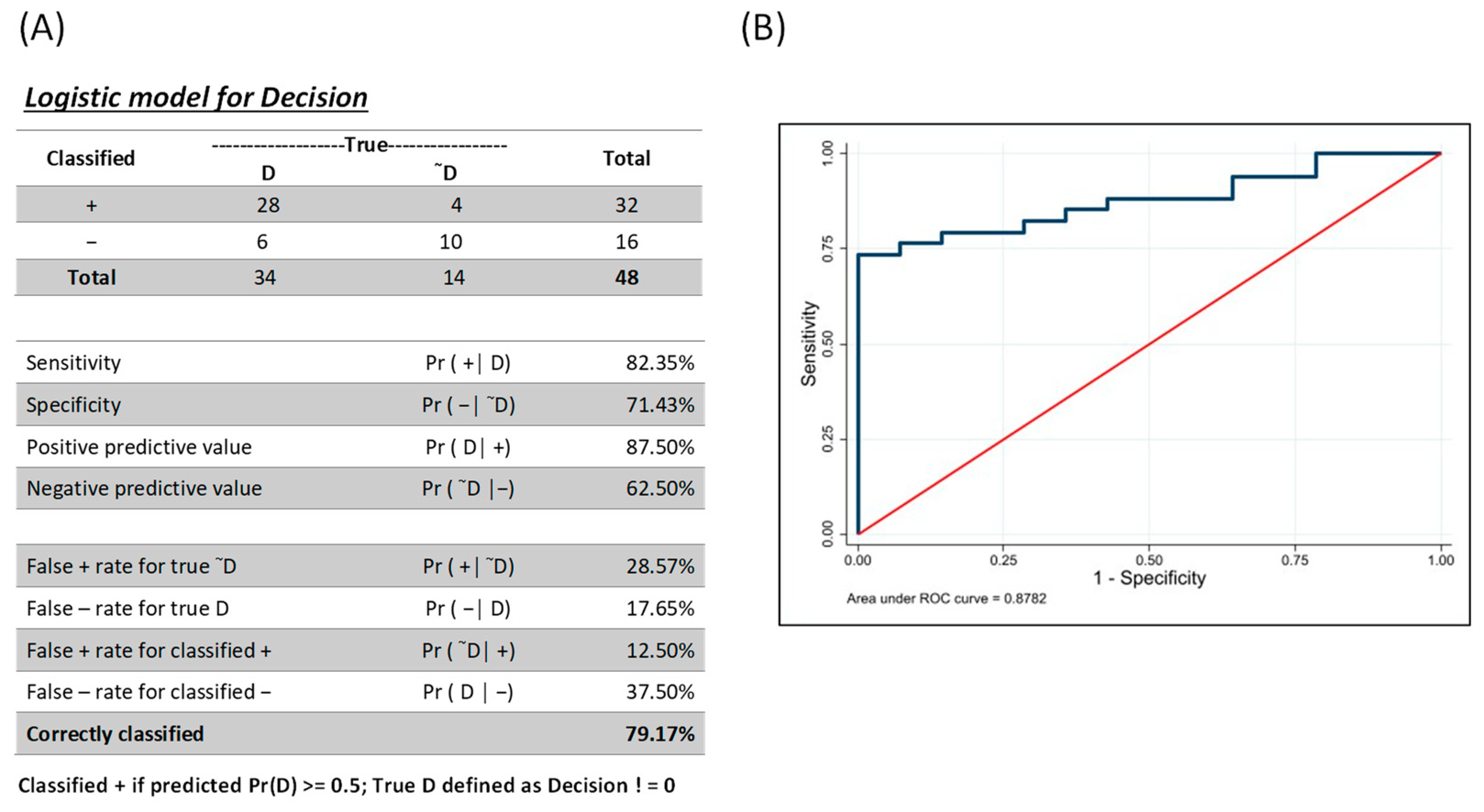
To determine whether a combination analysis of the MIG and MCP-1 biomarkers would increase the analyte performance, we generated a combined ROC using the lroc function and post-estimation for logistic regression in Stata (generation of the marker combinations based on se and sp filters) as described by [35]. Using a multiple logistic regression where all biomarkers were used as predictors, a resulting predicted probability of 0.5 was considered a case- or disease-positive observation (Figure 2). Using that as a cutoff, the model correctly classified 79.17% of the observations, with a sensitivity and specificity of 82.35% and 71.43%, respectively (Figure 2A). The ROC curve provided (Figure 2B) is a visual representation of all possible combinations of sensitivity and specificity from these inputs, with a possible AUC of 87.82%.
The ROC curve of MIG and MCP-1 markers in combination is presented in Figure 2B. Our results demonstrated that the combinatorial analysis did not improve the overall performance when compared to the MIG performance alone.

4. Discussion

The control of Leishmania parasites within the host cell is mediated by innate and adaptive immune responses [36]. Following infection, both pro- and anti-inflammatory cytokines and chemokines are secreted, which have dynamic effects in stimulating immune cells to react against Leishmania [37,38].
Previous studies showed that following antigen stimulation in vitro, PBMCs of symptomatic patients with VL exhibited a predominantly type 2 response, whereas cells of AVL+ individuals produced a mixed cytokine profile [9]. This mixed response ensures both effective control of parasite proliferation and preservation of immune homeostasis by blocking an excessive cellular response [11]. A mixed cytokine pattern (IFN-γ produced by CD8+ and CD4+ cells and IL-5 produced by CD4+) was also associated with asymptomatic L. infantum infection [39]. On the other hand, recently, a scoping review (selected 106 articles) debated whether the cellular immunity of asymptomatic VL was associated with a T1-type cellular response, with elevated levels of IFN-γ, TNF, IL-2, IP-10, IP-10 (CXCL10), MIG (CXCL9), MCP-1 (CCL2), neopterin, and soluble CD40 ligand, when low levels of IL-10, IL-4, and IL-17 were detected [40].
In the present work, cytokine/chemokine levels were assessed in a unique AVL+ cohort of U.S deployers. Although some chemokines/cytokines (from the large panel assessed) were not detected, the levels of specific chemokines were higher in AVL+ participants, especially MIG, MCP-1, and IL-8, suggesting their utility as biomarkers for identifying asymptomatically infected individuals.
Our results demonstrated that Monokine induced by Gamma (MIG) had the best performance when comparing the three identified biomarkers. MIG (or CXCL9) is a chemokine induced by IFN-γ and has the potential to provide amplification of the IFN-γ signal. Ritter and Korber [41] demonstrated the key role of MIG in the self-healing of localized cutaneous leishmaniasis, confirming that lesions exhibited a strong expression of Th1-associated chemokines, such as CCL2/MCP-1, CXCL9/MIG, and CXCL10/IP-10. More recently, it was shown that IL-8, IP-10, and MIG were robust markers for identifying asymptomatic VL from L. infantum (Spain) and L. donovani (Bangladesh) endemic areas [42]. MIG was reported as 100% sensitive and specific for those with VL [42]. However, previous studies demonstrated that MIG can also be produced at different disease stages (active VL, VL after treatment, asymptomatic) and may not be specific to AVL [43].
Our data suggest that MCP-1 and IL-8 also perform adequately as AVL+ biomarkers, although not as well as MIG. MCP-1 can activate anti-Leishmania macrophage-killing mechanisms and was studied as a biomarker of cure in cutaneous leishmaniasis [4]. High MCP-1 expression in the skin is described in localized, self-healing cutaneous lesions [44], but no detectable MCP-1 is found in diffuse, non-healing cutaneous lesions [41,45]. Our findings extend those of others, as MCP-1 was identified as a potential biomarker for asymptomatic individuals exposed to L. infantum and was proposed to measure VL treatment efficacy [19].
Among our healthy AVL+ cohort, SLA-stimulated mononuclear cells produced elevated IL-8/CXCL8 chemokine levels. Neutrophils are stimulated by IL-8-inducing chemotaxis during systemic inflammation, so IL-8 is broadly used as a biomarker of diverse inflammatory processes. Interleukin-8 also induces granulocyte degranulation [46]. Activation of human keratinocytes in response to L. infantum was associated with upregulated transcripts for IL-6, IL8, and CXCL5 that may limit the replication of intracellular parasites [47]. A reduced neutrophil count during active VL is associated with lower IL-8 levels in serum [48]. Tasew et al. [49] observed that in response to L. donovani and lipopolysaccharide stimulation, cultured whole blood from those with active VL showed significantly lower secretion of IL-8 and IP-10 compared to endemic healthy controls. Among malnourished individuals with active VL, L. donovani stimulation was associated with significantly less IL-8 and MIP-1α production [50]. Polymorphisms at the IL-8-251 position are associated with impaired IL-8 activity and the development of active VL in Iranian individuals [51], but this was not demonstrated in Brazilian patients with VL, where the T allele in position -509 of the TGFB1 gene was associated with both symptomatic and asymptomatic VL compared to Leishmania skin test-negative individuals [52]. In Bangladesh, those with symptomatic VL and asymptomatic “subclinical” VL had 14–19% rates of elevated IL-8, while those recently treated or “preclinical”, meaning within one year they developed symptomatic VL, had no IL-8 circulating [53]. Among 40 Spanish L. infantum AVL and 12 Bangladesh L. donovani AVL, IL-8/CXCL8 was associated with an AUC of 0.96 in stimulated plasma and 0.87 in dried plasma spots, although MIG/CXCL9 and IP-10/CXCL10 performed better [43].
The combined ROC is a user-friendly, reliable analytical tool to support researchers in the selection of optimal marker combination(s) through an interactive interface. It allows users to make choices based on the evaluation of all possible markers, double-filter scoring on sensitivity and specificity, selection of the best-performing combinations, and visualization of their receiver operating characteristic (ROC) curves. Based on that, we decided to evaluate the combination of our best biomarkers; however, for AVL+ biomarkers, it does not improve the sensitivity/specificity.
Our study has a few limitations due to including a relatively modest number of participants and measuring the cytokine/chemokine levels at 72 h post-stimulation, thus not accounting for cytokines peaking at earlier timelines (e.g. IL-4 typically secreted at 24 h). Moreover, although described as a powerful tool, the Luminex® platform does not have a high dynamic range, so cytokines secreted at low levels are not usually detected. We discussed in a prior report [24] that there may be some limited cross-reactivity in immune responses to our Leishmania antigen between species. In addition, the studied population was assessed after an average of 10 years after deployment to Iraq, so it could potentially be studied when waning immune responses. Despite these limitations, our study was unique as the cohort of participants had limited exposure to Leishmania during deployment and no continuous reinfection opportunities (as compared to living in endemic areas).
Depicting the biological implications of asymptomatic infection is critical for planning and evaluating VL control measures in endemic regions as well as developing strategies for the individual with infection. We identified that 64% of this cohort had antibodies to the saliva of the sand fly Phlebotomus alexandri, a VL vector in Iraq [25], suggesting that U.S. Forces had high exposure to the vector. Quantifying circulating immunological biomarkers as an alternative strategy for monitoring asymptomatic visceral infections will improve the identification of people at risk of developing VL disease. During a 16-year surveillance period among members of the U.S. Armed Forces, 25 active cases of visceral leishmaniasis were reported, representing 1.2% of the total leishmaniasis cases [54]. Late reactivated visceral leishmaniasis presenting as a lingular ulcer was described in a U.S. service member deployed during Operation Desert Storm in 1991 and Operation Iraqi Freedom (2002–2003) [55]. Therefore, the potential biomarkers described in our study can provide a further method to detect risk for activation of VL among U.S. veterans that is less complex than the multiple diagnostic strategies (serology, IGRA, qPCR) we employed in our surveillance for asymptomatic VL. The persistence of these chemokines, on average nearly a decade after infection in Iraq, raises theoretical concerns for accelerated immunoaging and risk of T cell exhaustion with persistent inflammatory stimuli.
These identified markers, MIG, MCP-1, and IL-8, may be useful to determine the risk of developing overt VL in individuals. Monitoring biomarkers may be used for individual or population-level surveillance, in this case for asymptomatic VL [56]. These biomarkers can be used as susceptibility/risk biomarkers that indicate the potential for developing a disease in a person who does not currently have a clinically apparent disease [57]. U.S. servicemembers are not tested for visceral leishmaniasis in the absence of symptoms outside of research studies, and there are no leishmaniasis restrictions on the donation of blood or other tissues except if VL has been confirmed with validated testing, such as invasive tissue biopsies. To further develop MIG as a biomarker, qualification would require concrete characterization of the biomarker to the outcome of interest, testing large numbers of the millions who deployed and following them over decades to determine who activates VL. Given that our biomarkers require cells, the Department of Defense’s serum repository would not suffice, although perhaps the Million Veterans Program of the Department of Veterans Affairs genomic material collection could be leveraged. Reproducibility and a better understanding of sources of variability in chemokine measurement, values over time in the same individual, analytic validation, and assessing specificity for Leishmania infantum would be necessary for this to become a clinically available marker. The applicability for those who deployed in the past and are now starting immunosuppression seem to be a target population at particular risk for activation. The natural history of asymptomatic VL continues to be incompletely studied, something that could provide information to buttress treatment mitigation strategies and understand the context of its use.
In conclusion, our results highlight that MIG is a promising biomarker for the identification of asymptomatic visceral leishmaniasis using peripheral blood samples. Moreover, IL-8 and MCP-1 may be elevated in AVL as well. Thus, understanding immune responses in asymptomatic infections can contribute to the development of new point-of-care tests and biomarkers that could have clinical applications.

Supplementary Materials

The following supporting information can be downloaded at: https://www.mdpi.com/article/10.3390/pathogens12050705/s1, Table S1: Chemokine/Cytokine production in supernatants of PBMC culture from participants.

Author Contributions

Conceptualization: F.F.d.A., I.L.-N. and N.A.; Methodology: F.F.d.A., I.L.-N. and N.K.; Software: S.R.; Validation: F.F.d.A., I.L.-N., N.K. and S.R.; Formal Analysis: F.F.d.A., I.L.-N. and S.R.; Investigation: F.F.d.A., I.L.-N., N.K. and N.A.; Resources: R.M. and N.A.; Data Curation: N.A. and R.M.; Writing—Original Draft Preparation: F.F.d.A., I.L.-N. and N.A.; Writing—Review & Editing: F.F.d.A., I.L.-N., N.K., S.R., R.M. and N.A.; Visualization: F.F.d.A., I.L.-N. and N.A.; Supervision: N.A.; Project Administration: N.A.; Funding Acquisition: N.A. All authors have read and agreed to the published version of the manuscript.

Funding

Funding for this project was obtained from the Global Emerging Infectious Disease Surveillance Branch, Armed Forces Health Surveillance Division PROMIS ID P0024_17_HS.

Institutional Review Board Statement

This study was conducted according to the guidelines of the Declarations of Helsinki, and the research protocol was approved by the Institutional Review Boards (IRB) of the Uniformed Services University (USU) MED 83-2979, April 2015, Walter Reed National Military Medical Center (WRNMMC) 412208-1, May 2015, and the William Beaumont Army Medical Center (WBAMC) 16–29, which deferred to USU approval.

Informed Consent Statement

Written informed consent was obtained from all participants involved in the study.

Data Availability Statement

Additional data supporting the reported analyses are available upon request from the corresponding author.

Conflicts of Interest

The authors declare no conflict of interest.

References

  1. World Health Organization. Leishmania. 2023. Available online: https://www.who.int/health-topics/leishmaniasis (accessed on 15 April 2023).
  2. Singh, S.; Kumari, V.; Singh, N. Predicting kala-azar disease manifestations in asymptomatic patients with latent Leishmania donovani infection by detection of antibody against recombinant K39 antigen. Clin. Diagn. Lab. Immunol. 2002, 9, 568–572. [Google Scholar] [CrossRef] [PubMed]
  3. Bimal, S.; Singh, S.K.; Das, V.N.R.; Sinha, P.K.; Gupta, A.K.; Bhattacharya, S.K.; Das, P. Leishmania donovani: Effect of therapy on expression of CD2 antigen and secretion of macrophage migration inhibition factor by T-cells in patients with visceral leishmaniasis. Exp. Parasitol. 2005, 111, 130–132. [Google Scholar] [CrossRef] [PubMed]
  4. Kalani, M.; Choopanizadeh, M.; Pourabbas, B.; Pouladfar, G.; Asaee, S.; Ghalati, E.G.; Moravej, A. Dynamic alterations and durability of T helper 22 and its corresponding cytokines following treatment in pediatric visceral leishmaniasis. Cytokine 2021, 144, 155579. [Google Scholar] [CrossRef] [PubMed]
  5. Carvalho, E.M.; Bacellar, O.; Barral, A.; Badaro, R.; Johnson, W.D., Jr. Antigen-specific immunosuppression in visceral leishmaniasis is cell mediated. J. Clin. Investig. 1989, 83, 860–864. [Google Scholar] [CrossRef]
  6. Kharazmi, A.; Kemp, K.; Ismail, A.; Gasim, S.; Gaafar, A.; Kurtzhals, J.A.; Hassan, A.M.E.; Theander, T.G.; Kemp, M. T-cell response in human leishmaniasis. Immunol. Lett. 1999, 65, 105–108. [Google Scholar] [CrossRef]
  7. Ferrua, B.; Luci, C.; Fichoux, Y.L.; Paul, A.; Marty, P. Imprinting of BALB/c mice with low Leishmania infantum parasite dose markedly protects spleen against high-dose challenge. Vaccine 2006, 24, 589–596. [Google Scholar] [CrossRef]
  8. Gouvea, V.L.; de Assis, T.S.M.; Orsini, M.; da Silva, A.R.; de Souza, G.F.; Caligiorne, R.; da Silva, A.C.L.; Peruhype-Magalhães, V.; Marciano, A.P.V.; Martins-Filho, O.A.; et al. Combined diagnostic methods identify a remarkable proportion of asymptomatic Leishmania (Leishmania) chagasi carriers who present modulated cytokine profiles. Trans. R Soc. Trop. Med. Hyg. 2008, 102, 548–555. [Google Scholar] [CrossRef]
  9. Peruhype-Magalhaes, V.; Martins-Filho, O.A.; Prata, A.; Silva, L.A.; Rabello, A.; Teixeira-Carvalho, A.; Figueiredo, R.M.; Guimarães-Carvalho, S.F.; Ferrari, T.C.A.; Correa-Oliveira, R. Immune response in human visceral leishmaniasis: Analysis of the correlation between innate immunity cytokine profile and disease outcome. Scand. J. Immunol. 2005, 62, 487–495. [Google Scholar] [CrossRef]
  10. Peruhype-Magalhaes, V.; Martins-Filho, O.A.; Prata, A.; Silva, L.A.; Rabello, A.; Teixeira-Carvalho, A.; Figueiredo, R.M.; Guimarães-Carvalho, S.F.; Ferrari, T.C.A.; Van Weyenbergh, J.; et al. Mixed inflammatory/regulatory cytokine profile marked by simultaneous raise of interferongamma and interleukin-10 and low frequency of tumour necrosis factoralpha(+) monocytes are hallmarks of active human visceral Leishmaniasis due to Leishmania chagasi infection. Clin. Exp. Immunol. 2006, 146, 124–132. [Google Scholar] [CrossRef]
  11. Machado, C.M.; Martins, T.C.; Colturato, I.; Leite, M.S.; Simione, A.J.; Souza, M.P.; Mauad, M.A.; Colturato, V.R. Epidemiology of neglected tropical diseases in transplant recipients. Review of the literature and experience of a Brazilian HSCT center. Rev. Inst. Med. Trop. Sao Paulo 2009, 51, 309–324. [Google Scholar] [CrossRef]
  12. Rodrigues, V.; Cordeiro-da-Silva, A.; Laforge, M.; Silvestre, R.; Estaquier, J. Regulation of immunity during visceral Leishmania infection. Parasit. Vectors 2016, 9, 118. [Google Scholar] [CrossRef] [PubMed]
  13. Saporito, L.; Giammanco, G.M.; De Grazia, S.; Colomba, C. Visceral leishmaniasis: Host-parasite interactions and clinical presentation in the immunocompetent and in the immunocompromised host. Int. J. Infect. Dis. 2013, 17, e572–e576. [Google Scholar] [CrossRef] [PubMed]
  14. Oghumu, S.; Lezama-Dávila, C.M.; Isaac-Márquez, A.P.; Satoskar, A.R. Role of chemokines in regulation of immunity against leishmaniasis. Exp. Parasitol. 2010, 126, 389–396. [Google Scholar] [CrossRef] [PubMed]
  15. Faleiro, R.J.; Kumar, R.; Hafner, L.M.; Engwerda, C.R. Immune regulation during chronic visceral leishmaniasis. PLoS Negl. Trop. Dis. 2014, 8, e2914. [Google Scholar] [CrossRef]
  16. Singh, O.P.; Gidwani, K.; Kumar, R.; Nylén, S.; Jones, S.L.; Boelaert, M.; Sacks, D.; Sundara, S. Reassessment of immune correlates in human visceral leishmaniasis as defined by cytokine release in whole blood. Clin. Vac. Immunol. 2012, 19, 961–966. [Google Scholar] [CrossRef]
  17. Ibarra-Meneses, A.V.; Carrillo, E.; Sanchez, C.; García-Martínez, J.; López Lacomba, D.; San Martin, J.V.; Alves, F.; Alvar, J.; Moreno, J. Interleukin-2 as a marker for detecting asymptomatic individuals in areas where Leishmania infantum is endemic. Clin. Microbiol. Infect. 2016, 22, 739. [Google Scholar] [CrossRef]
  18. Ibarra-Meneses, A.V.; Ghosh, P.; Hossain, F.; Chowdhury, R.; Mondal, D.; Alvar, J.; Moreno, J.; Carrillo, E. IFN-γ, IL-2, IP-10, and MIG as biomarkers of exposure to Leishmania spp., and of cure in human visceral leishmaniasis. Front. Cell. Infect. Microbiol. 2017, 7, 200. [Google Scholar] [CrossRef]
  19. Ibarra-Meneses, A.V.; Sanchez, C.; Alvar, J.; Moreno, J.; Carrillo, E. Monocyte chemotactic protein 1 in plasma from soluble Leishmania antigen-stimulated whole blood as a potential biomarker of the cellular immune response to Leishmania infantum. Front. Immunol. 2017, 8, 1208. [Google Scholar] [CrossRef]
  20. Adem, E.; Tajebe, F.; Getahun, M.; Kiflie, A.; Diro, E.; Hailu, A.; Shkedy, Z.; Mengesha, B.; Mulaw, T.; Atnafu, S. Successful treatment of human visceral leishmaniasis restores antigen-specific IFN-gamma, but not IL-10 production. PLoS Negl. Trop. Dis. 2016, 10, e0004468. [Google Scholar] [CrossRef]
  21. Schaefer, K.U.; Kurtzhals, J.A.; Gachihi, G.S.; Muller, A.S.; Kager, P.A. A prospective sero-epidemiological study of visceral leishmaniasis in Baringo District, Rift Valley Province, Kenya. Trans. R Soc. Trop. Med. Hyg. 1995, 89, 471–475. [Google Scholar] [CrossRef]
  22. Sinha, P.K.; Bimal, S.; Pandey, K.; Singh, S.K.; Ranjan, A.; Kumar, N.; Lal, C.S.; Barman, S.B.; Verma, R.B.; Jeyakumar, A.; et al. A community-based, comparative evaluation of direct agglutination and rK39 strip tests in the early detection of subclinical Leishmania donovani infection. Ann. Trop. Med. Parasitol. 2008, 102, 119–125. [Google Scholar] [CrossRef] [PubMed]
  23. Goswami, R.P.; Bairagi, B.; Kundu, P.K. rK39 strip test-easy, reliable and cost-effective field diagnosis for visceral leishmaniasis in India. J. Assoc. Physicians India 2003, 51, 759–761. [Google Scholar] [PubMed]
  24. Mody, R.M.; Lakhal-Naouar, I.; Sherwood, J.E.; Koles, N.L.; Shaw, D.; Bigley, D.P.; Co, E.-M.A.; Copeland, N.K.; Jagodzinski, L.L.; Mukbel, R.M. Asymptomatic Visceral Leishmania infantum Infection in US Soldiers Deployed to Iraq. Clin. Infect. Dis. 2019, 68, 2036–2044. [Google Scholar] [CrossRef] [PubMed]
  25. Lakhal-Naouar, I.; Mukbel, R.; DeFraites, R.F.; Mody, R.M.; Massoud, L.N.; Shaw, D.; Co, E.M.; Sherwood, J.E.; Kamhawi, S.; Aronson, N.E. The human immune response to saliva of Phlebotomus alexandri, the vector of visceral leishmaniasis in Iraq, and its relationship to sand fly exposure and infection. PLoS Negl. Trop. Dis. 2021, 15, e0009378. [Google Scholar] [CrossRef]
  26. Beasley, E.A.; Mahachi, K.G.; Petersen, C.A. Possibility of Leishmania transmission via Lutzomyia spp. sand flies within the USA and implications for human and canine autochthonous Infection. Curr. Trop. Med. Rep. 2022, 9, 160–168. [Google Scholar] [CrossRef]
  27. Curtin, J.M.; Aronson, N.E. Leishmaniasis in the United States: Emerging Issues in a Region of Low Endemicity. Microorganisms 2021, 9, 578. [Google Scholar] [CrossRef]
  28. Vallur, A.C.; Reinhart, C.; Mohamath, R.; Goto, Y.; Ghosh, P.; Mondal, D.; Duthie, M.S.; Reed, S.G. Accurate Serodetection of Asymptomatic Leishmania donovani Infection by Use of Defined Antigens. J. Clin. Microbiol. 2016, 54, 1025–1030. [Google Scholar] [CrossRef]
  29. Vallur, A.C.; Duthie, M.S.; Reinhart, C.; Tutterrow, Y.; Hamano, S.; Bhaskar, K.R.H.; Coler, R.; Mondal, D.; Reed, S. Biomarkers for intracellular pathogens: Establishing tools as vaccine and therapeutic endpoints for visceral leishmaniasis. Clin. Microbiol. Infect. 2014, 20, O374–O383. [Google Scholar] [CrossRef]
  30. Berens, R.L.; Brun, R.; Krassner, S.M. A simple monophasic medium for axenic culture of hemoflagellates. J. Parasitol. 1976, 62, 360–365. [Google Scholar] [CrossRef]
  31. Ramer-Tait, A.E.; Lei, S.M.; Bellaire, B.H.; Beetham, J.K. Differential surface deposition of complement proteins on logarithmic and stationary phase Leishmania chagasi promastigotes. J. Parasitol. 2012, 98, 1109–1116. [Google Scholar] [CrossRef]
  32. Scott, P.; Pearce, E.; Natovitz, P.; Sher, A. Vaccination against cutaneous leishmaniasis in a murine model. II. Immunologic properties of protective and nonprotective subfractions of soluble promastigote extract. J. Immunol. 1987, 139, 3118–3125. [Google Scholar] [CrossRef] [PubMed]
  33. Marovich, M.A.; Mascola, J.R.; Eller, M.; Louder, M.K.; Caudrelier, P.A.; El-Habib, R.; Ratto-Kim, S.; Cox, J.H.; Currier, J.R.; Levine, B.L.; et al. Preparation of Clinical-Grade Recombinant Canarypox–Human Immunodeficiency Virus Vaccine–Loaded Human Dendritic Cells. J. Infect. Dis. 2002, 186, 1242–1252. [Google Scholar] [CrossRef] [PubMed]
  34. StataCorp. Stata Statistical Software; Release 17; StataCorp LLC: College Station, TX, USA, 2021. [Google Scholar]
  35. Mazzara, S.; Rossi, R.L.; Grifantini, R.; Donizetti, S.; Abrignani, S.; Bombaci, M. CombiROC: An interactive web tool for selecting accurate marker combinations of omics data. Sci. Rep. 2017, 30, 45477. [Google Scholar] [CrossRef]
  36. Nylén, S.; Sacks, D. Interleukin-10 and the pathogenesis of human visceral leishmaniasis. Trends Immunol. 2007, 28, 378–384. [Google Scholar] [CrossRef] [PubMed]
  37. Lunney, J. Cytokines orchestrating the immune response. Rev. Sci. Tech. 1998, 17, 84–94. [Google Scholar] [CrossRef]
  38. Pitta, M.G.R.; Romano, A.; Cabantous, S.; Henri, S.; Hammad, A.; Kouriba, B.; Argiro, L.; El Kheir, M.; Bucheton, B.; Mary, C.; et al. IL-17 and IL-22 are associated with protection against human kala azar caused by Leishmania donovani. J. Clin. Investig. 2009, 119, 2379–2387. [Google Scholar] [CrossRef]
  39. Mary, C.; Auriault, V.; Faugère, B.; Dessein, A.J. Control of Leishmania infantum infection is associated with CD8(+) and gamma interferon- and interleukin-5-producing CD4(+) antigen-specific T cells. Infect. Immun. 1999, 67, 5559–5566. [Google Scholar] [CrossRef]
  40. Ibarra-Meneses, A.V.; Corbeil, A.; Wagner, V.; Onwuchekwa, C.; Fernandez-Prada, C. Identification of asymptomatic Leishmania infections: A scoping review. Parasit. Vectors 2022, 15, 5. [Google Scholar] [CrossRef]
  41. Ritter, U.; Korner, H. Divergent expression of inflammatory dermal chemokines in cutaneous leishmaniasis. Parasite Immunol. 2002, 24, 295–301. [Google Scholar] [CrossRef]
  42. Ibarra-Meneses, A.V.; Mondal, D.; Alvar, J.; Moreno, J.; Carrillo, E. Cytokines and chemokines measured in dried SLA-stimulated whole blood spots for asymptomatic Leishmania infantum and Leishmania donovani infection. Sci. Rep. 2017, 7, 17266. [Google Scholar] [CrossRef]
  43. Porcino, G.N.; Carvalho, K.S.S.; Braz, D.C.; Silva, V.C.; Costa, C.H.N.; Santos, I.K.F.M. Evaluation of methods for detection of asymptomatic individuals infected with Leishmania infantum in the state of Piauí, Brazil. PLoS Negl. Trop. Dis. 2019, 13, e0007493. [Google Scholar] [CrossRef] [PubMed]
  44. Valencia-Pacheco, G.; Loría-Cervera, E.N.; Sosa-Bibiano, E.I.; Canché-Pool, E.B.; Vargas-Gonzalez, A.; Melby, P.C.; Andrade-Narvaez, F.J. In situ cytokines (IL-4, IL-10, IL-12, IFN-γ) and chemokines (MCP-1, MIP-1α) gene expression in human Leishmania (Leishmania) mexicana infection. Cytokine 2014, 69, 56–61. [Google Scholar] [CrossRef] [PubMed]
  45. Moll, H. The role of chemokines and accessory cells in the immunoregulation of cutaneous leishmaniasis. Behring Inst. Mitt. 1997, 99, 73–78. [Google Scholar]
  46. Arango Duque, G.A.; Descoteaux, A. Macrophage Cytokines: Involvement in Immunity and Infectious Diseases. Front. Immunol. 2014, 5, 491. [Google Scholar] [CrossRef]
  47. Scorza, B.M.; Wacker, M.A.; Messingham, K.; Kim, P.; Klingelhutz, A.; Fairley, J.; Wilson, M.E. Differential Activation of Human Keratinocytes by Leishmania Species Causing Localized or Disseminated Disease. J. Investig. Dermatol. 2017, 137, 2149–2156. [Google Scholar] [CrossRef]
  48. Elshafie, A.I.; Hlin, E.; Håkansson, L.D.; Elghazali, G.; El Safi, S.H.; Rönnelid, J.; Venge, P. Activity and turnover of eosinophil and neutrophil granulocytes are altered in visceral leishmaniasis. Int. J. Parasitol. 2011, 41, 463–469. [Google Scholar] [CrossRef]
  49. Tasew, G.; Gadisa, E.; Abera, A.; Chanyalew, M.; Abebe, M.; Howe, R.; Ritter, U.; Aseffa, A.; Laskay, T. Whole blood-based in vitro culture reveals diminished secretion of pro-inflammatory cytokines and chemokines in visceral leishmaniasis. Cytokine 2021, 145, 155246. [Google Scholar] [CrossRef]
  50. Kumar, V.; Bimal, S.; Singh, S.K.; Chaudhary, R.; Das, S.; Lal, C.; Pandey, K.; Das, V.R.; Das, P. Leishmania donovani: Dynamics of L. donovani evasion of innate immune cell attack due to malnutrition in visceral leishmaniasis. Nutrition 2014, 30, 449–458. [Google Scholar] [CrossRef]
  51. Hajilooi, M.; Abasi, M.; Bazmani, A.; Ahmadi, A.; Matini, M.; Solgi, G.; Sardarian, K. Evaluation of interleukin-8-251 t/a polymorphisms in visceral leishmaniasis. J. Res. Health Sci. 2015, 15, 59–61. [Google Scholar]
  52. Frade, A.F.; de Oliveira, L.C.; Costa, D.L.; Costa, C.H.N.; Aquino, D.; Van Weyenbergh, J.; Barral-Netto, M.; Barral, A.; Kalil, J.; Goldberg, A.C. TGFB1 and IL8 gene polymorphisms and susceptibility to visceral leishmaniasis. Infect. Genet. Evol. 2011, 11, 912–916. [Google Scholar] [CrossRef]
  53. Kurkjian, K.M.; Mahmutovic, A.J.; Kellar, K.L.; Haque, R.; Bern, C.; Secor, W.E. Multiplex analysis of circulating cytokines in the sera of patients with different clinical forms of visceral leishmaniasis. Cytometry A. 2006, 69, 353–358. [Google Scholar] [CrossRef] [PubMed]
  54. Stahlman, S.; Williams, V.F.; Taubman, S.B. Incident diagnoses of leishmaniasis, active and reserve components, U.S. Armed Forces, 2001–2016. Med. Surveill. Mon. Rep. (MSMR) 2017, 24, 2–7. [Google Scholar]
  55. Mazumder, S.A.; Pandey, S.; Brewer, S.C.; Baselski, V.S.; Weina, P.J.; Land, M.A.; Fleckenstein, J.M. Lingual Leishmaniasis Complicating Visceral Disease. J. Travel Med. 2010, 17, 212–214. [Google Scholar] [CrossRef] [PubMed]
  56. FDA-NIH Biomarker Working Group. BEST (Biomarkers, EndpointS, and Other Tools) Resource Silver Spring (MD): Food and Drug Administration (US); Monitoring Biomarker; National Institutes of Health (US): Bethesda, MD, USA, 2016. Available online: https://www.ncbi.nlm.nih.gov/books/NBK402282/ (accessed on 25 January 2021).
  57. FDA-NIH Biomarker Working Group. BEST (Biomarkers, EndpointS, and Other Tools) Resource Silver Spring (MD): Food and Drug Administration (US); Susceptibility/Risk Biomarker; National Institutes of Health (US): Bethesda, MD, USA, 2016. Available online: https://www.ncbi.nlm.nih.gov/books/NBK402288/ (accessed on 27 August 2020).
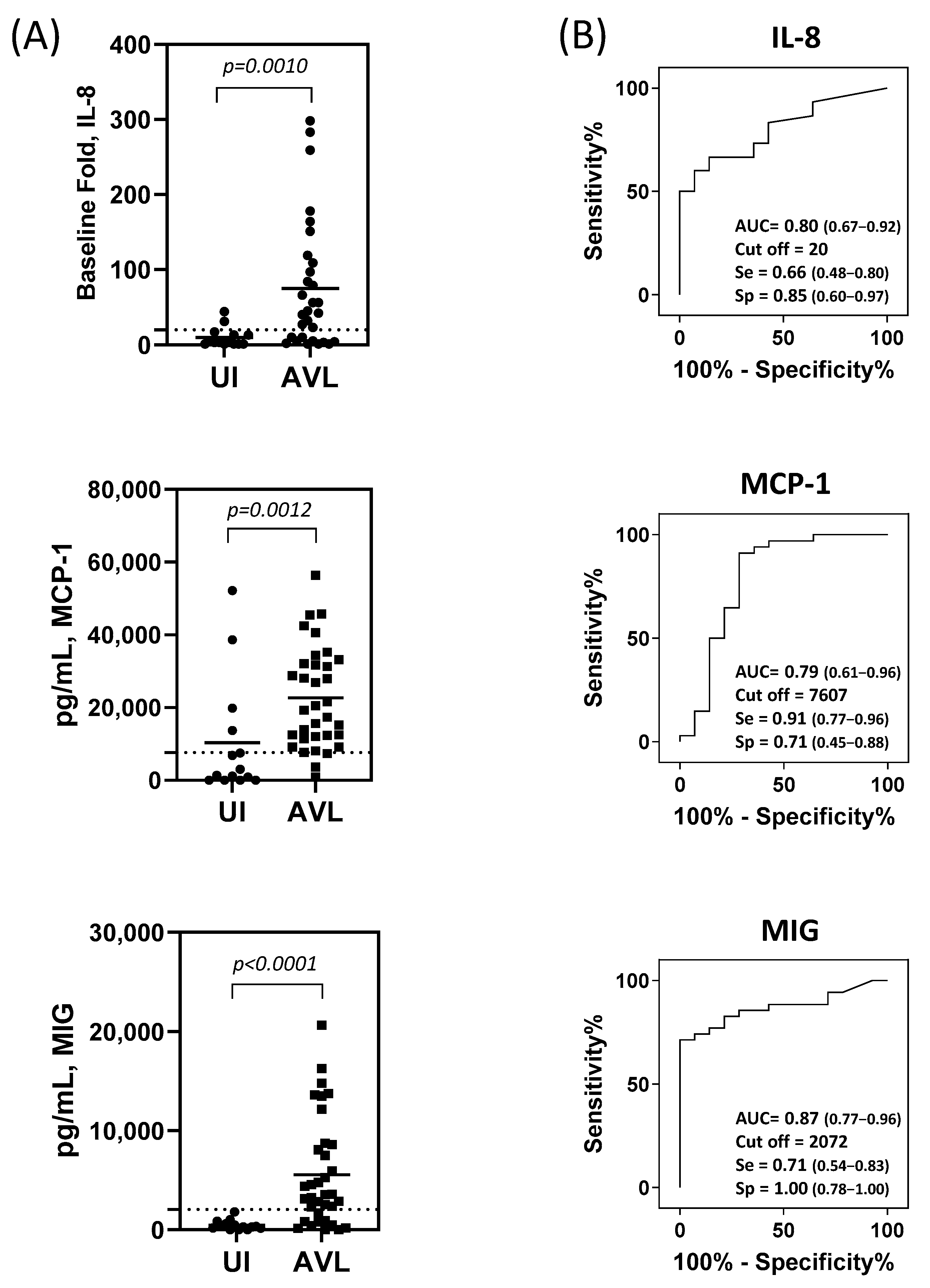
Figure 1. Chemokine production in supernatants of cultured PBMCs from study participants. (A) Production of chemokines following 72 h of SLA stimulation of PBMC from individuals with asymptomatic visceral leishmaniasis (AVL+) and uninfected (UI, AVL negative) military healthcare beneficiaries. Biomarkers IL-8 (UI, n =14 and AVL, n = 30); MCP-1 (UI, n =14 and AVL, n = 34), and MIG (UI, n =14 and AVL, n = 35). Horizontal bars represent the mean concentration for each biomarker. Dashed lines represent the cut-off. The identification of the cut-point value requires a simultaneous assessment of sensitivity and specificity. A cut-point will be referred to as optimal when the point classifies most of the individuals correctly. (B) Receiver operating characteristics curve analyses to determine the performance of these analytes as potential biomarkers for AVL. AUC = area under the curve, se = sensitivity and sp = specificity. Statistical analyses were performed using Mann–Whitney U test.
Pathogens 12 00705 g001
Figure 2. Combinations of MIG and MCP-1 markers identified by combined ROC. (A) Logistic model for decision showing the fraction of predictions using a cutoff of predicted probability 0.5 (FN: false negative; FP: false positives; TN: true negative; TP: true positive) and the percentage of correctly classified. (B) Receiver operating characteristic curve analysis to determine the performance of these analytes combined as potential biomarkers for AVL. AUC = area under the curve.
Pathogens 12 00705 g002
Table 1. Characteristics of Study Participants.
PopulationAVL Negative Controls
(n = 14)
AVL+ Participants
(n = 35)
Mean age, years (range)35.1 (25–57)40.7 (29–58)
Sex
Female
Male

5 (35.7%)
9 (64.3%)

3 (8.6%)
32 (91.4%)
Race/Ethnicity
African American2 (14.3%)5 (14.3%)
White
 Hispanic/Latino05 (14.3%)
 Not Hispanic/Latino9 (64.3%)22 (62.8%)
Other3 (21.4%)3 (8.6%)
Positive Leishmania Assay
IGRA+
ELISA+
qPCR+

negative
negative
negative

27 (77.2%)
6 (17.1%)
2 (5.7%)
IGRA = IFN-γ release assay; ELISA = enzyme-linked immunosorbent assay; qPCR = quantitative polymerase chain reaction.
Disclaimer/Publisher’s Note: The statements, opinions and data contained in all publications are solely those of the individual author(s) and contributor(s) and not of MDPI and/or the editor(s). MDPI and/or the editor(s) disclaim responsibility for any injury to people or property resulting from any ideas, methods, instructions or products referred to in the content.