Article

COVID-19 Is a Confounder of Increased Candida Airway Colonisation

by
Margaux Froidefond
1,2,
Jacques Sevestre
1,2,
Hervé Chaudet
1,2 and
Stéphane Ranque
1,2,*
1
Faculté des Sciences Médicales et Paramédicales, Aix Marseille University, IRD, AP-HM, SSA, VITROME, 13005 Marseille, France
2
IHU Méditerranée Infection, 19-21 Boulevard Jean Moulin, 13005 Marseille, France
*
Author to whom correspondence should be addressed.
Pathogens 2023, 12(3), 463; https://doi.org/10.3390/pathogens12030463
Submission received: 18 January 2023 / Revised: 10 March 2023 / Accepted: 12 March 2023 / Published: 15 March 2023

Abstract

:
An increased incidence of invasive fungal infection was reported in SARS-CoV-2-infected patients hospitalised in the intensive care unit. However, the impact of COVID-19 on Candida airway colonisation has not yet been assessed. This study aimed to test the impact of several factors on Candida airway colonisation, including SARS-CoV-2 infection. We conducted a two-pronged monocentric retrospective study. First, we analysed the prevalence of positive yeast culture in respiratory samples obtained from 23 departments of the University Hospital of Marseille between 1 January 2018 and 31 March 2022. We then conducted a case-control study, comparing patients with documented Candida airway colonisation to two control groups. We observed an increase in the prevalence of yeast isolation over the study period. The case-control study included 300 patients. In the multivariate logistic regression, diabetes, mechanical ventilation, length of stay in the hospital, invasive fungal disease, and the use of antibacterials were independently associated with Candida airway colonisation. The association of SARS-CoV-2 infection with an increased risk of Candida airway colonisation is likely to be a consequence of confounding factors. Nevertheless, we found the length of stay in the hospital, mechanical ventilation, diabetes, and the use of antibacterials to be statistically significant independent risk factors of Candida airway colonisation.

1. Introduction

In December 2019, the first case of SARS-CoV-2 infection was described in Wuhan, China, heralding the emergence of a global pandemic responsible for 546 million cases and 6.3 million deaths [1,2]. The morbidity associated with COVID-19 is heterogeneous, ranging from asymptomatic infections to acute respiratory distress syndrome (ARDS), requiring urgent hospitalisation in an intensive care unit (ICU) [3]. ICU hospitalisation and subsequent invasive therapies may generate infectious complications, potentially associated with significant morbidity and mortality [4].
Yeast isolation from respiratory samples is common, notably in ICU patients receiving mechanical ventilation [5,6]. Yeast colonisation has been reported in 30% of patients receiving ventilation over 48 h and in up to 50% of patients diagnosed with ventilator-associated pneumonia (VAP) [7]. Similarly, recent studies report such respiratory colonisation in up to 21% of patients with COVID-19 [8]. The known risk factors for Candida airway colonisation include exposure to broad-spectrum antibiotics, the use of corticosteroids and immunosuppressants, the use of mechanical ventilation, neutropaenia, and diabetes [9,10]. Some studies have also reported that the duration of mechanical ventilation and the length of stay in the ICU are associated with a poor clinical outcome [11,12]. However, data regarding the impact of SARS-CoV-2 infection on respiratory tract colonisation by Candida yeast are scarce. This study aimed to assess the association of risk factors, particularly SARS-CoV-2 infection, with Candida respiratory tract colonisation. A secondary objective was to assess the effect of systemic antibiotic treatments on Candida airway colonisation.

2. Methods

2.1. Study Design

We conducted a two-pronged, single-centre retrospective study. The first approach involved a longitudinal observational study based on the “Méditerranée Infection Data Warehousing and Surveillance” (MIDAS) database. This programme, hosted at the Institut Hospitalo-Universitaire (IHU) Méditerranée Infection, combines syndromic and conventional surveillance and collects data from all the samples received at the microbiology laboratory, any subsequent tests performed, and the results obtained, on a weekly basis [13]. We analysed the prevalence of yeast isolation in respiratory samples from patients hospitalised in 21 units at the Assistance Publique—Hôpitaux de Marseille (AP-HM) university hospital of Marseille (France) from 1 January 2018 to 31 March 2022. The second approach was a case-control study comparing patients with Candida airway colonisation to two groups of control patients, who had no positive respiratory sample yeast culture.

2.2. Inclusion Criteria

The patients included in the study between 1 April 2021 and 30 November 2021 were required to meet the following inclusion criteria:
Be over 18 years old.
Be hospitalised in the AP-HM during the study period, with at least three respiratory samples obtained for the culture.

2.3. Exclusion Criteria

Patients under 18 years of age, patients without respiratory samples, or patients who objected to the use of personal data for research purposes were excluded from the analysis.

2.4. Case Definition

Candida airway colonisation cases were defined by three or more respiratory samples growing yeasts on culture and obtained on separate days.

2.5. Control Definitions

The patients included in the control groups had to meet the following criteria:
Absence of yeast cultured from at least three respiratory samples obtained on separate days.
The positive control (BactC) group included patients with a positive bacterial culture (Haemophilus influenzae, Klebsiella pneumoniae, Streptococcus pneumoniae, Pseudomonas aeruginosa...) in a respiratory sample.
The negative control (NegC) group included patients for whom respiratory sample cultures were negative, i.e., neither bacteria nor yeast were cultured.

2.6. Data Collection

In the first part of the study, the respiratory sample culture results from 21 units of the AP-HM were collected through the MIDAS programme. In the second part of the study, clinical data were collected through the hospital computerised medical records. The demographic information collected included: sex, age, medical history (obesity, high blood pressure (HBP), heart disease, diabetes, smoking, chronic respiratory failure, chronic renal failure with a global filtration rate (GFR) < 60 mL/min/1.73 m2), a history of haematological disease, cancer, a history of transplantation, immunosuppression, and the use of immunosuppressive therapy (long-term corticosteroid therapy, cancer chemotherapy, or anti-rejection therapy for transplant patients).
The data collected on the hospital stay included: length of stay, hospitalisation in the ICU or medical ward, diagnosis of SARS-CoV-2 infection with identification of the variant, use of oxygen therapy and delivery technique (low-dose oxygen therapy, high-flow nasal cannula (HFNC), non-invasive ventilation (NIV), mechanical ventilation (MV)), use of extracorporeal membrane oxygenation (ECMO), use of enteral or parenteral nutrition, abdominal surgery during hospitalisation, the administration of antibacterial, antiviral, or antifungal antibiotics, diagnosis of invasive fungal disease during the stay, detection of respiratory viruses, the type of respiratory samples obtained (bronchoalveolar lavage (BAL), tracheobronchial aspiration (TBA), sputum cytobacteriological examination (CBES)), the use of immunomodulatory therapies (notably, dexamethasone, methylprednisolone, and tocilizumab), and the patient’s outcome (deceased or alive). The time from admission to isolation of the first yeast in a respiratory sample was noted in patients in the case group. Similarly, for the BactC group, the time between admission and the first positive bacterial culture of respiratory samples was recorded.

2.7. Statistical Analysis

The case-control study data were analysed using SAS 9.2 for Windows (SAS Institute Inc., Cary, NC, USA). Continuous variables were expressed as the mean (SD), while categorical variables were expressed as proportions and percentages. Continuous variables were compared using ANOVA. Categorical variables were compared using the Chi square or Fisher’s exact tests, as required. All statistical tests were two-sided, with a p < 0.05 significance level. Univariate and multivariate unconditional logistic regression analyses were performed to estimate odds ratios (ORs) with a 95% confidence interval (CI). All covariates with a p < 0.20 significance level in the univariate analysis were included in the multivariate analyses. Three multivariate logistical regression models were computed: the case group was compared either to (i) all controls (including both the NegC and BactC groups), (ii) only the BactC, or (iii) only the NegC groups. For each control group model, a stepwise selection was performed to retain the most parsimonious model, including the covariates that displayed an independent statistically significant effect on the risk of airway fungal colonisation.

3. Results

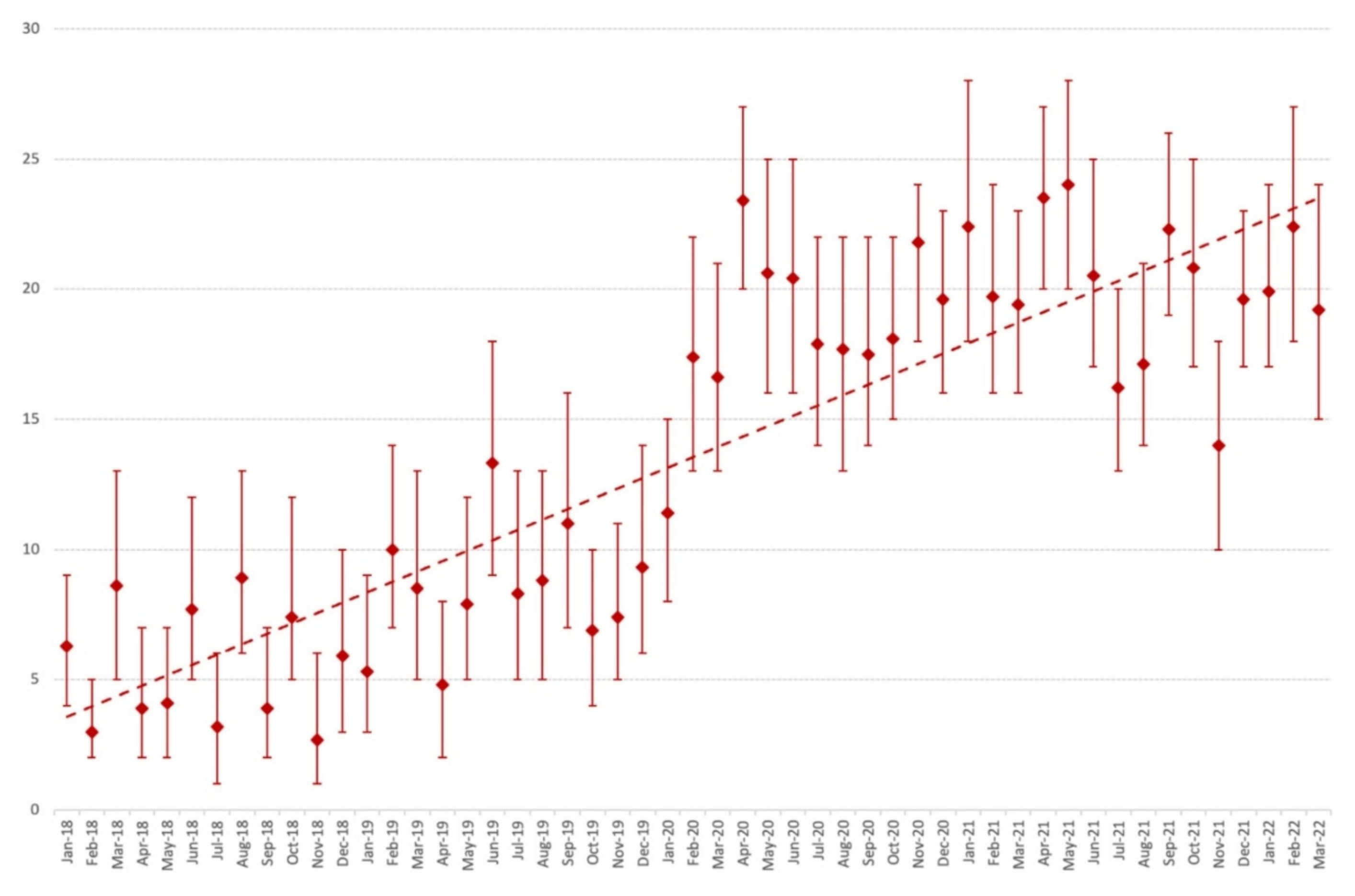
3.1. Time Trends in the Prevalence of Yeast-Positive Culture in Respiratory Samples

Between 1 January 2018 and 31 March 2022, a total of 17,408 respiratory samples were collected, of which 12,881 grew bacteria and/or fungi in culture. Most (17,102 (98.2%)) of the samples collected over the study period originated from ICU patients. Yeast was isolated in 3593 samples, amounting to 28% of the positive cultures. The distribution of yeast species during this period is described in Figure 1.
Of those samples, 2658 (74%) grew C. albicans, 272 (8%) grew C. tropicalis, 217 (6%) grew C. glabrata, 123 (3%) grew C. dubliniensis, and 115 (3%) grew C. parapsilosis. The prevalence of yeast isolation increased from February 2020, concomitantly with the onset of the COVID-19 epidemic in Marseille, with a predominance of C. albicans (17% of yeasts isolated). In April 2020, up to 23% of culture samples grew C. albicans. This trend continued over the following year, with C. albicans being isolated from 24% of respiratory samples in May 2021. A relative decrease in the prevalence of C. albicans occurred in July 2021 (16%), August 2021 (17%), and November 2021 (14%). Finally, in 2022, 20% of the samples were positive for C. albicans in January, 22% in February, and 19% in March. This time trend is illustrated in Figure 2 and Figure 3.

3.2. Patient Demographics

Between 1 April 2021 and 30 November 2021, 300 patients (100 in each group) were included in the case-control study (Table 1). The three groups were homogeneous regarding age (p = 0.8752) and sex (p = 0.7605). A total of 80 patients (80%) in the case group were admitted to the ICU, compared to 62 (62%) and 18 (18%) patients in the BactC and NegC groups, respectively (p < 10−4). Comorbidities were homogenously distributed among the three groups. Of the 79 patients with a history of chronic respiratory failure, 32 had chronic obstructive pulmonary disease (COPD), 18 had cystic fibrosis, 8 had diffuse interstitial lung disease, 7 had pulmonary fibrosis, 6 had bronchial dilatation, 6 had emphysema, 1 had severe asthma, and 1 had respiratory failure consecutive to pulmonary tuberculosis. Diabetes was statistically significantly (p < 10−4) more frequent in the case group (31%) compared to the BactC and NegC groups—26% and 7%, respectively. In contrast, solid malignancies were rarer (p = 0.0114) in the case group (7%) compared to the BactC and NegC groups—16% and 22%, respectively. Similarly, immunosuppression was rarer (p = 0.0212) in the case group (17%) compared to the BactC and NegC groups—31% and 33%, respectively. Among immunocompromised patients, 42 had a history of organ transplantation, including 33 lung transplant patients, 16 had a history of chronic inflammatory or autoimmune disease, 12 were receiving cancer chemotherapy, 7 had been treated for a haematological disease, and 4 were infected with HIV.

3.3. Characteristics of Hospital Stay

The use of parenteral nutrition (p = 0.1697) and oxygen delivery devices, including low-dose oxygen (p = 0.2129), HNFC (p = 0.6004), and non-invasive ventilation (p = 0.2529), was homogenously distributed within the three groups. In contrast to BAL (p = 0.8788) and CBES (p = 0.9505), TBA was statistically significantly most often performed in the case group (p < 10−4).
Regarding the factors associated with the ICU, MV (p < 10−4), ECMO (p < 10−4), central venous catheter (p < 10−4), abdominal surgery during hospitalisation (p = 0.0323), and parenteral nutrition implementation (p < 10−4) were statistically significantly most often observed in the case group patients. Similarly, the mean hospital stay was longer (p < 10−4) in the case group, with 46 (±37) days compared to 26 (±22) and 9 (± 8) days for the BactC and NegC groups, respectively. Case-fatality rates were also higher (p = 0.0032), with 29 (29%) fatal outcomes in the case group compared to 23 (23%) and 10 (10%) in the BactC and NegC groups, respectively.

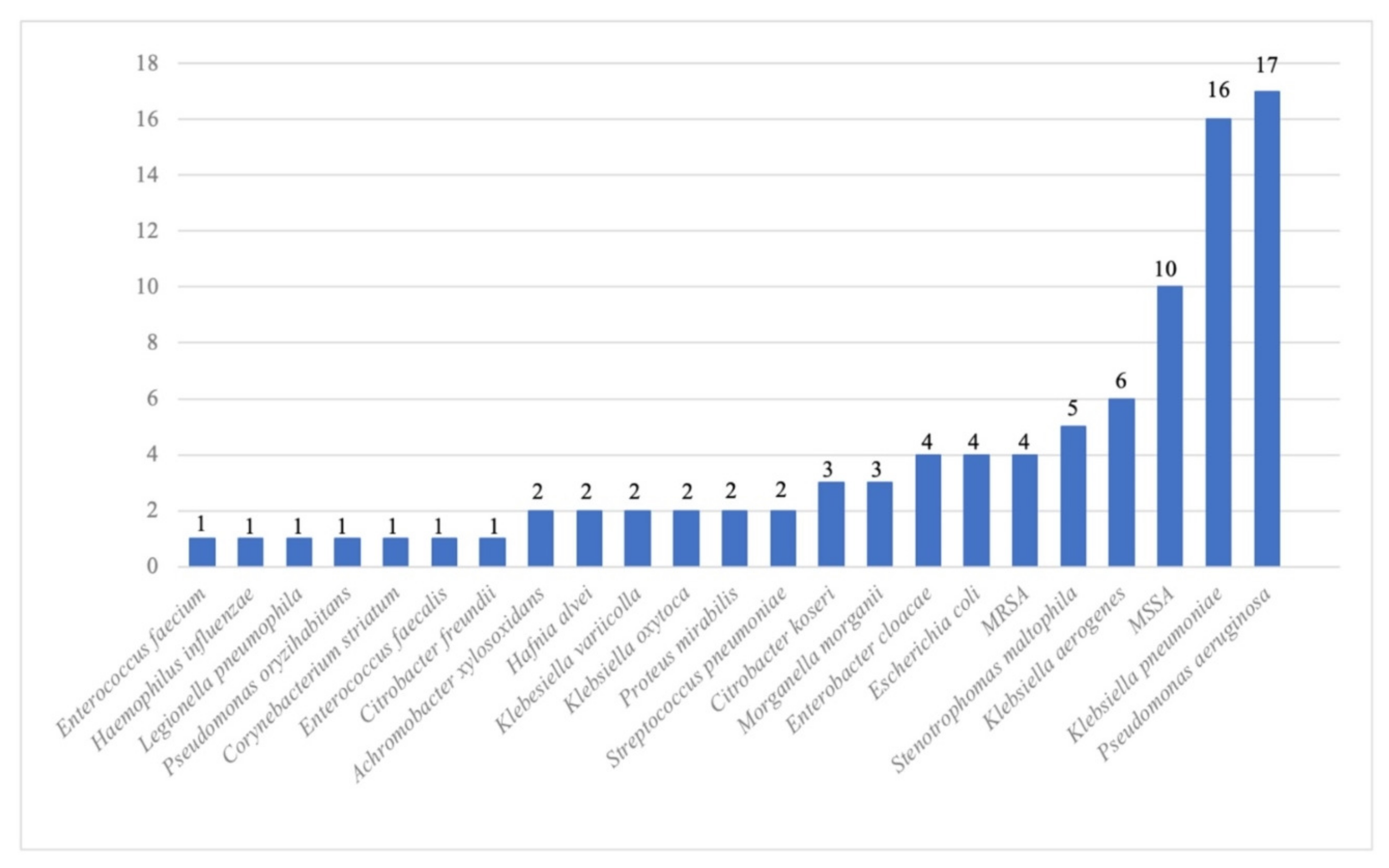
3.4. Microbiological Characteristics

A total of 53 (53%) patients in the case group had SARS-Co-V-2 infection, compared with 29% and 17% in the BactC and NegC groups, respectively (p < 10−4). The mean timescale between hospital admission and the first culture-positive respiratory sample was seven days in both the Case and BactC patients. Within the Candida genus (Figure 4), C. albicans (57%) was predominantly isolated, followed by C. glabrata (17%), C. dubliniensis (7%), Kluyveromyces marxianus (5%), and C. tropicalis (4%).
Of the 91 respiratory samples with a positive bacterial culture in the case group patients, the most frequent bacterial coinfection involved Enterobacteriaceae (47%), including Klebsiella pneumoniae (18%), followed by Pseudomonas aeruginosa (19%) and methicillin-susceptible Staphylococcus aureus (11%) (Figure 5). In the BactC group patients, 138 respiratory specimens yielded Enterobacteriaceae (40%), including E. coli (11%) and Klebsiella sp. (9%), 28% yielded P. aeruginosa, and 17% yielded methicillin-susceptible S. aureus. The distribution of bacterial species is illustrated in Figure S1. The use of antibacterial therapy was significantly more frequent (p < 10−4) in the case and BactC patients (88% each) compared to those in the NegC group (35%).
A viral respiratory infection was more frequently diagnosed (p < 10−4) in patients in the case group (25%) compared to patients in the BactC (13%) and NegC (1%) groups. Antiviral treatment was initiated during the hospital stay in 27 (27%) patients in the case group compared to 12% and 1% in the BactC and the NegC groups, respectively (p < 10−4).
Invasive fungal disease (IFD) occurred in 16 patients (16%) in the case group (five candidaemia, ten pulmonary aspergillosis, and one C. albicans pleurisy). In the BactC group, fungal infection was documented in three patients (one candidaemia and two pulmonary aspergilloses). No fungal infections were documented in the NegC group (p < 10−4). Twenty-six (26%) patients in the case group received systemic antifungal therapy, compared with 5% and 2% in the BactC and the NegC groups, respectively (p < 10−4). Notably, two patients in the NegC group received pre-emptive antifungal therapy.

3.5. Description of Candidaemia Cases

The clinical characteristics and microbiological findings of the five case group patients who developed candidaemia during their ICU stay are presented in Table 2. Comparatively, one patient from the BactC group developed C. parapsilosis candidaemia. This patient was 53 years old, without comorbidities at admission, and had been hospitalised for 46 days in the ICU for polytrauma management. He was intubated, had no documented SARS-CoV-2 infection, and had not had abdominal surgery. No Candida colonisation had been documented in previous samples. The initial caspofungin antifungal treatment was then switched to fluconazole. Additionally, a documented Enterobacter cloacae and K. pneumoniae VAP was treated with piperacillin-tazobactam.

3.6. Documented SARS-CoV-2 Infection

A SARS-Co-V-2 infection was diagnosed in 53 (53%) patients in the case group, compared to 29% and 17% in the BactC and NegC groups, respectively. The Alpha variant was the most frequently involved. It was documented in 26 patients from the case group, 9 patients from the BactC group, and 10 patients from the NegC group. The Delta variant was detected in 13 patients in the case group, 9 patients in the BactC group, and 1 patient in the NegC group (Figure S2). The Alpha variant was predominant during April and May, whereas the Delta variant became predominant from July 2021. The monthly distribution of variants between April and November 2021 is shown in Figure S3.
The immunomodulatory treatments used against SARS-CoV-2 are summarised in Table 3. Dexamethasone treatment was more frequently (p < 10−4) used in the case group patients (53%), compared with 28% and 10% in the BactC and NegC groups, respectively. Methylprednisolone was initiated in 37%, compared with 16% and 3% in the BactC and NegC groups, respectively (p < 10−4). Tocilizumab was homogenously (p = 0.6045) prescribed in the case (6%) and BactC (7%) groups.

3.7. Exposure to Antibacterials

The exposure to antibacterials in each patient group is summarised in Table S1. Piperacillin-tazobactam was the most frequent antibacterial used. It was administered in 44% of patients in the case group, compared with 34% and 14% in the BactC and NegC groups, respectively. Carbapenem was administered in 35% of the case group patients. In the BactC group patients, cefotaxime, ceftriaxone (23%), and group A penicillins (23%) were the most used after piperacillin-tazobactam. Penicillin A was administered in 13% of the NegC group patients.

3.8. Multivariate Analysis

The results of the multivariate analyses are summarised in Table 4. The first analysis, comparing the case group and both control groups (BactC+NegC), found a statistically significant association between Candida airway colonisation and diabetes, mechanical ventilation, length of hospital stay, the development of invasive fungal disease, and the use of some antibacterial antibiotics, including carbapenems, cefepime, and piperacillin/tazobactam. When compared to the BactC group, Candida airway colonisation was statistically significantly associated with mechanical ventilation, diabetes, length of hospital stay, and the use of cefepime and carbapenems. In contrast, a history of solid malignancy appeared to be a protective factor. When compared to the NegC group, Candida airway colonisation was statistically significantly associated with the length of hospitalisation, antifungal treatment, and linezolid administration.

4. Discussion

Our first finding was a dramatic increase in the number of respiratory samples analysed at the AP-HM university hospital microbiology laboratory since March 2020, in comparison with the two previous years. Most of the respiratory samples originated from ICU patients, and the prevalence of positive yeast (mainly Candida spp.) culture increased in parallel. This increased prevalence of Candida airway colonisation was concomitant to the SARS-CoV-2 epidemic and roughly correlated with the successive epidemic waves in France, notably in March and April 2020, the last quarter of 2020, from February to May 2021, in August and September 2021, and from December 2021 to January 2022 [14]. One explanation for this increased number of respiratory samples is probably the rising number of patients admitted to the ICU due to severe SARS-CoV-2 infection since March 2020. Some 106,000 patients with COVID-19 were admitted to ICUs between 1 March 2020 and 30 June 2021 in France. Comparatively, 19,000 patients were admitted to French ICUs due to severe influenza between 2014 and 2019 [15]. The documentation of possible respiratory coinfections in ICU patients is probably the main reason for this increase in respiratory samples. Most patients admitted to the ICU received mechanical ventilation and were thus exposed to VAP [8].
The distribution of yeast species, C. albicans (57%) and C. glabrata (17%), in the respiratory sample cultures that we observed was in line with previous reports [5]. Similarly, the seven-day timeframe between admission and the first positive Candida culture that we observed was comparable to the 6 ± 1.6-day timeframe reported by Hedderwick et al. in 16 ICU patients [16].
Several studies have assessed the risk of candidaemia in SARS-CoV-2-infected patients [17,18,19,20,21]. We tested whether SARS-CoV-2 infection was an independent factor of Candida airway colonisation. In our univariate analysis, SARS-CoV-2 infection was statistically significantly more frequently diagnosed in patients with Candida spp. airway colonisation. However, SARS-CoV-2 infection is likely to be a confounding factor, since its independent effect was not statistically significant in the multivariate analysis. Likewise, patients with Candida spp. airway colonisation were significantly more frequently hospitalised in the ICU, which is in line with previous studies that reported 50% to 86% Candida colonisation rates in patients who had a prolonged stay in the ICU [22,23,24,25]. However, this association was not statistically significant in the multivariate analysis, which highlighted ICU-related factors such as mechanical ventilation as independent risk factors of Candida spp. airway colonisation.
In the univariate analysis, diabetes, mechanical ventilation, ECMO, central venous catheters, abdominal surgery, length of hospitalisation, and antibacterial therapy were associated with colonisation. A fatal outcome occurred more frequently in colonised patients. However, the multivariate analysis only found diabetes, length of hospitalisation, mechanical ventilation, and antibacterial therapy to be associated with Candida colonisation.
Regarding mechanical ventilation, Arastehfar et al. reported respiratory Candida colonisation in 20% of patients after 48 h of mechanical ventilation [18]. In line with our findings, several studies reported the association of a protracted hospital stay and mechanical ventilation with Candida airway colonisation [5,19,26]. Keeping with our findings, Chakraborti et al. and Erami et al. identified diabetes as a risk factor for Candida airway colonisation [10,27]. However, in contrast to Erami et al., who identified solid malignancies, chronic renal failure, and pre-existing cardiovascular diseases as Candida airway colonisation risk factors, none of these factors were significant in our study [10]. In contrast, we found that a solid malignancy history protected from Candida airway colonisation. This debatable result could be explained by the heterogeneity of the patient population in each study. The predominance of ICU patients in our study represents a recruitment bias because patients with advanced cancer are generally not admitted to the ICU. The role of chronic renal failure remains a matter of debate [16].
In our study, the use of antibacterials was statistically significantly associated with an increased risk of Candida airway colonisation. Subgroup analyses identified some antibacterials that were more frequently associated with a risk of colonisation—notably, carbapenems and cefepime. These results are aligned with those of Delisle et al. and Charles et al., who reported an increased use of antibacterials in Candida-colonised patients [12,28]. Notably, these two studies reported on broad-spectrum antibacterials but did not specify which antibacterial classes were used.
The development of invasive fungal disease and the use of antifungal therapy were associated with Candida colonisation when compared to non-colonised (NegC) patients with culture-negative respiratory specimens. One explanation might be that patients in the NegC group were less severe and less likely to be treated with systemic antifungals than ICU patients. Five fungaemia occurred in the group of patients colonised with Candida (5%), whereas only one occurred in the non-colonised patient group. These data are in line with several previous studies [29]. Moreover, the relevance of assessing Candida colonisation in patients at risk of invasive candidiasis has been demonstrated by Pittet et al. [22].
The use of high-dose corticosteroids (dexamethasone and methylprednisolone) was not shown to be an independent risk factor in our multivariate analysis. Indeed, its effect on Candida colonisation remains under debate [16]. Likewise, in contrast to other studies, we did not find that tocilizumab was an independent risk factor for Candida colonisation, which might be due to the relatively small number of patients treated with it [23,24].
Our multivariate analysis did not show a difference in mortality between the colonised and non-colonised groups. It should be noted that the association between Candida colonisation and ICU patients’ mortality is disputed, with conflicting reports in the literature [12].
Among the colonised patients in our study, only one had a documented Candida lung infection. Candida pneumonia is a rare infection. In their study on 25 autopsied patients, El-Biary et al. reported 10 who were colonised with Candida, only 2 of whom had histologically documented pneumonia [30]. Similarly, among 232 autopsied ICU patients, Meersseman et al. reported histologically documented pneumonia in 135 (58%). Of these, 77 (57%) had Candida spp. isolated from pre-mortem respiratory samples, and none displayed histological features of Candida pneumonia.
The limitations of our study include its monocentric design, which means that our results can only be extrapolated to other settings with caution. Retrospective studies are also known to be prone to bias. Nevertheless, our findings are strengthened by our choice to define colonisation through the positive culture of three distinct respiratory samples, whereas other studies define colonisation on the basis of a single positive sample [29,31]. This is likely to increase the robustness of our findings. Furthermore, our choice to use distinct patient control groups allowed us to highlight robust risk factors, which have an impact on a fairly heterogeneous group of patients.

5. Conclusions

Both the incidence and prevalence of Candida-positive respiratory samples increased in parallel to the SARS-CoV-2 epidemic. However, SARS-CoV-2 infection is likely to be a confounding factor rather than an independent risk factor. While SARS-CoV-2 infection was statistically significantly more frequently diagnosed in patients with Candida airway colonisation in our univariate analysis, the length of hospitalisation, the use of mechanical ventilation, diabetes, and the use of antibacterial antibiotics, but not SARS-CoV-2 infection, were independently associated with Candida airway colonisation in our multivariate analysis. Notably, respiratory Candida colonisation was significantly associated with a sevenfold increase in the risk of developing invasive fungal disease—in particular, fungaemia.

Supplementary Materials

The following supporting information can be downloaded at: https://www.mdpi.com/article/10.3390/pathogens12030463/s1, Figure S1: Distribution of the bacterial species cultured from respiratory samples in the BactC (patients with a positive bacterial culture in a respiratory sample) group between 1 April and 30 November 2021. Figure S2: Distribution of SARS-CoV-2 variants in patients in the three study groups (Cases, Candida airway colonisation; BactC, patients with a positive bacterial culture in a respiratory sample; and NegC, patients for whom respiratory sample cultures were negative) from 1 April to 30 November 2021 (Not available: we found no data on the type of variant). Figure S3: Monthly distribution of the SARS-CoV-2 variants in the three groups of patients from 1 April to 30 November 2021. Table S1: Antibacterials used in the case and control groups: BactC (patients with positive bacterial culture) and NegC (patients with negative respiratory sample cultures).

Author Contributions

Conceptualisation, S.R.; methodology, J.S., M.F. and S.R.; validation, S.R. and H.C.; formal analysis, M.F., J.S. and S.R.; investigation, M.F.; resources, S.R. and H.C.; data curation, M.F., J.S., H.C. and S.R.; writing—original draft preparation, M.F. and J.S.; writing—review and editing, J.S. and S.R.; visualisation, S.R.; supervision, S.R.; project administration, S.R. All authors have read and agreed to the published version of the manuscript.

Funding

This research did not receive any specific grant from funding agencies in the public, commercial, or not-for-profit sectors.

Institutional Review Board Statement

This study was approved by the IHU Méditerranée Infection ethics committee (No. 2022-019) and registered in the AP-HM Data Protection Registry (No 2022-38).

Informed Consent Statement

Patient consent was waived according to the French law, because in this retrospective study, patients’ personal and medical data were collected during care and extracted from the hospital database. Patients have been informed of the possible use of their data collected during care for research purpose and they have the possibility to oppose this by reporting it to the DPO of the AP-HM.

Data Availability Statement

The data presented in this study are openly available in the IHU Méditerranée Infection site at https://doi.org/10.35081/0dm0-bj28, reference “COVID-19-Is-a-Confounder-of-Increased-Candida-Airway-Colonisation Dataset”.

Conflicts of Interest

The authors have no conflict of interest to declare.

References

  1. WHO. Coronavirus (COVID-19) Dashboard|WHO Coronavirus (COVID-19) Dashboard with Vaccination Data. Available online: https://covid19.who.int/data (accessed on 9 July 2022).
  2. Coronavirus: Chiffres clés et évolution de la COVID-19 en France et dans le Monde. Available online: https://www.santepubliquefrance.fr/dossiers/coronavirus-covid-19/coronavirus-chiffres-cles-et-evolution-de-la-covid-19-en-france-et-dans-le-monde (accessed on 9 July 2022).
  3. Huang, C.; Wang, Y.; Li, X.; Ren, L.; Zhao, J.; Hu, Y.; Zhang, L.; Fan, G.; Xu, J.; Gu, X.; et al. Clinical Features of Patients Infected with 2019 Novel Coronavirus in Wuhan, China. Lancet 2020, 395, 497–506. [Google Scholar] [CrossRef] [PubMed] [Green Version]
  4. Jeong, S.; Lee, N.; Park, Y.; Kim, J.; Jeon, K.; Park, M.-J.; Song, W. Prevalence and Clinical Impact of Coinfection in Patients with Coronavirus Disease 2019 in Korea. Viruses 2022, 14, 446. [Google Scholar] [CrossRef] [PubMed]
  5. Azoulay, E.; Timsit, J.-F.; Tafflet, M.; de Lassence, A.; Darmon, M.; Zahar, J.-R.; Adrie, C.; Garrouste-Orgeas, M.; Cohen, Y.; Mourvillier, B.; et al. Candida Colonization of the Respiratory Tract and Subsequent Pseudomonas Ventilator-Associated Pneumonia. Chest 2006, 129, 110–117. [Google Scholar] [CrossRef] [PubMed] [Green Version]
  6. Vincent, J.-L. The Prevalence of Nosocomial Infection in Intensive Care Units in Europe: Results of the European Prevalence of Infection in Intensive Care (EPIC) Study. JAMA 1995, 274, 639. [Google Scholar] [CrossRef] [PubMed]
  7. Hamet, M.; Pavon, A.; Dalle, F.; Pechinot, A.; Prin, S.; Quenot, J.-P.; Charles, P.-E. Candida Spp. Airway Colonization Could Promote Antibiotic-Resistant Bacteria Selection in Patients with Suspected Ventilator-Associated Pneumonia. Intensive Care Med. 2012, 38, 1272–1279. [Google Scholar] [CrossRef]
  8. Hughes, S.; Troise, O.; Donaldson, H.; Mughal, N.; Moore, L.S.P. Bacterial and Fungal Coinfection among Hospitalized Patients with COVID-19: A Retrospective Cohort Study in a UK Secondary-Care Setting. Clin. Microbiol. Infect. 2020, 26, 1395–1399. [Google Scholar] [CrossRef]
  9. Pendleton, K.M.; Huffnagle, G.B.; Dickson, R.P. The Significance of Candida in the Human Respiratory Tract: Our Evolving Understanding. Pathog. Dis. 2017, 75, ftx029. [Google Scholar] [CrossRef] [Green Version]
  10. Erami, M.; Raiesi, O.; Momen-Heravi, M.; Getso, M.I.; Fakhrehi, M.; Mehri, N.; Yarahmadi, M.; Amiri, S.; Raissi, V.; Hashemi, S.J. Clinical Impact of Candida Respiratory Tract Colonization and Acute Lung Infections in Critically Ill Patients with COVID-19 Pneumonia. Microb. Pathog. 2022, 166, 105520. [Google Scholar] [CrossRef]
  11. Huang, D.; Qi, M.; Hu, Y.; Yu, M.; Liang, Z. The Impact of Candida Spp Airway Colonization on Clinical Outcomes in Patients with Ventilator-Associated Pneumonia: A Systematic Review and Meta-Analysis. Am. J. Infect. Control 2020, 48, 695–701. [Google Scholar] [CrossRef]
  12. Delisle, M.-S.; Williamson, D.R.; Perreault, M.M.; Albert, M.; Jiang, X.; Heyland, D.K. The Clinical Significance of Candida Colonization of Respiratory Tract Secretions in Critically Ill Patients. J. Crit. Care 2008, 23, 11–17. [Google Scholar] [CrossRef]
  13. Kaba, L.; Giraud-Gatineau, A.; Jimeno, M.-T.; Rolain, J.-M.; Colson, P.; Raoult, D.; Chaudet, H. Consequences of the COVID-19 Outbreak Lockdown on Non-Viral Infectious Agents as Reported by a Laboratory-Based Surveillance System at the IHU Méditerranée Infection, Marseille, France. JCM 2021, 10, 3210. [Google Scholar] [CrossRef]
  14. SPF. COVID-19: Point épidémiologique du 4 mars. 2020. Available online: https://www.santepubliquefrance.fr/maladies-et-traumatismes/maladies-et-infections-respiratoires/infection-a-coronavirus/documents/bulletin-national/covid-19-point-epidemiologique-du-4-mars-2020 (accessed on 29 August 2022).
  15. COVID-19: Prise En Charge Des Patients En Soins Critiques Au Cours Des Trois Premières Vagues de l’épidémie | Direction de La Recherche, Des Études, de l’évaluation et Des Statistiques. Available online: https://drees.solidarites-sante.gouv.fr/publications-communique-de-presse/etudes-et-resultats/covid-19-prise-en-charge-des-patients-en (accessed on 3 September 2022).
  16. Hedderwick, S.A.; Lyons, M.J.; Liu, M.; Vazquez, J.A.; Kauffman, C.A. Epidemiology of Yeast Colonization in the Intensive Care Unit. Eur. J. Clin. Microbiol. Infect. Dis. 2000, 19, 663–670. [Google Scholar] [CrossRef] [Green Version]
  17. Kayaaslan, B.; Kaya Kalem, A.; Asilturk, D.; Kaplan, B.; Dönertas, G.; Hasanoglu, I.; Eser, F.; Korkmazer, R.; Oktay, Z.; Ozkocak Turan, I.; et al. Incidence and Risk Factors for COVID-19 Associated Candidemia (CAC) in ICU Patients. Mycoses 2022, 65, 508–516. [Google Scholar] [CrossRef]
  18. Arastehfar, A.; Carvalho, A.; Nguyen, M.H.; Hedayati, M.T.; Netea, M.G.; Perlin, D.S.; Hoenigl, M. COVID-19-Associated Candidiasis (CAC): An Underestimated Complication in the Absence of Immunological Predispositions? J. Fungi 2020, 6, 211. [Google Scholar] [CrossRef]
  19. Avkan-Oğuz, V.; Çelİk, M.; Eren-Kutsoylu, O.Ö.; Nazli, A.; Uğur, Y.L.; Taylan, A.; Ergan, B.; Irmak, Ç.; Duğral, E.; Özkütük, A.A. Fungal Colonization and Infections in Patients with COVID-19 in Intensive Care Units: A Real-Life Experience at a Tertiary-Care Hospital. Respir. Med. Res. 2022, 82, 100937. [Google Scholar] [CrossRef]
  20. Blaize, M.; Raoelina, A.; Kornblum, D.; Kamus, L.; Lampros, A.; Berger, M.; Demeret, S.; Constantin, J.-M.; Monsel, A.; Mayaux, J.; et al. Occurrence of Candidemia in Patients with COVID-19 Admitted to Five ICUs in France. J. Fungi 2022, 8, 678. [Google Scholar] [CrossRef]
  21. Gangneux, J.-P.; Dannaoui, E.; Fekkar, A.; Luyt, C.-E.; Botterel, F.; De Prost, N.; Tadié, J.-M.; Reizine, F.; Houzé, S.; Timsit, J.-F.; et al. Fungal Infections in Mechanically Ventilated Patients with COVID-19 during the First Wave: The French Multicentre MYCOVID Study. Lancet Respir. Med. 2022, 10, 180–190. [Google Scholar] [CrossRef]
  22. Pittet, D.; Monod, M.; Suter, P.M.; Frenk, E.; Auckenthaler, R. Candida Colonization and Subsequent Infections in Critically III Surgical Patients. Ann. Surg. 1994, 220, 751–758. [Google Scholar] [CrossRef]
  23. Eggimann, P.; Garbino, J.; Pittet, D. Epidemiology of Candida Species Infections in Critically Ill Non-Immunosuppressed Patients. Lancet Infect. Dis. 2003, 3, 685–702. [Google Scholar] [CrossRef]
  24. Tran, L.T.; Auger, P.; Marchand, R.; Carrier, M.; Pelletier, C. Epidemiological Study of Candida Spp. Colonization in Cardiovascular Surgical Patients. Mycoses 1997, 40, 169–173. [Google Scholar] [CrossRef]
  25. Blot, S.I.; Vandewoude, K.H.; Hoste, E.A.; Colardyn, F.A. Effects of Nosocomial Candidemia on Outcomes of Critically Ill Patients. Am. J. Med. 2002, 113, 480–485. [Google Scholar] [CrossRef] [PubMed]
  26. Delisle, M.-S.; Williamson, D.R.; Albert, M.; Perreault, M.M.; Jiang, X.; Day, A.G.; Heyland, D.K. Impact of Candida Species on Clinical Outcomes in Patients with Suspected Ventilator-Associated Pneumonia. Can. Respir. J. 2011, 18, 131–136. [Google Scholar] [CrossRef] [PubMed] [Green Version]
  27. Chakraborti, A.; Jaiswal, A.; Verma, P.K.; Singhal, R. A Prospective Study of Fungal Colonization and Invasive Fungal Disease in Long-Term Mechanically Ventilated Patients in a Respiratory Intensive Care Unit. Indian J. Crit. Care Med. 2018, 22, 597–601. [Google Scholar] [CrossRef] [PubMed]
  28. Charles, P.E.; Dalle, F.; Aube, H.; Doise, J.M.; Quenot, J.P.; Aho, L.S.; Chavanet, P.; Blettery, B. Candida Spp. Colonization Significance in Critically Ill Medical Patients: A Prospective Study. Intensive Care Med. 2005, 31, 393–400. [Google Scholar] [CrossRef]
  29. León, C.; Álvarez-Lerma, F.; Ruiz-Santana, S.; León, M.Á.; Nolla, J.; Jordá, R.; Saavedra, P.; Palomar, M. The EPCAN Study Group Fungal Colonization and/or Infection in Non-Neutropenic Critically Ill Patients: Results of the EPCAN Observational Study. Eur. J. Clin. Microbiol. Infect. Dis. 2009, 28, 233–242. [Google Scholar] [CrossRef]
  30. el-Ebiary, M.; Torres, A.; Fàbregas, N.; de la Bellacasa, J.P.; González, J.; Ramirez, J.; del Baño, D.; Hernández, C.; Jiménez de Anta, M.T. Significance of the Isolation of Candida Species from Respiratory Samples in Critically Ill, Non-Neutropenic Patients. An Immediate Postmortem Histologic Study. Am. J. Respir. Crit. Care Med. 1997, 156, 583–590. [Google Scholar] [CrossRef]
  31. Magill, S.S.; Swoboda, S.M.; Johnson, E.A.; Merz, W.G.; Pelz, R.K.; Lipsett, P.A.; Hendrix, C.W. The Association between Anatomic Site of Candida Colonization, Invasive Candidiasis, and Mortality in Critically Ill Surgical Patients. Diagn. Microbiol. Infect. Dis. 2006, 55, 293–301. [Google Scholar] [CrossRef]
Figure 1. Distribution of yeast species cultured from respiratory samples taken at the university hospital of Marseille (AP-HM) from 1 January 2018 to 31 March 2022.
Pathogens 12 00463 g001
Figure 2. Evolution of the yeast rates (and 95% confidence intervals) among culture-positive respiratory specimens at the university hospital of Marseille (AP-HM), from 1 January 2018 to 31 March 2022.
Pathogens 12 00463 g002
Figure 3. Evolution of C. albicans rates (and 95% confidence intervals) among culture-positive respiratory specimens from 1 January 2018 to 31 March 2022.
Pathogens 12 00463 g003
Figure 4. Distribution of the Candida species cultured from respiratory samples taken from the Candida airway colonisation group from 1 April to 30 November 2021 at the university hospital of Marseille (AP-HM).
Pathogens 12 00463 g004
Figure 5. Distribution of the bacterial species cultured from respiratory samples in the Candida airway colonised patients’ group from 1 April to 31 November 2021.
Pathogens 12 00463 g005
Table 1. Characteristics of the 300 patients included in the case, BactC, and NegC control groups between 1 April and 30 November 2021 at AP-HM.
n (%)Case
(n = 100)
Bbact
(n = 100)
NegC
(n = 100)
p *
Demographic characteristics
Males66 (66)64 (64)61 (61)0.7605
Age, years 57 ± 1658 ± 1658 ± 150.8752
Intensive care unit80 (80)62 (62)18 (18)<10−4
Hospital length of stay, days 46 ± 3726 ± 229 ± 8<0.0001
Comorbidities
BMI
BMI < 2533 (44)46 (56)51 (59)0.1283
25 < BMI < 3019 (25)21(25)17 (20)0.6033
30 ≤ BMI < 3516 (21)9 (11)10 (12)0.1200
35 ≤ BMI < 407 (9)3 (4)4 (5)0.2691
40 ≥ BMI0 (0)3 (4)2 (2)0.2659
High blood pressure42 (42)39 (39)31 (31)0.2511
Cardiovascular disease15 (15)20 (20)14 (14)0.4695
Diabetes31 (31)26 (26)7 (7)<10−4
Smoking25 (25)21 (21)28 (28)0.5149
Chronic respiratory failure28 (28)28 (28)23 (23)0.6508
Chronic renal failure 10 (10)15 (15)7 (7)0.1801
Solid cancer7 (7)16 (16)22 (22)0.0114
Haematological disease0 (0)3 (3)4 (4)0.1493
Transplantation11 (11)19 (19)14 (14)0.2712
Immunosuppression17 (17)31 (31)33 (33)0.0212
Immunosuppressive treatments §15 (15)28 (28)26 (26)0.0629
Ventilation/oxygen therapy
Low-dose oxygen therapy6 (6)13 (13)12 (12)0.2129
HFNC1 (1)2 (2)3 (3)0.6004
Non-invasive ventilation1 (1)5 (5)3 (3)0.2529
Mechanical ventilation80 (80)56 (56)15 (15)<10−4
ECMO22 (22)13 (13)1 (1)<10−4
Nutrition77 (77)53 (53)14 (14)
Enteral nutrition76 (76)50 (50)14 (14)<10−4
Parenteral nutrition1 (1)3 (3)0 (0)0.1697
Central venous catheter79 (79)58 (58)15 (15)<10−4
Abdominal surgery10 (10)2 (2)4 (4)0.0323
SARS-CoV-2 infection53 (53)29 (29)17 (17)<10−4
Antibiotic therapy88 (88)88 (88)35 (35)<10−4
Antifungals26 (26)5 (5)2 (2)<10−4
Antivirals27 (27)12 (12)1 (1)<10−4
Nature of respiratory sample
BAL78 (78)76 (76)75 (75)0.8788
TBA70 (70)43 (43)12 (12)<10−4
CBES27 (27)26 (26)28 (28)0.9505
Respiratory viral infection25 (25)13 (13)1 (1)<10−4
Fungal infection16 (16)3 (3)0 (0)<10−4
Fatality, n (%)29 (29)23 (23)10 (10)0.0032
* Statistically significant differences appear in bold font. : mean ± standard deviation; BMI: body mass index; HFNC: high-flow nasal cannula oxygen therapy; : GFR < 60 mL/min/1.73 m2; §: long-term corticosteroid therapy, chemotherapy, or antirejection therapy in organ transplant recipients.
Table 2. Clinical and mycological characteristics as well as treatments and outcomes of patients in the case group who developed candidaemia between 1 April and 30 November 2021 at AP-HM.
Patients12345
Demographic characteristics
GenderMaleFemaleMaleMaleFemale
Age (years)6268467269
Known immunosuppressionNoAnti-TNF α treatmentNoNoNo
Comorbidities *HBPDiab, CVOb, SmoHBP, Diab, SmoHBP, Ob
Clinical characteristics
Length of stay (days)120108724972
Mechanical ventilationYesYesYesYesYes
ECMO **YesNoNoNoNo
SARS-CoV-2YesYesNoYesNo
Abdominal surgeryNoYesYesNoNo
Mycological culture
Respiratory colonisationC. albicansC. albicansC. glabrataC. glabrataC. utilis
Other colonised siteUrineUrineNoNoUrine
Blood cultures (BC)C. albicansK. marxianusC. glabrataC. glabrataC. metapsilosis
No. of positive BCs32232
Treatments and outcome
Antifungal treatments ***CaspCaspCasp, VoricoCaspCasp, Fluco
OutcomeAliveAliveAliveAliveAlive
* HBP, high blood pressure; anti-TNF α, anti-Tumour Necrosis Factor alpha; Diab, diabetes; CV, cardiovascular disease; Ob, obesity; Smo, smoking. ** ECMO, extracorporeal membrane oxygenation.*** Casp, Caspofungin; Vorico, voriconazole; Fluco, fluconazole.
Table 3. Immunomodulatory treatments used from 1 April to 30 November 2021 against SARS-CoV-2 infection in the case and control groups: BactC (patients with positive bacterial culture) and NegC (patients with negative respiratory sample cultures).
CharacteristicsCases = n (%)BactC = n (%)NegC = n (%)p
SARS-CoV-2 53 (53)29 (29)17 (17)<10−4
Intensive care unit51 (51)18 (18)17 (17)<10−4
Immunomodulatory treatments
Dexamethasone53 (53)28 (28)10 (10)<10−4
Methylprednisolone37 (37)16 (16)3 (3)<10−4
Tocilizumab6 (6)7 (7)0 (0)0.6045
Table 4. Multivariate unconditional logistical regression analysis of factors associated with Candida colonisation. Analyses compared the case group with both or either of the control groups BactC (patients with positive bacterial culture) and NegC (patients with negative respiratory sample cultures).
Cases vs. BactC + NegCCases vs. BactCCases vs. NegC
OR [95% CI] *, pOR [95%CI], pOR, [95% CI], p
Oxacillin IV4.42 [1.05–18.62], 0.0427--
Ceftazidime2.30 [1.13–4.66)], 0.0213--
Cefepime2.83 [1.42–1.64], 0.003017.66 [3.95–79.06], 0.0002-
Piperacillin/tazobactam1.99 [1.14–3.45], 0.0148--
Linezolid--2.57 [1.23–5.36], 0.0120
Carbapenems3.00 [1.58–5.68], 0.000722.84 [5.18–100.61], <10−4
Length of stay1.02 [1.01–1.03], <10−41.06 [1.02–1.09], 0.00071.02 [1.01–1.03], 0.0021
Diabetes2.12 [1.10–4.07], 0.02487.44 [2.85–19.65], 0.0006-
MV 3.48 [1.79–6.74], 0.00027.48 [2.85–19.65], <10−4-
Fungal coinfection6.71 [1.67–26.97], 0.0073--
Solid malignancies-0.22 [0.06–0.73], 0.0138-
Antifungal--4,17 [0.46–11.96], 0.0078
* OR [95%CI]: odds ratio and 95% confidence interval, MV: mechanical ventilation.
Disclaimer/Publisher’s Note: The statements, opinions and data contained in all publications are solely those of the individual author(s) and contributor(s) and not of MDPI and/or the editor(s). MDPI and/or the editor(s) disclaim responsibility for any injury to people or property resulting from any ideas, methods, instructions or products referred to in the content.

