Article

Artemisia afra and Artemisia annua Extracts Have Bactericidal Activity against Mycobacterium tuberculosis in Physiologically Relevant Carbon Sources and Hypoxia

by
Bushra Hafeez Kiani
,
Maria Natalia Alonso
,
Pamela J. Weathers
and
Scarlet S. Shell
*
Department of Biology and Biotechnology, Worcester Polytechnic Institute, Worcester, MA 01609, USA
*
Author to whom correspondence should be addressed.
These authors contributed equally to this paper.
Pathogens 2023, 12(2), 227; https://doi.org/10.3390/pathogens12020227
Submission received: 25 December 2022 / Revised: 25 January 2023 / Accepted: 27 January 2023 / Published: 1 February 2023
(This article belongs to the Special Issue Drugs Repurposing for Multi-Drug Resistant Bacterial Infections)

Abstract

:
Mycobacterium tuberculosis (Mtb) is a deadly pathogen and causative agent of human tuberculosis, causing ~1.5 million deaths every year. The increasing drug resistance of this pathogen necessitates novel and improved treatment strategies. A crucial aspect of the host–pathogen interaction is bacterial nutrition. In this study, Artemisia annua and Artemisia afra dichloromethane extracts were tested for bactericidal activity against Mtb strain mc26230 under hypoxia and various infection-associated carbon sources (glycerol, glucose, and cholesterol). Both extracts showed significant bactericidal activity against Mtb, regardless of carbon source. Based on killing curves, A. afra showed the most consistent bactericidal activity against Mtb for all tested carbon sources, whereas A. annua showed the highest bactericidal activity in 7H9 minimal media with glycerol. Both extracts retained their bactericidal activity against Mtb under hypoxic conditions. Further investigations are required to determine the mechanism of action of these extracts and identify their active constituent compounds.

1. Introduction

Tuberculosis (TB), one of the major fatal diseases of humanity, still poses a major health, social, and economic burden worldwide and mainly occurs in low- and middle-income countries. TB remains a major worldwide health challenge, even 130 years after the discovery of the causative agent Mycobacterium tuberculosis (Mtb), causing 10.6 million reported cases and 1.6 million deaths in 2021 [1].
A major challenge of TB is that standard treatment regimens require six months of multi-drug therapy. Drug-resistant TB requires significantly longer treatment regimens, with additionally incapacitating side effects and diminishing results with a limited number of second-line drugs including linezolid, bedaquiline, delamanid, and pretomanid [2,3,4,5]. One contributor to the persistence of TB is the formation of hypoxic granulomas, where Mtb can survive for long periods of time in non-growing states in which it is phenotypically tolerant to most drugs [6,7]. The mechanisms by which Mtb can persist in the host are complex and likely include both low drug penetration and reduced drug efficacy within the granuloma. Immune stressors and the granuloma structure produce populations of Mtb in slow or non-growing states or with altered physiology, causing reduced sensitivity to most antibiotics, termed tolerance. Given this drug tolerance, treatment with the first-line TB antibiotics leads to a significant decrease in viable bacteria, but often fails to sterilize [8,9]. Surviving bacteria likely provide opportunities for the acquisition of genetically encoded drug resistance. The emergence of drug resistance demands the discovery of new drugs and combinations to improve TB therapy at all stages of the disease.
The development of novel therapies is linked with an understanding of different survival strategies by bacterial pathogens to overcome stress. Bacterial nutrition is a key component of host–pathogen interaction in TB [10,11] and, thus, is useful in drug development. The infection pattern of bacterial pathogens and the interaction between drugs and Mtb nutritional requirements may greatly affect disease progression. Lovewell et al. [12] previously reported that Mtb uses lipids of the host stored in functional lipid bodies during infection and intracellular replication, suggesting the importance of lipids during infection. Although lipids are Mtb’s major carbon source in vivo, their low solubility and the presence of multiple carbon sources led to the consideration that Mtb may use additional carbon sources during infection [13,14,15]. Different carbon sources are co-catabolized by Mtb in vitro [15,16,17], demonstrating that Mtb can use multiple carbon sources including glucose, glycerol, and cholesterol. Recent work [18] confirmed that, during infection, mycobacteria use multiple carbon sources.
Plant extracts have been used for treating various diseases for millennia and about 100,000 plant species have medicinal value [19,20], often without any adverse side effects on human health and the environment [21]. Plant specialty molecules (previously termed plant secondary metabolites; PSM) can affect microbial cells in several ways, including the disruption of membrane function and structure, the interruption of DNA/RNA synthesis and function, interference with intermediary metabolism, the induction of coagulation of cytoplasmic constituents, and the interruption of normal cell communication [22,23,24,25,26]. Antibiotics currently used as therapies to treat bacterial diseases rely on various mechanisms of bacterial growth inhibition and bacterial killing. The development of novel therapies using plants requires a better understanding of the mechanisms of the antimicrobial activity of plant compounds [27].
Many species of the genus Artemisia (Figure 1) have different proven medicinal properties and are used for treating diseases such as malaria, cancer, and hepatitis [28,29,30], and have even shown efficacy against COVID-19 [31,32,33,34]. Traditional treatments led to the testing of different Artemisia species and their extracts against several pathogens, including mycobacteria in vitro and in vivo in a murine model of tuberculosis [35,36]. Artemisia annua and Artemisia afra are used globally to treat fever and cough, which are the common symptoms of many diseases including TB [37]. A. annua produces the antimalarial drug artemisinin (ART), which also has antitubercular activity [38,39,40]. Both ART and its derivative artesunate were observed to be effective against Mtb in vitro as well as in a rat-infected model [40]. In another study, a mycobactin–artemisinin conjugate was designed which exhibited activity against sensitive as well as drug-resistant strains of mycobacteria at a much lower concentration. The conjugate promoted the initiation of radical reactions, which were predicted to be central to the mechanism of action [41]. ART also targets a key survival pathway for Mtb during non-replicating persistence, by blocking the two-component regulatory system DosRST under hypoxic conditions in vitro [39,42]. Although it produces little to no ART, A. afra also had significant bactericidal activity against Mtb [38,43,44].
A. annua and A. afra dichloromethane (DCM) extracts were previously shown to have strong bactericidal activity against M. tuberculosis [38], suggesting that these plants contain bactericidal compounds beyond ART. To our knowledge, however, there are no reports regarding the efficacy of A. annua or A. afra against Mtb in the presence of different carbon sources and under hypoxia. In the present study, the efficacy of extracts of A. annua and A. afra against Mtb was measured in different carbon sources and under hypoxic conditions to evaluate the potential of these plant extracts or their constituent compounds for the treatment of TB.

2. Methodology

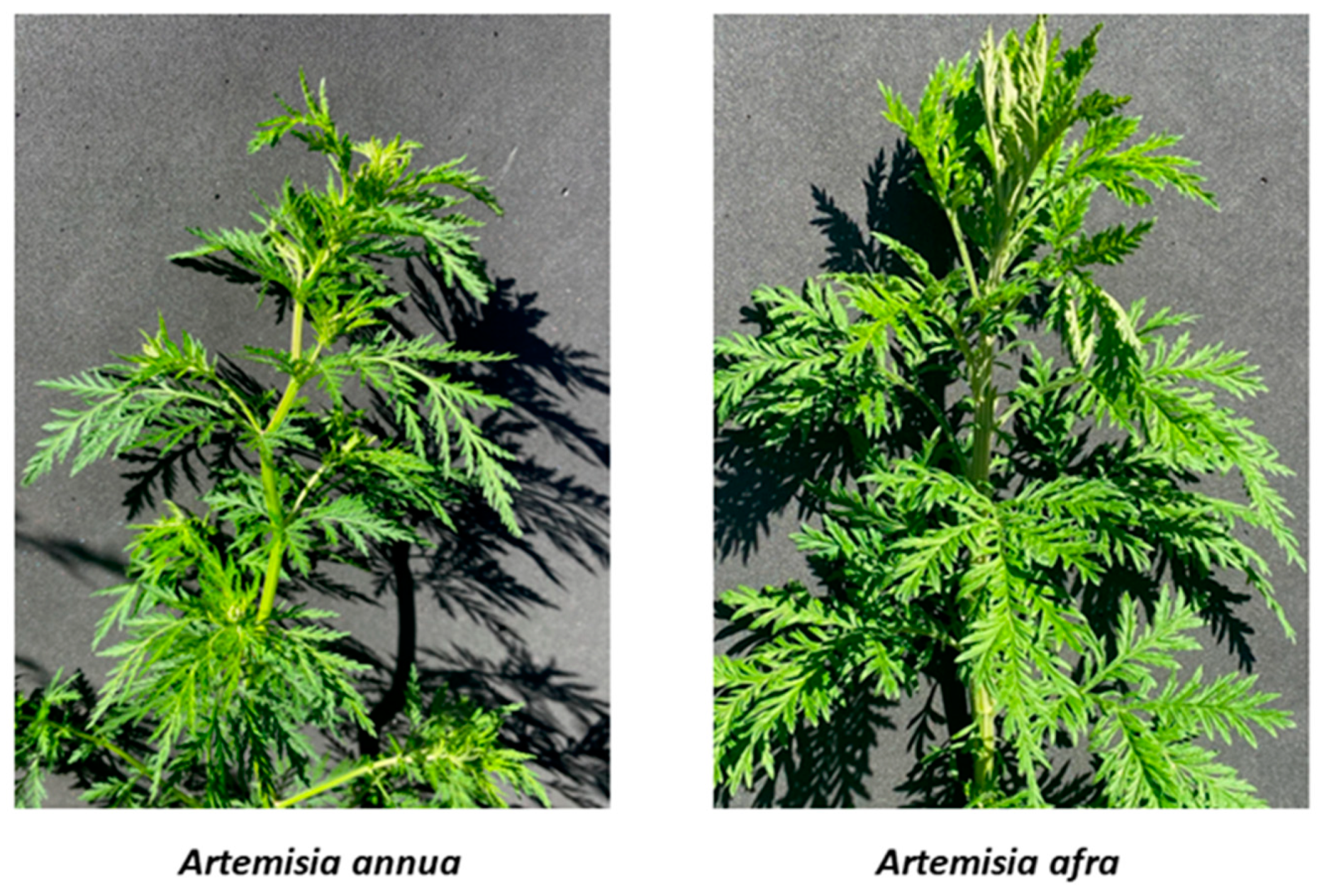
2.1. Plant Materials

Dried leaves of Artemisia annua L. cv. SAM (MASS 317314) were harvested from plants field-grown in Stow, MA from rooted cuttings as described [45]. Dried leaves of Artemisia afra Jacq. ex Willd. cv. MAL (originated from Malawi; FTG181107; batch B#1RbA.10.12.20) were obtained from Atelier Temenos LLC in Homestead, FL. The vouchers for other cultivars of A. afra are SEN (LG0019529), LUX (MNHNL17730), and PAR (LG0019528).

2.2. Preparation of Plant Extracts

Dried plant leaves were processed as previously described to produce DCM extracts [38]. DCM extracts were pooled and dried under N2 as previously detailed [46], and ART was analyzed using GC-MS as described [47]. Hot water tea infusions were also prepared as detailed in Desrosiers et al. [46].

2.3. Bacterial Strains and Culture Conditions

Mycobacterium tuberculosis (Mtb) strain mc26230 (ΔpanCD, ΔRD1 [48]) was grown at 37 °C and 200 rpm in Middlebrooks 7H9 broth, supplemented with 10% OADC (0.5 g/L oleic acid, 50 g/L bovine serum albumin fraction V, 20 g/L glucose, 8.5 g/L sodium chloride, and 40 mg/L catalase), 0.2% glycerol, 0.05% Tween 80, and 24 μg/mL pantothenate. Where specified, Mtb was grown in minimal media (0.5 g/L asparagine, 1 g/L KH2PO4, 2.5 g/L Na2HPO4, 50 mg/L ferric ammonium citrate, 0.5 g/L MgSO4·7H2O, 0.5 mg/L CaCl2, and 0.1 mg/L ZnSO4) supplemented with 0.1% tyloxapol, 24 μg/mL pantothenate, and 0.1% glycerol or 0.1% glucose or 0.1% cholesterol. Middlebrook 7H10 supplemented with 0.5% glycerol, OADC, and 24 μg/mL pantothenate was used to grow Mtb on solid media. When cultures were plated for CFU, they were first serially diluted by a series of six 10-fold dilutions. Multiple dilutions were plated, and colonies were counted for dilutions that yielded multiple colonies, spaced apart well enough for high-confidence counting.

2.4. Determination of the Minimum Inhibitory Concentration (MIC)

We previously reported MICs of Mtb strain mc26230 for DCM extracts of A. annua cv. SAM and A. afra cv. SEN using a resazurin microtiter assay (REMA) [38]. Using the same method, we also measured the MIC for A. afra cv. MAL and remeasured A. afra cv. SEN as well as the water extracts (tea infusions) of A. annua cv. SAM. For both MICs and killing assays, Artemisia extract concentrations are expressed as the leaf dry mass from which the extract was obtained. In each case, the DCM extract obtained from 2.94 g of dry leaf mass was resuspended in 1 mL DMSO and the concentration of the resulting solution was expressed as 2.94 g/mL.

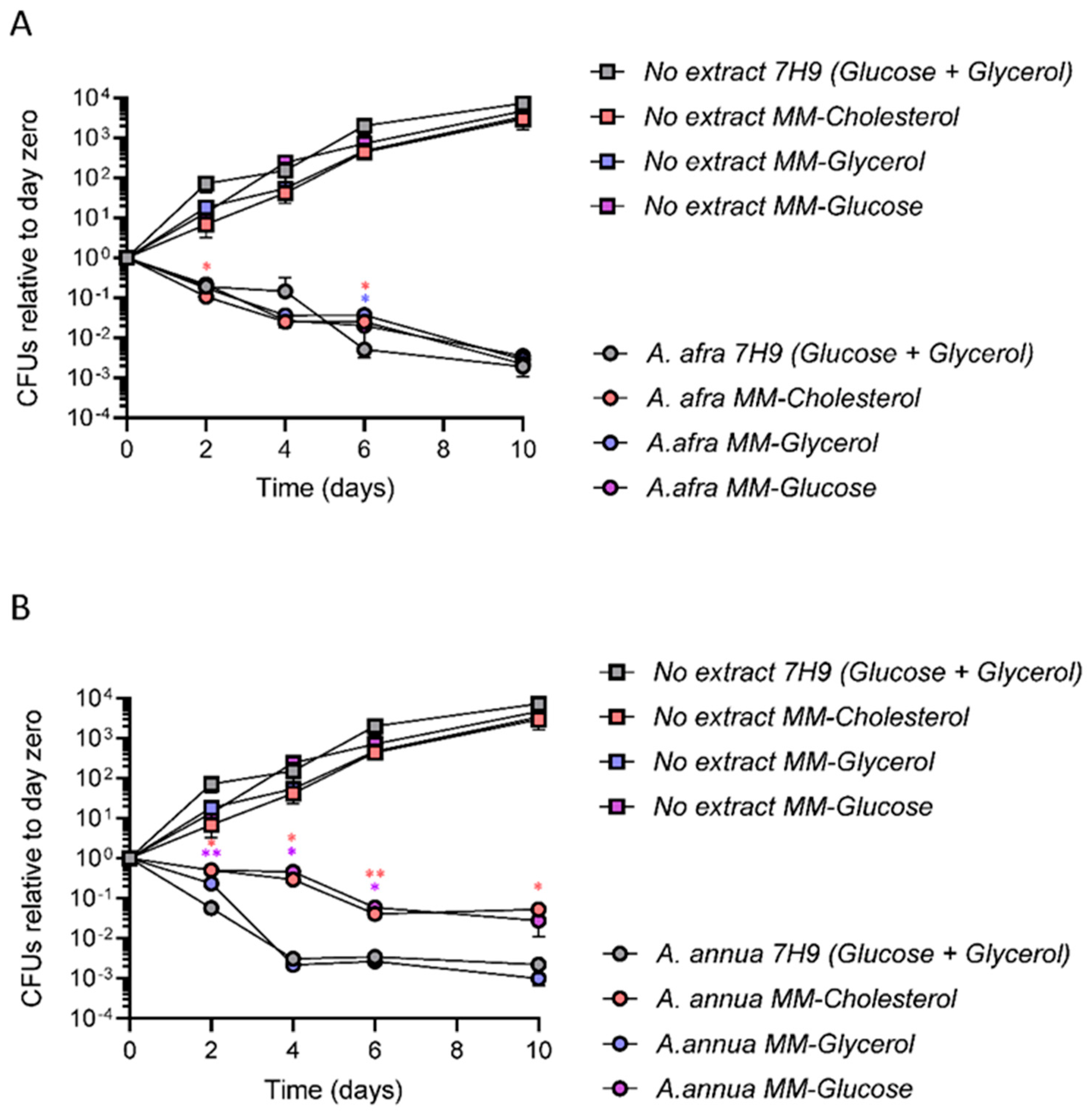
2.5. A. afra and A. annua Bactericidal Activity against Mtb in Different Carbon Sources

Mtb was grown in 7H9 media with OADC and glycerol as described above and when reaching OD600 = 0.8, cells were centrifuged, washed twice with PBS, and resuspended to OD600 = 0.8 in 7H9 with OADC and glycerol, or in minimal media containing 0.1% glycerol, 0.1% glucose, or 0.1% cholesterol. After 48 h, each culture was adjusted to a final OD600 of 0.1. Mtb suspensions were inoculated in triplicate with a final concentration of extract two times the MIC values, as determined for A. annua cv. SAM (MIC = extract from 4.5 mg dried leaves/mL, 2× MIC = extract from 9 mg dried leaves/mL) or A. afra cv. MAL (MIC = extract from 2.5 dried leaves/mL, 2× MIC = extract from 5 mg dried leaves/mL). Controls consisting of media with Mtb without added extracts were included, also in triplicate. In all cases, the final volume in each conical tube was 5 mL. Mtb cultures were incubated, shaking at 250 rpm and 37 °C. Mtb samples were taken from each liquid culture at days 0, 2, 4, 6 and 10 of incubation and then serially diluted and plated on 7H10 solid medium in the absence of plant extract. After 15–20 days of growth on 7H10 solid medium in the absence of extract, the resulting Mtb colonies were counted and the CFU/mL was calculated. CFU relative to day 0 were calculated as (CFU from culture on day x)/(CFU from culture on day 0).

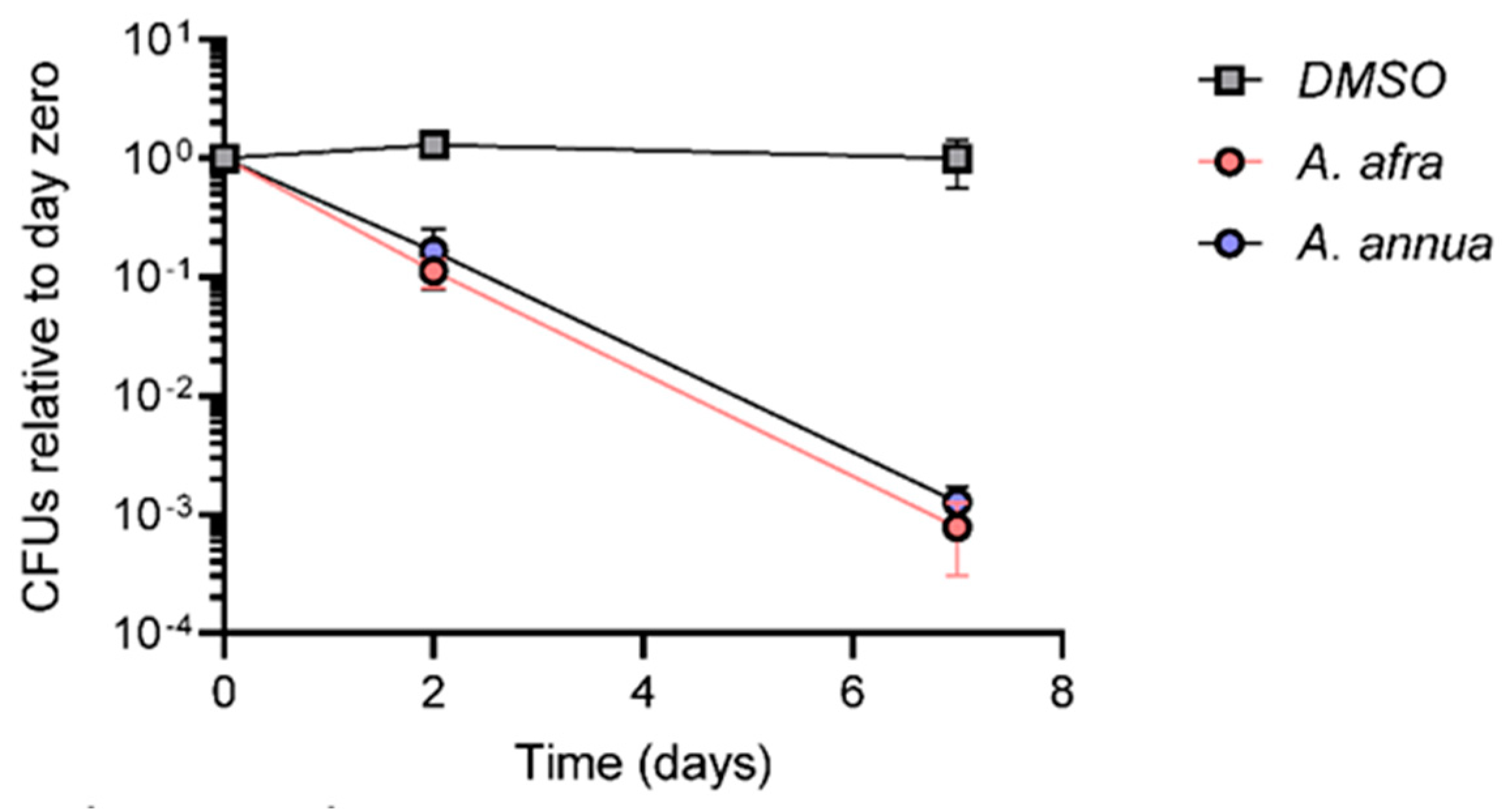
2.6. A. afra and A. annua Bactericidal Activity against Mtb under Hypoxia

Mtb was grown in 7H9 broth as above, but in this case supplemented with 10% ADC (50 g/L bovine serum albumin fraction V, 20 g/L glucose, 8.5 g/L sodium chloride, and 40 mg/L catalase) rather than OADC, at 200 rpm at 37 °C. Seed cultures were normalized to an OD600 = 0.1 in 17 mL using fresh media without antibiotics in a rubber stopper-sealed serum bottle (28 mL total bottle volume). The bottles were sealed with chlorobutyl rubber lids and aluminum caps, and then cultures were grown at 37 °C and 120 rpm to generate hypoxic conditions, as was previously reported [49]. Methylene blue in separate indicator cultures was used as an indicator of hypoxic conditions, and discoloration consistently occurred after eight days. ODs plateaued at approximately 0.35–0.4. Six days after hypoxia was established (14 days after sealing), cultures were treated with two times the MIC values as determined for A. annua cv. SAM (9 mg/mL) or A. afra cv. MAL (5 mg/mL) extracts. The extracts were injected aseptically using a 0.3 mm syringe to minimize the introduction of oxygen. The same volume of DMSO was also injected under the same conditions to use as a control. The hypoxic Mtb cultures were incubated with shaking at 120 rpm and 37 °C for 2 or 7 days and finally plated on solid media as previously described. Mtb cultures were also plated 14 days after sealing, before adding the respective drugs. All conditions were tested in triplicate, and colonies were counted after 15–20 days.

2.7. Statistical Analysis

Statistics were performed in GraphPad Prism 9.2.0. CFUs in kill curve experiments were compared by ANOVA and Dunnett’s multiple comparisons test.

3. Results

3.1. Determination of the Minimum Inhibitory Concentration (MIC)

Various plant extracts were tested for their ability to inhibit the growth of Mtb. Both hot water extracts and DCM extracts of both Artemisia sp. showed strong growth inhibitory effects against Mtb (Table 1). MICs were expressed as the dry leaf mass from which the extracts were obtained. Hot water extract MICs ranged from 1.3 to 1.7 mg/mL for the A. afra cultivars SEN, PAR and LUX; A. annua cv. SAM was 1.9 mg/mL (Table 1). A hot water extract of A. afra cv. MAL was not measured. MICs for DCM extracts of A. afra cv. SEN ranged from 4.8 to 10 mg/mL in replicate experiments, and the MIC for A. afra cv. MAL was consistently 2.5 mg/mL (Table 1). ART content of A. annua cv. SAM was 17.08 mg/g dry leaf mass, but was undetectable in A. afra cv. MAL. Since, in contrast to SEN, MAL was ART-free and readily available as a clonal source in the US, experiments were continued using the MAL cultivar. Considering that MICs of DCM extracts were not substantially different from the hot water extract MICs, yet allowed extracts to be more concentrated, further experiments were conducted using the DCM extracts.

3.2. A. afra and A. annua Bactericidal Activity against Mtb in Different Carbon Sources

DCM extracts of A. annua cv SAM and A. afra cv MAL were tested for their bactericidal activities against Mtb strain mc26230 (pantothenate auxotroph and RD1 deletion) in minimal media containing each of three different carbon sources individually: glycerol, glucose, and cholesterol. Cultures grown in 7H9 medium containing a mixture of glucose, glycerol, and oleic acid were tested in parallel. Untreated Mtb grew at equivalent rates and to equivalent final yields in all three single carbon sources, with a trend towards more growth in 7H9, as expected due to the latter having been developed as a medium to support maximal Mtb growth (Figure 2). Both extracts were bactericidal in all carbon sources. However, the extent of bactericidal activity differed according to carbon source in some cases. A. afra extracts showed similar bactericidal activity in all of the tested carbon sources. There were only small differences that were inconsistent across time-points in its bactericidal activity when Mtb was grown in glycerol, glucose, or cholesterol, or in 7H9 with both glucose and glycerol (Figure 2). However, the bactericidal activity of the A. annua extract was lower when Mtb was grown in cholesterol or glucose in comparison to growth in glycerol or 7H9 with both glucose and glycerol, suggesting different effects of carbon metabolism on the bactericidal activities of the two extracts (Figure 2).

3.3. A. afra and A. annua Bactericidal Activity against Mtb under Hypoxia

Considering that Mtb survives under hypoxic conditions within lung granulomas during natural infection, A. annua cv. SAM and A. afra cv. MAL extracts were tested for their bactericidal activity against Mtb strain mc26230 grown under hypoxic conditions. Cultures were grown in 7H9 medium containing a mixture of glucose and glycerol and sealed in bottles with a headspace:culture volume ratio of 0.6 at an OD of 0.1. Extracts and DMSO were added 14 days after sealing the bottles, by injection through the rubber lid to minimize the introduction of oxygen. Over seven days after injection, the viable population of Mtb remained relatively constant in cultures treated with only DMSO. We observed a small increase in the number of viable cells on Day 2 after DMSO treatment, which may be due to minimal oxygen introduction during drug injection. However, the viable Mtb cell population declined by one and three orders of magnitude at 2 and 7 d, respectively, after the injection of A. afra or A. annua DCM extracts into the hypoxic cultures (Figure 3). It was therefore concluded that A. afra and A. annua have similar bactericidal effects under hypoxic conditions.

4. Discussion

To our knowledge, this is the first study showing that Artemisia extracts can kill Mtb regardless of the carbon source used for its growth, and when Mtb is in a hypoxia-induced state of growth arrest. This is important because Mtb metabolizes a variety of different sources of carbon that it likely encounters during infection [15,16,17,50,51,52]. The results in this study showed that growth rates and yields were similar in glucose, glycerol, and cholesterol, and the A. afra cv. MAL extract was equally efficacious in all three conditions. However, the A. annua cv. SAM extract efficacy differed among carbon sources, suggesting that there may be two different mechanisms of killing action in different carbon sources in response to each Artemisia sp.
Plants have a long history of providing bioactive compounds for vital and novel therapeutics [53,54]. Many previous studies reported the effect of A. annua and A. afra extracts against the growth of Mtb [35,36,38,43,44]. Most prior studies were in vitro; however, Ntutela et al. [44] also included rodent testing. Although those in vitro tests showed a DCM extract MIC of 290 µg/mL against Mtb H37Rv, and a subfraction activity (fraction C8) that at 2 µg/mL was nearly 100 times more potent than the original extract, there was no efficacy of either the DCM or hot water extract in vivo in Mtb-infected mice (Table 1).
The extraction methods used in Ntutela et al. [44]’s studies may be a reason for the loss of activity in vivo. For example, 200 g of dried leaves were boiled in 4 L water for 30 min, filtered and then freeze dried to yield a hot water infusion. The traditional hot water extraction was boiled for 5–10 min at 5 g/L or steeped for 5–10 min in the boiled water. In our unpublished studies, increasing the dry mass of the Artemisia:water ratio beyond 10 g/L results in >50% loss of extractables. Furthermore, increasing the leaf g/L beyond ~10 g/L causes a significant decline in extractables [55]; e.g., ART recovery declined from 62 to 29% when leaf:hot water ratios increased from 20 to 50 g/L. The Ntutela et al. study used a water extract of 50 g/L with 30 min boiling and lyophilized to dryness. In our experience with A. annua, ART was not fully recovered when our lyophilized tea infusion was reconstituted in water, resulting in about a 50% loss, so for experiments we never use reconstituted lyophilized tea (Supplemental Figure S1). When compared to our tea MIC of ~1.5 mg/mL, the Ntutela et al. [44] water extract had neither in vitro nor in vivo activity (Table 1), results consistent with a possible loss of activity of the water extract in vivo. A similar argument can be made for the DCM extract of Ntutela et al. [44], because they extracted at 2 g/10 mL, then twice more but each at half the original solvent volume (Table 1). In contrast, we extracted 1 g/20 mL DCM for 30 min in a sonicating water bath at room temperature and extracted twice again using the same volumes [38]. One might question whether the extracts even reached the lungs of the treated mice; however, an ADME study of rats treated per os with A. annua showed that a considerable amount of the marker drug ART reached the lungs [46]. Finally, Ntutela et al. [44] did not gavage their animals with the Artemisia extracts, but instead mixed them into the feed for ad libidum consumption, which compromised dosing.
Our results revealed major survival differences of Mtb in different carbon sources when exposed to A. annua and A. afra extracts. The efficacy of the A. afra extract in cholesterol is particularly important because cholesterol plays a pivotal role in the infectivity and virulence of Mtb [56]. Mtb uses cholesterol as a major carbon source during infection and any other compounds or carbon sources that hinder cholesterol metabolism can inhibit Mtb growth, resulting in carbon starvation and metabolic intoxication and subsequently causing an imbalance in the central metabolism [57]. Chang and Guan confirmed that cholesterol and fatty acid are the main carbon sources that Mtb uses during infection [57]. However, some drugs have variable efficacy depending on the carbon source in use. For example, Kalia and collaborators reported that glycerol supplementation interfered with the potency of drugs targeting cytochrome bc1:aa3 in mycobacteria [18]. According to the studies of Bellerose et al., 2019 [58], Mtb mutants that lack glycerol catabolism due to variations in homopolymeric regions in the glycerol kinase gene (glpK) are associated with drug resistance in clinical isolates and are less susceptible to treatments. Further, they analyzed the efficacy of glycerol metabolism and other carbon sources (fatty acid and cholesterol) in determining the effect of different drugs (INH, RIF, and moxifloxacin) against Mtb. They found that all drugs were almost 50% less efficient in the absence of glycerol catabolism. That study speculated that glycerol significantly increases the efficacy of different drugs and enhances their efficacy. Highlighting that characteristic, a drug development effort encountered promising lead compounds when screening for activity in glycerol, but found they lacked activity against Mtb grown in other carbon sources [59].
Hypoxia and the gradual depletion of oxygen is a key element in granuloma development in human TB and an important consideration in the design of therapeutics useful for treating tuberculosis [60]. Several in vitro models to obtain non-replicating M. tuberculosis have been developed and are based on reducing oxygen availability and nutrient starvation. One of the most used methods is the Wayne model, in which non-replicating Mtb is obtained by gradually adapting stirred aerobic cultures to hypoxia through a self-generated oxygen depletion gradient [61]. Our method was an adaptation of the Wayne model.
Several lines of evidence link the inhibition of Mtb growth/metabolism with hypoxic conditions within the host. Mtb can survive long periods of hypoxia but is an obligate aerobe for growth. Tuberculosis infections are preferentially associated with the most oxygen-rich sites in the body [62], suggesting that reduced levels of O2 may limit Mtb growth in vivo. This may explain why recrudescent tuberculosis occurs most often in the upper lobes of the lung, the single most-oxygenated region of the body. According to Lim et al. [63], phosphoenolpyruvate (PEP) is almost completely depleted in Mtb under hypoxic conditions. A loss of PEP reduces PEP–carbon flux toward multiple pathways essential for the replication of Mtb. Metabolomic profiles of Mtb collected under hypoxia showed an accumulation of intermediates in glycolysis and the reductive branch of the TCA cycle, with a reciprocal depletion of PEP and oxidative branch intermediates of the TCA cycle, such as α-ketoglutarate. Under hypoxia, PEP depletion may affect multiple cellular metabolic processes that are involved in Mtb metabolic remodeling [51].
The impact of hypoxia during TB treatment is likely tremendous, because non-growing Mtb is less sensitive to most drugs. For example, isoniazid (INH) is one among the four first-line drugs used in the treatment of tuberculosis, and in vitro kills actively growing bacilli, but it possesses little or no activity against Mtb under conditions of nutrient starvation or progressive oxygen depletion [64,65]. In contrast, we found that A. annua and A. afra extracts were bactericidal to Mtb under hypoxia. Thus, these Artemisia sp. may maintain efficacy against Mtb in vivo where it is exposed to hypoxic conditions.

5. Conclusion

Mycobacterium tuberculosis is a deadly pathogen, causing disease in more than 10 million people annually. In this study, Mtb was killed by A. annua and A. afra extracts when grown in glycerol, glucose, or cholesterol as sole carbon sources and under hypoxic conditions. Hence, it is important to understand the metabolism of Mtb under alternative carbon sources and under hypoxia in the presence of extracts/drugs, given its consequences on infection, antibiotic efficacy, and potential in novel therapeutic development. It is important to unlock the underlying mechanism of how Artemisia extracts affect the growth of Mtb in different carbon sources and in hypoxia. This study can serve as a basis for further developing that understanding for future potential use against this multi drug resistant pathogen.

Supplementary Materials

The following supporting information can be downloaded at: https://www.mdpi.com/article/10.3390/pathogens12020227/s1, Figure S1: TLC showing loss of components of Artemisia annua tea upon lyophilization and reconstitution in water. Figure S2: A. afra and A. annua bactericidal activity against Mtb in different carbon sources.

Author Contributions

Conceptualization, B.H.K., M.N.A., P.J.W. and S.S.S.; validation, B.H.K., M.N.A., P.J.W. and S.S.S.; formal analysis, B.H.K. and M.N.A.; investigation, B.H.K. and M.N.A.; resources, P.J.W. and S.S.S.; writing—original draft preparation, B.H.K., M.N.A.; writing—review and editing, B.H.K., M.N.A., P.J.W. and S.S.S.; supervision, P.J.W. and S.S.S.; project administration, S.S.S.; funding acquisition, P.J.W. and S.S.S. All authors have read and agreed to the published version of the manuscript.

Funding

This research received no external funding.

Data Availability Statement

All data have been provided in the manuscript and its supplemental material.

Acknowledgments

The authors thank Worcester Polytechnic Institute and The American Association of University Women (AAUW) for supporting B. H. Kiani. Thank you to Tianbi Zhang for providing the MIC values from tea infusions. Thank you to James Kishlar of Atelier Temenos, Homestead FL, USA, for the photos provided in Figure 1. Award number NIH-1R21AI151481-01 provided to S. S. and P. J. W. from the National Institute for Allergy and Infectious Diseases funded this study. The content is solely the responsibility of the authors and does not necessarily represent the official views of the National Institute for Allergy and Infectious Diseases or the National Institutes of Health.

Conflicts of Interest

The authors declare they have no conflict of interest.

Abbreviations

ARTartemisinin
DCMdichloromethane
MALMalawi cultivar of Artemisia afra
PSMplant specialty metabolites (aka secondary metabolites)
SAMUS cultivar of A. annua
SENSenegal cultivar of A. afra

References

  1. WHO. Global Tuberculosis Report 2022. 2022. Available online: https://www.who.int/publications/i/item/9789240061729 (accessed on 2 December 2022).
  2. Keam, S.J. Pretomanid: First approval. Drugs 2019, 79, 1797–1803. [Google Scholar] [CrossRef] [PubMed]
  3. Mokrousov, I.; Akhmedova, G.; Polev, D.; Molchanov, V.; Vyazovaya, A. Acquisition of bedaquiline resistance by extensively drug-resistant Mycobacterium tuberculosis strain of Central Asian Outbreak clade. Clin. Microbiol. Infect. 2019, 25, 1295–1297. [Google Scholar] [CrossRef] [PubMed]
  4. Polsfuss, S.; Hofmann-Thiel, S.; Merker, M.; Krieger, D.; Niemann, S.; Rüssmann, H.; Schönfeld, N.; Hoffmann, H.; Kranzer, K. Emergence of low-level delamanid and bedaquiline resistance during extremely drug-resistant tuberculosis treatment. Clin. Infect. Dis. 2019, 69, 1229–1231. [Google Scholar] [CrossRef] [PubMed]
  5. Ryan, N.J.; Lo, J.H. Delamanid: First global approval. Drugs 2014, 74, 1041–1045. [Google Scholar] [CrossRef] [PubMed]
  6. Singh, P.R.; Vijjamarri, A.K.; Sarkar, D. Metabolic switching of Mycobacterium tuberculosis during hypoxia is controlled by the virulence regulator PhoP. J. Bacteriol. 2020, 202, e00705-19. [Google Scholar] [CrossRef]
  7. Parbhoo, T.; Mouton, J.M.; Sampson, S.L. Phenotypic adaptation of Mycobacterium tuberculosis to host-associated stressors that induce persister formation. Front. Cell. Infect. Microbiol. 2022, 12, 956607. [Google Scholar] [CrossRef]
  8. Samuels, A.N.; Wang, E.R.; Harrison, G.A.; Valenta, J.C.; Stallings, C.L. Understanding the contribution of metabolism to Mycobacterium tuberculosis drug tolerance. Front. Cell. Infect. Microbiol. 2022, 12, 958555. [Google Scholar] [CrossRef]
  9. Kreutzfeldt, K.M.; Jansen, R.S.; Hartman, T.E.; Gouzy, A.; Wang, R.; Krieger, I.V.; Zimmerman, M.D.; Gengenbacher, M.; Sarathy, J.P.; Xie, M.; et al. CinA mediates multidrug tolerance in Mycobacterium tuberculosis. Nat. Commun. 2022, 13, 2203. [Google Scholar] [CrossRef]
  10. Ehrt, S.; Schnappinger, D.; Rhee, K.Y. Metabolic principles of persistence and pathogenicity in Mycobacterium tuberculosis. Nat. Rev. Microbiol. 2018, 16, 496–507. [Google Scholar] [CrossRef]
  11. Olive, A.J.; Sassetti, C.M. Metabolic crosstalk between host and pathogen: Sensing, adapting and competing. Nat. Rev. Microbiol. 2016, 14, 221–234. [Google Scholar] [CrossRef] [PubMed]
  12. Lovewell, R.R.; Sassetti, C.M.; VanderVen, B.C. Chewing the fat: Lipid metabolism and homeostasis during M. tuberculosis infection. Curr. Opin. Microbiol. 2016, 29, 30–36. [Google Scholar] [CrossRef]
  13. Serafini, A.; Tan, L.; Horswell, S.; Howell, S.; Greenwood, D.J.; Hunt, D.M.; Phan, M.-D.; Schembri, M.; Monteleone, M.; Montague, C.R.; et al. Mycobacterium tuberculosis requires glyoxylate shunt and reverse methylcitrate cycle for lactate and pyruvate metabolism. Mol. Microbiol. 2019, 112, 1284–1307. [Google Scholar] [CrossRef] [PubMed]
  14. Noy, T.; Vergnolle, O.; Hartman, T.E.; Rhee, K.Y.; Jacobs, W.R.; Berney, M.; Blanchard, J.S. Central Role of Pyruvate Kinase in Carbon Co-catabolism of Mycobacterium tuberculosis. J. Biol. Chem. 2016, 291, 7060–7069. [Google Scholar] [CrossRef] [PubMed]
  15. de Carvalho, L.P.S.; Fischer, S.M.; Marrero, J.; Nathan, C.; Ehrt, S.; Rhee, K.Y. Metabolomics of Mycobacterium tuberculosis reveals compartmentalized co-catabolism of carbon substrates. Chem. Biol. 2010, 17, 1122–1131. [Google Scholar] [CrossRef] [PubMed]
  16. Koh, E.-I.; Oluoch, P.O.; Ruecker, N.; Proulx, M.K.; Soni, V.; Murphy, K.C.; Papavinasasundaram, K.; Reames, C.J.; Trujillo, C.; Zaveri, A.; et al. Chemical–genetic interaction mapping links carbon metabolism and cell wall structure to tuberculosis drug efficacy. Proc. Natl. Acad. Sci. USA 2022, 119, e2201632119. [Google Scholar] [CrossRef] [PubMed]
  17. Zimmermann, M.; Kogadeeva, M.; Gengenbacher, M.; McEwen, G.; Mollenkopf, H.-J.; Zamboni, N.; Kaufmann, S.H.E.; Sauer, U. Integration of metabolomics and transcriptomics reveals a complex diet of Mycobacterium tuberculosis during early macrophage infection. MSystems 2017, 2, e00057-17. [Google Scholar] [CrossRef]
  18. Kalia, N.P.; Shi Lee, B.; Ab Rahman, N.B.; Moraski, G.C.; Miller, M.J.; Pethe, K. Carbon metabolism modulates the efficacy of drugs targeting the cytochrome bc1: aa3 in Mycobacterium tuberculosis. Sci. Rep. 2019, 9, 8608. [Google Scholar] [CrossRef] [PubMed]
  19. Schmidt, T.J.; Khalid, S.A.; Romanha, A.; Alves, T.M.d.A.; Biavatti, M.W.; Brun, R.; Da Costa, F.B.; De Castro, S.L.; Ferreira, V.F.; De Lacerda, M.V.G.; et al. The potential of secondary metabolites from plants as drugs or leads against protozoan neglected diseases-part II. Curr. Med. Chem. 2012, 19, 2128–2175. [Google Scholar] [CrossRef]
  20. Veeresham, C. Natural products derived from plants as a source of drugs. J. Adv. Pharm. Technol. Res. 2012, 3, 200. [Google Scholar] [CrossRef]
  21. Behl, T.; Rocchetti, G.; Chadha, S.; Zengin, G.; Bungau, S.; Kumar, A.; Mehta, V.; Uddin, S.; Khullar, G.; Setia, D.; et al. Phytochemicals from plant foods as potential source of antiviral agents: An overview. Pharmaceuticals 2021, 14, 381. [Google Scholar] [CrossRef]
  22. Bouhdid, S.; Abrini, J.; Amensour, M.; Zhiri, A.; Espuny, M.; Manresa, A. Functional and ultrastructural changes in Pseudomonas aeruginosa and Staphylococcus aureus cells induced by Cinnamomum verum essential oil. J. Appl. Microbiol. 2010, 109, 1139–1149. [Google Scholar] [CrossRef] [PubMed]
  23. Di Pasqua, R.; Hoskins, N.; Betts, G.; Mauriello, G. Changes in membrane fatty acids composition of microbial cells induced by addiction of thymol, carvacrol, limonene, cinnamaldehyde, and eugenol in the growing media. J. Agric. Food Chem. 2006, 54, 2745–2749. [Google Scholar] [CrossRef] [PubMed]
  24. Garvey, M.I.; Rahman, M.M.; Gibbons, S.; Piddock, L.J. Medicinal plant extracts with efflux inhibitory activity against Gram-negative bacteria. Int. J. Antimicrob. Agents 2011, 37, 145–151. [Google Scholar] [CrossRef] [PubMed]
  25. Williams, P. Quorum sensing, communication and cross-kingdom signalling in the bacterial world. Microbiology 2007, 153, 3923–3938. [Google Scholar] [CrossRef]
  26. Zhang, L.; Kong, Y.; Wu, D.; Zhang, H.; Wu, J.; Chen, J.; Ding, J.; Hu, L.; Jiang, H.; Shen, X. Three flavonoids targeting the β-hydroxyacyl-acyl carrier protein dehydratase from Helicobacter pylori: Crystal structure characterization with enzymatic inhibition assay. Protein Sci. 2008, 17, 1971–1978. [Google Scholar] [CrossRef]
  27. Kohanski, M.A.; Dwyer, D.J.; Collins, J.J. How antibiotics kill bacteria: From targets to networks. Nat. Rev. Microbiol. 2010, 8, 423–435. [Google Scholar] [CrossRef]
  28. Abad, M.J.; Bedoya, L.M.; Apaza, L.; Bermejo, P. The Artemisia L. genus: A review of bioactive essential oils. Molecules 2012, 17, 2542–2566. [Google Scholar] [CrossRef] [PubMed]
  29. Ekiert, H.; Klimek-Szczykutowicz, M.; Rzepiela, A.; Klin, P.; Szopa, A. Artemisia species with high biological values as a potential source of medicinal and cosmetic raw materials. Molecules 2022, 27, 6427. [Google Scholar] [CrossRef]
  30. Trendafilova, A.; Moujir, L.M.; Sousa, P.M.; Seca, A.M. Research advances on health effects of edible Artemisia species and some sesquiterpene lactones constituents. Foods 2020, 10, 65. [Google Scholar] [CrossRef]
  31. Nair, M.S.; Huang, Y.; Fidock, D.A.; Polyak, S.J.; Wagoner, J.; Towler, M.J.; Weathers, P. Artemisia annua L. extracts inhibit the in vitro replication of SARS-CoV-2 and two of its variants. J. Ethnopharmacol. 2021, 274, 114016. [Google Scholar] [CrossRef]
  32. Nair, M.S.; Huang, Y.; Fidock, D.A.; Towler, M.; Weathers, P. Artemisia annua L. hot-water extracts show potent activity in vitro against Covid-19 variants including delta. J. Ethnopharmacol. 2022, 284, 114797. [Google Scholar] [CrossRef] [PubMed]
  33. Nair, M.S.; Huang, Y.; Weathers, P. SARS-CoV-2 omicron variants succumb in vitro to Artemisia annua hot water extracts. bioRxiv. 2022, 22, 501141. [Google Scholar] [CrossRef]
  34. Nie, C.; Trimpert, J.; Moon, S.; Haag, R.; Gilmore, K.; Kaufer, B.B.; Seeberger, P.H. In vitro efficacy of Artemisia extracts against SARS-CoV-2. Virol. J. 2021, 18, 182. [Google Scholar] [CrossRef]
  35. Cantrell, C.; Fischer, N.; Urbatsch, L.; McGuire, M.; Franzblau, S. Antimycobacterial crude plant extracts from South, Central, and North America. Phytomedicine 1998, 5, 137–145. [Google Scholar] [CrossRef]
  36. Uba, A.; Ibrahim, K.; Agbo, E.; Makinde, A. In vitro inhibition of Mycobacterium smegmatis and Mycobacterium tuberculosis by some Nigerian Medicinal Plants. East Cent. Afr. J. Pharm. Sci. 2003, 6, 15–19. [Google Scholar] [CrossRef]
  37. Thring, T.; Weitz, F. Medicinal plant use in the Bredasdorp/Elim region of the Southern Overberg in the Western Cape Province of South Africa. J. Ethnopharmacol. 2006, 103, 261–275. [Google Scholar] [CrossRef]
  38. Martini, M.; Zhang, T.; Williams, J.; Abramovitch, R.; Weathers, P.; Shell, S. Artemisia annua and Artemisia afra extracts exhibit strong bactericidal activity against Mycobacterium tuberculosis. J. Ethnopharmacol. 2020, 262, 113191. [Google Scholar] [CrossRef]
  39. Zheng, H.; Colvin, C.J.; Johnson, B.K.; Kirchhoff, P.D.; Wilson, M.; Jorgensen-Muga, K.; Larsen, S.D.; Abramovitch, R.B. Inhibitors of Mycobacterium tuberculosis DosRST signaling and persistence. Nat. Chem. Biol. 2017, 13, 218–225. [Google Scholar] [CrossRef]
  40. Choi, W.H. Novel Pharmacological Activity of Artesunate and Artemisinin: Their Potential as Anti-Tubercular Agents. J. Clin. Med. 2017, 6, 30. [Google Scholar] [CrossRef]
  41. Miller, M.J.; Walz, A.J.; Zhu, H.; Wu, C.; Moraski, G.; Mollmann, U.; Tristani, E.M.; Crumbliss, A.L.; Ferdig, M.T.; Checkley, L.; et al. Design, synthesis, and study of a mycobactin-artemisinin conjugate that has selective and potent activity against tuberculosis and malaria. J. Am. Chem. Soc. 2011, 133, 2076–2079. [Google Scholar] [CrossRef] [Green Version]
  42. Zheng, H.; Williams, J.T.; Aleiwi, B.; Ellsworth, E.; Abramovitch, R.B. Inhibiting Mycobacterium tuberculosis DosRST signaling by targeting response regulator DNA binding and sensor kinase heme. ACS Chem. Biol. 2019, 15, 52–62. [Google Scholar] [CrossRef] [PubMed]
  43. Mativandlela, S.P.N.; Meyer, J.J.M.; Hussein, A.A.; Houghton, P.J.; Hamilton, C.J.; Lall, N. Activity against Mycobacterium smegmatis and M. tuberculosis by extract of South. African medicinal plants. Phytother. Res. Int. J. Devoted Pharmacol. Toxicol. Eval. Nat. Prod. Deriv. 2008, 22, 841–845. [Google Scholar]
  44. Ntutela, S.; Smith, P.; Matika, L.; Mukinda, J.; Arendse, H.; Allie, N.; Estes, D.M.; Mabusela, W.; Folb, P.; Steyn, L.; et al. Efficacy of Artemisia afra phytotherapy in experimental tuberculosis. Tuberculosis 2009, 89, S33–S40. [Google Scholar] [CrossRef]
  45. Weathers, P.J.; Towler, M.J. Changes in key constituents of clonally propagated Artemisia annua L. during preparation of compressed leaf tablets for possible therapeutic use. Ind. Crops Prod. 2014, 62, 173–178. [Google Scholar] [PubMed]
  46. Desrosiers, M.R.; Mittelman, A.; Weathers, P.J. Dried leaf Artemisia annua improves bioavailability of artemisinin via cytochrome P450 inhibition and enhances artemisinin efficacy downstream. Biomolecules 2020, 10, 254. [Google Scholar] [CrossRef]
  47. Kane, N.F.; Kiani, B.H.; Desrosiers, M.R.; Towler, M.J.; Weathers, P.J. Artemisia extracts differ from artemisinin effects on human hepatic CYP450s 2B6 and 3A4 in vitro. J. Ethnopharmacol. 2022, 298, 115587. [Google Scholar] [CrossRef]
  48. Sambandamurthy, V.K.; Derrick, S.C.; Hsu, T.; Chen, B.; Larsen, M.H.; Jalapathy, K.V.; Chen, M.; Kim, J.; Porcelli, S.A.; Chan, J.; et al. Mycobacterium tuberculosis ΔRD1 ΔpanCD: A safe and limited replicating mutant strain that protects immunocompetent and immunocompromised mice against experimental tuberculosis. Vaccine 2006, 24, 6309–6320. [Google Scholar] [CrossRef]
  49. Martini, M.C.; Zhou, Y.; Sun, H.; Shell, S.S. Defining the Transcriptional and Post-transcriptional Landscapes of Mycobacterium smegmatis in Aerobic Growth and Hypoxia. Front. Microbiol. 2019, 10, 591. [Google Scholar] [CrossRef]
  50. Gould, T.A.; Van De Langemheen, H.; Muñoz-Elías, E.J.; McKinney, J.D.; Sacchettini, J.C. Dual role of isocitrate lyase 1 in the glyoxylate and methylcitrate cycles in Mycobacterium tuberculosis. Mol. Microbiol. 2006, 61, 940–947. [Google Scholar] [CrossRef]
  51. Marrero, J.; Rhee, K.Y.; Schnappinger, D.; Pethe, K.; Ehrt, S. Gluconeogenic carbon flow of tricarboxylic acid cycle intermediates is critical for Mycobacterium tuberculosis to establish and maintain infection. Proc. Natl. Acad. Sci. USA 2010, 107, 9819–9824. [Google Scholar] [CrossRef]
  52. Muñoz-Elías, E.J.; McKinney, J.D. Mycobacterium tuberculosis isocitrate lyases 1 and 2 are jointly required for in vivo growth and virulence. Nat. Med. 2005, 11, 638–644. [Google Scholar] [CrossRef] [PubMed] [Green Version]
  53. Elisha, I.L.; Botha, F.S.; McGaw, L.J.; Eloff, J.N. The antibacterial activity of extracts of nine plant species with good activity against Escherichia coli against five other bacteria and cytotoxicity of extracts. BMC Complement. Altern. Med. 2017, 17, 133. [Google Scholar] [CrossRef] [PubMed]
  54. Jamkhande, P.G.; Pathan, S.K.; Wadher, S.J. In silico PASS analysis and determination of antimycobacterial, antifungal, and antioxidant efficacies of maslinic acid in an extract rich in pentacyclic triterpenoids. Int. J. Mycobacteriol. 2016, 5, 417–425. [Google Scholar] [CrossRef] [PubMed]
  55. van der Kooy, F.; Verpoorte, R. The content of artemisinin in the Artemisia annua tea infusion. Planta Med. 2011, 77, 1754–1756. [Google Scholar] [CrossRef] [PubMed]
  56. Abuhammad, A. Cholesterol metabolism: A potential therapeutic target in Mycobacteria. Br. J. Pharmacol. 2017, 174, 2194–2208. [Google Scholar] [CrossRef]
  57. Chang, D.P.S.; Guan, X.L. Metabolic versatility of Mycobacterium tuberculosis during infection and dormancy. Metabolites 2021, 11, 88. [Google Scholar] [CrossRef]
  58. Bellerose, M.M.; Baek, S.H.; Huang, C.C.; Moss, C.E.; Koh, E.I.; Proulx, M.K.; Smith, C.M.; Baker, R.E.; Lee, J.S.; Eum, S.; et al. Common Variants in the Glycerol Kinase Gene Reduce Tuberculosis Drug Efficacy. MBio 2019, 10, e00663-19. [Google Scholar] [CrossRef]
  59. Pethe, K.; Sequeira, P.C.; Agarwalla, S.; Rhee, K.; Kuhen, K.; Phong, W.Y.; Patel, V.; Beer, D.; Walker, J.R.; Duraiswamy, J.; et al. A chemical genetic screen in Mycobacterium tuberculosis identifies carbon-source-dependent growth inhibitors devoid of in vivo efficacy. Nat. Commun. 2010, 1, 57. [Google Scholar] [CrossRef]
  60. Gibson, S.E.; Harrison, J.; Cox, J.A. Modelling a silent epidemic: A review of the in vitro models of latent tuberculosis. Pathogens 2018, 7, 88. [Google Scholar] [CrossRef]
  61. Sohaskey, C.D.; Voskuil, M.I. In vitro models that utilize hypoxia to induce non-replicating persistence in Mycobacteria. In Mycobacteria Protocols; Springer: Cham, Switzerland, 2015; pp. 201–213. [Google Scholar]
  62. Adler-Shohet, F.C.; Low, J.; Carson, M.; Girma, H.; Singh, J. Management of latent tuberculosis infection in child contacts of multidrug-resistant tuberculosis. Pediatr. Infect. Dis. J. 2014, 33, 664–666. [Google Scholar] [CrossRef]
  63. Lim, J.; Lee, J.J.; Lee, S.-K.; Kim, S.; Eum, S.-Y.; Eoh, H. Phosphoenolpyruvate depletion mediates both growth arrest and drug tolerance of Mycobacterium tuberculosis in hypoxia. Proc. Natl. Acad. Sci. USA 2021, 118, e2105800118. [Google Scholar] [CrossRef] [PubMed]
  64. Raghunandanan, S.; Jose, L.; Kumar, R.A. Dormant Mycobacterium tuberculosis converts isoniazid to the active drug in a Wayne’s model of dormancy. J. Antibiot. 2018, 71, 939–949. [Google Scholar] [CrossRef] [PubMed]
  65. Iacobino, A.; Piccaro, G.; Giannoni, F.; Mustazzolu, A.; Fattorini, L. Mycobacterium tuberculosis is selectively killed by rifampin and rifapentine in hypoxia at neutral pH. Antimicrob. Agents Chemother. 2016, 61, e02296-16. [Google Scholar] [CrossRef] [PubMed] [Green Version]
Figure 1. Artemisia annua and Artemisia afra. Photos courtesy of James Kishlar of Atelier Temenos.
Pathogens 12 00227 g001
Figure 2. Comparative analysis of Mtb growth and plant extract bactericidal activity in different carbon sources. At day 0, log-phase cultures grown in the indicated carbon sources were back-diluted to OD600 0.1 in the same media and treated with either the indicated extract or with the extract vehicle DMSO. At the time-points indicated on the x axis, aliquots from each culture were removed, diluted, and plated on extract-free solid 7H10 media to determine the number of viable CFU. CFU counts for extract-treated cultures were compared to Day 0 and to each other by 2-way ANOVA. All samples from Day 2 onward were significantly different than Day 0 (adjusted p < 0.05). Samples grown in minimal media with individual carbon sources that were significantly different from corresponding samples grown in 7H9 are indicated with stars. * = adjusted p < 0.05; ** = adjusted p < 0.01. (A). Comparative growth/death of Mtb in the presence of 5 mg/mL A. afra extracts in all carbon sources. (B). Comparative growth/death of Mtb in the presence 9 mg/mL A. annua extracts in all carbon sources. The following are the sole carbon sources used in this experiment: 7H9 + glucose and glycerol, minimal medium (MM) + glycerol, MM + glucose, and MM + cholesterol. Data are the average from three independent experiments. Plots showing the growth of Mtb in each individual carbon source in the presence of each plant extract (n = 3) ±SD are provided in the supplementary material (Supplemental Figure S2).
Pathogens 12 00227 g002
Figure 3. Comparative analysis of A. afra and A. annua extract bactericidal activities against Mtb under hypoxic conditions. Quantities of 5 mg/mL A. afra extract, 9 mg/mL A. annua extract, or vehicle control were added by syringe to Mtb cultures that had previously been slowly depleted of oxygen over 14 days. At the time-points indicated on the x axis after adding extracts or vehicle, cultures were opened, diluted, and plated on extract-free 7H10 plates to determine viable CFU. N = 3 cultures per time-point; bars = ±SD. Extract-treated samples at Days 2 and 7 all had significantly lower CFU than samples at time 0 (2-way ANOVA with Dunnett’s multiple comparisons test, p < 0.0001). Samples treated with A. afra did not differ significantly from samples treated with A. annua at any time-point (p > 0.05).
Pathogens 12 00227 g003
Table 1. A. afra and A. annua comparison of extraction and inhibition data.
CultivarPart Tested aSolvent bMtb Inhibition cExtraction MethodTest System dRef. e
Artemisia annua
SAMLD4.5 mg/mL1 g/20 mL DCM, 30 min in sonicating water bath, rt, repeat twice, pooled, dry under N2
5 g/L water, boiled 10 min, cooled, filtered, −20 °C storage
IThis study
W1.9 mg/mLI
NSL + StM5 mg/mL1 kg/2.5 L, 72 h rt, repeat twice, pool, rot. evap.
100 g/500 mL, 72 h rt, then vacuum dry
I[36]
W5 mg/mLI
NSL + StD77% inhib. by 100 µg/mL extract500 g/UNK vol solvent, sit at rt 12–48 hI[35]
Artemisia afra
SENLD4.8 mg/mL1 g/20 mL DCM, 30 min in sonicating water bath, rt, repeat twice, pooled, dry under N2I[38]
SENLD5–10 mg/mL1 g/20 mL DCM, 30 min in sonicating water bath, rt, repeat twice, pooled, dry under N2
5 g/L water, boiled 10 min, cooled, filtered, −20 °C storage
IThis study
MALD2.5 mg/mL
SENW1.3 mg/mL
PARW1.7 mg/mL
LUXW1.5 mg/mL
NSLD290 µg/mL1 kg/10 L, rt 3 h stir, sit overnight, filter, rot. evap., repeat twice w 5 L DCM, pool evap. extracts
200 g/4 L, 30 min boil, filter, freeze dry.
I[44]
NAM
WNAM
NSLENA50 g/500 mL, 24 h rt, repeat twice, pool, rot. evap.I[43]
a L, leaves; St, leaves + stems, in all cases dried and then ground prior to extraction. b Solvents: D, dichloromethane, E, ethanol; M, methanol; W, water. c All reported as MICs, if known, otherwise % inhibition. Mass refers to the amount of dry leaf mass used to produce extract, unless otherwise indicated. d M, in vivo mouse; I, in vitro. e Results obtained in this study are indicated as such. Results with numbered references were reported in those studies and are reproduced in this table to facilitate comparison of results from different studies. NS, not specified; NM, not measured; NA, no activity; rt, room temperature.
Disclaimer/Publisher’s Note: The statements, opinions and data contained in all publications are solely those of the individual author(s) and contributor(s) and not of MDPI and/or the editor(s). MDPI and/or the editor(s) disclaim responsibility for any injury to people or property resulting from any ideas, methods, instructions or products referred to in the content.

