
Genomic Mutations in SARS-CoV-2 Genome following Infection in Syrian Golden Hamster and Associated Lung Pathologies

 ,
,  and
and
Abstract
:1. Introduction
2. Materials and Methods
2.1. Cell Lines and Virus Propagation
2.2. SARS-CoV-2 Infection in Syrian Golden Hamsters
2.3. Determination of Viral Load from the Lung Samples
2.4. Next-Generation Sequencing (NGS) to Study Mutation in the Viral RNA
2.5. Immune Response against SARS-CoV-2 Infection in Hamsters
2.6. Lung Histopathology
2.7. Statistical Analysis
3. Results
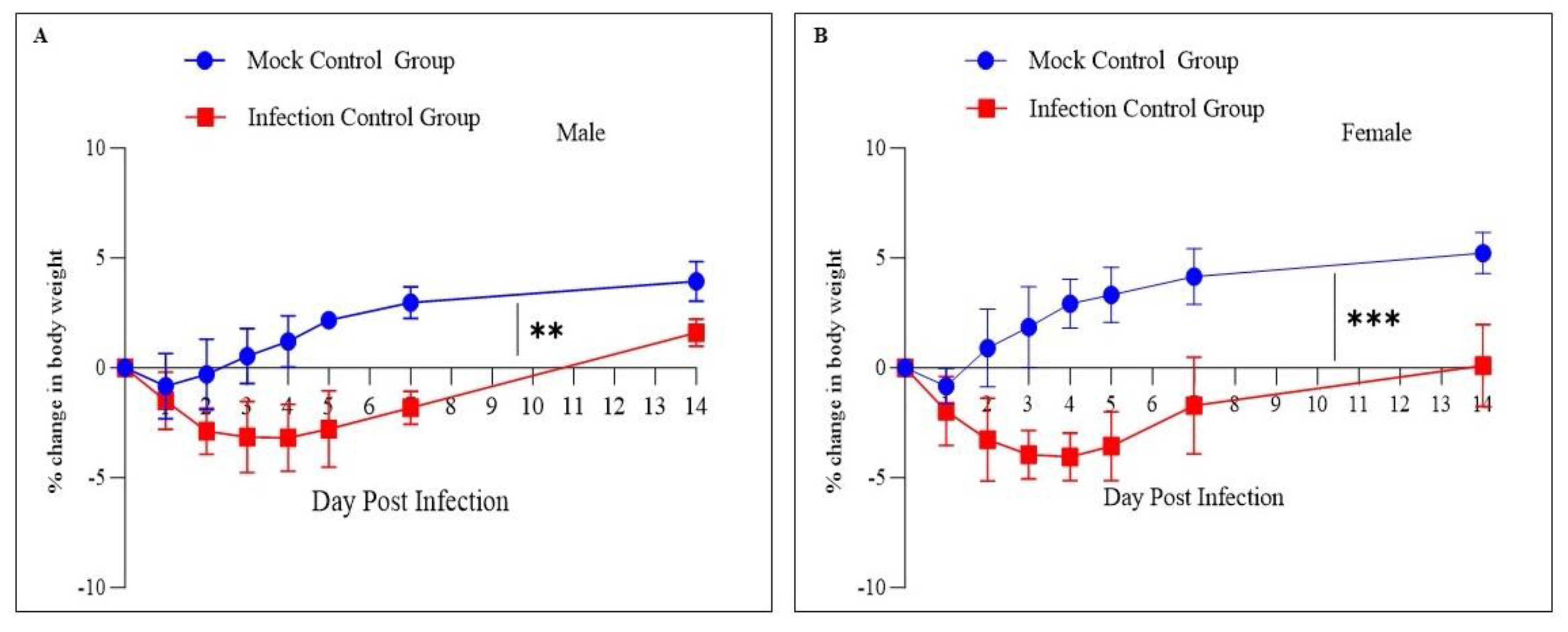
3.1. Change in Body Weight Remained Similar in Both Sexes
3.2. Whole-Genome Sequencing of Viral RNA Showed Mutations in the Spike Protein
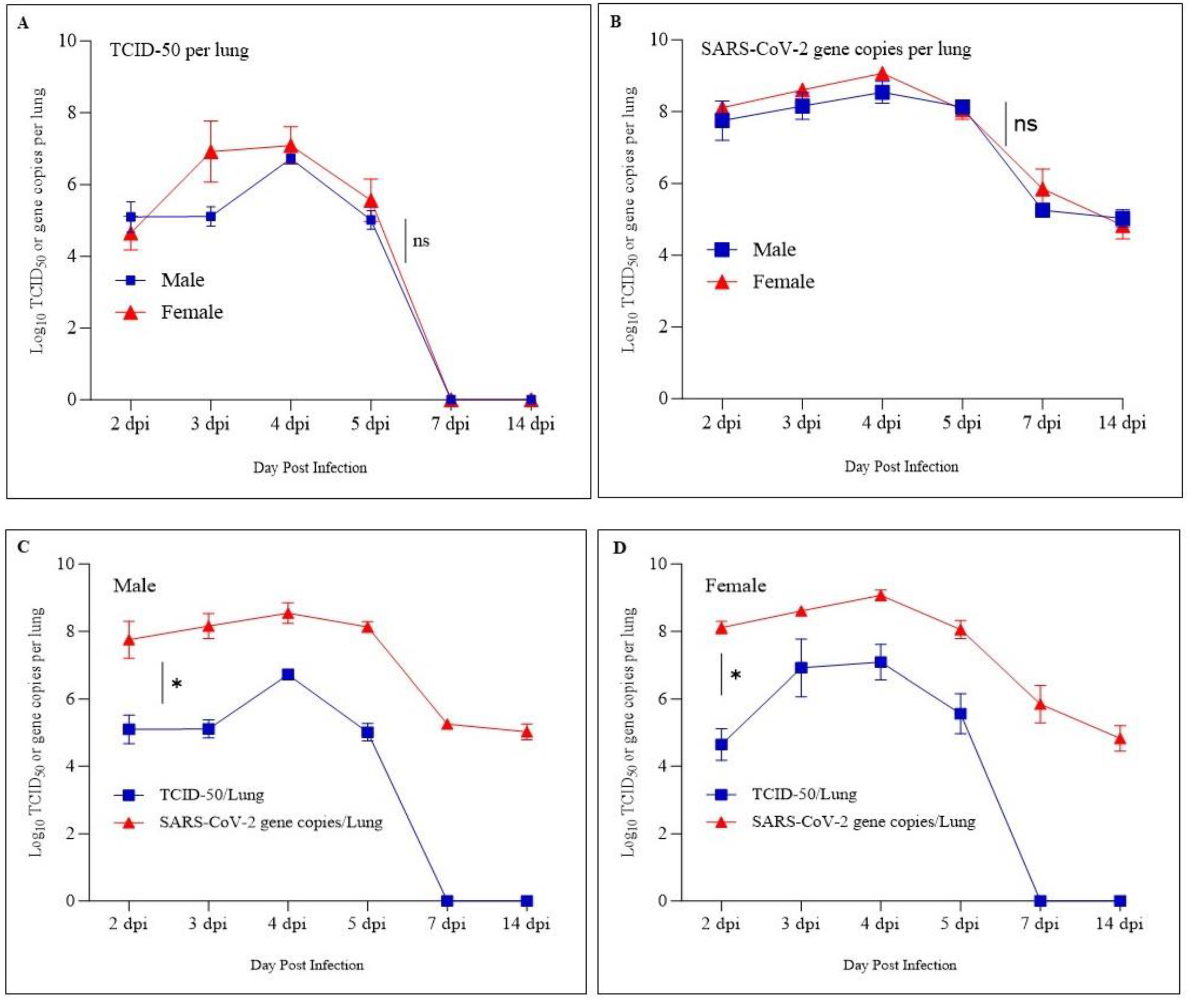
3.3. Lung Viral Load Revealed No Significant Difference between Male and Female Hamsters
3.4. Immune Response against SARS-CoV-2 Has Been Detected in Infected Hamsters as Early as 4 dpi
3.5. Significant Lung Pathology Has Been Observed as Early as 2 dpi in SARS-CoV-2-Infected Hamsters
4. Discussion
5. Conclusions
Supplementary Materials
Author Contributions
Funding
Institutional Review Board Statement
Informed Consent Statement
Data Availability Statement
Acknowledgments
Conflicts of Interest
References
- Graham, R.L.; Baric, R.S. Recombination, Reservoirs, and the Modular Spike: Mechanisms of Coronavirus Cross-Species Transmission. J. Virol. 2010, 84, 3134–3146. [Google Scholar] [CrossRef]
- Anthony, S.J.; Gilardi, K.; Menachery, V.D.; Goldstein, T.; Ssebide, B.; Mbabazi, R.; Navarrete-Macias, I.; Liang, E.; Wells, H.; Hicks, A.; et al. Further Evidence for Bats as the Evolutionary Source of Middle East Respiratory Syndrome Coronavirus. mBio 2017, 8, e00373-17. [Google Scholar] [CrossRef]
- Hu, B.; Ge, X.; Wang, L.-F.; Shi, Z. Bat Origin of Human Coronaviruses. Virol. J. 2015, 12, 221. [Google Scholar] [CrossRef] [PubMed]
- Huynh, J.; Li, S.; Yount, B.; Smith, A.; Sturges, L.; Olsen, J.C.; Nagel, J.; Johnson, J.B.; Agnihothram, S.; Gates, J.E.; et al. Evidence Supporting a Zoonotic Origin of Human Coronavirus Strain NL63. J. Virol. 2012, 86, 12816–12825. [Google Scholar] [CrossRef]
- Murgolo, N.; Therien, A.G.; Howell, B.; Klein, D.; Koeplinger, K.; Lieberman, L.A.; Adam, G.C.; Flynn, J.; McKenna, P.; Swaminathan, G.; et al. SARS-CoV-2 Tropism, Entry, Replication, and Propagation: Considerations for Drug Discovery and Development. PLoS Pathog. 2021, 17, e1009225. [Google Scholar] [CrossRef] [PubMed]
- Huang, C.; Wang, Y.; Li, X.; Ren, L.; Zhao, J.; Hu, Y.; Zhang, L.; Fan, G.; Xu, J.; Gu, X.; et al. Clinical Features of Patients Infected with 2019 Novel Coronavirus in Wuhan, China. Lancet 2020, 395, 497–506. [Google Scholar] [CrossRef] [PubMed]
- Zhou, Z.; Ren, L.; Zhang, L.; Zhong, J.; Xiao, Y.X.; Jia, Z.; Guo, L.; Yang, J.; Wang, C.; Jiang, S.; et al. Heightened Innate Immune Responses in the Respiratory Tract of COVID-19 Patients. Cell Host Microbe 2020, 27, 883–890.e2. [Google Scholar] [CrossRef] [PubMed]
- Sun, G.; Cui, Q.; Garcia, G.; Wang, C.; Zhang, M.; Arumugaswami, V.; Riggs, A.D.; Shi, Y. Comparative Transcriptomic Analysis of SARS-CoV-2 Infected Cell Model Systems Reveals Differential Innate Immune Responses. Sci. Rep. 2021, 11, 17146. [Google Scholar] [CrossRef] [PubMed]
- Krause, P.R.; Fleming, T.R.; Longini, I.M.; Peto, R.; Briand, S.; Heymann, D.L.; Beral, V.; Snape, M.D.; Rees, H.; Ropero, A.-M.; et al. SARS-CoV-2 Variants and Vaccines. N. Engl. J. Med. 2021, 385, 179–186. [Google Scholar] [CrossRef] [PubMed]
- WHO. Update on Omicron. 2021. Available online: https://www.who.int/fr/news/item/28-11-2021-update-on-omicron (accessed on 5 November 2023).
- Mohandas, S.; Dhruv Yadav, P.; Shete, A.; Nyayanit, D.; Sapkal, G.; Lole, K.; Gupta, N. SARS-CoV-2 Delta Variant Pathogenesis and Host Response in Syrian Hamsters. Viruses 2021, 13, 1773. [Google Scholar] [CrossRef] [PubMed]
- Sheahan, T.; Sims, A.; Zhou, S.; Graham, R.; Hill, C.; Leist, S.; Schäfer, A.; Dinnon, K.; Montgomery, S.; Agostini, M.; et al. An Orally Bioavailable Broad-Spectrum Antiviral Inhibits SARS-CoV-2 and Multiple Endemic, Epidemic and Bat Coronavirus. bioRxiv 2020. [Google Scholar] [CrossRef]
- Kaptein, S.J.; Jacobs, S.; Langendries, L.; Seldeslachts, L.; ter Horst, S.; Liesenborghs, L.; Hens, B.; Vergote, V.; Heylen, E.; Maas, E.; et al. Antiviral Treatment of SARS-CoV-2-Infected Hamsters Reveals a Weak Effect of Favipiravir and a Complete Lack of Effect for Hydroxychloroquine. bioRxiv 2020. [Google Scholar] [CrossRef]
- Muñoz-Fontela, C.; Dowling, W.E.; Funnell, S.G.P.; Gsell, P.S.; Riveros-Balta, A.X.; Albrecht, R.A.; Andersen, H.; Baric, R.S.; Carroll, M.W.; Cavaleri, M.; et al. Animal Models for COVID-19. Nature 2020, 586, 509–515. [Google Scholar] [CrossRef] [PubMed]
- Liu, Y.; Hu, G.; Wang, Y.; Ren, W.; Zhao, X.; Ji, F.; Zhu, Y.; Feng, F.; Gong, M.; Ju, X.; et al. Functional and Genetic Analysis of Viral Receptor ACE2 Orthologs Reveals a Broad Potential Host Range of SARS-CoV-2. Proc. Natl. Acad. Sci. USA 2021, 118, e2025373118. [Google Scholar] [CrossRef]
- Roberts, A.; Vogel, L.; Guarner, J.; Hayes, N.; Murphy, B.; Zaki, S.; Subbarao, K. Severe Acute Respiratory Syndrome Coronavirus Infection of Golden Syrian Hamsters. J. Virol. 2005, 79, 503–511. [Google Scholar] [CrossRef]
- Iwatsuki-Horimoto, K.; Nakajima, N.; Ichiko, Y.; Sakai-Tagawa, Y.; Noda, T.; Hasegawa, H.; Kawaoka, Y. Syrian Hamster as an Animal Model for the Study of Human Influenza Virus Infection. J. Virol. 2018, 92, 1–14. [Google Scholar] [CrossRef] [PubMed]
- Mehla, R.; Kokate, P.; Bhosale, S.R.; Vaidya, V.; Narayanan, S.; Shandil, R.K.; Singh, M.; Rudramurthy, G.R.; Naveenkumar, C.N.; Bharathkumar, K.; et al. A Live Attenuated COVID-19 Candidate Vaccine for Children: Protection against SARS-CoV-2 Challenge in Hamsters. Vaccines 2023, 11, 255. [Google Scholar] [CrossRef] [PubMed]
- Chan, J.F.W.; Zhang, A.J.; Yuan, S.; Poon, V.K.M.; Chan, C.C.S.; Lee, A.C.Y.; Chan, W.M.; Fan, Z.; Tsoi, H.W.; Wen, L.; et al. Simulation of the Clinical and Pathological Manifestations of Coronavirus Disease 2019 (COVID-19) in a Golden Syrian Hamster Model: Implications for Disease Pathogenesis and Transmissibility. Clin. Infect. Dis. 2020, 71, 2428–2446. [Google Scholar] [CrossRef]
- Case, J.B.; Bailey, A.L.; Kim, A.S.; Chen, R.E.; Diamond, M.S. Growth, Detection, Quantification, and Inactivation of SARS-CoV-2. Virology 2020, 548, 39–48. [Google Scholar] [CrossRef]
- Jureka, A.S.; Silvas, J.A.; Basler, C.F. Propagation, Inactivation, and Safety Testing of SARS-CoV-2. Viruses 2020, 12, 622. [Google Scholar] [CrossRef]
- Rudramurthy, G.R.; Shandil, R.K.; Narayanana, S. In-Vitro Screening of Repurposed Drug Library against Severe Acute Respiratory Syndrome Coronavirus-2. Med. Res. Arch. 2023, 11, 1–13. [Google Scholar] [CrossRef]
- Reed, L.J.; Muench, H. A Simple Method of Estimating Fifty per Cent Endpoints. Am. J. Epidemiol. 1938, 27, 493–497. [Google Scholar] [CrossRef]
- Dhakal, S.; Ruiz-Bedoya, C.A.; Zhou, R.; Creisher, P.S.; Villano, J.S.; Littlefield, K.; Castillo, J.R.; Marinho, P.; Jedlicka, A.E.; Ordonez, A.A.; et al. Sex Differences in Lung Imaging and SARS-CoV-2 Antibody Responses in a COVID-19 Golden Syrian Hamster Model. mBio 2021, 12, e0097421. [Google Scholar] [CrossRef]
- Yuan, S.; Yin, X.; Meng, X.; Chan, J.F.W.; Ye, Z.W.; Riva, L.; Pache, L.; Chan, C.C.Y.; Lai, P.M.; Chan, C.C.S.; et al. Clofazimine Broadly Inhibits Coronaviruses Including SARS-CoV-2. Nature 2021, 593, 418–423. [Google Scholar] [CrossRef] [PubMed]
- Osterrieder, N.; Bertzbach, L.D.; Dietert, K.; Abdelgawad, A.; Vladimirova, D.; Kunec, D.; Hoffmann, D.; Beer, M.; Gruber, A.D.; Trimpert, J. Age-Dependent Progression of SARS-CoV-2 Infection in Syrian Hamsters. Viruses 2020, 12, 779. [Google Scholar] [CrossRef]
- Plunkard, J.; Mulka, K.; Zhou, R.; Tarwater, P.; Zhong, W.; Lowman, M.; Wong, A.; Pekosz, A.; Villano, J. SARS-CoV-2 Variant Pathogenesis Following Primary Infection and Reinfection in Syrian Hamsters. mBio 2023, 14, e0007823. [Google Scholar] [CrossRef] [PubMed]
- Lee, A.C.Y.; Zhang, A.J.; Chan, J.F.W.; Li, C.; Fan, Z.; Liu, F.; Chen, Y.; Liang, R.; Sridhar, S.; Cai, J.P.; et al. Oral SARS-CoV-2 Inoculation Establishes Subclinical Respiratory Infection with Virus Shedding in Golden Syrian Hamsters. Cell Rep. Med. 2020, 1, 100121. [Google Scholar] [CrossRef] [PubMed]
- Imai, M.; Iwatsuki-Horimoto, K.; Hatta, M.; Loeber, S.; Halfmann, P.J.; Nakajima, N.; Watanabe, T.; Ujie, M.; Takahashi, K.; Ito, M.; et al. Syrian Hamsters as a Small Animal Model for SARS-CoV-2 Infection and Countermeasure Development. Proc. Natl. Acad. Sci. USA 2020, 117, 16587–16595. [Google Scholar] [CrossRef] [PubMed]
- O’Donnell, K.L.; Pinski, A.N.; Clancy, C.S.; Gourdine, T.; Shifflett, K.; Fletcher, P.; Messaoudi, I.; Marzi, A. Pathogenic and Transcriptomic Differences of Emerging SARS-CoV-2 Variants in the Syrian Golden Hamster Model. EBioMedicine 2021, 73, 103675. [Google Scholar] [CrossRef]
- Boudewijns, R.; Thibaut, H.J.; Kaptein, S.J.F.; Li, R.; Vergote, V.; Seldeslachts, L.; Van Weyenbergh, J.; De Keyzer, C.; Bervoets, L.; Sharma, S.; et al. STAT2 Signaling Restricts Viral Dissemination but Drives Severe Pneumonia in SARS-CoV-2 Infected Hamsters. Nat. Commun. 2020, 11, 5838. [Google Scholar] [CrossRef]
- Sia, S.F.; Yan, L.M.; Chin, A.W.H.; Fung, K.; Choy, K.T.; Wong, A.Y.L.; Kaewpreedee, P.; Perera, R.A.P.M.; Poon, L.L.M.; Nicholls, J.M.; et al. Pathogenesis and Transmission of SARS-CoV-2 in Golden Hamsters. Nature 2020, 583, 834–838. [Google Scholar] [CrossRef] [PubMed]
- Gruber, A.D.; Firsching, T.C.; Trimpert, J.; Dietert, K. Hamster Models of COVID-19 Pneumonia Reviewed: How Human Can They Be? Vet. Pathol. 2022, 59, 528–545. [Google Scholar] [CrossRef]
- Tostanoski, L.H.; Wegmann, F.; Martinot, A.J.; Loos, C.; McMahan, K.; Mercado, N.B.; Yu, J.; Chan, C.N.; Bondoc, S.; Starke, C.E.; et al. Ad26 Vaccine Protects against SARS-CoV-2 Severe Clinical Disease in Hamsters. Nat. Med. 2020, 26, 1694–1700. [Google Scholar] [CrossRef] [PubMed]
- Mulka, K.R.; Beck, S.E.; Solis, C.V.; Johanson, A.L.; Queen, S.E.; McCarron, M.E.; Richardson, M.R.; Zhou, R.; Marinho, P.; Jedlicka, A.; et al. Progression and Resolution of Severe Acute Respiratory Syndrome Coronavirus 2 (SARS-CoV-2) Infection in Golden Syrian Hamsters. Am. J. Pathol. 2022, 192, 195–207. [Google Scholar] [CrossRef]
- Ogando, N.S.; Dalebout, T.J.; Zevenhoven-Dobbe, J.C.; Limpens, R.W.A.L.; van der Meer, Y.; Caly, L.; Druce, J.; de Vries, J.J.C.; Kikkert, M.; Barcena, M.; et al. SARS-Coronavirus-2 Replication in Vero E6 Cells: Replication Kinetics, Rapid Adaptation and Cytopathology. J. Gen. Virol. 2020, 101, 925–940. [Google Scholar] [CrossRef] [PubMed]
- Chen, Y.; Liu, M.-Q.; Luo, Y.; Jiang, R.-D.; Si, H.-R.; Zhu, Y.; Li, B.; Shen, X.-R.; Lin, H.-F.; Zhao, K.; et al. Genetic Mutation of SARS-CoV-2 during Consecutive Passages in Permissive Cells. Virol. Sin. 2021, 36, 1073–1076. [Google Scholar] [CrossRef] [PubMed]
- Huang, Y.; Yang, C.; Xu, X.F.; Xu, W.; Liu, S.W. Structural and Functional Properties of SARS-CoV-2 Spike Protein: Potential Antivirus Drug Development for COVID-19. Acta Pharmacol. Sin. 2020, 41, 1141–1149. [Google Scholar] [CrossRef]
- Wrapp, D.; Wang, N.; Corbett, K.S.; Goldsmith, J.A.; Hsieh, C.L.; Abiona, O.; Graham, B.S.; McLellan, J.S. Cryo-EM Structure of the 2019-NCoV Spike in the Prefusion Conformation. Science 2020, 367, 1264–1269. [Google Scholar] [CrossRef]
- Harvey, W.T.; Carabelli, A.M.; Jackson, B.; Gupta, R.K.; Thomson, E.C.; Harrison, E.M.; Ludden, C.; Reeve, R.; Rambaut, A.; Peacock, S.J.; et al. SARS-CoV-2 Variants, Spike Mutations and Immune Escape. Nat. Rev. Microbiol. 2021, 19, 409–424. [Google Scholar] [CrossRef]
- Greaney, A.J.; Loes, A.N.; Crawford, K.H.D.; Starr, T.N.; Malone, K.D.; Chu, H.Y.; Bloom, J.D. Comprehensive Mapping of Mutations in the SARS-CoV-2 Receptor-Binding Domain That Affect Recognition by Polyclonal Human Plasma Antibodies. Cell Host Microbe 2021, 29, 463–476.e6. [Google Scholar] [CrossRef]
- Weisblum, Y.; Schmidt, F.; Zhang, F.; DaSilva, J.; Poston, D.; Lorenzi, J.C.C.; Muecksch, F.; Rutkowska, M.; Hoffmann, H.H.; Michailidis, E.; et al. Escape from Neutralizing Antibodies 1 by SARS-CoV-2 Spike Protein Variants. eLife 2020, 9, e61312. [Google Scholar] [CrossRef]
- Baum, A.; Fulton, B.O.; Wloga, E.; Copin, R.; Pascal, K.E.; Russo, V.; Giordano, S.; Lanza, K.; Negron, N.; Ni, M.; et al. Antibody Cocktail to SARS-CoV-2 Spike Protein Prevents Rapid Mutational Escape Seen with Individual Antibodies. Science 2020, 369, 1014–1018. [Google Scholar] [CrossRef]
- European Centre for Disease Prevention and Control. SARS-CoV-2 Variant Mutations Conferring Reduced Susceptibility to Antiviral Drugs and Monoclonal Antibodies: A Non-Systematic Literature Review for Surveillance Purposes; European Centre for Disease Prevention and Control: Stockholm, Sweden, 2023. [Google Scholar]
- Sonnleitner, S.T.; Prelog, M.; Sonnleitner, S.; Hinterbichler, E.; Halbfurter, H.; Kopecky, D.B.C.; Almanzar, G.; Koblmüller, S.; Sturmbauer, C.; Feist, L.; et al. Cumulative SARS-CoV-2 Mutations and Corresponding Changes in Immunity in an Immunocompromised Patient Indicate Viral Evolution within the Host. Nat. Commun. 2022, 13, 2560. [Google Scholar] [CrossRef] [PubMed]
- John, G.; Sahajpal, N.S.; Mondal, A.K.; Ananth, S.; Williams, C.; Chaubey, A.; Rojiani, A.M.; Kolhe, R. Next-Generation Sequencing (Ngs) in Covid-19: A Tool for Sars-Cov-2 Diagnosis, Monitoring New Strains and Phylodynamic Modeling in Molecular Epidemiology. Curr. Issues Mol. Biol. 2021, 43, 845–867. [Google Scholar] [CrossRef] [PubMed]
- Roy, S.; Coldren, C.; Karunamurthy, A.; Kip, N.S.; Klee, E.W.; Lincoln, S.E.; Leon, A.; Pullambhatla, M.; Temple-Smolkin, R.L.; Voelkerding, K.V.; et al. Standards and Guidelines for Validating Next-Generation Sequencing Bioinformatics Pipelines: A Joint Recommendation of the Association for Molecular Pathology and the College of American Pathologists. J. Mol. Diagn. 2018, 20, 4–27. [Google Scholar] [CrossRef] [PubMed]
- Wadapurkar, R.M.; Vyas, R. Computational Analysis of next Generation Sequencing Data and Its Applications in Clinical Oncology. Inform. Med. Unlocked 2018, 11, 75–82. [Google Scholar] [CrossRef]
- Bin Mahmood, T.; Saha, A.; Hossan, M.I.; Mizan, S.; Arman, S.M.A.S.; Chowdhury, A.S. A next Generation Sequencing (NGS) Analysis to Reveal Genomic and Proteomic Mutation Landscapes of SARS-CoV-2 in South Asia. Curr. Res. Microb. Sci. 2021, 2, 100065. [Google Scholar] [CrossRef]
- Liu, T.; Chen, Z.; Chen, W.; Chen, X.; Hosseini, M.; Yang, Z.; Li, J.; Ho, D.; Turay, D.; Gheorghe, C.P.; et al. A Benchmarking Study of SARS-CoV-2 Whole-Genome Sequencing Protocols Using COVID-19 Patient Samples. iScience 2021, 24, 102892. [Google Scholar] [CrossRef]
- Jacot, D.; Pillonel, T.; Greub, G.; Bertelli, C. Assessment of SARS-CoV-2 Genome Sequencing: Quality Criteria and Low-Frequency Variants. J. Clin. Microbiol. 2021, 59, e0094421. [Google Scholar] [CrossRef] [PubMed]
- Kebschull, J.M.; Zador, A.M. Sources of PCR-Induced Distortions in High-Throughput Sequencing Data Sets. Nucleic Acids Res. 2015, 43, e143. [Google Scholar] [CrossRef]


 Represent the areas of congestion and hemorrhage and
  represent lung consolidation.
Represent the areas of congestion and hemorrhage and represent lung consolidation.
represent lung consolidation.
Represent the areas of congestion and hemorrhage and represent lung consolidation.
 ), and an accumulation of eosinophilic edematous exudate (
 ). Moderate to marked broncho-interstitial pneumonia with alveolar damage and inflammatory cell infiltration (
 ), hemorrhage (
 ), and hyperplasia of the bronchial epithelium (
 ) at 3 and 4 dpi in both male and female hamsters. Severe broncho-interstitial pneumonia with a marked increase in lung cellularity with hyperplasia of Type II alveolar cells (
 ) was observed at 7 dpi. Mild congestion (
 ), infiltration of inflammatory cells (
 ), and mild hyperplasia of the bronchial epithelium (
 ) were observed at 14 dpi.
), and an accumulation of eosinophilic edematous exudate (). Moderate to marked broncho-interstitial pneumonia with alveolar damage and inflammatory cell infiltration (), hemorrhage (), and hyperplasia of the bronchial epithelium () at 3 and 4 dpi in both male and female hamsters. Severe broncho-interstitial pneumonia with a marked increase in lung cellularity with hyperplasia of Type II alveolar cells () was observed at 7 dpi. Mild congestion (), infiltration of inflammatory cells (), and mild hyperplasia of the bronchial epithelium () were observed at 14 dpi.
) were observed at 14 dpi.
), and an accumulation of eosinophilic edematous exudate (). Moderate to marked broncho-interstitial pneumonia with alveolar damage and inflammatory cell infiltration (), hemorrhage (), and hyperplasia of the bronchial epithelium () at 3 and 4 dpi in both male and female hamsters. Severe broncho-interstitial pneumonia with a marked increase in lung cellularity with hyperplasia of Type II alveolar cells () was observed at 7 dpi. Mild congestion (), infiltration of inflammatory cells (), and mild hyperplasia of the bronchial epithelium () were observed at 14 dpi.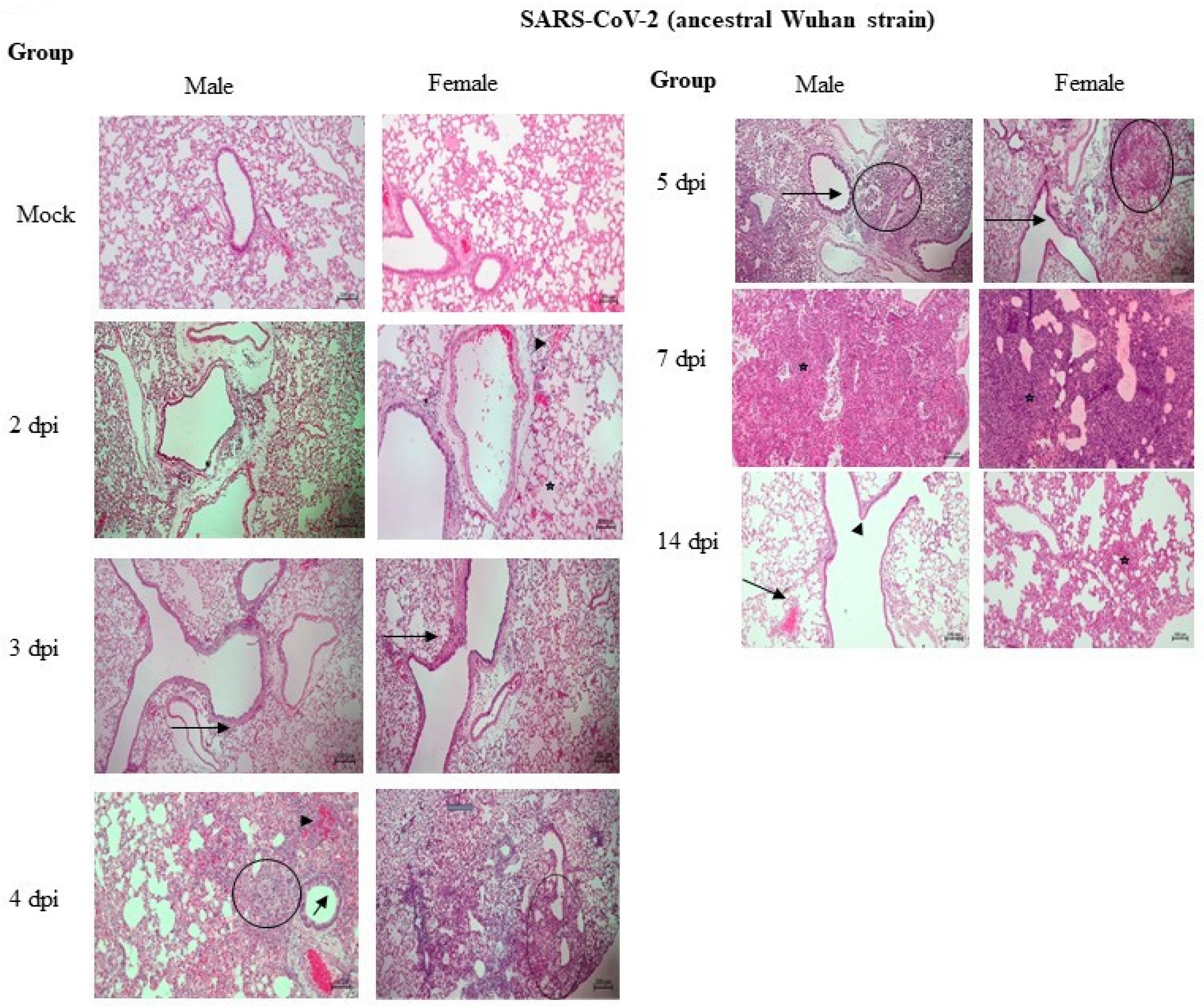

| Sample | Reference Sequence | Total Length of Consensus (bp) | No. of SNPs | Lineage Identified |
|---|---|---|---|---|
| Virus stock used in the infection of Hamsters | NC_045512.2 | 29903 | 8 | A |
| Virus isolated from the infected hamster (Male) | Virus stock (used in the infection of hamsters) | 29903 | 10 | A |
| Virus isolated from the infected hamster (Female) | 29903 | 10 | A |
| Position in Reference Sequence (NC_045512.2) | Reported Base in Reference Sequence (NC_045512.2) | Identified Alternate Base in Virus Stock (Used in the Infection of Hamsters) | Gene Name | Protein Change |
|---|---|---|---|---|
| 8782 | C | T | ORF1ab | Ser2839Ser |
| 18060 | C | T | ORF1ab | Leu5932Leu |
| 21759 | A | G | S | His66Arg |
| 22296 | A | G | S | His245Arg |
| 22482 | C | T | S | Thr307Ile |
| 23606 | C | T | S | Arg682Trp |
| 23607 | G | T | S | Arg682Leu |
| 28144 | T | C | ORF8 | Leu84Ser |
| Position in Stock Virus (Used in Infection) | Reported Base in Stock Virus (Used in Infection) | Identified Alternate Base from an Infected Animal (Male) | Identified Alternate Base from Infected Animal (Female) | Gene Name | Protein Change |
|---|---|---|---|---|---|
| 17827 | C | A | A | ORF1ab | Gln5855Lys |
| 21801 | A | G | G | S | Asp80Gly |
| 21849 | A | - | C | S | Glu96Ala |
| 22206 | A | G | G | S | Asp215Gly |
| 23014 | A | C | C | S | Glu484Asp |
| 23525 | C | T | T | S | His655Tyr |
| 24734 | C | T | T | S | His1058Tyr |
| 21759 | G | A | A | S | Arg66His |
| 22296 | G | A | G | S | Arg245His (Male) |
| 23606 | T | C | C | S | Trp682Arg |
| 23607 | T | G | G | S | Leu682Arg |
Disclaimer/Publisher’s Note: The statements, opinions and data contained in all publications are solely those of the individual author(s) and contributor(s) and not of MDPI and/or the editor(s). MDPI and/or the editor(s) disclaim responsibility for any injury to people or property resulting from any ideas, methods, instructions or products referred to in the content. |
© 2023 by the authors. Licensee MDPI, Basel, Switzerland. This article is an open access article distributed under the terms and conditions of the Creative Commons Attribution (CC BY) license (https://creativecommons.org/licenses/by/4.0/).
Share and Cite
Rudramurthy, G.R.; Naveenkumar, C.N.; Bharathkumar, K.; Shandil, R.K.; Narayanan, S. Genomic Mutations in SARS-CoV-2 Genome following Infection in Syrian Golden Hamster and Associated Lung Pathologies. Pathogens 2023, 12, 1328. https://doi.org/10.3390/pathogens12111328
Rudramurthy GR, Naveenkumar CN, Bharathkumar K, Shandil RK, Narayanan S. Genomic Mutations in SARS-CoV-2 Genome following Infection in Syrian Golden Hamster and Associated Lung Pathologies. Pathogens. 2023; 12(11):1328. https://doi.org/10.3390/pathogens12111328
Chicago/Turabian StyleRudramurthy, Gudepalya Renukaiah, Chakenahalli N. Naveenkumar, Kumaraswamy Bharathkumar, Radha K. Shandil, and Shridhar Narayanan. 2023. "Genomic Mutations in SARS-CoV-2 Genome following Infection in Syrian Golden Hamster and Associated Lung Pathologies" Pathogens 12, no. 11: 1328. https://doi.org/10.3390/pathogens12111328
APA StyleRudramurthy, G. R., Naveenkumar, C. N., Bharathkumar, K., Shandil, R. K., & Narayanan, S. (2023). Genomic Mutations in SARS-CoV-2 Genome following Infection in Syrian Golden Hamster and Associated Lung Pathologies. Pathogens, 12(11), 1328. https://doi.org/10.3390/pathogens12111328