Article

Linear Peptide Epitopes Derived from ErpP, p35, and FlaB in the Serodiagnosis of Lyme Disease

by
Paul M. Arnaboldi
1,2,*,
Adiya S. Katseff
1,
Mariya Sambir
1,2 and
Raymond J. Dattwyler
1,2
1
Department of Pathology, Microbiology, and Immunology, New York Medical College, Valhalla, NY 10595, USA
2
Biopeptides, Corp., East Setauket, NY 11733, USA
*
Author to whom correspondence should be addressed.
Pathogens 2022, 11(8), 944; https://doi.org/10.3390/pathogens11080944
Submission received: 14 July 2022 / Revised: 13 August 2022 / Accepted: 16 August 2022 / Published: 20 August 2022

Abstract

:
Lyme disease is the most common vector-borne disease in the northern hemisphere. Current serodiagnostics are insensitive in early infection. Sensitivity in these seroassays is compromised by the necessity to preserve specificity in the presence of cross-reactive epitopes in Borrelia burgdorferi target antigens. We evaluated the efficacy of using synthetic peptides containing epitopes unique to B. burgdorferi as antigen targets in a Lyme disease seroassay. We performed linear B cell epitope mapping of the proteins p35 (BBH32) and ErpP to identify unique epitopes. We generated peptides containing these newly identified linear epitope sequences along with previously identified epitopes from the antigens FlaB and VlsE and evaluated their diagnostic capabilities via ELISA using large serum sets. Single-epitope peptides, while specific, demonstrated insufficient sensitivity. However, when epitopes from FlaB, ErpP, or p35 were combined in tandem with an epitope from VlsE, the sensitivity of the assay was significantly increased without compromising specificity. The identification of additional unique epitopes from other B. burgdorferi antigens and the further development of a combined multi-peptide-based assay for the laboratory diagnosis of Lyme disease offers a way to address the poor specificity associated with the use of whole protein antigen targets and thus significantly improve the laboratory diagnosis of Lyme disease.

1. Background

Lyme disease, caused by pathogenic members of the Borrelia burgdorferi sensu lato genospecies, is the most common vector-borne disease in the United States and Europe [1,2]. The CDC estimates that there are more than 400,000 newly diagnosed cases of Lyme disease each year in the U.S. (http://www.cdc.gov/lyme/stats/humanCases.html accessed on 27 June 2022), with over 3 million diagnostic laboratory tests ordered each year [3,4]. Treatment of early Lyme disease with appropriate oral antibiotics is highly effective at preventing the development of disseminated sequelae in most cases. However, if the disease progresses, significant nervous system or musculoskeletal involvement can occur with the risk of permanent damage to the nervous and musculoskeletal systems [1,2,5]. Early detection and treatment are critical to ensure good patient outcomes; unfortunately, current serodiagnostics lack sensitivity in early Lyme disease detection precisely at the time when diagnosis and treatment have the maximum effect on the outcome. Erythema migrans (EM) is the characteristic skin lesion of Lyme disease. In Lyme-disease-endemic regions, EM is considered diagnostic [5,6,7,8]. However, roughly 20% of patients do not develop EM [9]. Further complicating the diagnosis is that EM is variable in appearance and may not be recognized. It is also fleeting and may be gone by the time a patient seeks medical care. Considering that 20% of patients per year (approximately 60,000 based on current CDC estimations) do not develop EM, and given the difficulty in accurately identifying all those who do develop it, sensitive and specific laboratory testing becomes critical to provide optimal patient outcomes.
Direct detection of B. burgdorferi using assays including culture and/or PCR has proven ineffective in the absence of EM [6,9,10,11]. The laboratory diagnosis of Lyme disease is based on indirect methods. The mainstays for laboratory diagnosis of Lyme disease are serologic assays that detect antibodies to B. burgdorferi antigens. Because of the lack of specificity in past and current serologic assays, a two-tier paradigm consisting of an EIA followed by an immunoblot, or more recently, another EIA is recommended by the CDC to mitigate false positivity [5,7,8,9,10,11,12,13]. The two-tier paradigm addressed the issue of specificity, but unfortunately, due to its stringency, it is insensitive at the time many patients with early LD seek initial medical care. A 2016 meta-analysis demonstrated a low sensitivity of 46.3% for early stage Lyme disease across all the studies evaluated, with sensitivity increasing to 89.7% and 99.4% in patients with early disseminated or late-stage disease, respectively [14]. Although protein-based assays can be made to be sensitive, protein antigens commonly contain cross-reactive epitopes; thus, increased sensitivity is limited by decreased specificity. Despite extensive research, no protein-based assay has overcome this specificity problem. The use of synthetic peptides containing linear epitopes unique to B. burgdorferi as antigen targets can improve specificity [15,16,17,18,19,20,21,22,23,24,25,26,27] by eliminating nonspecific, cross-reactive epitopes [8,11,12,15,28,29,30,31,32]. By removing cross-reactive epitopes, it is possible to improve specificity, which could lead to the development of single-tier assays that would allow for greater sensitivity. In the present study, we evaluated the diagnostic potential of synthetic peptides from three prominent B. burgdorferi antigens expressed during human infection, ErpP (OspE/F related protein P), p35, and FlaB. ErpP binds complement factor H and plasminogen and is thought to play a role in bacterial survival/immune evasion and dissemination [33,34,35,36]. The second prominent antigen, p35 (BBH32), is a surface lipoprotein of unknown function [37,38]. Finally, FlaB is a major constituent of the B. burgdorferi flagellum [39,40]. We assessed both IgM and IgG binding using a standard ELISA comparing the diagnostic potential of immunodominant epitopes from each protein both singularly and when conjugated to an immunogenic epitope-containing peptide derived from the B. burgdorferi protein VlsE [17].

2. Results

Epitope mapping of full-length ErpP and p35 revealed a single 15-AA peptide containing a linear epitope in each protein (Figure 1a,b). All eight patient sera used for epitope mapping detected p35(101–115). ErpP(51–65) was detected by four of the eight patient sera used for epitope mapping, and it was the only peptide that bound antibodies in multiple patient sera. We evaluated the efficacy of these peptides and a previously described 13AA peptide derived from Borrelia flagellin, FlaB(211–223), as antigen targets for the detection of IgM and IgG in a large panel of sera from patients with early (EM+) or late (LA+) Lyme disease in an ELISA format (Table 1). Sera from healthy individuals, patients with RA, or patients with syphilis were used as controls for nonspecificity (Table 2). Cutoffs were defined as 3SD and 2SD or positivity and equivocality, respectively, from the mean IgM or IgG binding to peptides in healthy control serum. In 13.1% and 6.3% of EM+ patient sera, respectively, p35(101–155) demonstrated positive binding of IgM and IgG. Equivocal levels of IgM and IgG were 9.0% and 13.9%, respectively. ErpP(51–65) performed marginally better, with positive binding of IgM and IgG occurring in 23.4% and 11.8% of EM+ patient sera, respectively. Equivocal binding was identical to that of p35(101–155). FlaB(211–223) detected positive levels of antibodies in a higher number of patient sera than the other two peptides; the positive detection of rate of IgM and IgG was 37.6% and 17.3%, respectively, with an equivocal detection rate of 3.0% (IgM) and 12.0% (IgG) (p < 0.01 FlaB(211–223) vs. p35(101–155) and ErpP(51–65) for IgM, and p < 0.05 FlaB(211–223) vs. p35(101–155) for IgG, FlaB(211–223) vs. ErpP(51–65), N.S for IgG).
To enhance antibody binding detection capability, each of the three epitopes was linked to a 17-AA VlsE peptide (modVlsE(275–291)) that we previously developed. This single-epitope was modified to represent a consensus sequence to minimize the impact of sequence variability found in VlsE among different Borrelia strains [17]. In 25.9% and 54.0% of EM+ patient samples, respectively, p35(101–115)-modVlsE(275–291) (pp35-mV) demonstrated significantly higher positive binding of IgM and IgG (IgM, p < 0.05; IgG, p < 0.0001 pp35-mv vs. p35), respectively, with equivocal detection of both isotypes in 10.8% of patients. ErpP(51–65)-modVlsE(275–291) (pErpP-mV) had positive detection rates of 43.2% and 51.1% for IgM and IgG, respectively, and equivocal detection rates of 8.6% (IgM) and 10.8% (IgG) (IgM, p < 0.005; IgG, p < 0.0001 pErpP-mv vs. pErpP). FlaB(211–223)-modVlsE(275–291) (pFlaB-mV) had the highest rates of detection, positively identifying 55.5% and 54.7% of IgM and IgG in EM+ patient sera and equivocal detection rates of 5.1% (IgM) and 3.6% (IgG) (IgM, p < 0.005; IgG, p < 0.0001 pErpP-mv vs. pErpP). Late Lyme disease antibody binding detection was better with dual-epitope peptides compared to single-epitope peptides (Table 1). IgG was the predominant isotype detected, as would be anticipated for late Lyme disease samples. IgG was positively detected in 95%, 85%, and 85% of LA serum incubated with pp35-mV, pErpP-mV, and pFlaB-mV, respectively. As with the other single-epitopes, modVlsE(275–291) (mV) alone had low sensitivity for detection of antibodies during early infection, positively identifying 12.0% and 26.5% of IgM and IgG in EM+ patient sera, with equivocal binding rates of 2.4% for IgM and 3.6% for IgG.
The serum incubated with pFlaB-mV had the highest rate of total positive antibody binding (either IgM or IgG, 62.8%) when compared to both pErpP-mV (59.0%) or pp35-mV (56.9%) (Table 1). However, pErpP-mV had the lowest rate of false-negative antibody binding (either IgM or IgG, 28.8%) when compared to the other two dual-epitope peptides (pFlaB-mV, 31.4%; pp35-mV, 32.9%) due to a higher rate of equivocal binding of patient antibodies (Table 1). Using ROC analysis to compare IgM and IgG binding in EM patient sera to all negative control sera (syphilis and RA patient sera, and sera from healthy individuals with no history of Lyme disease), specificity and sensitivity at 3SD from the mean antibody binding of healthy controls were determined (Table 3). Specificities for both IgM and IgG peptide binding were greater than 95% for all peptides, except for pErpP-mV, which had a specificity of 94.9% for IgM binding. This was due to IgM binding in 5 of 38 sera from patients with RA (Table 3). In total, the use of the tandem di-epitope-containing peptides significantly increased the serodetection of anti-Borrelial antibodies compared to single peptides with a minimal effect on specificity.

3. Discussion

In the present study, we performed epitope mapping of two B. burgdorferi proteins, p35 (BBH32) and ErpP, to identify linear B cell epitopes that might serve as sensitive and specific diagnostics for early and late infection. While some antigens we have evaluated contained numerous epitopes [20,24,26,41], these proteins each contained only one linear epitope that was identified by at least 50% of the serum samples used for mapping. We also evaluated a previously identified linear epitope from the B. burgdorferi primary flagellin protein, FlaB. This epitope was previously shown to be unique and specific for B. burgdorferi (as most of the flagellin sequence is highly conserved in other bacteria) [42]. Linear epitopes were pursued in this study because they can be easily synthesized; they are not dependent on potentially complex folded structures such as conformational epitopes, and they can be more easily screened for similar sequences in other antigens that may give rise to cross-reactive antibodies and increased nonspecificity. Though somewhat counterintuitive, linear epitopes do not need to be surface-exposed to be useful diagnostically, as they may become exposed as antigens are shed and/or degraded during infection. The C6 epitope is an example of this, as it is not surface-exposed in the folded VlsE protein on the bacterial surface [21]. ErpP(51–65), p35(101–115), and FlaB(211–223) were not very sensitive for the detection of antibodies when used as single-epitope peptides. However, combining the epitopes with mVlsE(275–291), a modified version of the C6 peptide epitope [17], significantly increased antibody binding (p < 0.05) compared to the single-epitope peptides, with a notable increase in IgG binding. In most cases, the inclusion of the second epitope had only a modest effect on specificity compared to the individual epitopes without mVlsE(275–291) (Table 2). Single- and dual-epitope-containing peptides were screened using Lyme disease patient sera obtained from patients with physician-diagnosed Lyme disease. The pattern of antibody binding was as would be expected; the early Lyme disease sera pool (EM+) contained a mixture of IgM and IgG seropositive and seronegative patients, while the late Lyme pool contained predominantly IgG seropositive patients. This indicates that the peptides could detect disease over the course of infection. We have identified peptide epitopes in the past that were only effective in detecting early infection [19]. There is a critical need for improved diagnostics for Lyme disease. Current diagnostic assays are insensitive during early infection, the phase of the disease when treatment is most effective at preventing the dissemination of B. burgdorferi and the development of sequelae. Our data support that single-epitope peptides are suboptimal when used alone but that peptides can be combined to improve sensitivity while still maintaining good specificity. Though the sensitivity of the di-epitope peptides was not high enough to justify their use as single-assay targets for serodiagnosis, they could serve as one of a group of components, including as part of a multiplex-based assay [25]. Our results provide support for the identification of additional peptide antigens and the further development of combined multi-peptide-based assays for the laboratory diagnosis of Lyme disease.

4. Materials and Methods

4.1. Patient Samples

A total of 145 early Lyme disease sera were collected at first presentation with erythema migrans from patients under informed consent with the approval of the institutional review boards of Stony Brook University in Stony Brook, NY (n = 21), New York Medical College in Westchester, NY (n = 74), and Gundersen-Lutheran Medical Center in La Crosse, WI (n = 50). Samples were accumulated over the course of the last 30 years. The 50 samples from Gundersen-Lutheran Medical Center in La Crosse, WI, had a clinician-documented EM lesion of > 4 cm, appropriate epidemiologic history (e.g., tick bite or exposure), and were seropositive according to a whole-cell ELISA (VIDAS by BioMerieux, Durham, NC, USA). We were not provided with the clinical laboratory results for the other deidentified Lyme disease patients beyond the fact that the patients were EM+. A total of 20 late Lyme disease samples were collected from patients at Gundersen-Lutheran Medical Center in La Crosse, WI, with Lyme arthritis (LA) (n = 20) that had one or more episodes of swollen joints, appropriate epidemiologic history, and positive reactivity using a whole-cell ELISA (VIDAS). The regions where the samples were collected, lower New York and Wisconsin, are highly endemic areas for Lyme disease. Sera from healthy volunteers (n = 64) were collected in New Mexico, which is not endemic for Lyme disease, and were purchased from Creative Testing Solutions (Tempe, AZ, USA). A total of 80 sera from patients with rheumatoid arthritis (RA, n = 40, rheumatoid factor status unknown) or who were Rapid Plasma Reagin positive (RPR+, first-tier test for syphilis, n- = 35) were purchased from Bioreclamation LLC (Westbury, NY, USA). These samples were collected in a region endemic for Lyme disease (the northeastern US). A total of 34 of the 35 RPR+ sera used in this study had positive or equivocal antibody levels against Treponema pallidum by ELISA (Abnova, Walnut, CA, USA). Some serum samples are not represented in all data sets because they were fully consumed during experimentation.

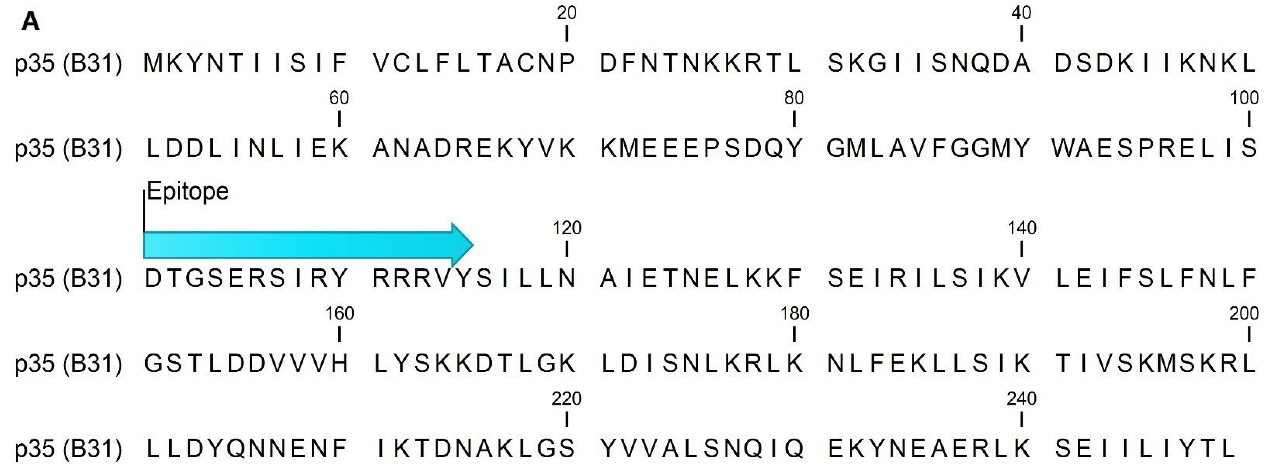
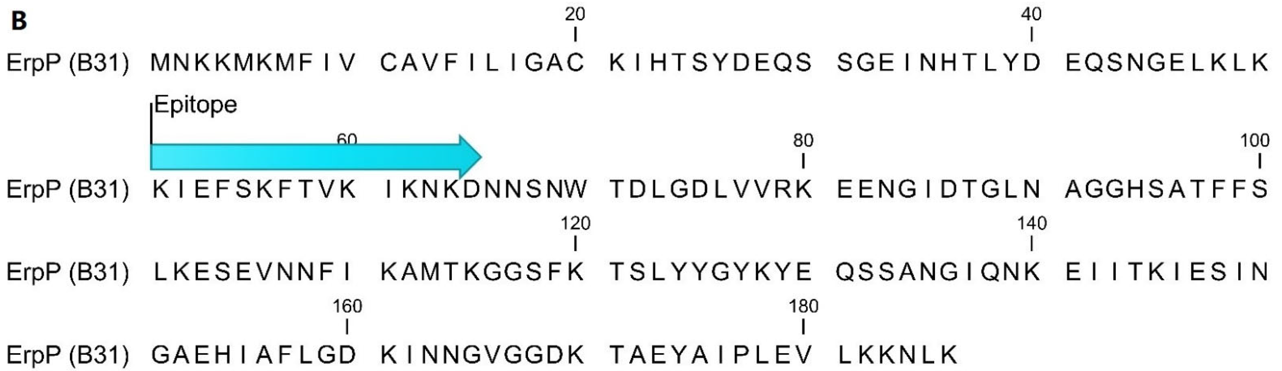
4.2. Epitope Mapping and Peptide Sequences

Overlapping peptide libraries (ProImmune, Oxford, UK), consisting of 15-AA long peptides overlapping by 10-AA, were generated using sequences of p35 (BBH32) (accession# WP_010256558.1) and ErpP (accession # WP_010883886.1) from the B31 strain of Borrelia burgdorferi sensu stricto. ProImmune, Inc. performed the epitope mapping under contract using their proprietary ProArray Ultra technology, as previously described [19]. Sera from 8 patients with physician-diagnosed EM+ early Lyme disease that were seropositive by two-tier testing, with an IgG immunoblot sensitivity of 9-10, of 10 bands were used for the epitope mapping. Using the 17-mer B31 sequence previously described, mod-VlsE(275–291) was engineered. [17]. It includes substitutions at AA278 (D→ N), AA282 (A→ V), and AA290 (M→ V), which represent natural sequence variability found in Borrelia strains other than B31. It is similar in sequence to the IR6 epitope of VlsE, which has been shown to be sensitive as a first- or second-tier assay target [16,27]. FlaB(211–223) is a previously described immunodominant peptide of the Borrelia Flagella [42] contained within a region of the protein previously identified to have little cross-reactivity with flagella from other species [39,40]. Peptides: p35(101–115): DTGSERSIRYRRRVY, ErpP(51–65): KIEFSKFTVKIKNKD, FlaB(211–223): (C)VQEGVQQEGAQQP, and mod-VlsE(275–291): MKKNDQIVAAIALRGVA were synthesized by Lifetein (Hillsborough, NJ, USA). Dual-epitope peptides were synthesized containing three glycine (G) residues between two epitope sequences.

4.3. ELISA

ELISAs were performed as previously described [19], using the following conditions: plates were coated with 10 μg/ml of peptides, sera were incubated at a 1:100 dilution, and antibody binding was detected with a 1:8000 dilution of anti-human IgM (μ-chain specific) antibodies or a 1:5000 dilution of HRP-labeled goat anti-human IgG (γ-chain specific) antibodies (Southern Biotech, Birmingham, AL, USA). Cutoffs for positive and equivocal antibody binding were defined as >3SD or >2SD but <3SD from the mean absorbance of healthy controls, respectively. IgM and IgG absorbance less than 2SD from the mean of healthy controls were considered serologically negative.

4.4. Statistics

Statistical analyses were performed using Prism 7.0 software (GraphPad, La Jolla, CA, USA). We analyzed categorical data with a Chi-square test. We calculated the sensitivity and specificity of each peptide by receiver operating characteristic (ROC) analysis comparing Lyme patient serum IgM and IgG binding to that in all negative controls. The cutoff value used for comparing sensitivity and specificity was 3 standard deviations (SD) above the mean of the healthy controls. We considered p values of less than 0.05 statistically significant.

Author Contributions

P.M.A.: Conceptualization, Methodology, Analysis, Investigation, Writing, Supervision, and Funding Acquisition. A.S.K.: Methodology, Analysis, and Writing. M.S.: Methodology and Investigation. R.J.D.: Conceptualization, Writing, and Funding Acquisition. All authors have read and agreed to the published version of the manuscript.

Funding

This work was supported by the National Institute of Allergy and Infectious Disease Grants: AI102435, AI120364, and AI122399.

Institutional Review Board Statement

The study was conducted in accordance with the Declaration of Helsinki, and approved by the Institutional Review Board of New York Medical College (protocol code 11658, 11920 and 03/31/15 and 07/11/16, respectively), Gundersen Lutheran Mecical Center, and Stony Brook University.

Informed Consent Statement

Informed consent was obtained from all subjects, and samples were deidentified.

Data Availability Statement

Not applicable.

Acknowledgments

The authors would like to thank Steven M. Callister and Dean A. Jobe of the Microbiology Research and Molecular Diagnostics Laboratories of Gundersen Health System, Wisconsin, and Mary Petzke, of New York Medical College, who generously provided Lyme disease serum samples.

Conflicts of Interest

Some of the peptides described in this study are protected under U.S. patent number 7887815B2 and U.S. provisional patent application nos. 14376409 and 15102002, all owned by Biopeptides, Corp. These patents could serve as a future source of funding for Biopeptides, Corp. R.J.D. is a shareholder in Biopeptides, Corp. P.M.A. and M.S have research appointments with Biopeptides Corp. A.S.K. reports no conflicts.

References

  1. Steere, A.C. Lyme disease. N. Engl. J. Med. 2001, 345, 115–125. [Google Scholar] [CrossRef] [PubMed]
  2. Steere, A.C.; Sikand, V.K. The presenting manifestations of Lyme disease and the outcomes of treatment. N. Engl. J. Med. 2003, 348, 2472–2474. [Google Scholar] [CrossRef] [PubMed]
  3. Hinckley, A.F.; Connally, N.P.; Meek, J.I.; Johnson, B.J.; Kemperman, M.M.; Feldman, K.A.; White, J.L.; Mead, P.S. Lyme disease testing by large commercial laboratories in the United States. Clin. Infect. Dis. 2014, 59, 676–681. [Google Scholar] [CrossRef] [PubMed]
  4. Nelson, C.A.; Saha, S.; Kugeler, K.J.; Delorey, M.J.; Shankar, M.B.; Hinckley, A.F.; Mead, P.S. Incidence of Clinician-Diagnosed Lyme Disease, United States, 2005–2010. Emerg. Infect. Dis. 2015, 21, 1625–1631. [Google Scholar] [CrossRef]
  5. Wormser, G.P.; Dattwyler, R.J.; Shapiro, E.D.; Halperin, J.J.; Steere, A.C.; Klempner, M.S.; Krause, P.J.; Bakken, J.S.; Strle, F.; Stanek, G.; et al. The clinical assessment, treatment, and prevention of lyme disease, human granulocytic anaplasmosis, and babesiosis: Clinical practice guidelines by the Infectious Diseases Society of America. Clin. Infect. Dis. 2006, 43, 1089–1134. [Google Scholar] [CrossRef]
  6. AnonymousCase definitions for infectious conditions under public health surveillance. Centers for Disease Control and Prevention. MMWR Recomm. Rep. 1997, 46, 1–55.
  7. Berger, B.W. Erythema migrans. Clin. Dermatol. 1993, 11, 359–362. [Google Scholar] [CrossRef]
  8. Nowakowski, J.; Schwartz, I.; Liveris, D.; Wang, G.; Aguero-Rosenfeld, M.E.; Girao, G.; McKenna, D.; Nadelman, R.B.; Cavaliere, L.F.; Wormser, G.P. Lyme Disease Study Group Laboratory diagnostic techniques for patients with early Lyme disease associated with erythema migrans: A comparison of different techniques. Clin. Infect. Dis. 2001, 33, 2023–2027. [Google Scholar] [CrossRef] [Green Version]
  9. Aguero-Rosenfeld, M.E.; Wang, G.; Schwartz, I.; Wormser, G.P. Diagnosis of lyme borreliosis. Clin. Microbiol. Rev. 2005, 18, 484–509. [Google Scholar] [CrossRef] [Green Version]
  10. Centers for Disease Control and Prevention (CDC). Recommendations for test performance and interpretation from the Second National Conference on Serologic Diagnosis of Lyme Disease. MMWR Morb. Mortal. Wkly. Rep. 1995, 44, 590–591. [Google Scholar]
  11. Coulter, P.; Lema, C.; Flayhart, D.; Linhardt, A.S.; Aucott, J.N.; Auwaerter, P.G.; Dumler, J.S. Two-year evaluation of Borrelia burgdorferi culture and supplemental tests for definitive diagnosis of Lyme disease. J. Clin. Microbiol. 2005, 43, 5080–5084. [Google Scholar] [CrossRef] [PubMed] [Green Version]
  12. Wormser, G.P.; Aguero-Rosenfeld, M.E.; Nadelman, R.B. Lyme disease serology: Problems and opportunities. JAMA 1999, 282, 79–80. [Google Scholar] [CrossRef] [PubMed]
  13. Mead, P.; Petersen, J.; Hinkley, A. Updated CDC Recommendation for Serologic Diagnosis of Lyme Disease. MMWR Morb. Mortal. Wkly. Rep. 2019, 68, 703. [Google Scholar] [CrossRef] [Green Version]
  14. Waddell, L.A.; Greig, J.; Mascarenhas, M.; Harding, S.; Lindsay, R.; Ogden, N. The Accuracy of Diagnostic Tests for Lyme Disease in Humans, A Systematic Review and Meta-Analysis of North American Research. PLoS ONE 2016, 11, e0168613. [Google Scholar] [CrossRef] [PubMed] [Green Version]
  15. Bacon, R.M.; Biggerstaff, B.J.; Schriefer, M.E.; Gilmore, R.D.; Philipp, M.T.; Steere, A.C.; Wormser, G.P.; Marques, A.R.; Johnson, B.J.B. Serodiagnosis of Lyme Disease by Kinetic Enzyme-Linked Immunosorbent Assay Using Recombinant VlsE1 or Peptide Antigens of Borrelia burgdorferi Compared with 2-Tiered Testing Using Whole-Cell Lysates. J. Infect. Dis. 2003, 187, 1187–1199. [Google Scholar] [CrossRef] [PubMed] [Green Version]
  16. Liang, F.T.; Steere, A.C.; Marques, A.R.; Johnson, B.J.; Miller, J.N.; Philipp, M.T. Sensitive and specific serodiagnosis of Lyme disease by enzyme-linked immunosorbent assay with a peptide based on an immunodominant conserved region of Borrelia burgdorferi vlsE. J. Clin. Microbiol. 1999, 37, 3990–3996. [Google Scholar] [CrossRef] [Green Version]
  17. Gomes-Solecki, M.J.; Meirelles, L.; Glass, J.; Dattwyler, R.J. Epitope length, genospecies dependency, and serum panel effect in the IR6 enzyme-linked immunosorbent assay for detection of antibodies to Borrelia burgdorferi. Clin. Vaccine Immunol. 2007, 14, 875–879. [Google Scholar] [CrossRef] [Green Version]
  18. Heikkila, T.; Huppertz, H.I.; Seppala, I.; Sillanpaa, H.; Saxen, H.; Lahdenne, P. Recombinant or peptide antigens in the serology of Lyme arthritis in children. J. Infect. Dis. 2003, 187, 1888–1894. [Google Scholar] [CrossRef]
  19. Arnaboldi, P.M.; Seedarnee, R.; Sambir, M.; Callister, S.M.; Imparato, J.A.; Dattwyler, R.J. Outer surface protein C peptide derived from Borrelia burgdorferi sensu stricto as a target for serodiagnosis of early lyme disease. Clin. Vaccine Immunol. 2013, 20, 474–481. [Google Scholar] [CrossRef] [Green Version]
  20. Signorino, G.; Arnaboldi, P.M.; Petzke, M.M.; Dattwyler, R.J. Identification of OppA2 Linear Epitopes as Serodiagnostic Markers for Lyme Disease. Clin. Vaccine Immunol. 2014, 21, 704–711. [Google Scholar] [CrossRef] [Green Version]
  21. Embers, M.E.; Jacobs, M.B.; Johnson, B.J.; Philipp, M.T. Dominant epitopes of the C6 diagnostic peptide of Borrelia burgdorferi are largely inaccessible to antibody on the parent VlsE molecule. Clin. Vaccine Immunol. 2007, 14, 931–936. [Google Scholar] [CrossRef] [Green Version]
  22. Sillanpaa, H.; Lahdenne, P.; Sarvas, H.; Arnez, M.; Steere, A.; Peltomaa, M.; Seppala, I. Immune responses to borrelial VlsE IR6 peptide variants. Int. J. Med. Microbiol. 2007, 297, 45–52. [Google Scholar] [CrossRef] [PubMed]
  23. Tjernberg, I.; Sillanpaa, H.; Seppala, I.; Eliasson, I.; Forsberg, P.; Lahdenne, P. Antibody responses to borrelia IR(6) peptide variants and the C6 peptide in Swedish patients with erythema migrans. Int. J. Med. Microbiol. 2009, 299, 439–446. [Google Scholar] [CrossRef] [PubMed] [Green Version]
  24. Arnaboldi, P.M.; Sambir, M.; Dattwyler, R.J. Decorin binding proteins A and B in the serodiagnosis of Lyme disease in North America. Clin. Vaccine Immunol. 2014, 21, 1426–1436. [Google Scholar] [CrossRef] [PubMed] [Green Version]
  25. Lahey, L.J.; Panas, M.W.; Mao, R.; Delanoy, M.; Flanagan, J.; Binder, S.R.; Rebman, A.W.; Montoya, J.G.; Soloski, M.J.; Steere, A.C.; et al. Development of a multi-antigen panel for improved detection of Borrelia burgdorferi infection in early Lyme disease. J. Clin. Microbiol. 2015, 53, 3834–3841. [Google Scholar] [CrossRef] [Green Version]
  26. Arnaboldi, P.M.; Dattwyler, R.J. Cross-Reactive Epitopes in Borrelia burgdorferi p66. Clin. Vaccine Immunol. 2015, 22, 840–843. [Google Scholar] [CrossRef] [Green Version]
  27. Baarsma, M.E.; Schellekens, J.; Meijer, B.C.; Brandenburg, A.H.; Souilljee, T.; Hofhuis, A.; Hovius, J.W.; van Dam, A.P. Diagnostic parameters of modified two-tier testing in European patients with early Lyme disease. Eur. J. Clin. Microbiol. Infect. Dis. 2020, 39, 2143–2152. [Google Scholar] [CrossRef]
  28. Liang, F.T.; Yan, J.; Mbow, M.L.; Sviat, S.L.; Gilmore, R.D.; Mamula, M.; Fikrig, E. Borrelia burgdorferi changes its surface antigenic expression in response to host immune responses. Infect. Immun. 2004, 72, 5759–5767. [Google Scholar] [CrossRef] [Green Version]
  29. Gomes-Solecki, M.J.; Wormser, G.P.; Schriefer, M.; Neuman, G.; Hannafey, L.; Glass, J.D.; Dattwyler, R.J. Recombinant assay for serodiagnosis of Lyme disease regardless of OspA vaccination status. J. Clin. Microbiol. 2002, 40, 193–197. [Google Scholar] [CrossRef] [Green Version]
  30. Gomes-Solecki, M.J.; Wormser, G.P.; Persing, D.H.; Berger, B.W.; Glass, J.D.; Yang, X.; Dattwyler, R.J. A first-tier rapid assay for the serodiagnosis of Borrelia burgdorferi infection. Arch. Intern. Med. 2001, 161, 2015–2020. [Google Scholar] [CrossRef] [Green Version]
  31. Robertson, J.; Guy, E.; Andrews, N.; Wilske, B.; Anda, P.; Granstrom, M.; Hauser, U.; Moosmann, Y.; Sambri, V.; Schellekens, J.; et al. A European multicenter study of immunoblotting in serodiagnosis of lyme borreliosis. J. Clin. Microbiol. 2000, 38, 2097–2102. [Google Scholar] [CrossRef] [PubMed]
  32. Dattwyler, R.J.; Arnaboldi, P.M. Comparison of Lyme Disease Serologic Assays and Lyme Specialty Laboratories. Clin. Infect. Dis. 2014, 59, 1711–1713. [Google Scholar] [CrossRef] [PubMed] [Green Version]
  33. Brissette, C.A.; Haupt, K.; Barthel, D.; Cooley, A.E.; Bowman, A.; Skerka, C.; Wallich, R.; Zipfel, P.F.; Kraiczy, P.; Stevenson, B. Borrelia burgdorferi infection-associated surface proteins ErpP, ErpA, and ErpC bind human plasminogen. Infect. Immun. 2009, 77, 300–306. [Google Scholar] [CrossRef] [Green Version]
  34. Bykowski, T.; Woodman, M.E.; Cooley, A.E.; Brissette, C.A.; Wallich, R.; Brade, V.; Kraiczy, P.; Stevenson, B. Borrelia burgdorferi complement regulator-acquiring surface proteins (BbCRASPs): Expression patterns during the mammal-tick infection cycle. Int. J. Med. Microbiol. 2008, 298 (Suppl. 1), 249–256. [Google Scholar] [CrossRef] [PubMed] [Green Version]
  35. Alitalo, A.; Meri, T.; Lankinen, H.; Seppala, I.; Lahdenne, P.; Hefty, P.S.; Akins, D.; Meri, S. Complement inhibitor factor H binding to Lyme disease spirochetes is mediated by inducible expression of multiple plasmid-encoded outer surface protein E paralogs. J. Immunol. 2002, 169, 3847–3853. [Google Scholar] [CrossRef] [Green Version]
  36. Nowalk, A.J.; Gilmore, R.D., Jr.; Carroll, J.A. Serologic proteome analysis of Borrelia burgdorferi membrane-associated proteins. Infect. Immun. 2006, 74, 3864–3873. [Google Scholar] [CrossRef] [PubMed] [Green Version]
  37. Liang, F.T.; Nelson, F.K.; Fikrig, E. DNA microarray assessment of putative Borrelia burgdorferi lipoprotein genes. Infect. Immun. 2002, 70, 3300–3303. [Google Scholar] [CrossRef] [Green Version]
  38. Dowdell, A.S.; Murphy, M.D.; Azodi, C.; Swanson, S.K.; Florens, L.; Chen, S.; Zückert, W.R. Comprehensive Spatial Analysis of the Borrelia burgdorferi Lipoproteome Reveals a Compartmentalization Bias toward the Bacterial Surface. J. Bacteriol. 2017, 199, e00658-16. [Google Scholar] [CrossRef] [Green Version]
  39. Jiang, W.; Luft, B.J.; Schubach, W.; Dattwyler, R.J.; Gorevic, P.D. Mapping the major antigenic domains of the native flagellar antigen of Borrelia burgdorferi. J. Clin. Microbiol. 1992, 30, 1535–1540. [Google Scholar] [CrossRef] [Green Version]
  40. Robinson, J.M.; Pilot-Matias, T.J.; Pratt, S.D.; Patel, C.B.; Bevirt, T.S.; Hunt, J.C. Analysis of the humoral response to the flagellin protein of Borrelia burgdorferi: Cloning of regions capable of differentiating Lyme disease from syphilis. J. Clin. Microbiol. 1993, 31, 629–635. [Google Scholar] [CrossRef] [Green Version]
  41. Toumanios, C.; Prisco, L.; Dattwyler, R.J.; Arnaboldi, P.M. Linear B Cell Epitopes Derived from the Multifunctional Surface Lipoprotein BBK32 as Targets for the Serodiagnosis of Lyme Disease. mSphere 2019, 4, e00111-19. [Google Scholar] [CrossRef] [PubMed] [Green Version]
  42. Yu, Z.; Carter, J.M.; Sigal, L.H.; Stein, S. Multi-well ELISA based on independent peptide antigens for antibody capture. Application to Lyme disease serodiagnosis. J. Immunol. Methods 1996, 198, 25–33. [Google Scholar] [CrossRef]
Figure 1. Sequence of p35 (accession WP_010256558.1) (A) and ErpP (accession WP_010883886.1) (B) from the B31 strain of B. burgdorferi demonstrating the location of the epitope identified by epitope mapping.
Pathogens 11 00944 g001aPathogens 11 00944 g001b
Table 1. Positive and equivocal antibody binding to single- and dual-epitope peptides in early and late Lyme disease serum.
Early Lyme (EM+) 1 (n = as Indicated)Late Lyme (LA) 2 (n = as Indicated)
IgMIgGIgM + IgG 3IgMIgGIgM + IgG
p35(101–115)Positive 419/145
13.1%
9/144
6.3%
24/145
16.6%
0/20
0.0%
3/20
15.0%
3/20
15.0%
Equivocal 513/145
9.0%
20/144
13.9%
25/145
17.2%
1/20
5.0%
7/20
35.0%
7/20
35.0%
Negative 6113/145
77.9%
115/144
79.9%
96/145
66.2%
19/20
95.0%
10/20
50.0%
10/20
50.0%
pp35-mVPositive36/139
25.9%
75/139
54.0%
78/139
56.9%
2/20
10.0%
19/20
95.0%
19/20
95.0%
Equivocal15/139
10.8%
15/139
10.8%
14/139
10.2%
0/20
0.0%
1/20
5.0%
1/20
5.0%
Negative88/139
63.3%
49/139
35.3%
45/139
32.8%
18/20
90.0%
0/20
0.0%
0/20
0.0%
ErpP(51–65)Positive34/145
23.4%
17/144
11.8%
45/145
31.0%
3/20
15.0%
9/20
45.0%
10/20
50.0%
Equivocal13/145
9.0%
20/144
13.9%
22/145
15.2%
2/20
10.0%
2/20
10.0%
3/20
15.0%
Negative98/145
67.6%
107/144
74.3%
78/145
53.8%
15/20
75.0%
9/20
45.0%
7/20
35.0%
pErpP-mVPositive60/139
43.2%
71/139
51.1%
82/139
59.0%
1/20
5.0%
17/20
85.0%
17/20
85.0%
Equivocal12/139
8.6%
15/139
10.8%
17/139
12.2%
1/20
5.0%
1/20
5.0%
1/20
5.0%
Negative67/139
48.2%
53/139
38.1%
40/139
28.8%
18/20
90.0%
2/20
10.0%
2/20
10.0%
FlaB(211–223)Positive50/133
37.6%
23/133
17.3%
60/133
45.1%
2/20
10.0%
15/20
75.0%
15/20
75.0%
Equivocal4/133
3.0%
16/133
12.0%
10/133
7.5%
0/20
0.0%
1/20
5.0%
1/20
5.0%
Negative79/133
59.4%
94/133
70.7%
63/133
47.4%
18/20
90.0%
4/20
20.0%
4/20
20.0%
pFlaB-mVPositive76/137
55.5%
75/137
54.7%
86/137
62.8%
7/20
35.0%
17/20
85.0%
17/20
85.0%
Equivocal7/137
5.1%
5/137
3.6%
9/137
6.6%
3/20
15.0%
2/20
10.0%
2/20
10.0%
Negative54/137
39.4%
57/137
41.6%
42/137
30.7%
10/20
50.0%
1/20
5.0%
1/20
5.0%
mVlsE(275–291) (mV)Positive10/83
12.0%
22/83
26.5%
23/83
27.7%
1/19
5.3%
13/19
68.4%
13/19
68.4%
Equivocal2/83
2.4%
3/83
3.6%
3/83
3.6%
0/19
0.0%
1/19
5.3%
1/19
5.3%
Negative71/83
85.6%
58/83
69.9%
57/83
68.7%
18/19
94.7%
5/19
26.3%
5/19
26.3%
1-Sera from patients obtained during the first visit with EM; 2-sera from patients with one or more episodes of swollen joints, appropriate epidemiologic history, and positive reactivity by whole-cell ELISA (VIDAS); 3-positive for either IgM, IgG, or both; 4-greater than 3SD from the mean of healthy control sera; 5-greater than 2SD but less than 3SD from the mean of healthy control sera; 6-less than 2SD from the mean of healthy control sera.
Table 2. Positive and equivocal antibody binding to single- and dual-epitope peptides in healthy control, RA, and syphilis patient serum.
Healthy Controls 1RA 2Syphilis 3
IgMIgGIgM + IgG 4IgMIgGIgM + IgGIgMIgGIgM + IgG
p35(101–115)Positive 50/64
0.0%
0/64
0.0%
0/64
0.0%
0/40
0.0%
2/40
5.0%
2/40
5.0%
1/35
2.9%
3/35
8.6%
4/35
11.4%
Equivocal 62/64
3.1%
2/64
3.1%
4/64
6.3%
0/40
0.0%
3/40
7.5%
3/40
7.5%
1/35
2.9%
6/35
17.1%
7/35
20.0%
Negative 762/64
96.9%
62/64
96.9%
60/64
93.8%
40/40
100.0%
35/40
87.5%
35/40
87.5%
33/35
94.3%
26/35
74.3%
24/35
68.6%
pp35-mVPositive0/64
0.0%
0/64
0.0%
0/64
0.0%
1/38
2.6%
2/38
5.3%
3/38
7.9%
0/34
0.0%
4/34
11.8%
4/34
11.8%
Equivocal3/64
4.7%
4/64
6.3%
6/64
9.4%
2/38
5.3%
6/38
15.8%
8/38
21.1%
0/34
0.0%
8/34
23.5%
8/34
23.5%
Negative61/64
95.3%
60/64
93.8%
58/64
90.6%
35/38
92.1%
30/38
78.9%
27/38
71.1%
34/34
100.0%
22/34
64.7%
22/34
64.7%
ErpP(51–65)Positive0/64
0.0%
0/64
0.0%
0/64
0.0%
0/40
0.0%
0/40
0.0%
0/40
0.0%
2/35
5.7%
1/35
2.9%
3/35
8.6%
Equivocal3/64
4.7%
1/64
1.6%
4/64
6.3%
4/40
10.0%
1/40
2.6%
5/40
12.8%
0/35
0.0%
3/35
8.6%
3/35
8.6%
Negative61/64
95.3%
63/64
98.4%
60/64
93.8%
36/40
90.0%
38/40
97.4%
34/40
87.2%
33/35
94.3%
31/35
88.6%
29/35
82.9%
pErpP-mVPositive1/64
1.6%
1/64
1.6%
1/64
1.6%
5/38
13.2%
0/38
0.0%
5/38
13.2%
1/34
2.9%
1/34
2.9%
2/34
5.9%
Equivocal4/64
6.3%
2/64
3.1%
6/64
9.4%
2/38
5.3%
2/38
5.3%
4/38
10.5%
3/34
8.8%
0/34
0.0%
3/34
8.8%
Negative59/64
92.2%
61/64
95.3%
57/64
89.1%
31/38
81.6%
36/38
94.7%
29/38
76.3%
30/34
88.2%
33/34
97.1%
29/34
85.3%
FlaB(211–223)Positive1/64
1.6%
0/64
0.0%
1/64
1.6%
0/44
0.0%
2/44
4.5%
2/44
4.5%
0/34
0.0%
2/34
5.9%
2/34
5.9%
Equivocal2/64
3.1%
2/64
3.1%
3/64
4.7%
1/44
2.3%
1/44
2.3%
2/44
4.5%
1/34
2.9%
3/34
8.8%
4/34
11.8%
Negative61/64
95.3%
62/64
96.9%
60/64
93.8%
43/44
97.7%
41/44
93.2%
40/44
90.9%
33/34
97.1%
29/34
85.3%
28/34
82.4%
pFlaB-mVPositive2/64
3.1%
2/64
3.1%
4/64
6.3%
2/40
4.2%
1/40
2.1%
3/40
6.3%
1/32
3.1%
1/32
3.1%
2/32
6.3%
Equivocal1/64
1.6%
2/64
3.1%
3/64
4.7%
5/40
10.4%
1/40
2.1%
6/40
12.5%
1/32
3.1%
1/32
3.1%
2/32
6.3%
Negative61/64
95.3%
60/64
93.7%
57/64
89.1%
41/40
85.4%
46/40
95.8%
39/40
81.3%
30/32
93.7%
30/32
93.7%
28/32
87.5%
mVlsE(275–291) (mV)Positive0/61
0.0%
1/61
1.6%
1/61
1.6%
0/30
0.0%
0/30
0.0%
0/30
0.0%
0/20
0.0%
4/20
20.0%
4/20
20.0%
Equivocal4/61
6.6%
2/61
3.3%
5/61
8.2%
0/30
0.0%
0/30
0.0%
0/30
0.0%
0/20
0.0%
3/20
15.0%
3/20
15.0%
Negative57/61
93.4%
58/61
95.1%
55/61
90.2%
30/30
100.0%
30/30
100.0%
30/30
100.0%
20/20
100.0%
13/20
65.0%
13/20
65.0%
1-Sera from healthy individuals living in an area not endemic for Lyme disease (New Mexico); 2-sera from patients with rheumatoid arthritis (RF factor unknown); 3-sera from patients with syphilis; 4-positive for either IgM, IgG, or both; 5-greater than 3SD from the mean of healthy control sera; 6-greater than 2SD but less than 3SD from the mean of healthy control sera; 7-less than 2SD from the mean of healthy control sera.
Table 3. Sensitivity and specificity of single- and dual-epitope peptides in ELISA 1.
IgMIgG
SensitivitySpecificitySensitivitySpecificity
p35(101–115)13.8%99.3%6.9%96.4%
pp35-mV35.3%99.3%54.0%95.6%
ErpP(51–65)24.1%98.6%12.5%99.3%
pErpP-mV43.2%94.9%51.8%98.5%
FlaB(211–223)37.6%98.6%17.3%97.2%
pFlaB-mV56.2%96.5%54.7%97.9%
mVlsE(275–291) (mV)12.1%99.1%26.5%95.5%
1-Specificity and sensitivity were determined using ROC analysis comparing antibody binding in early Lyme patient sera to negative control sera (healthy control, RA, and syphilis) at >3SD from the mean of healthy control sera.
Publisher’s Note: MDPI stays neutral with regard to jurisdictional claims in published maps and institutional affiliations.

