
First Molecular Confirmation of Treponema spp. in Lesions Consistent with Digital Dermatitis in Chilean Dairy Cattle


Abstract
1. Introduction
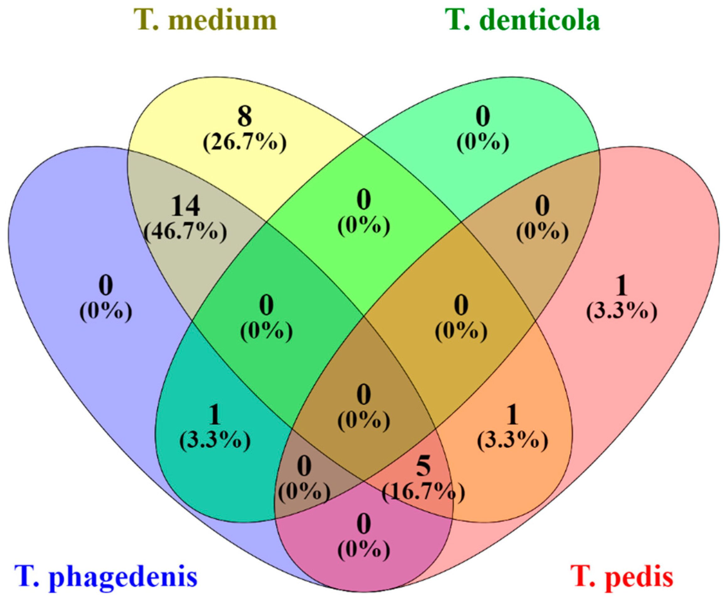
2. Results
3. Discussion
4. Materials and Methods
4.1. Study Design
4.2. Study Population
4.3. Collection of Samples
4.4. Bacteriological Analysis of Lesions Biopsies
4.4.1. Extraction of DNA
4.4.2. Molecular Confirmation
4.4.3. Descriptive Data Analysis
Author Contributions
Funding
Institutional Review Board Statement
Data Availability Statement
Acknowledgments
Conflicts of Interest
References
- Sullivan, L.E.; Evans, N.J.; Blowey, R.W.; Grove-White, D.H.; Clegg, S.R.; Duncan, J.S.; Carter, S.D. A molecular epidemiology of treponemes in beef cattle digital dermatitis lesions and comparative analyses with sheep contagious ovine digital dermatitis and dairy cattle digital dermatitis lesions. Vet Microbiol. 2015, 178, 77–87. [Google Scholar] [CrossRef]
- Moreira, T.F.; Facury Filho, E.J.; Carvalho, A.U.; Strube, M.L.; Nielsen, M.W.; Klitgaard, K.; Jensen, T.K. Pathology and bacteria related to digital dermatitis in dairy cattle in all year-round grazing system in Brazil. PLoS ONE 2018, 13, e0193870. [Google Scholar] [CrossRef] [PubMed]
- Guccione, J.; Carcasole, C.; Alsaaod, M.; D’Andrea, L.; Di Loria, A.; De Rosa, A.; Ciaramella, P.; Steiner, A. Assessment of foot health and animal welfare: Clinical findings in 229 dairy Mediterranean Buffaloes (Bubalus bubalis) affected by foot disorders. BMC Vet. Res. 2016, 14, 107. [Google Scholar] [CrossRef] [PubMed]
- Clegg, S.R.; Mansfield, K.G.; Newbrook, K.; Sullivan, L.E.; Blowey, R.W.; Carter, S.D.; Evans, N.J. Isolation of digital dermatitis treponemes from hoof lesions in Wild North American Elk (Cervus elaphus) in Washington State, USA. J. Clin. Microbiol. 2015, 53, 88–94. [Google Scholar] [CrossRef]
- Hoby, S.; Jensen, T.K.; Brodard, I.; Gurtner, C.; Eicher, R.; Steiner, A.; Kuhnert, P.; Alsaaod, M. Detection of treponemes in digital dermatitis lesions of captive European bison (Bison bonasus). PLoS ONE 2021, 9, 16. [Google Scholar] [CrossRef]
- Demirkan, I.; Güzel, N. An outbreak of digital dermatitis in Turkish dairy cattle. Indian Vet. J. 2004, 81, 1331–1333. [Google Scholar]
- Vermunt, J.J.; Hill, F.I. Papillomatous digital dermatitis in a Holstein-Friesian bull. N. Z. Vet. J. 2004, 52, 99–101. [Google Scholar] [CrossRef] [PubMed]
- Cramer, G.; Lissemore, K.D.; Guard, C.L.; Leslie, K.E.; Kelton, D.F. Herd-level risk factors for seven different foot lesions in Ontario Holstein cattle housed in tie stalls or free stalls. J. Dairy Sci. 2009, 92, 1404–1411. [Google Scholar] [CrossRef] [PubMed]
- Rodriguez-Lainz, A.; Melendez-Retamal, P.; Hird, D.W.; Read, D.H. Papillomatous digital dermatitis in Chilean dairies and evaluation of a screening method. Prev. Vet. Med. 1998, 37, 197–207. [Google Scholar] [CrossRef]
- Flor, E.; Tadich, N. Claudicaciones en vacas de rebaños lecheros grandes y pequeños del sur de Chile. Arch. Med. Vet. 2008, 40, 125–134. [Google Scholar] [CrossRef][Green Version]
- Wilson-Welder, J.H.; Alt, D.P.; Nally, J.E. Digital dermatitis in cattle: Current bacterial and immunological findings. Animals 2015, 5, 1114–1135. [Google Scholar] [CrossRef] [PubMed]
- Marcatili, P.; Nielsen, M.W.; Sicheritz-Pontén, T.; Jensen, T.K.; Schafer-Nielsen, C.; Boye, M.; Nielsen, M.; Klitgaard, K. A novel approach to probe host-pathogen interactions of bovine digital dermatitis, a model of a complex polymicrobial infection. BMC Genom. 2016, 17, 987. [Google Scholar] [CrossRef]
- Alsaaod, M.; Locher, I.; Jores, J.; Grimm, P.; Brodard, I.; Steiner, A.; Kuhnert, P. Detection of specific treponema species and dichelobacter nodosus from digital dermatitis (Mortellaro’s disease) lesions in Swiss cattle. Schweiz Arch. Tierh. 2019, 161, 207–215. [Google Scholar] [CrossRef] [PubMed]
- Han, S.; Mansfield, K.G.; Bradway, D.S.; Besser, T.E.; Read, D.H.; Haldorson, G.J.; Alt, D.P.; Wilson-Welder, J.H. Treponeme-Associated Hoof Disease of Free-Ranging Elk (Cervus elaphus) in Southwestern Washington State, USA. Vet. Pathol. 2019, 56, 118–132. [Google Scholar] [CrossRef] [PubMed]
- Mamuad, L.L.; Seo, B.J.; Faruk, M.S.A.; Espiritu, H.M.; Jin, S.J.; Kim, W.I.; Lee, S.S.; Cho, Y.I. Treponema spp., the dominant pathogen in the lesion of bovine digital dermatitis and its characterization in dairy cattle. Vet. Microbiol. 2020, 245, 108696. [Google Scholar] [CrossRef]
- Laven, R.A.; Logue, D.N. Treatment strategies for digital dermatitis for the UK. Vet. J. 2006, 171, 79–88. [Google Scholar] [CrossRef]
- Refaai, W.; van Aert, M.; Abd El-Aal, A.M.; Behery, A.E.; Opsomer, G. Infectious diseases causing lameness in cattle with a main emphasis on digital dermatitis (Mortellaro disease). Livest. Sci. 2013, 156, 53–63. [Google Scholar] [CrossRef]
- Clegg, S.R.; Bell, J.; Ainsworth, S.; Blowey, R.W.; Bell, N.J.; Carter, S.D.; Evans, N.J. Isolation of digital dermatitis treponemes from cattle hock skin lesions. Vet. Dermatol. 2016, 27, 106–112.e29. [Google Scholar] [CrossRef]
- Speers, D.J. Clinical applications of molecular biology for infectious diseases. Clin. Biochem. Rev. 2006, 27, 39–51. [Google Scholar]
- Evans, N.J.; Brown, J.M.; Demirkan, I.; Murray, R.D.; Vink, W.D.; Blowey, R.W.; Hart, C.A.; Carter, S.D. Three unique groups of spirochetes isolated from digital dermatitis lesions in UK cattle. Vet. Microbiol. 2008, 130, 141–150. [Google Scholar] [CrossRef]
- Evans, N.J.; Brown, J.M.; Demirkan, I.; Singh, P.; Getty, B.; Timofte, D.; Vink, W.D.; Murray, R.D.; Blowey, R.W.; Birtles, R.J.; et al. Association of unique, isolated treponemes with bovine digital dermatitis lesions. J. Clin. Microbiol. 2009, 47, 689–696. [Google Scholar] [CrossRef] [PubMed]
- Akin, I.; Akin, T. Economic impact of digital dermatitis treatment on a dairy farm: An application of the break-even analysis. Cienc. Rural 2018, 48, 8. [Google Scholar] [CrossRef]
- Alonso, M.E.; González-Montaña, J.R.; Lomillos, J.M. Consumers’ Concerns and Perceptions of Farm Animal Welfare. Animals 2020, 10, 385. [Google Scholar] [CrossRef] [PubMed]
- Cook, N. LifeStep—A Lesion Oriented, Life Cycle Approach to Preventing Lameness in Dairy Herds. In Proceedings of the KvaegKongress, Herning, Denmark, 1 February 2016; pp. 1–11. Available online: https://www.landbrugsinfo.dk/kvaeg/dansk-kvaeg-kongres/sider/bilag-kk-nigel-b-cook-02.pdf (accessed on 20 January 2022).
- Willis, S.G.; Smith, K.S.; Dunn, V.L.; Gapter, L.A.; Riviere, K.H.; Riviere, G.R. Identification of seven Treponema species in health- and disease-associated dental plaque by nested PCR. J. Clin. Microbiol. 1999, 37, 867–869. [Google Scholar] [CrossRef] [PubMed]
- Wilson-Welder, J.H.; Elliott, M.K.; Zuerner, R.L.; Bayles, D.O.; Alt, D.P.; Stanton, T.B. Biochemical and molecular characterization of Treponema phagedenis-like spirochetes isolated from a bovine digital dermatitis lesion. BMC Microbiol. 2013, 13, 280. [Google Scholar] [CrossRef] [PubMed]
- Beninger, C.; Naqvi, S.A.; Naushad, S.; Orsel, K.; Luby, C.; Derakhshani, H.; Khafipour, E.; De Buck, J. Associations between digital dermatitis lesion grades in dairy cattle and the quantities of four Treponema species. Vet. Res. 2018, 49, 10–12. [Google Scholar] [CrossRef]
- Zinicola, M.; Lima, F.; Lima, S.; Machado, V.; Gomez, M.; Döpfer, D.; Guard, C.; Bicalho, R. Altered microbiomes in bovine digital dermatitis lesions, and the gut as a pathogen reservoir. PLoS ONE 2015, 10, 1–23. [Google Scholar] [CrossRef]
- Krull, A.C.; Shearer, J.K.; Gorden, P.J.; Cooper, V.L.; Phillips, G.J.; Plummera, P.J. Deep sequencing analysis reveals temporal microbiota changes associated with development of bovine digital dermatitis. Infect. Immun. 2014, 82, 3359–3373. [Google Scholar] [CrossRef]
- Klitgaard, K.; Foix Bretó, A.; Boye, M.; Jensen, T.K. Targeting the treponemal microbiome of digital dermatitis infections by high-resolution phylogenetic analyses and comparison with fluorescent in situ hybridization. J. Clin. Microbiol. 2013, 51, 2212–2219. [Google Scholar] [CrossRef]


| ID | Farm | Lameness Prev. % | DD Prev. % | Treponema | T. Phagedenis | T. Medium | T. Denticola | T. Pedis |
|---|---|---|---|---|---|---|---|---|
| 31 | 7 | 20 | 10 | + | + | + | − | + |
| 582 | 2 | + | + | + | − | − | ||
| 1033 | 11 | 10 | 7 | + | + | + | − | + |
| 1359 | 14 | 8 | 5 | + | + | + | − | + |
| 1746 | 1 | 15 | 15 | + | + | + | − | − |
| 1784 | 10 | 7 | 6 | + | + | + | − | − |
| 2152 | 7 | 20 | 10 | + | + | + | − | + |
| 222-7986 | 1 | 15 | 15 | + | + | + | − | − |
| 3065 | 10 | 7 | 6 | + | + | − | + | − |
| 3310 | 6 | + | + | + | − | − | ||
| 4213 | 14 | 8 | 5 | + | + | + | − | − |
| 4258 | 13 | 7 | 6 | + | + | + | − | − |
| 4352 | 7 | 20 | 10 | + | + | + | − | − |
| 4552 | 7 | 20 | 10 | + | + | + | − | − |
| 5415 | 8 | 7 | 6 | + | + | + | − | − |
| 5841 | 14 | 8 | 5 | + | − | + | − | − |
| 5877 | 14 | 8 | 5 | + | − | + | − | − |
| 7871 | 12 | 5 | 5 | + | + | + | − | − |
| 8865 | 5 | 15 | 15 | + | − | + | − | − |
| 9235 | 3 | 7 | 6 | + | − | + | − | − |
| 943-6669 | 14 | 8 | 5 | + | + | + | − | − |
| 9456 | 3 | 7 | 6 | + | − | + | − | + |
| 9505 | 7 | 20 | 10 | + | − | + | − | − |
| 9508 | 7 | 20 | 10 | + | − | + | − | − |
| 9542 | 4 | + | − | + | − | − | ||
| 9585 | 7 | 20 | 10 | + | − | + | − | − |
| 9766 | 14 | 8 | 5 | + | − | − | − | + |
| Percentage | 100% | 63% | 93% | 3% | 22% |
Publisher’s Note: MDPI stays neutral with regard to jurisdictional claims in published maps and institutional affiliations. |
© 2022 by the authors. Licensee MDPI, Basel, Switzerland. This article is an open access article distributed under the terms and conditions of the Creative Commons Attribution (CC BY) license (https://creativecommons.org/licenses/by/4.0/).
Share and Cite
Canales, N.; Bustamante, H.; Wilson-Welder, J.; Thomas, C.; Ramirez, E.; Salgado, M. First Molecular Confirmation of Treponema spp. in Lesions Consistent with Digital Dermatitis in Chilean Dairy Cattle. Pathogens 2022, 11, 510. https://doi.org/10.3390/pathogens11050510
Canales N, Bustamante H, Wilson-Welder J, Thomas C, Ramirez E, Salgado M. First Molecular Confirmation of Treponema spp. in Lesions Consistent with Digital Dermatitis in Chilean Dairy Cattle. Pathogens. 2022; 11(5):510. https://doi.org/10.3390/pathogens11050510
Chicago/Turabian StyleCanales, Nivia, Hedie Bustamante, Jennifer Wilson-Welder, Cristian Thomas, Emilio Ramirez, and Miguel Salgado. 2022. "First Molecular Confirmation of Treponema spp. in Lesions Consistent with Digital Dermatitis in Chilean Dairy Cattle" Pathogens 11, no. 5: 510. https://doi.org/10.3390/pathogens11050510
APA StyleCanales, N., Bustamante, H., Wilson-Welder, J., Thomas, C., Ramirez, E., & Salgado, M. (2022). First Molecular Confirmation of Treponema spp. in Lesions Consistent with Digital Dermatitis in Chilean Dairy Cattle. Pathogens, 11(5), 510. https://doi.org/10.3390/pathogens11050510