Review

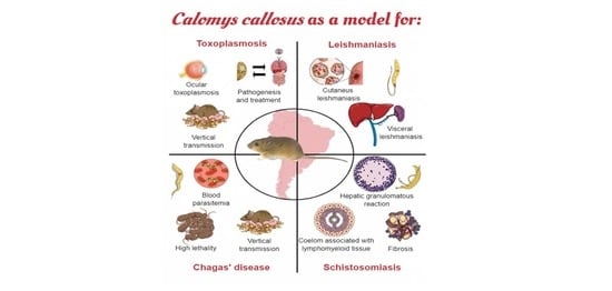
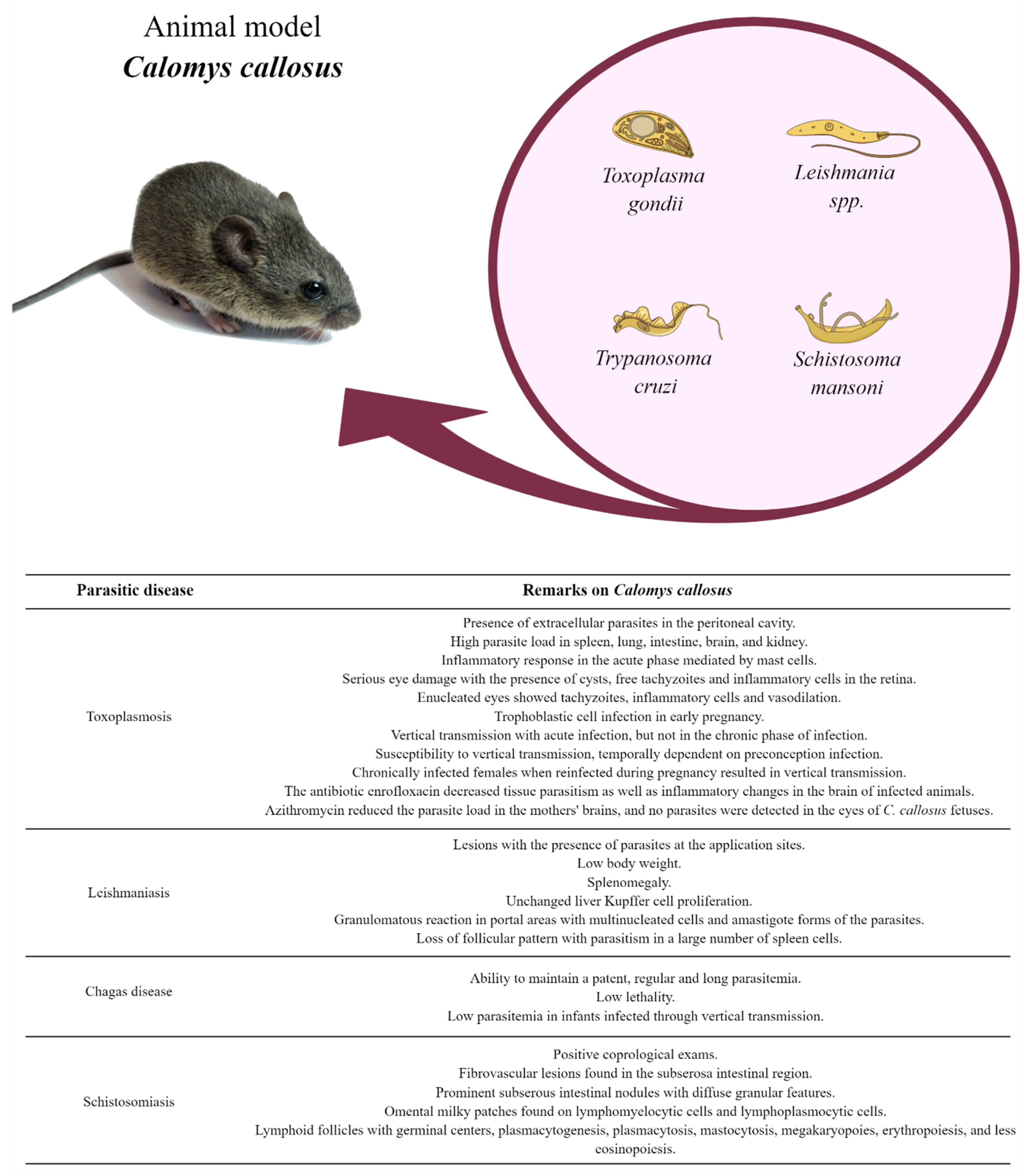
Calomys callosus: An Experimental Animal Model Applied to Parasitic Diseases Investigations of Public Health Concern

by
Rafael Borges Rosa
1,
Mylla Spirandelli da Costa
1,
Samuel Cota Teixeira
2,
Emilene Ferreira de Castro
1,
Willyenne Marília Dantas
3,
Eloisa Amália Vieira Ferro
2 and
Murilo Vieira da Silva
1,*
1
Rodents Animal Facilities Complex, Federal University of Uberlandia, Uberlandia 38400-902, Brazil
2
Institute of Biomedical Sciences, Federal University of Uberlandia, Uberlandia 38405-318, Brazil
3
Institute Aggeu Magalhães, Fiocruz Pernambuco, Recife 50740-465, Brazil
*
Author to whom correspondence should be addressed.
Pathogens 2022, 11(3), 369; https://doi.org/10.3390/pathogens11030369
Submission received: 2 February 2022 / Revised: 7 March 2022 / Accepted: 14 March 2022 / Published: 17 March 2022
(This article belongs to the Special Issue Advances in Parasitic Diseases)

Abstract

:
The appearance and spread of parasitic diseases around the world aroused the interest of the scientific community to discover new animal models for improving the quality and specificity of surveys. Calomys callosus is a rodent native to South America, an easy handling model, with satisfactory longevity and reproducibility. C. callosus is susceptible to toxoplasmosis and can be used as experimental model for the study the pathogenesis, treatment, vertical transmission, and ocular toxoplasmosis. C. callosus can also be used to study cutaneous and visceral leishmaniasis, as the animals present cutaneous lesions, as well as parasites in the organs. C. callosus has epidemiological importance in Chagas disease, and since it is a Trypanosoma cruzi natural host in which rodents show high parasitemia and lethality, they are also effective as a model of congenital transmission. In the study of schistosomiasis, Schistosoma mansoni was proven to be a C. callosus natural host; thus, this rodent is a great model for fibrosis, hepatic granulomatous reaction, and celloma associated with lymphomyeloid tissue (CALT) during S. mansoni infection. In this review, we summarize the leading studies of parasitic diseases that used C. callosus as a rodent experimental model, describing the main uses and characteristics that led them to be considered an effective model.

Graphical Abstract

1. Introduction

Calomys callosus is a rodent species that belongs to the Cricetidae family, is native to South America, and is similar in size to mice [1,2,3,4]. The introduction of C. callosus as a laboratory animal occurred in 1967 by Petter et al. [5]. Since they were bred for this purpose, these rodents have characteristics such as easy handling, satisfactory longevity, and reproducibility [6,7,8]. Currently the biology, behavior, and anatomy of C. callosus are well known, as the species has been established in experimental laboratories and is considered an effective model to be used in infectious disease studies [5,9,10].
Studies with C. callosus showed that these animals are considered an adequate experimental model for investigating the dynamics of the acute phase and the acquired, ocular, and congenital forms of toxoplasmosis. Research has also shown that C. callosus is a tolerant natural host to Trypanosoma cruzi infection, in addition to being considered an alternative model in leishmaniasis studies since it can be naturally infected by Schistosoma mansoni [11,12,13,14,15,16,17,18].
Despite all these works involving the use of C. callosus as a great model for the study infectious diseases of public health concern, this rodent is still underused. In this review, we summarize the main studies of parasitic diseases (Supplementary Table S1) that used C. callosus as a rodent experimental model and discuss the principal uses and characteristics that led these animals to be considered an effective model.
Our review has as its main objective the presentation of the applicability of the experimental model C. callosus in the study of different parasitic diseases of public health importance—diseases that burden health systems and cause significant mortality. Therefore, aiming to illustrate this variability in the application of the model, we used four parasites with marked pathophysiological differences, being one Apicomplexa, two Trypanosomatids, and one Helminth).

2. Toxoplasmosis

Toxoplasma gondii, an obligate intracellular protozoan parasite, is the etiologic agent of toxoplasmosis, one of the most common zoonotic food-borne infection that affects up to one-third of the global human population [19]. Acquired infection with T. gondii is usually asymptomatic in healthy individuals; however, the primary infection or reactivation of chronic infection, caused by this opportunistic human pathogen, can cause severe clinical features in immunocompromised individuals, human fetuses, and newborns [20,21].
T. gondii has a notable ability to infect efficiently target organs, including brain, eyes, and placenta [22], which configures a limiting factor for obtaining infected human material. Thus, it is mandatory to use experimental animal models capable of mimicking the course of toxoplasmosis in order to elucidate several aspects of the mechanisms responsible for pathological lesions associated with the clinical disease.
In this context, a considerable number of studies have shown the rodent C. callosus to be an efficient experimental model for investigating the underlying mechanisms associated with T. gondii infection. For this reason, we consider it appropriate to compile these studies addressing a current view, for the first time, of the remarkable findings involving the use of C. callosus as an experimental model, especially for understanding the acute phase and the acquired, ocular, and congenital forms of the toxoplasmosis.

2.1. Acute Phase of Toxoplasmosis

The first study addressing the use of the C. callosus as a promising model to investigate the experimental infection by T. gondii was published in 1998 by Favoreto-Junior et al. In this work, C. callosus intraperitoneally infected by T. gondii tachyzoites of RH strain were demonstrated to be highly susceptible to parasitic infection, which was characterized by a massive presence of extracellular parasites in the peritoneal cavity and by a high parasite load in spleen, lung, intestine, brain, and kidney [23]. In a later study, C. callosus was demonstrated to be an interesting tool for investigating the role of mast cells in the acute phase of inflammatory response caused by T. gondii [24]. Thus, these early works showing C. callosus as a host with susceptibility to T. gondii infection initiated new ways of investigating in depth the parasitic acute infection pathogenesis, as well as the host immune response during toxoplasmosis. In this scenario, Franco et al. (2014) studied the different rate of susceptibility between C. callosus females and males during acute infection of Brazilian atypical T. gondii strains. These authors demonstrated that this rodent is susceptible to parasite infection with gender differences in severity of infection, regardless of T. gondii strain [25].

2.2. Acquired and Congenital Ocular Toxoplasmosis

In humans, T. gondii is an important cause of ocular disease in immunosuppressed and immunocompetent individuals, especially in newborns [26]. The ocular toxoplasmosis is one of the most common clinical manifestations caused by T. gondii infection, which results in retinochoroiditis in 75–80% of cases, while acquired systemic infection can cause ocular disease in 1% to 3% of individuals, specifically the unilateral form [27,28]. However, although ocular lesions in patients with acquired toxoplasmosis appear to be uncommon, several studies have reported a high incidence of ocular disease during toxoplasmosis outbreaks [29,30]. Despite the importance, the knowledge about the complex pathogenesis of ocular toxoplasmosis is still unclear [31], thus, highlighting the urgency to find new study models for understanding this disease.
In this scenario, Pereira et al. (1999) demonstrated that males and virgin females (acquired form) and pregnant females (congenital form) of C. callosus orally infected with T. gondii cysts (ME49 strain) developed severe ocular lesions, which were associated with the presence of cysts, free tachyzoites, and inflammatory cells in the retina. Interestingly, as shown in humans, the authors reported that most animals with acquired systemic toxoplasmosis had unilateral ocular cysts post-infection [32].
About the pathogenesis of ocular toxoplasmosis, it was shown that C. callosus enucleated eyes presented a high number of mast cells after intraperitoneal or conjunctival inoculation with T. gondii tachyzoites (RH strain) [33]. Surprisingly, some studies have implicated the presence of ocular mast cells and their connection to hypersensitivity reactions with development of human ocular toxoplasmosis [31,34]. Thus, due to both the difficulty in obtaining human eyes removed from patients with ocular toxoplasmosis and the great similarity in comparison with human samples, the rodent C. callosus has emerged as a remarkable experimental model for studying mechanisms associated with ocular diseases, especially ocular toxoplasmosis.

2.3. Congenital Toxoplasmosis

The search for experimental models for assessing congenital toxoplasmosis have been explored since the 1950s [35,36]. In humans, the vertical transmission of T. gondii from the mother to the fetus can result in fetal death, abortion, or severe clinical manifestation in infected fetuses and newborns, including neurologic damage, neurocognitive deficits, or chorioretinitis, among others [21,37]. In this context, a pioneering study by Ferro et al. (1999), using C. callosus as experimental model, demonstrated that T. gondii is capable of infecting trophoblast cells in the early stage of pregnancy, highlighting the trophoblast role as a possible route of access from maternal to embryonic tissues [38].
It was reported that T. gondii vertical transmission occurs in acutely infected pregnant C. callosus, but not in the infection’s chronic phase [11]. Furthermore, as shown in pregnant women, the susceptibility to T. gondii vertical transmission is temporally dependent on preconceptional infection in C. callosus—i.e., parasitic infection acquired during or just before pregnancy can result in vertical transmission in this model [39]. For years, several studies showed that primary T. gondii infection is capable of leading a life-long immunity that can prevent vertical transmission [40,41]. However, some studies have reported cases of congenital toxoplasmosis in fetuses to immunocompetent mothers who had been infected with the parasite before conception [42,43,44,45]. In line with this discussion, Franco et al. (2015) showed that C. callosus females chronically infected with T. gondii when reinfected during pregnancy resulted in vertical transmission [46].
Despite the existence of some drugs for treating human congenital toxoplasmosis, it is widely known that these compounds are associated with serious side effects to both mothers and fetuses [20,47]. Moreover, the treatment options available for congenital toxoplasmosis suppress the active infection but do not cure the latent infection [48,49]. Thus, the conventional therapy is still limited, which affects mortality and quality of life on pregnancy and newborn health [50]. For this reason, the search for new bioactive substances for congenital toxoplasmosis treatment gathered great interest in the past few decades [51,52,53,54]. In this scenario, some studies have suggested the use of C. callosus as an interesting experimental model for assessing the efficacy of new possible compounds that have remarkable activities for controlling T. gondii infection, as well as for inhibiting vertical transmission [55,56,57].

3. Leishmaniasis

Leishmaniasis is an infectious disease caused by a protozoa parasite, Leishmania sp., that affects economically vulnerable populations and represents a significant problem in public health because it is the second most neglected tropical disease in mortality and seventh in disability [58]. Leishmania parasites are transmitted through the bites of infected female phlebotomine sandflies to more than 70 animal species, including humans [59].
Highly endemic regions such as Algeria, Brazil, Iraq, Somalia, and Sudan have more than 1 billion people who are at risk of infection. It is estimated that there are 700,000–1.2 million new cases and 14,000–40,000 deaths from the disease each year worldwide. Available data from 2018 show that out of the 200 countries and territories that reported to the World Health Organization, 97 (49%) were considered endemic for leishmaniasis [60]. In other words, it is a widespread concern around the world.
Leishmaniasis presents itself in three clinical forms: visceral leishmaniasis (VL)—affects the internal organs and causing more systemic symptoms such as fever, swelling of liver and spleen, weight loss, and petechiae; cutaneous leishmaniasis (CL)—is the most common form and affects the skin, causing a small lump or sores to appear at the bite site; and mucocutaneous leishmaniasis—leads to partial or total destruction of mucous membranes of the nose, mouth, and throat [61].

3.1. Visceral Leishmaniasis (VL)

Also known as kala-azar, this leishmaniasis clinical form is fatal if not treated in over 95% of cases, and it presents irregular bouts of fever, weight loss, spleen and liver enlargement, and anemia. The locations with higher incidence are Brazil, East Africa, and India. An estimated 50,000 to 90,000 new cases of VL occur worldwide annually, but only 25% to 45% are reported to WHO. It remains one of the neglected parasitic diseases, with outbreak and mortality potential. In 2019, more than 90% of new cases reported to WHO occurred in 10 countries: Brazil, Ethiopia, Eritrea, India, Iraq, Kenya, Nepal, Somalia, South Sudan, and Sudan [60].
Experiments performed using Leishmania infantum, the etiological agent of VL, demonstrated that after 3 months of infection C. callosus presented hepato and splenomegaly, also parasite amastigote forms were found intra (macrophages) and extracellularly in the bone marrow, liver and spleen smears of infected rodents [17]. C. callosus could also be useful as an experimental animal model for VL, due to histopathological and visceral alterations promoted after infection.

3.2. Cutaneous Leishmaniasis (CL)

CL is the most common leishmaniasis form and causes skin lesions, mainly ulcers on exposed body parts, leaving life-long scars and serious disability or stigma. About 95% of CL cases occur in the Americas, the Mediterranean basin, the Middle East, and Central Asia. In 2019, over 87% of new CL cases occurred in 10 countries: Afghanistan, Algeria, Brazil, Colombia, Iran (Islamic Republic of), Iraq, Libya, Pakistan, the Syrian Arab Republic, and Tunisia. It is estimated that between 600,000 to 1 million new cases occur worldwide annually [60].
Mello (1978) investigated the infection of some parasitic strains, including Leishmania amazonensis, isolated from the wound of a patient affected by CL. C. callosus presented lesions at the application sites from the 40th day post-infection, and the parasite presence was confirmed by lesion smear [16]. This rodent has the ability to reproduce CL lesions, proving to be a decent model for the study of this leishmaniasis clinical form.
Despite the fact that mice and hamsters are still the most commonly used rodents as experimental animal models in leishmaniosis studies [62,63,64,65] and that there are few studies using C. callosus, here we demonstrate that C. callosus should be potentially used as an experimental model in leishmaniasis research [16,17].

4. Chagas Disease

Chagas disease, also known as American trypanosomiasis, is a potentially fatal disease caused by the protozoan parasite Trypanosoma cruzi [66]. It is estimated that 6 million people worldwide are infected, mainly in the endemic areas of 21 countries in Latin America. Mortality associated with the disease is higher than other parasitic diseases, causing approximately 10,000 deaths per year, in addition to more than 75 million people who are exposed to the risk of contracting the infection [67].
T. cruzi was discovered and described for the first time by the Brazilian physician and scientist Carlos Chagas, who investigated the hematophagous Panstrongylus megistus. Monkeys of the species Callithrix penicillata were exposed to the bite of vectors collected in the field, and it was found through hematological analysis that the animals were infected with the parasite of the genus Trypanosoma, whose life cycle was later elucidated and then identified as the species T. cruzi, now known as the etiologic agent of Chagas disease [66,68].
The main forms of Chagas disease transmission are contact with infected triatomines feces after being bitten and oral ingestion of contaminated food with parasites from infected triatomines (açai, sugar cane, etc.); congenital transmission occurs by the parasites passage from women infected with T. cruzi to their infants during pregnancy or childbirth, by blood transfusion or organ transplantation from infected donors to healthy recipients, and by accidental contact by injured skin or mucous membranes with contaminated material during handling in the laboratory [69].
Chagas disease has two distinct phases: acute and chronic. The prognosis and symptoms for these phases are different [70]. The acute phase is characterized by trypomastigotes presence circulating in the peripheral blood, prolonged fever, headache, severe weakness, and swelling of the face and legs; physiological changes are edema, lymphadenopathy, anemia, hepatosplenomegaly, abnormalities in the electrocardiogram (ECG), involvement of the central nervous system, and death [71]. The chronic phase usually starts with an asymptomatic period; if progressing, individuals may have cardiac involvement, fever, cardiomegaly, apical aneurysms, and/or ECG abnormalities. In addition, fever, megaesophagus, megacolon due to myenteric plexus destruction, and constipation [72].
For the last 50 years, the drugs of choice have been benznidazole and nifurtimox for T. cruzi infection treatment. However, they have a long period of treatment and a cure rate around 70%, in addition to high liver and kidney toxicity and being contraindicated for pregnant women. Adverse reactions such as allergic dermatitis, peripheral neuropathy, anorexia, weight loss, and insomnia are some of the main reasons why many patients drop out of treatment. The search for new drug treatments involves more effective and less toxic drugs to replace the usual and also studies to create a vaccine have been developed [68].
C. callosus has epidemiological importance in Chagas disease since it is a T. cruzi natural host and can be considered a good experimental laboratory model due to its high permissiveness to T. cruzi infection and its ability to maintain a patient, regular, and long-term parasitemia [73,74,75]. Although Rattus rattus, Mus musculus, and the opossums of the genus Didelphis spp. can become infected with T. cruzi, as demonstrated in several studies, they do not always represent the most important mammalian species for the maintenance of the parasite [76].
Historically, the natural occurrence of T. cruzi in Calomys rodent was first described in 1976 by Basso et al., who reported the parasite in Calomys musculinus captured in a rural area of the province of Córdoba, Argentina [77]. In Brazil, Mello and Teixeira (1977) reported the presence of T. cruzi in wild Calomys expulsus captured in Formosa, state of Goiás [78], and also identified a flagellated protozoan belonging to the sub-genus Herpetosoma in C. callosus wild. After evaluating other studies, the authors considered the parasite found in C. callosus to be the species Trypanosoma lewisi [79].
The collected strain of T. lewisi was experimentally inoculated into C. callosus under laboratory conditions, and subsequently, the blood of the animals was examined on alternate days. The evaluations showed that the animals were positive for the infection. It was also found that the rodents had high blood parasitemia that disappeared between 45 and 60 days after inoculation, demonstrating the ability to infect C. callosus [79].
Although Mello (1978) believed that there was a relationship of parasite–host specificity, it is currently known that T. Lewisi, although it has a higher rate of infection for Rattus rattus, is also capable of infecting several other wild rodent species [80]. The same has also been observed for primates [81], including man [82]. Therefore, it can be said that there is no parasite–host specificity.

4.1. Parasitemia and Lethality

In order to study C. callosus susceptibility to T. cruzi infection and characterize the prepatency period, rodents were infected with four different strains: R52, R64, R65, and M226. Daily blood tests were performed to detect trypomastigotes and follow the parasitemia evolution. R52, R64, and R65 strains present high parasitemia peaks and low lethality, while M226 strain showed low parasitemia peaks and lethality was zero [74]. Despite the different results between strains, the study showed positive results, concluding that the infective capacity was wielded in C. callosus regardless of strain and confirming this rodent as a good experimental model for Chagas disease [75].

4.2. Congenital Transmission

Mello and Borges (1981) also described the ability of C. callosus females to transmit the infection to their offspring. Female rodents in Chagas disease chronic phase started to reproduce, and all newborns were submitted to direct analysis of fresh blood and presented positive parasitemia. Although the transmission rate was low compared with intraperitoneal infection, in order to investigate T. cruzi in C. callosus different tissues, as in heart and leg muscle, infected animals have their organs collected between the 9th and 30th post infection day. Histopathology results presented lesions and parasite infiltration in cardiac and skeletal muscles [83]. Even with the low parasitism rate, it was possible to observe that there was T. cruzi congenital transmission and that these animals had infection sequelae, such as cardiac and muscle injuries. Thus, C. callosus can also be used as an animal model for congenital transmission for Chagas disease.

5. Schistosomiasis

Schistosomiasis is an infectious parasitic disease caused by trematode worms of Schistosoma genus. Considered a neglected disease, schistosomiasis transmission has been reported from 78 countries [84]. Studies on this parasite have been taking place for several decades, such as in Brazil, which since the 1950s has brought great contributions to the clinical and epidemiological aspects of the disease [85].
Schistosomiasis is a disease of tropical and subtropical areas, such as Africa, the Caribbean, Brazil, Venezuela, Suriname, China, Indonesia, Philippines, Cambodia, and Corsica, whose main form of transmission occurs with the penetration of the larva in the skin during contact with the infested water. After infection, the larvae develop into adult schistosomes that will live in the host’s blood [86]. There are five main species that cause schistosomiasis in humans—S. mansoni, S. japonicum, S. mekongi, S. guineensis (causing intestinal schistosomiasis), and S. haematobium (causing urogenital schistosomiasis)—and infection occurs in areas with low health coverage [87].
After larval skin penetration, the larvae develop into adult schistosomes, and adult worms live in the blood vessels and release eggs in the feces or urine to continue the lifecycle; transmission occurs when eggs, which hatch in water, contaminate freshwater sources [87].
Symptoms of schistosomiasis are caused by the reaction of the body to the Schistosoma eggs and include intestinal schistosomiasis, liver enlargement, accumulation of fluid in the peritoneal cavity, hypertension of the abdominal blood vessels, and spleen enlargement. Additionally, the condition of urogenital schistosomiasis features hematuria, fibrosis of the bladder and ureter, bladder cancer, and kidney damage and can lead to infertility. The chronic phase of schistosomiasis can result in death [88].
Due to the importance of this disease for world public health, different animal experimentation models have emerged to study the biology, immunology, and pathogenesis of schistosomiasis, such as rodents, lagomorphs, and primates of different species [89,90]. Rodents tend to be the animals of choice, essentially due to their easy availability, fecundity, and susceptibility to experimental infection [91]. An animal experimentation model discovered for investigating Schistosoma is C. callosus, first proven in an investigation of natural infection in 1979 with S. mansoni [18].
Swiss mice and the rodent C. callosus were used in a study to compare the prepatent period of Schistosoma infection. The animals were infected via the transcutaneous route, simulating a transdermal infection. The animals were exposed to the cercariae from human isolate for 45 min; the coprological method evaluation showed that the prepatent period observed in C. callosus was 43–44 days, while in Swiss mice it occurred 40 days after exposition [16]. The evaluation showed that C. callosus have a prepatent period equal to that of mice, which are experimental models known to be widely used in laboratories. Considering that the prepatent period occurs, on average, from thirty to sixty days after infection in human patients [92], the ability of C. callosus to present clinical signs similar to those observed in clinical cases of these patients is observed.
C. callosus was also shown to be a great fibrosis experimental model when infected with S. mansoni; between 55–160 days post infection, prominent intestinal subserous nodules with granular features were observed, and some had hemorrhage after their removal [93] and developed a severe hepatic granulomatous reaction, suggesting signs of hypersensitivity [94].
During schistosomiasis, C. callosus present an omental reactivity in milky spots with patches that represent subsidiary foci of lymphomyeloid tissue-associated coelom (CALT), which was also observed in mice [95,96]. S. mansoni worms live predominantly in mesenteric vessels, and consequently, their released products can reach the mesenteric and omental microcirculation, which leads to CALT [97].
A study using C. callosus was carried out to evaluate the effect of schistosomiasis in CALT during the acute, transient, and chronic phase of infection. The administration route was percutaneous, where S. mansoni cercariae were introduced. Omental milky spots were found in lymphomyelocytic (42–90 days post infection) and lymphoplasmacytic cells (160 days post infection); also, the milky spots are the preferred site of the germinal center, mast cell maturation, and proliferation in C. callosus, as observed in rats [98,99].
Administration routes that mimic natural infection and susceptibility to S. mansoni were demonstrated to be efficient through animal experimentation with C. callosus, confirming an important role of this rodent’s use in schistosomiasis research.

6. Challenges in Maintaining C. callosus as an Experimental Model

Considering the studies presented in this review, it is evident that C. callosus as an experimental model has great relevance for understanding the pathophysiology of several infectious diseases caused by parasites from different genera and species, thus contributing to the development of prophylactic and therapeutic measures. However, some factors such as legal issues associated with biosafety and the environment, animal welfare, breeding, transport, adaptation, and quarantine and maintenance in captivity can be obstacles, but not an impediment to the model’s use [100].
C. callosus is a wild rodent, and its capture and maintenance for scientific research purposes is directly related to the legal aspects of each country and must be strictly enforced. In Brazil, the competent authority is the Biodiversity Authorization and Information System (SISBIO), which can provide authorization for C. callosus capture. In addition, the experimental protocol must be evaluated and approved by the ethics committee [101].
After the approval of legally responsible authorities with ethical principles, species capture, maintenance, and breeding must be carried out in animal facilities. C. callosus must fulfill a quarantine period to ensure the health of the researchers and technicians who handle these animals. Thus, all biosafety protocols must be adopted, such as the use of collective and individual protection equipment, preventing the transmission of diseases and accidents [102].
Clinical evaluation and sanitary control must be performed by a veterinarian as a guarantee of animal health, as well as of the quality of the results obtained with their use in experimental tests. In the case of animals infected with zoonoses, they must be treated to eliminate the infection [103].
In addition to ethical issues related to animal welfare, for experimental results to be reproducible and reliable, an environmental enrichment program must be adopted, considering the physiological needs of the species to maintain homeostasis [104]. For this, we suggest that an ethogram be applied and that an intense literature review be carried out on the natural behaviors of the species prior to the implementation of the environmental enrichment program. Additionally, it is important that the animal facility has conditions to offer this species luminosity, temperature, humidity, and noise within the limits of comfort.
As for the other factors related to the maintenance and breeding of C. callosus, in a summarized way, a management approach similar to that used for other rodents such as rats and mice must be adopted. In daily handling, for example, avoid picking up animals by the tail—the use of tubes or cupped hands is highly recommended. When transporting, use boxes that guarantee safety and comfort to the animals [105,106].
To maintain the heterozygosity of the established colony and to avoid frequent captures of specimens in the wild, it is recommended to adopt mating systems used for outbreed colonies in animal facilities, such as random mate, Poiley method, and Falconer method [107,108]. These systems have their limitations, but they are widely accepted. Furthermore, it is possible to consider the adoption of inbreeding and establishment of an inbred lineage from the monitoring and characterization of allele frequency, which creates a sub-colony. In this case, it is necessary that this information be evidenced in the articles in order to contribute to the tests’ reproducibility.

7. Conclusions

Toxoplasmosis, Chagas disease, leishmaniasis, and schistosomiasis are parasitic diseases with worldwide impact. For a long time, the scientific community has been dedicated to study these parasitosis regarding their parasite biology, mechanism of infection, disease development, and novel treatments. For this, it is essential to use appropriate animal experimentation models to make the research accurate and specific. C. callosus, a species native from South America, is an animal model capable of efficiently replicating these parasitic diseases—a model with easy handling and with satisfactory longevity and reproducibility (Figure 1). Although, it is not widely used, these animals are considered effective models for carrying out any study in the parasitic diseases area involving these four main parasites.

Supplementary Materials

The following supporting information can be downloaded at: https://www.mdpi.com/article/10.3390/pathogens11030369/s1, Table S1: Current findings using C. callosus as an experimental model to study parasitic diseases.

Author Contributions

R.B.R., M.S.d.C., S.C.T., E.F.d.C. and W.M.D. participated in the search for articles to be reviewed and carried out the writing of the review. M.S.d.C. designated graphical abstract. E.A.V.F. and M.V.d.S. participated in the supervision, writing, and review of this paper. All authors have read and agreed to the published version of the manuscript.

Funding

This work was supported by the following Brazilian research funding agencies and institutions: Conselho Nacional de Desenvolvimento Científico e Tecnológico (CNPq) (10/2020), Coordenação de Aperfeiçoamento de Pessoal de Nível Superior (CAPES) (10/2002), and Universidade Federal de Uberlândia–Pró-reitoria de Pesquisa e Pós-graduação (UFU-PROPP) (04/2014).

Institutional Review Board Statement

Not applicable.

Informed Consent Statement

Not applicable.

Data Availability Statement

Not applicable.

Acknowledgments

The authors thank Loyane Bertagnolli Coutinho and Sandra Gabriela Klein for their technical assistance and Carlos Henrique de Carvalho for his institutional support.

Conflicts of Interest

The authors declare no conflict of interest.

References

  1. Moojen, J. Os Roedores do Brasil; Instituto Nacional do Livro: Rio de Janeiro, Brazil, 1952. [Google Scholar]
  2. Hershkovitz, P. Evolution of Neotropical Cricetine Rodents (Muridae) with Special Reference to the Phyllotine Group; Chicago Natural History Museum: Chicago, IL, USA, 1962. [Google Scholar]
  3. Massoia, E.; Fornes, A. Nuevos dados sobre la morfologia distribucion geografica y etoecologia de Calomys callosus (Rengger) (Rodentia Cricetidae). Physis 1965, 25, 325. [Google Scholar]
  4. Mello, D.A.; Moojen, L.E. Nota sobre uma coleção de roedores e marsupiais superiores de algumas regiões do cerrado do Brasil central. Rev. Bras. Pesqui Med. Biol. 1979, 12, 287–291. [Google Scholar] [PubMed]
  5. Petter, F.; de Karimi Almeida, C.R. A new laboratory rodent, Cricetida Calomys callosus. Comptes Rendus Hebd. Séances L’académie Sci. 1967, 265, 1974–1976. [Google Scholar]
  6. Karimi, Y.; Almeida, C.R.; Petter, F. Note sur Les Rongeurs du Nord.-Est du Brésil. Mammalia 1976, 40, 257–266. [Google Scholar] [CrossRef]
  7. Mello, D.A. Calomys Callosus Rengger, 1930, (Rodentia-Cricetidae): Sua caracterização, distribuição, biologia, criação e manenjo de uma cepa em laboratório. Mem. Inst. Oswaldo Cruz 1984, 79, 37–44. [Google Scholar] [CrossRef] [Green Version]
  8. Mello, D.A. Studies on reproduction and logevity of Calomys callosus (Rosentia, Cricetidae) under laboratory conditions. Rev. Bras. Biol. 1981, 41, 841–843. [Google Scholar]
  9. Justines, G.; Johnson, K.M. Observations on the laboratory breeding of the cricetine rodent Calomys callosus. Lab. Anim. Care 1970, 20, 57–60. [Google Scholar]
  10. Vaz-de-Lima, L.R.; Kipnis, A.; Kipnis, T.L.; Dias-da-Silva, W. The complement system of Calomys callosus, Rengger, 1830 (Rodentia, Cricetidae). Braz. J. Med. Biol. Res. 1992, 25, 161–166. [Google Scholar]
  11. Ferro, E.A.V.; Silva, D.A.O.; Bevilacqua, E.; Mineo, J.R. Effect of Toxoplasma gondii infection kinetics on trophoblast cell population in Calomys callosus, a model of congenital toxoplasmosis. Infect. Immun. 2002, 70, 7089–7094. [Google Scholar] [CrossRef] [Green Version]
  12. Andrade, S.G.; Kloetzel, J.K.; Borges, M.M.; Ferrans, V.J. Morphological aspects of the myocarditis and myositis in Calomys callosus experimentally infected with Trypanosoma cruzi: Fibrogenesis and spontaneous regression of fibrosis. Mem. Inst. Oswaldo Cruz 1994, 89, 379–393. [Google Scholar] [CrossRef] [Green Version]
  13. Borges, M.M.; De Andrade, S.G.; Pilatti, C.G.; do Prado Júnior, J.C.; Kloetzel, J.K. Macrophage activation and histopathological findings in Calomys callosus and Swiss mice infected with several strains of Trypanosoma cruzi. Mem. Inst. Oswaldo Cruz 1992, 87, 493–502. [Google Scholar] [CrossRef] [PubMed] [Green Version]
  14. Oliveira, L.; Borges, M.M.; Leal, R.C.; Assreuy, J.; Kloetzel, J.K. Nitric oxide involvement in experimental Trypanosoma cruzi infection in Calomys callosus and Swiss mice. Parasitol. Res. 1997, 83, 762–770. [Google Scholar] [PubMed]
  15. Prado, J.C.J.; Levy, A.M.; Leal, M.P.; Bernard, E.; Kloetzel, J.K. Influence of male gonadal hormones on the parasitemia and humoral response of male Calomys callosus infected with the Y strain of Trypanosoma cruzi. Parasitol. Res. 1999, 85, 826–829. [Google Scholar] [CrossRef] [PubMed]
  16. Mello, D.A. Infecção Experimental de Calomys Callosus (Rengger, 1830), (Cricetidae-Rodentia) a quatro espécies de parasitos. Rev. Bras. Med. Trop. 1978, 13, 5–9. [Google Scholar] [CrossRef]
  17. Mello, D.A.; Teixeira, M.L. Infecção Experimental de Calomys Callosus (Rodentia-Cricetidae) com Leishmania Donovani Chagasi (Laison, 1982). Rev. Saúde Pública 1984, 18, 4–8. [Google Scholar] [CrossRef] [Green Version]
  18. Coelho, P.M.; Dias, M.; Mayrink, W.; Magalhães, P.; Mello, M.N.; Costa, C.A. Wild reservoirs of Schistosoma mansoni from Caratinga, an endemic schistosomiasis area of Minas Gerais State, Brazil. Am. J. Trop. Med. Hyg. 1979, 28, 163–164. [Google Scholar] [CrossRef]
  19. Dubey, J.P.; Jones, J.L. Toxoplasma gondii infection in humans and animals in the United States. Int. J. Parasitol. 2008, 38, 1257–1278. [Google Scholar] [CrossRef]
  20. Montoya, J.G.; Liesenfeld, O. Toxoplasmosis. Lancet 2004, 363, 145–158. [Google Scholar] [CrossRef]
  21. Aguirre, A.A.; Longcore, T.; Barbieri, M.; Dabritz, H.; Hill, D.; Klein, P.N.; Lepczyk, C.; Lilly, E.L.; McLeod, R.; Milcarsky, J.; et al. The One Health Approach to Toxoplasmosis: Epidemiology, Control, and Prevention Strategies. EcoHealth 2019, 16, 378–390. [Google Scholar] [CrossRef] [Green Version]
  22. Harker, K.S.; Ueno, N.; Lodoen, M.B. Toxoplasma gondii Dissemination: A Parasite’s Journey through the Infected Host. Parasite Immunol. 2015, 37, 141–149. [Google Scholar] [CrossRef]
  23. Favoreto, S.; Ferro, E.A.V.; Clemente, D.; Silva, D.A.O.; Mineo, J.R. Experimental Infection of Calomys callosus (Rodentia, Cricetidae) by Toxoplasma gondii. Mem. Inst. Oswaldo Cruz 1998, 93, 103–107. [Google Scholar] [CrossRef] [PubMed] [Green Version]
  24. Ferreira, G.L.S.; Mineo, J.R.; Oliveira, J.G.; Ferro, E.A.V.; Souza, M.A.; Santos, A.A.D. Toxoplasma gondii and mast cell interactions in vivo and in vitro: Experimental infection approaches in Calomys callosus (Rodentia, Cricetidae). Microbes Infect. 2004, 6, 172–181. [Google Scholar] [CrossRef] [PubMed]
  25. Franco, P.S.; Ribeiro, M.; Lopes-Maria, J.B.; Costa, L.F.; Silva, D.A.O.; De Freitas Barbosa, B.; de Oliveira Gomes, A.; Mineo, J.R.; Ferro, E.A.V. Experimental infection of Calomys callosus with atypical strains of Toxoplasma gondii shows gender differences in severity of infection. Parasitol. Res. 2014, 113, 2655–2664. [Google Scholar] [CrossRef] [PubMed]
  26. Park, Y.H.; Nam, H.W. Clinical features and treatment of ocular toxoplasmosis. Korean J. Parasitol. 2013, 51, 393–399. [Google Scholar] [CrossRef]
  27. Montoya, J.G.; Remington, J.S. Toxoplasmic chorioretinitis in the setting of acute acquired toxoplasmosis. Clin. Infect. Dis. 1996, 23, 277–282. [Google Scholar] [CrossRef]
  28. Butler, N.J.; Furtado, J.M.; Winthrop, K.L.; Smith, J.R. Ocular toxoplasmosis II: Clinical features, pathology and management. Clin. Exp. Ophthalmol. 2013, 41, 95–108. [Google Scholar] [CrossRef] [Green Version]
  29. Burnett, A.J.; Shortt, S.G.; Isaac-Renton, J.; King, A.; Werker, D.; Bowie, W.R. Multiple cases of acquired toxoplasmosis retinitis presenting in an outbreak. Ophthalmology 1998, 105, 1032–1037. [Google Scholar] [CrossRef]
  30. Balasundaram, M.B.; Andavar, R.; Palaniswamy, M.; Venkatapathy, N. Outbreak of acquired ocular toxoplasmosis involving 248 patients. Arch. Ophthalmol. 2010, 128, 206–207. [Google Scholar] [CrossRef] [Green Version]
  31. Smith, J.R.; Ashander, L.M.; Arruda, S.L.; Cordeiro, C.A.; Lie, S.; Rochet, E.; Belfort, R., Jr.; Furtado, J.M. Pathogenesis of ocular toxoplasmosis. Prog. Retin. Eye Res. 2020, 81, 100882. [Google Scholar] [CrossRef]
  32. Pereira, M.D.F.; Silva, D.A.O.; Ferro, E.A.V.; Mineo, J.R. Acquired and Congenital Ocular Toxoplasmosis Experimentally Induced in Calomys callosus (Rodentia, Cricetidae). Mem. Inst. Oswaldo Cruz 1999, 94, 103–114. [Google Scholar] [CrossRef] [Green Version]
  33. Gil, C.D.; Mineo, J.R.; Smith, R.L.; Oliani, S.M. Mast cells in the eyes of Calomys callosus (Rodentia: Cricetidae) infected by Toxoplasma gondii. Parasitol. Res. 2002, 88, 557–562. [Google Scholar] [CrossRef] [PubMed]
  34. Joulia, R.; Gaudenzio, N.; Rodrigues, M.; Lopez, J.; Blanchard, N.; Valitutti, S.; Espinosa, E. Mast cells form antibody-dependent degranulatory synapse for dedicated secretion and defence. Nat. Commun. 2015, 6, 6174. [Google Scholar] [CrossRef] [PubMed] [Green Version]
  35. Cowen, D.; Wolf, A. Experimental Congenital Toxoplasmosis. Toxoplasmosis in the offspring of mice infected by the vaginal route. Incid. Manif. Dis. 1950, 92, 417–429. [Google Scholar]
  36. Hutchison, W.M.; Hay, J.; Siim, J.C. A study of cataract in murine congenital toxoplasmosis. Ann. Trop. Med. Parasitol. 1982, 76, 53–70. [Google Scholar] [CrossRef] [PubMed]
  37. Hampton, M.M. Congenital toxoplasmosis: A review. Neonatal Netw. 2015, 34, 274–278. [Google Scholar] [CrossRef] [PubMed]
  38. Ferro, E.A.V.; Bevilacqua, E.; Favoreto-Junior, S.; Silva, D.A.O.; Mortara, R.A.; Mineo, J.R. Calomys callosus (Rodentia: Cricetidae) trophoblast cells as host cells to Toxoplasma gondii in early pregnancy. Parasitol. Res. 1999, 85, 647–654. [Google Scholar] [CrossRef] [PubMed]
  39. Barbosa, B.F.; Silva, D.A.O.; Costa, I.N.; Pena, J.D.O.; Mineo, J.R.; Ferro, E.A.V. Susceptibility to Vertical Transmission of Toxoplasma gondii is Temporally Dependent on the Preconceptional Infection in Calomys callosus. Placenta 2007, 28, 624–630. [Google Scholar] [CrossRef]
  40. Abou-Bacar, A.; Pfaff, A.W.; Letscher-Bru, V.; Filisetti, D.; Rajapakse, R.; Antoni, E.; Villard, O.; Klein, J.-P.; Candolfi, E. Role of gamma interferon and T cells in congenital Toxoplasma transmission. Parasite Immunol. 2004, 26, 315–318. [Google Scholar] [CrossRef]
  41. Pfaff, A.W.; Abou-Bacar, A.; Letscher-Bru, V.; Villard, O.; Senegas, A.; Mousli, M.; Candolfi, E. Cellular and molecular physiopathology of congenital toxoplasmosis: The dual role of IFN-γ. Parasitology 2007, 134, 1895–1902. [Google Scholar] [CrossRef]
  42. Dao, A.; Fortier, B.; Soete, M.; Plenat, F.; Dubremetz, J.F. Successful reinfection of chronically infected mice by a different Toxoplasma gondii genotype. Int. J. Parasitol. 2001, 31, 63–65. [Google Scholar] [CrossRef]
  43. Freyre, A.; Falcón, J.; Méndez, J.; Rodriguez, A.; Correa, L.; Gonzalez, M. Toxoplasma gondii: Partial cross-protection among several strains of the parasite against congenital transmission in a rat model. Exp. Parasitol. 2006, 112, 8–12. [Google Scholar] [CrossRef] [PubMed]
  44. Elbez-Rubinstein, A.; Ajzenberg, D.; Dardé, M.L.; Cohen, R.; Dumètre, A.; Yera, H.; Gondon, E.; Janaud, J.-C.; Thulliez, P. Congenital toxoplasmosis and reinfection during pregnancy: Case report, strain characterization, experimental model of reinfection, and review. J. Infect. Dis. 2009, 199, 280–285. [Google Scholar] [CrossRef] [PubMed]
  45. Pezerico, S.B.; Langoni, H.; Da Silva, A.V.; Da Silva, R.C. Evaluation of Toxoplasma gondii placental transmission in BALB/c mice model. Exp. Parasitol. 2009, 123, 168–172. [Google Scholar] [CrossRef] [PubMed]
  46. Franco, P.S.; da Silva, N.M.; de Barbosa, B.F.; de Gomes, A.O.; Ietta, F.; Shwab, E.K.; Su, C.; Mineo, J.R.; Ferro, E.A.V. Calomys callosus chronically infected by Toxoplasma gondii clonal type II strain and reinfected by Brazilian strains is not able to prevent vertical transmission. Front. Microbiol. 2015, 6, 181. [Google Scholar] [CrossRef] [PubMed] [Green Version]
  47. Oz, H.S. Toxoplasmosis complications and novel therapeutic synergism combination of diclazuril plus atovaquone. Front. Microbiol. 2014, 5, 484. [Google Scholar] [CrossRef] [PubMed] [Green Version]
  48. Black, M.W.; Boothroyd, J.C. Lytic Cycle of Toxoplasma gondii. Microbiol. Mol. Biol. Rev. 2000, 64, 607–623. [Google Scholar] [CrossRef] [PubMed] [Green Version]
  49. Boothroyd, J.C.; Dubremetz, J.F. Kiss and spit: The dual roles of Toxoplasma rhoptries. Nat. Rev. Microbiol. 2008, 6, 79–88. [Google Scholar] [CrossRef]
  50. Bigna, J.J.; Tochie, J.N.; Tounouga, D.N.; Bekolo, A.O.; Ymele, N.S.; Simé, P.S.; Nansseu, J.R. Global, regional and national estimates of Toxoplasma gondii seroprevalence in pregnant women: A protocol for a systematic review and modelling analysis. BMJ Open 2019, 9, e030472. [Google Scholar] [CrossRef] [Green Version]
  51. Benoit-Vical, F.; Santillana-Hayat, M.; Kone-Bamba, D.; Mallie, M.; Derouin, F. Anti-Toxoplasma activity of vegetal extracts used in west african traditional medicine. Parasite 2000, 7, 3–7. [Google Scholar] [CrossRef] [Green Version]
  52. Youn, H.J.; Lakritz, J.; Kim, D.Y.; Rottinghaus, G.E.; Marsh, A.E. Anti-protozoal efficacy of medicinal herb extracts against Toxoplasma gondii and Neospora caninum. Vet. Parasitol. 2003, 116, 7–14. [Google Scholar] [CrossRef]
  53. Adeyemi, O.S.; Murata, Y.; Sugi, T.; Kato, K. Inorganic nanoparticles kill Toxoplasma gondii via changes in redox status and mitochondrial membrane potential. Int. J. Nanomed. 2017, 12, 1647–1661. [Google Scholar] [CrossRef] [PubMed] [Green Version]
  54. Teixeira, S.C.; de Souza, G.; Borges, B.C.; Araujo, T.E.; de Rosini, A.M.; Aguila, F.A.; Ambrósio, S.R.; Veneziani, R.C.S.; Bastos, J.K.; Silva, M.J.B.; et al. Copaifera spp. oleoresins impair Toxoplasma gondii infection in both human trophoblastic cells and human placental explants. Sci. Rep. 2020, 10, 15158. [Google Scholar] [PubMed]
  55. Costa, I.N.; Angeloni, M.B.; Santana, L.A.; Barbosa, B.F.; Silva, M.C.P.; Rodrigues, A.A.; Rostkowsa, C.; Magalhães, P.M.; Pena, J.D.O.; Silva, D.A.O.; et al. Azithromycin Inhibits Vertical Transmission of Toxoplasma gondii in Calomys callosus (Rodentia: Cricetidae). Placenta 2009, 30, 884–890. [Google Scholar] [CrossRef] [PubMed]
  56. Lopes, C.D.; Silva, N.M.; Ferro, E.A.V.; Sousa, R.A.; Firmino, M.L.; Bernardes, E.S.; Roque-Barreira, M.C.; Pena, J.D.O. Azithromycin reduces ocular infection during congenital transmission of toxoplasmosis in the Calomys callosus model. J. Parasitol. 2009, 95, 1005–1010. [Google Scholar] [CrossRef] [PubMed]
  57. Barbosa, B.F.; Gomes, A.O.; Ferro, E.A.V.; Napolitano, D.R.; Mineo, J.R.; Silva, N.M. Enrofloxacin is able to control Toxoplasma gondii infection in both in vitro and in vivo experimental models. Vet. Parasitol. 2012, 187, 44–52. [Google Scholar] [CrossRef]
  58. World Health Organization (WHO). Control of the Leishmaniases; WHO Technical Report Series 949; WHO: Geneva, Switzerland, 2010. [Google Scholar]
  59. Kassebaum, N.J.; Arora, M.; Barber, R.M.; Bhutta, Z.A.; Brown, J.; Carter, A.; Casey, D.C.; Charlson, F.J.; Coates, M.M.; Coggeshall, M.; et al. Global, regional, and national disability-adjusted life-years (DALYs) for 315 diseases and injuries and healthy life expectancy (HALE), 1990–2015: A systematic analysis for the Global Burden of Disease Study 2015. Lancet 2016, 388, 1603–1658. [Google Scholar] [CrossRef] [Green Version]
  60. World Health Organization (WHO). Global leishmaniasis surveillance, 2017–2018, and first report on 5 additional indicators. Wkly. Epidemiol. Rec. 2020, 95, 265–280. [Google Scholar]
  61. Mcgwire, B.S.; Satoskar, A.R. Leishmaniasis: Clinical syndromes and treatment. Q. J. Med. 2014, 107, 7–14. [Google Scholar] [CrossRef] [Green Version]
  62. Miao, J.; Chard, L.S.; Wang, Z.; Wang, Y. Syrian Hamster as an Animal Model for the Study on Infectious Diseases. Front. Immunol. 2019, 10, 2329. [Google Scholar] [CrossRef] [Green Version]
  63. Saini, S.; Rai, A.K. Hamster, a close model for visceral leishmaniasis: Opportunities and challenges. Parasite Immunol. 2020, 42, e12768. [Google Scholar] [CrossRef]
  64. Prianti, M.G.; Yokoo, M.; Saldanha, L.C.B.; Costa, F.A.L.; Goto, H. Leishmania (Leishmania) chagasi-infected mice as a model for the study of glomerular lesions in visceral leishmaniasis. Braz. J. Med. Biol. Res. 2007, 40, 819–824. [Google Scholar] [CrossRef] [PubMed] [Green Version]
  65. Loeuillet, C.; Bañuls, A.L.; Hide, M. Study of Leishmania pathogenesis in mice: Experimental considerations. Parasites Vectors 2016, 9, 144. [Google Scholar] [CrossRef] [PubMed] [Green Version]
  66. Chagas, C. Nova tripanozomiaze humana. Estudos sobre a morfologia e o ciclo evolutivo do Shizotrypanum cruzi n. gen., n. sp., agente etiologico de nova entidade morbida do homem. Mem. Inst. Oswaldo Cruz 1909, 1, 159. [Google Scholar] [CrossRef] [Green Version]
  67. World Health Organization (WHO). Chagas Disease (American Trypanosomiasis). 2021. Available online: https://www.who.int/health-topics/chagas-disease#tab=tab_1 (accessed on 4 November 2021).
  68. Lana, M.; Tafuri, W.L. Trypanosoma cruzi e Doença de Chagas. In Parasitology Humana; Neves, D.P., de Melo, A.L., Linardi, P.M., Vitor, R.w.A.V., Eds.; Atheneu: São Paulo, Brazil, 2012. [Google Scholar]
  69. Ministério da Saúde. Informações Técnicas sobre Doença de Chagas; Portal do Ministério da Saúde: Online, 2019.
  70. Kawaguchi, W.H.; Leonart, L.P.; Fachi, M.M.; Böger, B.; Pontarolo, R. Doença de Chagas: Do surgimento ao tratamento. J. Health Sci. Inst. Rev. Inst. Ciências Saúde 2019, 37, 182–189. [Google Scholar]
  71. Nguyen, T.; Waseem, M. Chagas Disease (American Trypanosomiasis). In StatPearls; StatPearls Publishing: Treasure Island, FL, USA, 2020. [Google Scholar]
  72. Vieira, J.L.; Távora, F.; Sobral, M.; Vasconcelos, G.G.; Almeida, G.; Fernandes, J.R.; Da Escóssia Marinho, L.L.; De Mendonça Trompieri, D.F.; De Souza Neto, J.D.; Mejia, J. Chagas Cardiomyopathy in Latin America Review. Curr. Cardiol. Rep. 2019, 21, 8. [Google Scholar] [CrossRef]
  73. Santos, C.D.; Toldo, M.P.A.; Levy, A.M.A.; Prado, J.C. Trypanosoma cruzi: Effects of social stress in Calomys callosus a natural reservoir of infection. Exp. Parasitol. 2008, 119, 197–201. [Google Scholar] [CrossRef]
  74. Mello, D.A.; Valin, E.; Teixeira, M.L. Alguns aspectos do comportamento de cepas silvestres de Trypanosoma cruzi em camundongos e Calomys callosus (Rodentia). Rev. Saude Publica 1979, 13, 314–325. [Google Scholar] [CrossRef] [Green Version]
  75. Borges, M.A.; Mello, D. Infectividade de cepas silvestres de Trypanosoma cruzi, mantidas em cultura, para Calomys callosus (Rodentia) e camundongos albinos. J. Trop. Pathol. 1980, 9, 2–5. [Google Scholar]
  76. Galvão, C. Vetores da Doença de Chagas No Brasil; Sociedade Brasileira de Zoologia: Curitiba, Brazil, 2014; ISBN 9788598203096. [Google Scholar]
  77. Basso, B.; Eraso, A.J.; Moretti, E.R.; Albesa, I.; Kravetz, F.O. Natural infection of Calomys musculinus (Rodentia, Cricetidae) by Trypanosoma cruzi. Rev. Argent. Microbiol. 1977, 9, 11–16. [Google Scholar]
  78. Mello, D.A.; Teixeira, M.L. Nota sobre a infecção natural de Calomys expulsus, Lund, 1841 (Cricetidae-Rodentia) pelo Trypanosoma cruzi. Rev. Saude Publica 1977, 11, 561–564. [Google Scholar] [CrossRef] [Green Version]
  79. Mello, D.A. Trypanosoma (Herpetosoma) mariae n. sp., isolated from Calomys callosus Rengger, 1830 (Rodentia-Cricetinae). Ann. Parasitol. Hum. Comparée 1978, 53, 345–350. [Google Scholar] [CrossRef] [PubMed]
  80. Dobigny, G.; Gauthier, P.; Houéménou, G.; Dossou, H.J.; Badou, S.; Etougbétché, J.; Tartard, C.; Truc, P. Spatio-temporal survey of small mammal-borne Trypanosoma lewisi in Cotonou, Benin, and the potential risk of human infection. Infect. Genet. Evol. 2019, 75, 103967. [Google Scholar] [CrossRef] [PubMed]
  81. Da Silva, F.M.; Marcili, A.; Ortiz, P.A.; Epifânio, S.; Campaner, M.; Shaw, J.J.; de Camargo, E.P.; Maria, M.; Teixeira, G. Análises filogenéticas, morfológicas e comportamentais apoiam a troca de hospedeiros de Trypanosoma (Herpetosoma) lewisi de ratos domésticos para primatas. Infect. Genet. Evol. 2010, 10, 522–529. [Google Scholar]
  82. Lun, Z.R.; Wen, Y.Z.; Uzureau, P.; Lecordier, L.; Lai, D.H.; Lan, Y.G.; Desquesnes, M.; Geng, G.-Q.; Yang, T.-B.; Zhou, W.-L.; et al. Resistance to normal human serum reveals Trypanosoma lewisi as an underestimated human pathogen. Mol. Biochem. Parasitol. 2015, 199, 58–61. [Google Scholar] [CrossRef] [PubMed]
  83. Mello, D.A.; Borges, M.M. Neonatal transmission of T. cruzi in Calomys callosus (rodentia). Trans. R. Soc. Trop. Med. Hyg. 1981, 75, 754–755. [Google Scholar] [CrossRef]
  84. Carvalho, O.S.; Coelho, P.M.Z.; Lenzi, H.L. Schistosoma Mansoni e Esquistossomose: Uma visão Multidisciplinar; Editora FIOCRUZ: Rio de Janeiro, Brazil, 2008. [Google Scholar]
  85. Andrade, Z.A. A esquistossomose no Brasil após quase um século de pesquisas. Rev. Soc. Bras. Med. Trop. 2002, 35, 509–513. [Google Scholar] [CrossRef] [PubMed] [Green Version]
  86. Chabasse, D.; Bertrand, G.; Leroux, J.P.; Gauthey, N.; Hocquet, P. Developmental bilharziasis caused by Schistosoma mansoni discovered 37 years after infestation. Bull. Soc. Pathol. Exot. Fil. 1985, 78, 643–647. [Google Scholar]
  87. Colley, D.G.; Bustinduy, A.L.; Secor, W.E.; King, C.H. Human schistosomiasis. Lancet 2014, 383, 2253–2264. [Google Scholar] [CrossRef]
  88. McManus, D.P.; Dunne, D.W.; Sacko, M.; Utzinger, J.; Vennervald, B.J.; Zhou, X.N. Schistosomiasis. Nat. Rev. Dis. Prim. 2018, 4, 13. [Google Scholar] [CrossRef]
  89. Andrade, Z.A.; Ramos, E.G.; Dos Reis, M.G. Pathology of Schistosomiasis mansoni in rabbits. Mem. Inst. Oswaldo Cruz 1988, 83, 323–333. [Google Scholar] [CrossRef] [Green Version]
  90. Cheever, A.W.; Lenzi, J.A.; Lenzi, H.L.; Andrade, Z.A. Experimental models of Schistosoma mansoni infection. Mem. Inst. Oswaldo Cruz 2002, 97, 917–940. [Google Scholar] [CrossRef] [PubMed]
  91. Farah, I.O.; Kariuki, T.M.; King, C.L.; Hau, J. An overview of animal models in experimental schistosomiasis and refinements in the use of non-human primates. Lab. Anim. 2001, 35, 205–212. [Google Scholar] [CrossRef] [PubMed] [Green Version]
  92. Silva, A.; Santana, L.; Jesus, A. A resposta imune na forma aguda da esquistossomose mansoni. In Schistosoma Mansoni & Esquistossomose. Uma Visão Multidisciplinar, 1st ed.; Carvalho, O.S., Coelho, P.M.Z., Lenzi, H.L., Eds.; Fiocruz: Rio de Janeiro, Brazil, 2008; pp. 546–568. ISBN 9788575413708. [Google Scholar]
  93. Lenzi, J.A.; Mota, E.M.; Pelajo-Machado, M.; Vale, L.S.; Vale, B.S.; Andrade, Z.A.; Lenzi, H.L. Intestinal fibrovascular nodules caused by Schistosoma mansoni infection in Calomys callosus Rengger, 1830 (Rodentia: Cricetidae): A model of concomitant fibrosis and angiogenesis. Mem. Inst. Oswaldo Cruz 2002, 97, 117–127. [Google Scholar] [CrossRef] [PubMed] [Green Version]
  94. Lenzi, J.A.; Mota, E.M.; Pelajo-Machado, M.; Paiva, R.A.; Lenzi, H.L. Calomys callosus: An alternative model to study fibrosis in Schistosomiasis mansoni. The pathology of the acute phase. Mem. Inst. Oswaldo Cruz 1995, 90, 311–318. [Google Scholar] [CrossRef] [PubMed] [Green Version]
  95. Lenzi, H.L.; Oliveira, D.N.; Borojevic, R.; Lenzi, J.A. Milky spots reaction to Schistosomal mansoni infection. Mem. Inst. Oswaldo Cruz 1992, 87 (Suppl. S5), 111–116. [Google Scholar] [CrossRef] [Green Version]
  96. Lenzi, H.L.; Oliveira, D.N.; Pelajo-Machado, M.; Borojevic, R.; Lenzi, J.A. Coelom-associated lymphomyeloid tissue (milky spots): Site of lymphoid and myelomonocytic cell generation. Braz. J. Med. Biol. Res. 1996, 29, 19–24. [Google Scholar]
  97. Lenzi, J.A.; Pelajo-Machado, M.; Mota, E.M.; Oliveira, D.N.; Panasco, M.S.; Andrade, Z.A.; Lenzi, H.L. Effects of Schistosoma mansoni Infection on Calomys callosus Coelom-associated Lymphomyeloid Tissue (Milky Spots). Mem. Inst. Oswaldo Cruz 1998, 93, 13–23. [Google Scholar] [CrossRef] [Green Version]
  98. Yong, L.C.; Watkins, S.; Wilhelm, D.L. The mast cell: Distribution and maturation in the peritoneal cavity of the adult rat. Pathology 1975, 7, 307–318. [Google Scholar] [CrossRef]
  99. Yong, L.C.; Watkins, S.G.; Boland, J.E. The mast cell: III. Distribution and maturation in various organs of the young rat. Pathology 1977, 11, 221–232. [Google Scholar] [CrossRef]
  100. Soulsbury, C.D.; Gray, H.E.; Smith, L.M.; Braithwaite, V.; Cotter, S.C.; Elwood, R.W.; Wilkinson, A.; Collins, L.M. The welfare and ethics of research involving wild animals: A primer. Methods Ecol. Evol. 2020, 11, 1164–1181. [Google Scholar] [CrossRef]
  101. Ministério do Meio Ambiente. Instrução Normativa nº 03, de 01 de setembro de 2014. Instituto Chico Mendes De Conservação Da Biodiversidade. (ONLINE). 2014. Available online: https://www.icmbio.gov.br/flonatapajos/images/stories/INSTRU%C3%87%C3%83O_NORMATIVA_ICMBio_N%C2%BA_3_DE_2014__com_retifica%C3%A7%C3%A3o_do_DOU18062015.pdf (accessed on 25 February 2022).
  102. Chen, P.H.; Trundy, R. Biosegurity in laboratory animal. Research facilities. In Biosecurity in Animal Pruction and Veterinary Medicine; CABI Publishing: São Paulo, Brazil, 2019. [Google Scholar]
  103. Guillen, J. FELASA guidelines and recommendations. J. Am. Assoc. Lab. Anim. Sci. 2012, 51, 311–321. [Google Scholar] [PubMed]
  104. Hutchinson, E.; Avery, A.; VandeWoude, S. Environmental enrichment for laboratory rodents. ILAR J. 2005, 46, 148–161. [Google Scholar] [CrossRef] [PubMed]
  105. Hurst, J.L.; West, R.S. Taming anxiety in laboratory mice. Nat. Methods 2010, 7, 825–826. [Google Scholar] [CrossRef] [PubMed]
  106. Gouveia, K.; Hurst, J.L. Reducing Mouse Anxiety during Handling: Effect of Experience with Handling Tunnels. PLoS ONE 2013, 8, e66401. [Google Scholar] [CrossRef] [PubMed] [Green Version]
  107. Poiley, S.M. A systematic method of breeder rotation for non-inbred laboratory animal colonies. Proc. Anim. Care Panel 1960, 10, 159–166. [Google Scholar]
  108. Falconer, D.S.; MCKAY, T.F.C. Introduction to quantitative genetics. In Quantitative Genetics; Pearson Prentice Hall: Harlow, UK, 1996; Volume 4, p. 480. [Google Scholar]
Figure 1. Remarkable points about Calomys callosus use as a model for animal experimentation. This figure lists the main experimental phenotypes observed in infected C. callosus according to different parasitic diseases such as toxoplasmosis, leishmaniasis, Chagas disease, and schistosomiasis. Created with MindtheGraph.com (accessed on 15 March 2022). All the pictures were taken by the authors.
Pathogens 11 00369 g001
Publisher’s Note: MDPI stays neutral with regard to jurisdictional claims in published maps and institutional affiliations.

