Review

Leishmaniasis Vaccines: Applications of RNA Technology and Targeted Clinical Trial Designs

1
HDT Bio, 1616 Eastlake Ave E, Seattle, WA 98102, USA
2
SENAI Institute of Innovation (ISI) in Health Advanced Systems (CIMATEC ISI SAS), University Center SENAI/CIMATEC, Salvador 41650-010, Bahia, Brazil
3
York Biomedical Research Institute, Hull York Medical School, University of York, York YO10 5DD, UK
*
Author to whom correspondence should be addressed.
Pathogens 2022, 11(11), 1259; https://doi.org/10.3390/pathogens11111259
Submission received: 31 August 2022 / Revised: 20 October 2022 / Accepted: 27 October 2022 / Published: 29 October 2022
(This article belongs to the Special Issue Leishmania & Leishmaniasis)

Abstract

:
Leishmania parasites cause a variety of discrete clinical diseases that present in regions where their specific sand fly vectors sustain transmission. Clinical and laboratory research indicate the potential of immunization to prevent leishmaniasis and a wide array of vaccine candidates have been proposed. Unfortunately, multiple factors have precluded advancement of more than a few Leishmania targeting vaccines to clinical trial. The recent maturation of RNA vaccines into licensed products in the context of COVID-19 indicates the likelihood of broader use of the technology. Herein, we discuss the potential benefits provided by RNA technology as an approach to address the bottlenecks encountered for Leishmania vaccines. Further, we outline a variety of strategies that could be used to more efficiently evaluate Leishmania vaccine efficacy, including controlled human infection models and initial use in a therapeutic setting, that could prioritize candidates before evaluation in larger, longer and more complicated field trials.

1. Introduction

Leishmania, obligate intracellular macrophage parasites, are an important genus of parasites that can affect humans and canines for which disease manifestation is dependent upon the infecting parasite species. Each Leishmania species demonstrates a geographic range that is naturally determined by the presence of their specific sand fly vector, and thus differing forms of leishmaniasis are dispersed across endemic regions. Given migration and that the parasites can be transferred in blood, however, cases are occasionally observed in non-endemic regions. Most infections remain asymptomatic (e.g., 90% of humans infected with L. donovani do not advance to symptoms) with a key element for parasite containment being an effective antigen-specific T cell response that can prevent advancement to, or resolution from, the diseased state [1,2]. Recovery from primary infection is typically associated with long term protection against reinfection, indicating the potential for generating lasting protection through the use of durable anti-Leishmania vaccines [1,2].
In addition to their own clinical significance, experimental Leishmania infection models have served as an important tool for general immunological understanding. Seminal work in the 1980s used these models to define the Th1/2 paradigm, identifying that experimental L. major infection becomes established then typically clears as antigen-specific Th1 cells develop in resistant C57BL/6 mice whereas infection continues unabated despite the Th2 cells that predominate in the susceptible BALB/c mice [3,4]. These classic models have also been used to define numerous subsets of CD4+ T cells and reveal genetic mechanisms involved in the development of both disease and adaptive immunity [5,6,7]. Beyond CD4+ T cells, there is evidence that CD8+ T cells also participate in protection in both experimental and physiological situations [8,9]. Thus, Leishmania infection provides a strong basis for the evaluation of T cell-inducing vaccines and determining their durability.

2. Potential Application of RNA Technology for Leishmania Vaccines

An increase in the clinical availability and use of antileishmanial drugs has been observed in recent years, with a positive impact observed in the reduced severity of disease especially in the most lethal visceral leishmaniasis manifestation[10]. Such treatment of leishmaniasis still presents with the classical challenges of any drug treatments, including the emergence of drug resistant parasites[11], however, and prevention through immunization appears both attainable and preferred. Unfortunately, despite the plethora of preclinical evaluations multiple factors have precluded advancement of more than a few Leishmania targeting vaccines to clinical trial. The sheer volume of potential vaccine platforms and antigen targets identified, the substantial economic impact of producing these as GMP-grade, and the lack of availability of safe and effective adjuvants with which to enhance or sustain responses, has led to reticence in advancing to trial candidates that appear to have ‘room for improvement’ [12,13,14,15]. Further, given that infection is reliant on sand fly vector transmission and sand fly populations are impacted by seasonal variation and micro-geography, pre-planning for enactment in regions with sufficiently high parasite transmission at time of trial is both difficult and unassured. The risk of over-estimating infection rates and inadvertently under-powering Leishmania vaccine trials that have disease prevention as an endpoint may be mitigated by a predictive controlled human infection model (CHIM).
RNA-based vaccines rapidly emerged in response to the COVID-19 pandemic largely because (a) the SARS-CoV-2 Spike protein was rationally selected as the target antigen given previous work on SARS and MERS [16], and (b) RNA vaccines could be made and released at GMP grade far more rapidly than subunit vaccines involving recombinant proteins. The inserted RNA sequence can be modified with relative ease and as the COVID-19 pandemic has continued with the evolution to SARS-CoV-2 variants-of-concern, vaccines have accordingly been quickly updated to allow evaluation of revised Spike antigen sequences [17,18,19]. Applying the same logic to Leishmania, where multiple antigens are known to afford at least some protection in animal models, it may be possible to evaluate leads, then quickly revise them, in response to emerging clinical data. Relative ease in the design and manufacture of nucleic acid-based vaccines also suggests the potential for inexpensive and somewhat generic production. One considerable logistical advantage of RNA-based vaccines over the majority of other platforms is that the RNA can be produced in a cell-free environment by in vitro transcription, removing the need for cultured cells in the manufacturing process and avoiding the quality and safety issues associated with their use. In this way, it is possible to perform simple downstream purification to provide both faster and more cost-effective manufacturing, and robust manufacturing processes have now been established for both mRNA and self-amplifying replicon RNA (repRNA) constructs. Thus, RNA vaccines possess an inherent nimbleness that recombinant proteins of defined subunit vaccines do not. Further, the composition of T cell responses elicited by different vaccine platforms are qualitatively distinct: the intracellular localization of RNA vaccines allows for MHC I presentation and generation of associated CD8+ T cell responses that is not typically observed in response to immunization with subunit vaccines (Figure 1). The utility of this is debated for COVID-19 in which a neutralizing antibody response is the most desired initial outcome, recent data indicates that generation of longer term T cell responses provides an extended benefit [20,21]. For chronic infections such as Leishmaniasis the generation of both CD4+ and CD8+ T cell responses may be the optimal profile for affording protection[22].

3. Current Challenges for Leishmania Vaccines

3.1. Development

Numerous diverse technological platforms have been explored as Leishmania vaccine candidates, including live-attenuated or whole-killed parasites (first generation), recombinant proteins (second generation) and DNA vaccines (third generation). Theavailability of genetic information has significantly aided the development process. Several live attenuated Leishmania species have been rendered by genetic modification of critical parasite virulence or survival genes, perhaps most notably the Centrin gene-deleted series that includes L. braziliensis, L. donovani, L. major and L. mexicana parasites [23]. Significant effort has also been focused on defining antigens associated with a protective immune response against Leishmania. Selected targets have been delivered as formulated protein, or as DNA sequences either alone or within vectors such as adenoviruses or even within Leishmania themselves, and measurements have included cellular immune responses and protection [23,24,25,26,27,28,29,30,31]. These studies clearly demonstrate that delivery of defined antigens (or antigenic sequences) in a manner that induces appropriate T cell responses affords protection in animals. Although single antigens may prove to be effective vaccines it is possible, especially when attempting to induce protection against multiple parasite species, that a multi-antigen approach would be desirable. Several defined subunit vaccines consisting of recombinant fusion proteins formulated with adjuvant that elicit protective Th1 responses have been developed [32,33,34]. Although this reduces manufacturing costs, further attempts should be made to address the practical aspects of vaccine production during early development. Case in point is the M72 tuberculosis vaccine candidate that was produced with a scientifically sound approach but which failed to advance beyond phase 2 clinical trials due to costs of production and limited availability of adjuvant components.
RNA technology has the potential to provide an effective and practical solution to vaccine development for a multitude of diseases, including many neglected tropical diseases [14]. RNA vaccine development requires only the target gene sequence be known and removes the need for pathogen culture or scaled recombinant protein production. Due to activation of various pattern-recognition receptors, RNA vaccines can be very immunogenic and have demonstrated a capacity for rapid induction of antibody responses to several emerging pathogens [16,35,36,37,38,39]. From their initial conception, by mimicking immunization with a live vaccine, nucleic acid vaccines, delivered virally, such as with viral replicon particles or similar systems have also held promise as an effective way to induce T cell immunity. While mRNA vaccines are translated directly from the incoming RNA molecules, introduction of repRNA into cells initiates ongoing biosynthesis of antigen-encoding RNA that results in dramatically increased transcription and hence greater protein yields for each RNA molecule delivered [40,41,42]. In addition, repRNA vaccines mimic an alphavirus infection in that viral-sensing stress factors are triggered and innate pathways are activated through Toll-like receptors (TLR) and retinoic acid inducible gene (RIG)-I to produce interferons, pro-inflammatory factors and chemotaxis of APCs, as well as promoting antigen cross-priming. As a consequence, repRNA typically elicit stronger immune responses than similar quantities of mRNA, or equivalent responses when provided at substantially lower doses [42].
The first studies of mRNA and repRNA in the context of immunization demonstrated antigen-specific cell-mediated and humoral-adaptive immune responses against the influenza A [43,44,45]. Unlike for the rapid initiation of antibody responses, however, mRNA and defined subunit vaccines (antigen and adjuvant) typically require multiple administrations over an extended period of time to raise effective T cell responses. Viral delivery of replicon RNA derived from the alphavirus genus has demonstrated potent CD8+ T cell responses, as for example in mice immunized with a naked repRNA derived from a vaccine strain of the alphavirus Venezuelan equine encephalitis virus (VEEV), TC-83, which has a long history in pre-clinical and, more recently, clinical development [46,47,48].
In contrast to the relative ease in generating responses in mice (where even naked RNA can generate immunity if larger enough doses are provided), several formulation strategies have failed upon evaluation in primates and humans [49]. Thus, although many candidates may appear valid in small animal models this “primate barrier” represents a critical hurdle to clinical use of RNA vaccines. Accordingly, different vehicles have been developed for the RNA to both protect the molecule from degradation and improve cell transfection/uptake [50,51,52]. In vitro mRNA transfection of various eukaryotic cells using a lipid nanoparticle (LNP) was demonstrated in 1989 and in 2007, de Jong et al. demonstrated that LNP encapsulated antigen can induce a strong immune response and enhance immune efficacy [53,54]. LNP have continued to be a key consideration in RNA vaccine development [40,55]. Clinical trials have demonstrated the dose-dependent safety and tolerability of LNP-based nucleic acid vaccines is suitable for a pandemic response, although improved safety profiles would appear to be desirable for non-emergency situations [56]. Allergic reactions have been attributed to polyethylene glycol and polysorbate excipients within these vaccines, and post-licensure safety monitoring has observed increased risk of myo- and pericarditis in recipients that have prompted updates to regulatory guidelines for trial conductance [57,58,59,60,61]. As an alternate delivery formulation, the LIONTM family of highly stable cationic emulsions was developed. LIONTM enables electrostatic association with RNA molecules when combined by a simple 1:1 (v/v) mixing step and the formulation is colloidally stable for at least 12 months when stored at 4 and 25 °C ([62], and unpublished data). Unlike unformulated repRNA, when formulated with LIONTM repRNA molecules are protected from RNase-catalyzed degradation and ongoing clinical trials (clinicaltrials.gov NCT05132907, NCT04844268) are supporting the safety of LIONTM formulated repRNA vaccines (unpublished data).
Building upon a long-term antigen selection program that encompassed cutaneous and visceral leishmaniasis patients, New and Old World leishmaniasis, and associated Leishmania species, the sequences of the LEISH-F2 and LEISH-F3+ fusion proteins were selected for evaluation as repRNA vaccine candidates capable of raising protective T cell responses. In the direct comparison of subunit and repRNA vaccines with these same antigen-inserts, the T cell responses elicited were qualitatively distinct: repRNA elicited a CD8+ T cell response that was not observed in animals immunized with subunit vaccine. Further, priming with the repRNA followed by boosting with subunit vaccine generated extremely potent CD4+ T cell responses (at levels greater than those achieved with either modality on its own) and provided immunity sufficient to protect against L. donovani challenge [63]. These data indicate the important impact that the antigen production/ presentation platform can have on immunity and suggest that RNA immunization can be used for the preferential induction of T cell responses. It should also be noted that our previous mouse validation study used naked RNA replicon (i.e., formulated only in a saline diluent), with the knowledge that a previous restrictive feature of naked RNA vaccines was the transition to use with a clinically appropriate formulation. As discussed previously, advancements in formulation technology strongly suggest that both the relative immunogenicity and stability of the target-specific RNA can be substantially enhanced by appropriate formulation. Efficient introduction of genetic material may also lead to an extended period of antigen presentation/persistence relative to the exogenous delivery of protein antigen achieved with defined subunit vaccines, and this may facilitate the generation of memory T cell responses. Continued monitoring of participants in COVID-19 vaccine trials will be informative in determining the durability of the T cell responses that both mRNA and self-amplifying/ replicon RNA platforms induce. Long term monitoring is also required to determine fulminant safety profiles for each RNA platform, with contextualization against other authorized vaccines (adenoviruses and defined subunit) to identify if any concerns are associated with the platform or are due to the immunizing Spike antigen.

3.2. Clinical Evaluation

Addressing the on-the-ground reality of inconsistent pockets of local Leishmania transmission within much larger overall endemic regions, establishing controlled human infection models (CHIM) provides an opportunity to evaluate vaccines in the context of assured infection rates, with a marked impact in terms of reducing study complexity and cost. For example, estimates suggest that use of a CHIM for sand fly transmitted Leishmania major may require as few as 30 subjects per arm to detect vaccine efficacy of 60%. This compares favorably to the need for many hundreds if not thousands of subjects in conventional natural exposure trials. CHIM studies, by virtue of the known time of exposure, also provide an excellent opportunity to identify immunological correlates of protection and to further understand disease pathogenesis. Such findings from CHIM studies can then be evaluated and used to interpret data within larger field trials. CHIM studies and prospective human infection studies exist for a diverse array of pathogens, including for SARS-CoV-2 [64,65,66,67,68,69,70]. Importantly, CHIM can have surprising outcomes, such as a CHIM for malaria that did not fulfill the hypothesis that strong cellular immune responses impacted parasite growth rates but rather directed focus to achieving sufficient antibody titers [71].
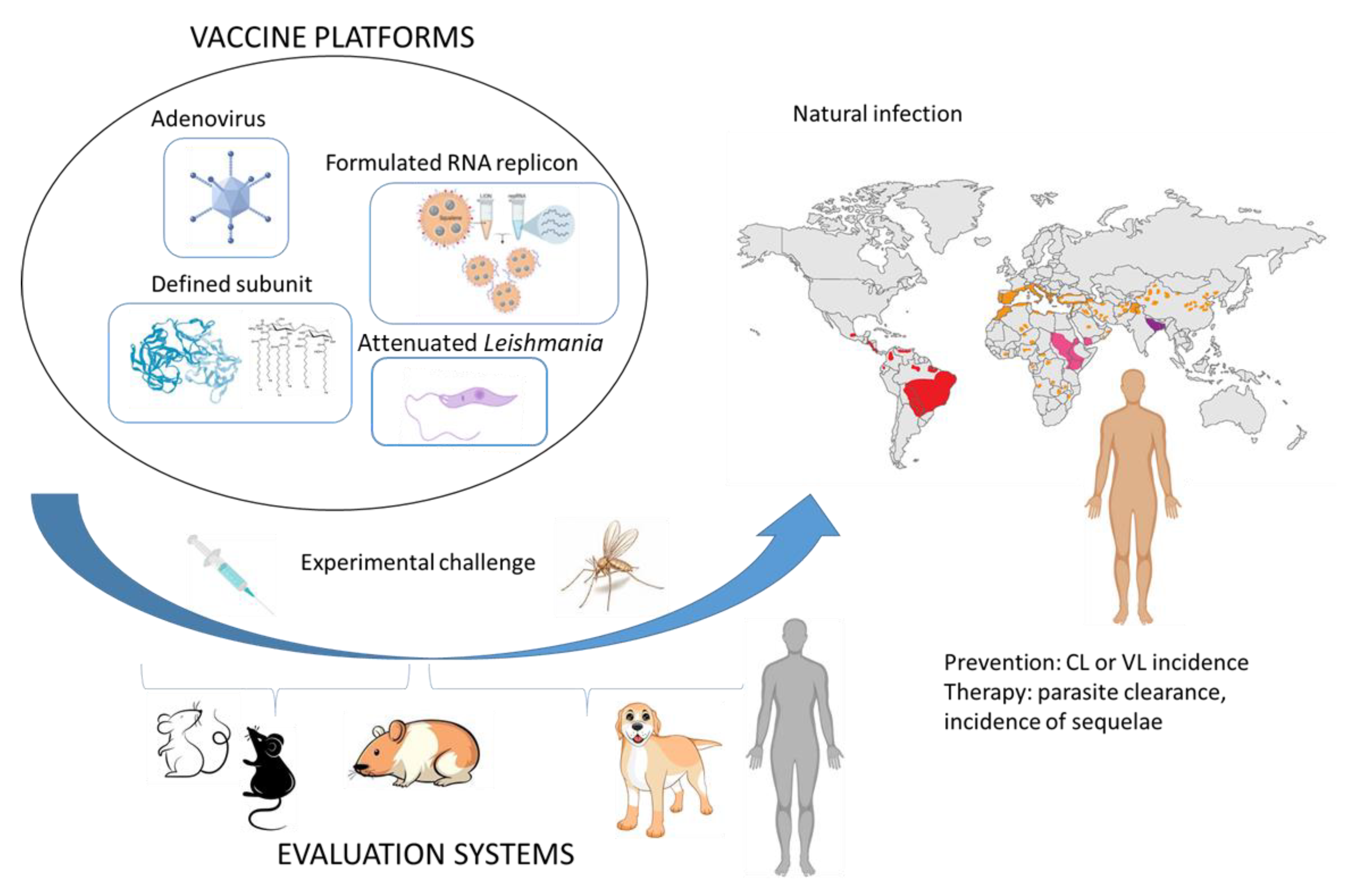
It is important to note that experimental infection of humans with Leishmania spp. is extremely well established for both needle challenge and sand fly initiated infection [72,73]. Many factors weigh on the decision to develop a CHIM, however, including (a) poor disease control and impact on morbidity and mortality; (b) lack of successful vaccines, despite a number of candidate vaccines/antigens in the development pipeline; (c) absence of effective treatments and/or evidence of drug resistance; and perhaps most important, (d) a defined and treatable pathogen strain or species relevant to clinical disease. Leishmaniasis appears to satisfy each of these criteria: despite vector control efforts, highly endemic regions persist and cause suffering; several Leishmania species have been characterized genetically and multiple antigens have been proposed as vaccine candidates; despite improving drugs, drug resistance has and continues to emerge; and finally, establishing well characterized, and preferably GMP compliant, parasite banks for clinical use is being addressed [74]. Beyond establishing the rational and required tools, several practical and safety concerns with any proposed Leishmania CHIM then present themselves. A study involving non-infected sand fly biting was used to establish parameters for challenge and importantly to gauge and incorporate public perceptions of this type of study into a challenge protocol ([75,76] and clinicaltrials.gov: NCT03999970). A clinical study to evaluate the reproducibility of a CHIM for sand fly transmitted cutaneous leishmaniasis has similarly gained ethical and institutional approval and is ongoing (clinicaltrials.gov: NCT04512742). Despite the upfront costs of establishing the CHIM, their use may ultimately represent a cost-effective strategy for prioritizing vaccine candidates because one of their most significant benefits may be in preventing candidates that perform poorly from advancement into large scale clinical trials where they would otherwise consume both investigators and potential recruits in endemic regions. It is important to emphasize that CHIM may not completely replace traditional efficacy trials as their limitations include the participation of individuals in non-endemic regions such that typical environmental pressures are different (i.e., continued or multiple low level exposures that do not establish infection but could influence underlying immunity), ethnic/ genetic varianaces, and, given that Leishmaniases are neglected tropical diseases that disproportionately impact the poor, socioeconomic status and underlying health status. If these Leishmania CHIM efforts are successful, however, they could well become an important stepping-stone for leishmaniasis candidate vaccines before evaluation in larger field trials (Figure 2). In addition, data from CHIM studies has directly led to vaccine licensure or vaccine usage policy changes and a similar approach might be applicable for vaccines against forms of leishmaniasis where field trials would be almost impossible [77].
An additional alternative, or adjunct, to awaiting primary infection and disease development is to evaluate vaccine candidates among infected individuals that can be provided a vaccine post-infection. Speed to, and completeness of, clinical cure and parasite resolution can then be used as an indication of efficacy. If the clinical course is mild and there is a “window of opportunity” that is ethically acceptable (such as was the case in the evaluation of the ChAd63-KH vaccine as a therapeutic for persistent post-kala azar dermal leishmaniasis (PKDL)) vaccines may be tested as stand alone treatments[26]. In more severe presentations, however, this can be confounded by the ethical requirement for provision of standard care to affected individuals and it is therefore important to establish that the vaccine of interest is compatible with chemotherapy. In the case of visceral leishmaniasis, an additional clinical endpoint of sequelae such as post-kala azar dermal leishmaniasis could be used.

4. Leverage of a Profitable Veterinary Application?

In contrast to the human situation, veterinary vaccines based on Leishmania parasite lysates have been advanced to approval for canine leishmaniasis (CanL). Injection of a vaccine comprising total antigens of Leishmania amazonensis plus saponin (LaSap) to infected dogs alleviated clinical symptoms and reduced parasite loads in the skin for at least 6 months [78]. Post-infection use of recombinant Leishmania A2 protein plus saponin (LeishTec®) has also indicated a 25% reduction in risk of developing CanL in asymptomatic dogs and a 70% reduction in mortality among younger dogs (<6 years old) [79]. Similarly, immunotherapeutic investigation of a vaccine comprising Leishmania braziliensis antigens and TLR4 agonist MPL (LBMPL vaccine) demonstrated reductions in both parasite burden and the intensity of disease in the treated dogs, accompanied by a blocking of their transmission of L. infantum to sand flies (observed in 66% (6 of 9) dogs evaluated 3 months later [80].
It is well established that infection and clinical status of each Leishmania-affected dog influences the response to treatment and study outcomes could therefore be impacted by infection level at time of treatment. Supplementation of subunit vaccines with MPL has promoted success, or contrarily has had no impact, in halting disease progression [81,82,83]. Another consideration is onset of immune exhaustion that precedes the transition from asymptomatic Leishmania infection to progressively worsening CanL [84]. Ex vivo incubation with combinations of TLR4, 7, and 7/8 agonists allowed the identification of robust Th1 cells from symptomatic dogs and further suggested the possibility that Leishmania-induced cellular exhaustion could be overcome by potent immunization [85]. The involvement of TLR7 in this is particularly relevant for repRNA vaccines that can engage this receptor [63].
A further complication in providing effective immune therapy may arise from the observation that even after ‘cure’ parasites are retained in very low numbers and maintain immune status. The hypothesis that allopurinol-induced parasite reduction in CanL-affected dogs would render animals more able to develop robust immunity to provide additional longer term control of infection, was tested by evaluating L. infantum infected dogs for clinical and parasitological outcomes following short-term treatment with allopurinol, either alone or in combination with a defined subunit vaccine. Dogs treated with allopurinol alone alleviated their CanL symptoms but had only a transient reduction in parasites that rebounded after a few months. Concomitant immunization with Leish-F2 + SLA-SE, however, not only improved clinical status but also elicited L. infantum clearance from lymphoid tissues and systemic organs in the long term (Figure 3 and [86]).

5. Conclusions

As we have outlined, RNA vaccines could address several current limitations in generating a truly field applicable vaccine for leishmaniasis. RNA vaccines can be made more cost-effectively than either defined subunit or vector-based vaccines and, given the growing familiarity and acceptance of RNA platforms with regulatory agencies, progression to trial is likely to become simpler and more rapid. In this way, we envision that trials can be planned and conducted in a manner more responsive to the identification of relatively small and transient “hot-spots”, permitting a much more efficient design for the evaluation of efficacy. As an alternative, proof-of-concept of safety and efficacy could be generated in human infection models or inferred among patients undergoing therapy.

Author Contributions

Conceptualization, M.S.D. and S.G.R.; writing—original draft preparation, M.S.D., P.M.K. and S.G.R.; writing—review and editing, M.S.D., B.A.S.M., R.B., P.M.K. and S.G.R. All authors have read and agreed to the published version of the manuscript.

Funding

The authors work on leishmaniasis has been funded by National Institute of Allergy and Infectious Diseases of the National Institutes of Health under Award Number R01AI025038 (to SGR), Bill and Melinda Gates Foundation (#631 and #39129; to SGR), Global Health Innovative Technology Fund (G2018–111 to MSD ), and CRDF Global (#62966 to MSD), the Wellcome Trust (WT 085879 and 108518 to PMK) and UK Medical Research Council (MR/P022030 to PMK).

Conflicts of Interest

The authors declare no conflict of interest. The content is solely the responsibility of the authors and does not necessarily represent the official views of the funding bodies.

References

  1. Brito, M.E.F.; Mendonça, M.G.; Gomes, Y.M.; Jardim, M.L.; Abath, F.G. Dynamics of the antibody response in patients with therapeutic or spontaneous cure of American cutaneous leishmaniasis. Trans. R. Soc. Trop. Med. Hyg. 2001, 95, 203–206. [Google Scholar] [CrossRef]
  2. Carvalho, E.M.; Correia Filho, D.; Bacellar, O.; Almeida, R.P.; Lessa, H.; Rocha, H. Characterization of the immune response in subjects with self-healing cutaneous leishmaniasis. Am. J. Trop. Med. Hyg. 1995, 53, 273–277. [Google Scholar] [CrossRef] [PubMed]
  3. Heinzel, F.P.; Schoenhaut, D.S.; Rerko, R.M.; Rosser, L.E.; Gately, M.K. Recombinant interleukin 12 cures mice infected with Leishmania major. J. Exp. Med. 1993, 177, 1505–1509. [Google Scholar] [CrossRef] [PubMed]
  4. Mosmann, T.R.; Cherwinski, H.; Bond, M.W.; Giedlin, M.A.; Coffman, R.L. Two types of murine helper T cell clone. I. Definition according to profiles of lymphokine activities and secreted proteins. J. Immunol. 1986, 136, 2348–2357. [Google Scholar]
  5. Reed, S.G.; Scott, P. T-cell and cytokine responses in leishmaniasis. Curr. Opin. Immunol. 1993, 5, 524–531. [Google Scholar] [CrossRef]
  6. Hutchins, A.S.; Artis, D.; Hendrich, B.D.; Bird, A.P.; Scott, P.; Reiner, S.L. Cutting Edge: A Critical Role for Gene Silencing in Preventing Excessive Type 1 Immunity. J. Immunol. 2005, 175, 5606–5610. [Google Scholar] [CrossRef] [Green Version]
  7. Alexander, J.; Brombacher, F. T Helper1/T Helper2 Cells and Resistance/Susceptibility to Leishmania Infection: Is This Paradigm Still Relevant? Front. Immunol. 2012, 3, 80. [Google Scholar] [CrossRef] [PubMed] [Green Version]
  8. Gautam, S.; Kumar, R.; Singh, N.; Singh, A.K.; Rai, M.; Sacks, D.; Sundar, S.; Nylén, S. CD8 T cell exhaustion in human visceral leishmaniasis. J. Infect. Dis. 2014, 209, 290–299. [Google Scholar] [CrossRef] [PubMed] [Green Version]
  9. Kaushal, H.; Bras-Gonçalves, R.; Negi, N.S.; Lemesre, J.L.; Papierok, G.; Salotra, P. Role of CD8(+) T cells in protection against Leishmania donovani infection in healed Visceral Leishmaniasis individuals. BMC Infect. Dis. 2014, 14, 653. [Google Scholar] [CrossRef] [Green Version]
  10. Den Boer, M.L.; Alvar, J.; Davidson, R.N.; Ritmeijer, K.; Balasegaram, M. Developments in the treatment of visceral leishmaniasis. Expert Opin. Emerg. Drugs 2009, 14, 395–410. [Google Scholar] [CrossRef] [Green Version]
  11. Salari, S.; Bamorovat, M.; Sharifi, I.; Almani, P.G.N. Global distribution of treatment resistance gene markers for leishmaniasis. J. Clin. Lab. Anal. 2022, 36, e24599. [Google Scholar] [CrossRef] [PubMed]
  12. Kaye, P.M.; Mohan, S.; Mantel, C.; Malhame, M.; Revill, P.; Le Rutte, E.; Pakash, V.; Layton, A.M.; Lacey, C.J.N.; Malvolti, S. Overcoming roadblocks in the development of vaccines for leishmaniasis. Expert Rev. Vaccines 2021, 20, 1419–1430. [Google Scholar] [CrossRef] [PubMed]
  13. Bottazzi, M.E.; Hotez, P.J. “Running the Gauntlet”: Formidable challenges in advancing neglected tropical diseases vaccines from development through licensure, and a “Call to Action”. Hum. Vacc. Immunother. 2019, 15, 2235–2242. [Google Scholar] [CrossRef] [Green Version]
  14. Versteeg, L.; Almutairi, M.M.; Hotez, P.J.; Pollet, J. Enlisting the mRNA Vaccine Platform to Combat Parasitic Infections. Vaccines 2019, 7, 122. [Google Scholar] [CrossRef] [Green Version]
  15. Mohan, S.; Revill, P.; Malvolti, S.; Malhame, M.; Sculpher, M.; Kaye, P.M. Estimating the global demand curve for a leishmaniasis vaccine: A generalisable approach based on global burden of disease estimates. PLoS Negl. Trop. Dis. 2022, 16, e0010471. [Google Scholar] [CrossRef]
  16. Corbett, K.S.; Edwards, D.K.; Leist, S.R.; Abiona, O.M.; Boyoglu-Barnum, S.; Gillespie, R.A.; Himansu, S.; Schäfer, A.; Ziwawo, C.T.; DiPiazza, A.T.; et al. SARS-CoV-2 mRNA vaccine design enabled by prototype pathogen preparedness. Nature 2020, 586, 567–571. [Google Scholar] [CrossRef]
  17. Branche, A.R.; Rouphael, N.G.; Diemert, D.D.; Falsey, A.R.; Losada, C.; Baden, L.R.; Frey, S.E.; Whitaker, J.A.; Little, S.J.; Anderson, E.J.; et al. SARS-CoV-2 Variant Vaccine Boosters Trial: Preliminary Analyses. MedRxiv 2022. [Google Scholar] [CrossRef]
  18. Ying, B.; Scheaffer, S.M.; Whitener, B.; Liang, C.Y.; Dmytrenko, O.; Mackin, S.; Wu, K.; Lee, D.; Avena, L.E.; Chong, Z.; et al. Boosting with variant-matched or historical mRNA vaccines protects against Omicron infection in mice. Cell 2022, 185, 1572–1587.e11. [Google Scholar] [CrossRef] [PubMed]
  19. Hawman, D.W.; Meade-White, K.; Archer, J.; Leventhal, S.; Wilson, D.; Shaia, C.; Randall, S.; Khandhar, A.P.; Hsiang, T.-Y.; Gale, M., Jr.; et al. SARS-CoV2 variant-specific replicating RNA vaccines protect from disease and pathology and reduce viral shedding following challenge with heterologous SARS-CoV2 variants of concern. BioRxiv 2021. [Google Scholar] [CrossRef]
  20. Brasu, N.; Elia, I.; Russo, V.; Montacchiesi, G.; Stabile, S.A.; De Intinis, C.; Fesi, F.; Gizzi, K.; Macagno, M.; Montone, M.; et al. Memory CD8+ T cell diversity and B cell responses correlate with protection against SARS-CoV-2 following mRNA vaccination. Nat. Immunol. 2022, 23, 1445–1456. [Google Scholar] [CrossRef] [PubMed]
  21. Wherry, E.J.; Barouch, D.H. T cell immunity to COVID-19 vaccines. Science 2022, 377, 821–822. [Google Scholar] [CrossRef] [PubMed]
  22. Rodrigues, L.S.; Barreto, A.S.; Bomfim, L.G.S.; Gomes, M.C.; Ferreira, N.L.C.; da Cruz, G.S.; Magalhães, L.; de Jesus, A.R.; Palatnik de Sousa, C.B.; Correa, C.B.; et al. Multifunctional, TNF-α and IFN-γ-Secreting CD4 and CD8 T Cells and CD8High T Cells Are Associated With the Cure of Human Visceral Leishmaniasis. Front. Immunol. 2021, 12, 773983. [Google Scholar] [CrossRef]
  23. Volpedo, G.; Bhattacharya, P.; Gannavaram, S.; Pacheco-Fernandez, T.; Oljuskin, T.; Dey, R.; Satoskar, A.R.; Nakhasi, H.L. The History of Live Attenuated Centrin Gene-Deleted Leishmania Vaccine Candidates. Pathogens 2022, 11, 431. [Google Scholar] [CrossRef]
  24. Oliveira BC, D.; Duthie, M.S.; Pereira, V.R.A. Vaccines for leishmaniasis and the implications of their development for American tegumentary leishmaniasis. Hum. Vaccines Immunother. 2019, 16, 919–930. [Google Scholar] [CrossRef]
  25. Duthie, M.S.; Reed, S.G. Not All Antigens Are Created Equally: Progress, Challenges, and Lessons Associated with Developing a Vaccine for Leishmaniasis. Clin. Vaccine Immunol. 2017, 24, e00108-17. [Google Scholar] [CrossRef] [Green Version]
  26. Younis, B.M.; Osman, M.; Khalil, E.A.; Santoro, F.; Furini, S.; Wiggins, R.; Keding, A.; Carraro, M.; Musa, A.E.A.; Abdarahaman, M.A.A.; et al. Safety and immunogenicity of ChAd63-KH vaccine in post-kala-azar dermal leishmaniasis patients in Sudan. Mol. Ther. 2021, 29, 2366–2377. [Google Scholar] [CrossRef] [PubMed]
  27. Osman, M.; Mistry, A.; Keding, A.; Gabe, R.; Cook, E.; Forrester, S.; Wiggins, R.; Di Marco, S.; Colloca, S.; Siani, L.; et al. A third generation vaccine for human visceral leishmaniasis and post kala azar dermal leishmaniasis: First-in-human trial of ChAd63-KH. PLoS Negl. Trop. Dis. 2017, 11, e0005527. [Google Scholar] [CrossRef] [PubMed] [Green Version]
  28. Kumari, S.; Samant, M.; Khare, P.; Misra, P.; Dutta, S.; Kolli, B.K.; Sharma, S.; Chang, K.P.; Dube, A. Photodynamic vaccination of hamsters with inducible suicidal mutants of Leishmania amazonensis elicits immunity against visceral leishmaniasis. Eur. J. Immunol. 2009, 39, 178–191. [Google Scholar] [CrossRef] [PubMed] [Green Version]
  29. Zhang, W.-W.; Zhang, W.W.; Karmakar, S.; Gannavaram, S.; Dey, R.; Lypaczewski, P.; Ismail, N.; Siddiqui, A.; Simonyan, V.; Oliveira, F.; et al. A second generation leishmanization vaccine with a markerless attenuated Leishmania major strain using CRISPR gene editing. Nat. Commun. 2020, 11, 3461. [Google Scholar] [CrossRef] [PubMed]
  30. Gomes, D.C.O.; Souza, B.L.D.S.C.; Schwedersky, R.P.; Covre, L.P.; de Matos Guedes, H.L.; Lopes, U.G.; Re, M.I.; Rossi-Bergmann, B. Intranasal immunization with chitosan microparticles enhances LACK-DNA vaccine protection and induces specific long-lasting immunity against visceral leishmaniasis. Microbes Infect. 2022, 24, 104884. [Google Scholar] [CrossRef]
  31. Santos, T.T.O.; Machado, A.S.; Ramos, F.F.; Oliveira-da-Silva, J.A.; Lage, D.P.; Tavares, G.S.; Mendonça, D.V.C.; Cardoso, M.S.; Siqueira, W.F.; Martins, V.T.; et al. Leishmania eukaryotic elongation Factor-1 beta protein is immunogenic and induces parasitological protection in mice against Leishmania infantum infection. Microb. Pathog. 2021, 151, 104745. [Google Scholar] [CrossRef]
  32. Bertholet, S.; Goto, Y.; Carter, L.; Bhatia, A.; Howard, R.F.; Carter, D.; Coler, R.N.; Vedvick, T.S.; Reed, S.G. Optimized subunit vaccine protects against experimental leishmaniasis. Vaccine 2009, 27, 7036–7045. [Google Scholar] [CrossRef] [Green Version]
  33. Coler, R.N.; Duthie, M.S.; Hofmeyer, K.A.; Guderian, J.; Jayashankar, L.; Vergara, J.; Rolf, T.; Misquith, A.; Lurance, J.D.; Raman, V.S.; et al. From mouse to man: Safety, immunogenicity and efficacy of a candidate leishmaniasis vaccine LEISH-F3+GLA-SE. Clin. Transl. Immunol. 2015, 4, e35. [Google Scholar] [CrossRef]
  34. Duthie, M.S.; Pereira, L.; Favila, M.; Hofmeyer, K.A.; Reed, S.J.; Metangmo, S.; Townsend, S.; Laurance, J.D.; Picone, A.; Misquith, A.; et al. A defined subunit vaccine that protects against vector-borne visceral leishmaniasis. NPJ Vaccines 2017, 2, 23. [Google Scholar] [CrossRef] [Green Version]
  35. Pardi, N.; Hogan, M.J.; Pelc, R.S.; Muramatsu, H.; Andersen, H.; DeMaso, C.R.; Dowd, K.A.; Sutherland, L.L.; Scearce, M.; Parks, R.; et al. Zika virus protection by a single low-dose nucleoside-modified mRNA vaccination. Nature 2017, 543, 248–251. [Google Scholar] [CrossRef] [Green Version]
  36. Pardi, N.; Weissman, D. RNA Vaccines, Methods and Protocols. Methods Mol. Biol. 2016, 1499, 143–153. [Google Scholar]
  37. Richner, J.M.; Himansu, S.; Dowd, K.A.; Butler, S.L.; Salazar, V.; Fox, J.M.; Julander, J.G.; Tang, W.W.; Shresta, S.; Pierson, T.C.; et al. Modified mRNA Vaccines Protect against Zika Virus Infection. Cell 2017, 168, 1114–1125. [Google Scholar] [CrossRef] [Green Version]
  38. Hekele, A.; Bertholet, S.; Archer, J.; Gibson, D.G.; Palladino, G.; Brito, L.A.; Otten, G.R.; Brazolli, M.; Buccato, S.; Bonci, A.; et al. Rapidly produced SAM((R)) vaccine against H7N9 influenza is immunogenic in mice. Emerg. Microbes Infect. 2013, 2, e52. [Google Scholar] [CrossRef] [PubMed]
  39. Leventhal, S.S.; Meade-White, K.; Rao, D.; Haddock, E.; Leung, J.; Scott, D.; Archer, J.; Randall, S.; Erasmus, J.H.; Feldmann, H.; et al. Replicating RNA vaccination elicits an unexpected immune response that efficiently protects mice against lethal Crimean-Congo hemorrhagic fever virus challenge. Ebiomedicine 2022, 82, 104188. [Google Scholar] [CrossRef] [PubMed]
  40. Blakney, A.K.; Ip, S.; Geall, A.J. An Update on Self-Amplifying mRNA Vaccine Development. Vaccines 2021, 9, 97. [Google Scholar] [CrossRef]
  41. Lundstrom, K. Replicon RNA Viral Vectors as Vaccines. Vaccines 2016, 4, 39. [Google Scholar] [CrossRef] [PubMed]
  42. Vogel, A.B.; Lambert, L.; Kinnear, E.; Busse, D.; Erbar, S.; Reuter, K.C.; Wicke, L.; Perkovic, M.; Beissert, T.; Hass, H.; et al. Self-Amplifying RNA Vaccines Give Equivalent Protection against Influenza to mRNA Vaccines but at Much Lower Doses. Mol. Ther. 2018, 26, 446–455. [Google Scholar] [CrossRef] [PubMed]
  43. Zhou, X.; Berglund, P.; Rhodes, G.; Parker, S.E.; Jondal, M.; Liljeström, P. Self-replicating Semliki Forest virus RNA as recombinant vaccine. Vaccine 1994, 12, 1510–1514. [Google Scholar] [CrossRef]
  44. Brazzoli, M.; Magini, D.; Bonci, A.; Buccato, S.; Giovani, C.; Kratzer, R.; Zurli, V.; Mangiavacchi, S.; Casini, D.; Brito, L.M.; et al. Induction of Broad-Based Immunity and Protective Efficacy by Self-amplifying mRNA Vaccines Encoding Influenza Virus Hemagglutinin. J. Virol. 2016, 90, 332–344. [Google Scholar] [CrossRef] [Green Version]
  45. Martinon, F.; Krishnan, S.; Lenzen, G.; Magné, R.; Gomard, E.; Guillet, J.G.; Lévy, J.P.; Meulien, P. Induction of virus-specific cytotoxic T lymphocytes in vivo by liposome-entrapped mRNA. Eur. J. Immunol. 1993, 23, 1719–1722. [Google Scholar] [CrossRef]
  46. Ljungberg, K.; Liljestrom, P. Self-replicating alphavirus RNA vaccines. Expert Rev. Vaccines 2015, 14, 177–194. [Google Scholar] [CrossRef]
  47. Strauss, J.H.; Strauss, E.G. The alphaviruses: Gene expression, replication, and evolution. Microbiol. Rev. 1994, 58, 491–562. [Google Scholar] [CrossRef]
  48. Atasheva, S.; Kim, D.Y.; Akhrymuk, M.; Morgan, D.G.; Frolova, E.I.; Frolov, I. Pseudoinfectious Venezuelan Equine Encephalitis Virus: A New Means of Alphavirus Attenuation. J. Virol. 2013, 87, 2023–2035. [Google Scholar] [CrossRef] [Green Version]
  49. Kowalski, P.S.; Rudra, A.; Miao, L.; Anderson, D.G. Delivering the Messenger: Advances in Technologies for Therapeutic mRNA Delivery. Mol. Ther. 2019, 27, 710–728. [Google Scholar] [CrossRef] [Green Version]
  50. Mintzer, M.A.; Simanek, E.E. Nonviral Vectors for Gene Delivery. Chem. Rev. 2009, 109, 259–302. [Google Scholar] [CrossRef]
  51. Martin, M.E.; Rice, K.G. Peptide-guided gene delivery. AAPS J. 2007, 9, E18–E29. [Google Scholar] [CrossRef] [PubMed] [Green Version]
  52. Pack, D.W.; Hoffman, A.S.; Pun, S.; Stayton, P.S. Design and development of polymers for gene delivery. Nat. Rev. Drug Discov. 2005, 4, 581–593. [Google Scholar] [CrossRef] [PubMed]
  53. Malone, R.W.; Felgner, P.L.; Verma, I.M. Cationic liposome-mediated RNA transfection. Proc. Natl. Acad. Sci. USA 1989, 86, 6077–6081. [Google Scholar] [CrossRef] [PubMed] [Green Version]
  54. De Jong, S.; Chikh, G.; Sekirov, L.; Raney, S.; Semple, S.; Klimuk, S.; Yuan, N.; Hope, M.; Cullis, P.; Tam, Y. Encapsulation in liposomal nanoparticles enhances the immunostimulatory, adjuvant and anti-tumor activity of subcutaneously administered CpG ODN. Cancer Immunol. Immunother. 2007, 56, 1251–1264. [Google Scholar] [CrossRef]
  55. Reichmuth, A.M.; Oberli, M.A.; Jaklenec, A.; Langer, R.; Blankschtein, D. mRNA vaccine delivery using lipid nanoparticles. Ther. Deliv. 2016, 7, 319–334. [Google Scholar] [CrossRef] [Green Version]
  56. Baden, L.R.; El Sahly, H.M.; Essink, B.; Kotloff, K.; Frey, S.; Novak, R.; Diemert, D.; Spector, S.A.; Rouphael, N.; Creech, C.B.; et al. Efficacy and Safety of the mRNA-1273 SARS-CoV-2 Vaccine. N. Engl. J. Med. 2021, 384, 403–416. [Google Scholar] [CrossRef]
  57. Greenhawt, M.; Abrams, E.M.; Shaker, M.; Chu, D.K.; Khan, D.; Akin, C.; Alqurashi, W.; Arkwright, P.; Baldwin, J.L.; Ben-Shoshan, M.; et al. The Risk of Allergic Reaction to SARS-CoV-2 Vaccines and Recommended Evaluation and Management: A Systematic Review, Meta-Analysis, GRADE Assessment, and International Consensus Approach. J. Allergy Clin. Immunol. Pract. 2021, 9, 3546–3567. [Google Scholar] [CrossRef]
  58. Mahdiabadi, S.; Rezaei, N. Anaphylaxis and allergic reactions to COVID-19 vaccines: A narrative review of characteristics and potential obstacles on achieving herd immunity. Health Sci. Rep. 2022, 5, e787. [Google Scholar] [CrossRef]
  59. Li, M.; Wang, X.; Feng, J.; Feng, Z.; Li, W.; Ya, B. Myocarditis or Pericarditis Following the COVID-19 Vaccination in Adolescents: A Systematic Review. Vaccines 2022, 10, 1316. [Google Scholar] [CrossRef]
  60. Massari, M.; Spila-Alegiani, S.; Morciano, C.; Spuri, M.; Marchione, P.; Felicetti, P.; Belleudi, C.; Poggi, F.R.; Lazzeretti, M.; Ercolanoni, M.; et al. Postmarketing active surveillance of myocarditis and pericarditis following vaccination with COVID-19 mRNA vaccines in persons aged 12 to 39 years in Italy: A multi-database, self-controlled case series study. PLoS Med. 2022, 19, e1004056. [Google Scholar] [CrossRef]
  61. Wong, H.L.; Hu, M.; Zhou, C.K.; Lloyd, P.C.; Amend, K.L.; Beachler, D.C.; Secora, A.; McMahill-Walraven, C.N.; Lu, Y.; Wu, Y.; et al. Risk of myocarditis and pericarditis after the COVID-19 mRNA vaccination in the USA: A cohort study in claims databases. Lancet 2022, 399, 2191–2199. [Google Scholar] [CrossRef]
  62. Erasmus, J.H.; Khandhar, A.P.; O’Connor, M.A.; Walls, A.C.; Hemann, E.A.; Murapa, P.; Archer, J.; Leventhal, S.; Fuller, J.T.; Lewis, T.B.; et al. An Alphavirus-derived replicon RNA vaccine induces SARS-CoV-2 neutralizing antibody and T cell responses in mice and nonhuman primates. Sci. Transl. Med. 2020, 12, eabc9396. [Google Scholar] [CrossRef] [PubMed]
  63. Duthie, M.S.; Van Hoeven, N.; MacMillen, Z.; Picone, A.; Mohamath, R.; Erasmus, J.; Hsu, F.-C.; Stinchcomb, D.T.; Reed, S.G. Heterologous Immunization with Defined RNA and Subunit Vaccines Enhances T Cell Responses That Protect against Leishmania donovani. Front. Immunol. 2018, 9, 2420. [Google Scholar] [CrossRef] [PubMed] [Green Version]
  64. Shah, S.K.; Miller, F.G.; Darton, T.C.; Duenas, D.; Emerson, C.; Lynch, H.F.; Jamrozik, E.; Jecker, N.S.; Kamuya, D.; Kapulu, M.; et al. Ethics of controlled human infection to address COVID-19. Science 2020, 368, 832–834. [Google Scholar] [CrossRef]
  65. Eyal, N.; Lipsitch, M.; Smith, P.G. Human challenge studies to accelerate coronavirus vaccine licensure. J. Infect. Dis. 2020, 221, 1752–1756. [Google Scholar] [CrossRef] [Green Version]
  66. Langenberg, M.C.; Hoogerwerf, M.A.; Koopman, J.P.R.; Janse, J.J.; Kos-van Oosterhoud, J.; Feijt, C.; Jochems, S.P.; De Dood, C.J.; van Schuijlenburg, R.; Ozir-Fazalalikhan, A.; et al. A controlled human Schistosoma mansoni infection model to advance novel drugs, vaccines and diagnostics. Nat. Med. 2020, 26, 326–332. [Google Scholar] [CrossRef]
  67. Kirkpatrick, B.D.; Whitehead, S.S.; Pierce, K.K.; Tibery, C.M.; Grier, P.L.; Hynes, N.A.; Larsson, C.J.; Saundayo, B.P.; Talaat, K.R.; Janiak, A.; et al. The live attenuated dengue vaccine TV003 elicits complete protection against dengue in a human challenge model. Sci. Transl. Med. 2016, 8, 330ra36. [Google Scholar] [CrossRef] [Green Version]
  68. Gould, V.M.; Francis, J.N.; Anderson, K.J.; Georges, B.; Cope, A.V.; Tregoning, J.S. Nasal IgA Provides Protection against Human Influenza Challenge in Volunteers with Low Serum Influenza Antibody Titre. Front. Microbiol. 2017, 8, 900. [Google Scholar] [CrossRef] [Green Version]
  69. Payne, R.O.; Griffin, P.M.; McCarthy, J.S.; Draper, S.J. Plasmodium vivax Controlled Human Malaria Infection—Progress and Prospects. Trends Parasitol. 2017, 33, 141–150. [Google Scholar] [CrossRef] [Green Version]
  70. Roestenberg, M.; Hoogerwerf, M.-A.; Ferreira, D.M.; Mordmüller, B.; Yazdanbakhsh, M. Experimental infection of human volunteers. Lancet Infect. Dis. 2018, 18, e312–e322. [Google Scholar] [CrossRef]
  71. Sheehy, S.H.; Duncan, C.J.; Elias, S.C.; Choudhary, P.; Biswas, S.; Halstead, F.D.; Collins, K.A.; Edwards, N.J.; Douglas, A.D.; Anagnostou, N.A.; et al. ChAd63-MVA–vectored Blood-stage Malaria Vaccines Targeting MSP1 and AMA1: Assessment of Efficacy against Mosquito Bite Challenge in Humans. Mol. Ther. 2012, 20, 2355–2368. [Google Scholar] [CrossRef] [PubMed] [Green Version]
  72. Parkash, V.; Kaye, P.M.; Layton, A.M.; Lacey, C.J. Vaccines against leishmaniasis: Using controlled human infection models to accelerate development. Expert Rev. Vaccines 2012, 20, 1407–1418. [Google Scholar] [CrossRef] [PubMed]
  73. Melby, P.C. Experimental Leishmaniasis in Humans: Review. Clin. Infect. Dis. 1991, 13, 1009–1017. [Google Scholar] [CrossRef] [PubMed]
  74. Ashwin, H.; Sadlova, J.; Vojtkova, B.; Becvar, T.; Lypaczewski, P.; Schwartz, E.; Greensted, E.; Van Bocxlaer, K.; Pasin, M.; Lipinski, K.S.; et al. Characterization of a new Leishmania major strain for use in a controlled human infection model. Nat. Commun. 2021, 12, 215. [Google Scholar] [CrossRef]
  75. Parkash, V.; Ashwin, H.; Sadlova, J.; Vojtkova, B.; Jones, G.; Martin, N.; Greensted, E.; Allgar, V.; Kamhawi, S.; Valenzuela, J.G.; et al. A clinical study to optimise a sand fly biting protocol for use in a controlled human infection model of cutaneous leishmaniasis (the FLYBITE study). Wellcome Open Res. 2021, 6, 168. [Google Scholar] [CrossRef]
  76. Parkash, V.; Jones, G.; Martin, N.; Steigmann, M.; Greensted, E.; Kaye, P.; Layton, A.M.; Lacey, C.J. Assessing public perception of a sand fly biting study on the pathway to a controlled human infection model for cutaneous leishmaniasis. Res. Involv. Engagem. 2021, 7, 33. [Google Scholar] [CrossRef]
  77. Giersing, B.K.; Porter, C.K.; Kotloff, K.; Neels, P.; Cravioto, A.; MacLennan, C.A. How can controlled human infection models accelerate clinical development and policy pathways for vaccines against Shigella? Vaccine 2019, 37, 4778–4783. [Google Scholar] [CrossRef]
  78. Viana, K.F.; Lacerda, G.; Teixeira, N.S.; Cangussu, A.S.R.; Aguiar, R.W.S.; Giunchetti, R.C. Therapeutic vaccine of killed Leishmania amazonensis plus saponin reduced parasite burden in dogs naturally infected with Leishmania infantum. Vet. Parasitol. 2018, 254, 98–104. [Google Scholar] [CrossRef]
  79. Toepp, A.; Larson, M.; Wilson, G.; Grinnage-Pulley, T.; Bennett, C.; Leal-Lima, A.; Anderson, B.; Parrish, M.; Anderson, M.; Fowler, H. Randomized, controlled, double-blinded field trial to assess Leishmania vaccine effectiveness as immunotherapy for canine leishmaniosis. Vaccine 2018, 36, 6433–6441. [Google Scholar] [CrossRef]
  80. Roatt, B.M.; Aguiar-Soares, R.D.D.O.; Reis, L.E.S.; Cardoso, J.M.D.O.; Mathias, F.A.S.; Brito, R.C.F.D.; da Silva, S.M.; De Figueiredo Gontijo, N.; Ferreira, S.; Valenzuela, J.G.; et al. A Vaccine Therapy for Canine Visceral Leishmaniasis Promoted Significant Improvement of Clinical and Immune Status with Reduction in Parasite Burden. Front. Immunol. 2017, 8, 217. [Google Scholar] [CrossRef] [Green Version]
  81. Gradoni, L.; Manzillo, V.F.; Pagano, A.; Piantedosi, D.; De Luna, R.; Gramiccia, M.; Scalone, A.; Di Muccio, T.; Oliva, G. Failure of a multi-subunit recombinant leishmanial vaccine (MML) to protect dogs from Leishmania infantum infection and to prevent disease progression in infected animals. Vaccine 2005, 23, 5245–5251. [Google Scholar] [CrossRef] [PubMed]
  82. Miret, J.; Nascimento, E.; Sampaio, W.; França, J.C.; Fujiwara, R.T.; Vale, A.; Dias, E.S.; Vieira, E.; Costa, R.T.; Mayrink, W.; et al. Evaluation of an immunochemotherapeutic protocol constituted of N-methyl meglumine antimoniate (Glucantime) and the recombinant Leish-110f + MPL-SE vaccine to treat canine visceral leishmaniasis. Vaccine 2008, 26, 1585–1594. [Google Scholar] [CrossRef] [PubMed]
  83. Moreno, J.; Nieto, J.; Masina, S.; Cañavate, C.; Cruz, I.; Chicharro, C.; Carrillo, E.; Napp, S.; Reymond, C.; Kaye, P.M.; et al. Immunization with H1, HASPB1 and MML Leishmania proteins in a vaccine trial against experimental canine leishmaniasis. Vaccine 2007, 25, 5290–5300. [Google Scholar] [CrossRef] [PubMed] [Green Version]
  84. Esch, K.J.; Juelsgaard, R.; Martinez, P.A.; Jones, D.E.; Petersen, C.A. Programmed death 1-mediated T cell exhaustion during visceral leishmaniasis impairs phagocyte function. J. Immunol. 2013, 191, 5542–5550. [Google Scholar] [CrossRef] [Green Version]
  85. Schaut, R.G.; Grinnage-Pulley, T.L.; Esch, K.J.; Toepp, A.J.; Duthie, M.S.; Howard, R.F.; Reed, S.G.; Petersen, C.A. Recovery of antigen-specific T cell responses from dogs infected with Leishmania (L.) infantum by use of vaccine associated TLR-agonist adjuvant. Vaccine 2016, 34, 5225–5234. [Google Scholar] [CrossRef] [Green Version]
  86. Nascimento, L.F.; Miranda DF, H.; Moura, L.D.; Pinho, F.A.; Werneck, G.L.; Khouri, R.; Reed, S.G.; Duthie, M.S.; Barral, A.; Barral-Netto, M.; et al. Allopurinol therapy provides long term clinical improvement, but additional immunotherapy is required for sustained parasite clearance, in L. infantum-infected dogs. Vaccine X 2020, 4, 100048. [Google Scholar] [CrossRef]
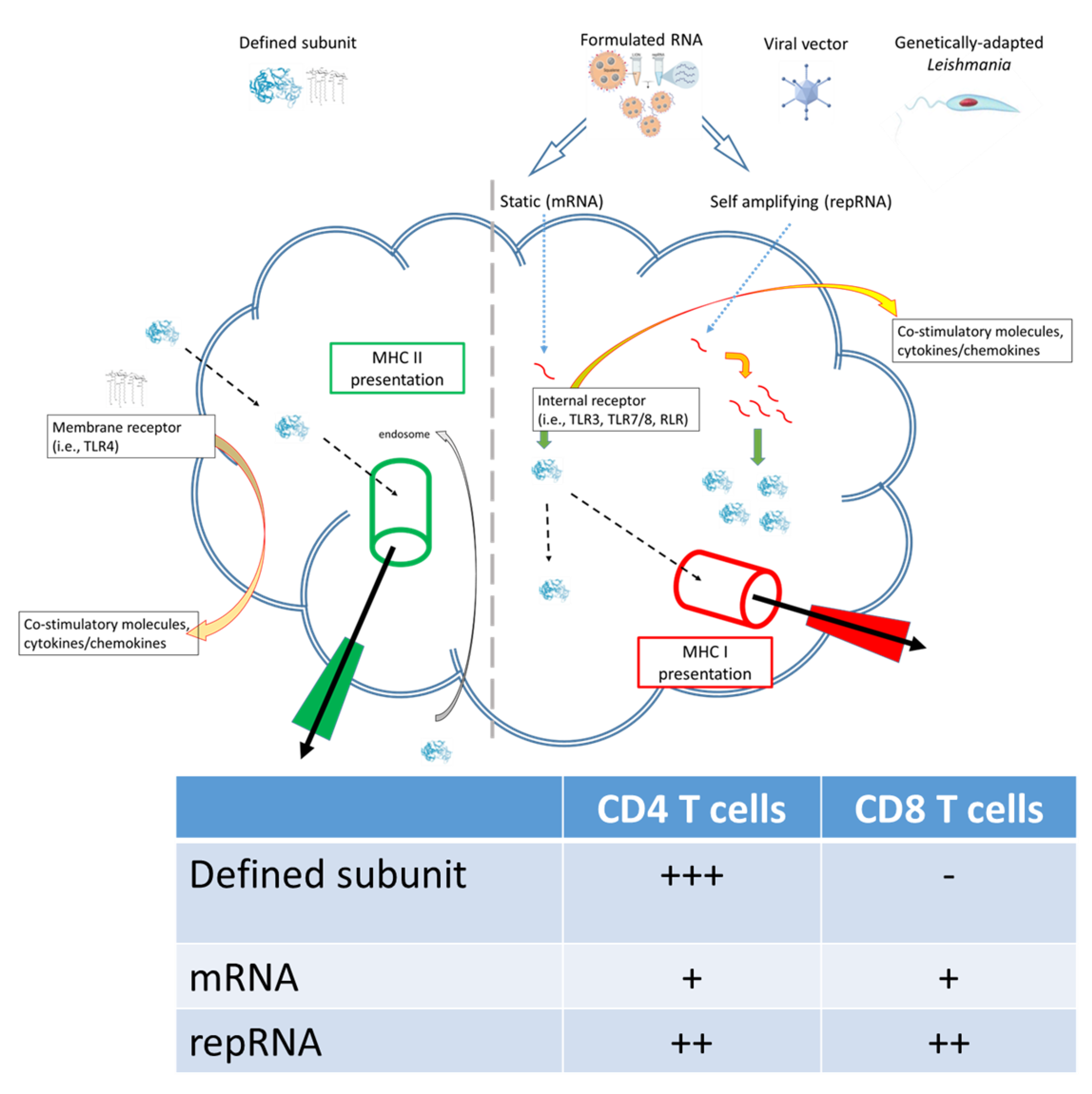
Figure 1. Subunit and RNA replicon vaccines can generate different quality of immune response.
Pathogens 11 01259 g001
Figure 2. Vaccine classes and how they can be evaluated for efficacy against Leishmaniasis.
Pathogens 11 01259 g002
Figure 3. Addition of vaccine to drug treatment generates sustained L. infantum clearance. Infected dogs were treated with either drug (allopurinol) alone or drug plus immunization (Leish-F2 + SLA-SE) then one year later L. infantum burden determined in the indicated organs were determined. Adapted from original data published in [86].
Pathogens 11 01259 g003
Publisher’s Note: MDPI stays neutral with regard to jurisdictional claims in published maps and institutional affiliations.