Article

Multiplex PCR and Sequence Analysis to Investigate Genetic Diversity of Fasciola Isolates from Cattle and Sheep in Turkey

1
Department of Parasitology, Faculty of Veterinary Medicine, University of Firat, 23119 Elazig, Turkey
2
Department of Parasitology, Faculty of Veterinary Medicine, Bingol University, 12000 Bingol, Turkey
3
National Institute of Parasitic Diseases, Chinese Center for Disease Control and Prevention, (Chinese Center for Tropical Diseases Research), Shanghai 200025, China
4
Key Laboratory of Parasite and Vector Biology, National Health Commission of the People’s Republic of China, Shanghai 200025, China
5
World Health Organization Collaborating Center for Tropical Diseases, Shanghai 200025, China
6
Department of Biosciences, COMSATS University Islamabad (CUI), Park Road, ChakhShazad, Islamabad 45550, Pakistan
7
The School of Global Health, Chinese Center for Tropical Diseases Research, Shanghai Jiao Tong University School of Medicine, Shanghai 200240, China
*
Authors to whom correspondence should be addressed.
Pathogens 2022, 11(11), 1235; https://doi.org/10.3390/pathogens11111235
Submission received: 19 September 2022 / Revised: 19 October 2022 / Accepted: 19 October 2022 / Published: 26 October 2022

Abstract

:
Fasciolosis is a highly prevalent helminthic infection caused by Fasciola hepatica and F. gigantica. With the aim of identifying hybrid Fasciola flukes, multiplex PCR was performed to amplify the pepck gene. Furthermore, to determine Fasciola haplotypes, mitochondrial nad1 gene was amplified and sequenced, and phylogenetic analyses were performed. Adult Fasciola isolates were collected from 51 cattle and 51 sheep, genomic DNA was isolated, and one-step multiplex PCR was subsequently performed to amplify pepck. Isolates that generated a 510 bp band were identified as F. gigantica, those that generated a 241 bp band were identified as F. hepatica, and those that generated both bands were identified as hybrid (aspermic) flukes. Multiplex PCR data identified four isolates as F. gigantica and 84 as F. hepatica. Fourteen hybrid isolates (five cattle and nine sheep) were identified. On unidirectional DNA sequence analysis of nad1 PCR products, three sequences were identified as F. gigantica and 99 as F. hepatica. In addition, only 4 of 87 haplotypes detected for F. hepatica nad1 sequences were found to be previously reported, while the remaining 83 are unique to this study. To summarize, this study is the first to report the existence of hybrid Fasciola flukes and 83 unique haplotypes of F. hepatica in Turkey.

1. Introduction

Fasciolosis is a parasitic liver infection mainly caused by Fasciola hepatica and F. gigantica, as well as by other digenetic trematodes in the Fasciolidae family [1]. According to The Centers for Disease Control and Prevention estimates, F. hepatica is prevalent in >70 countries across all continents, except Antarctica [2]. In a study focusing on the genetic characterization of F. hepatica in Argentina, 21 adult parasites were obtained from naturally infected bovine livers. Six haplotypes were detected with mitochondrial cytochrome c oxidase subunit 1 (mt-CO1), four haplotypes with mitochondrial NADH dehydrogenase subunit 4 (nad4), and three haplotypes with nad5, while no haplotypes were detected with ribosomal internal transcribed spacer 1 (ITS1) [3]. Furthermore, as per their analysis, the mt-CO1 gene fragment was the most variable marker. Another study reported the detection of 37 mitochondrial NADH dehydrogenase subunit 1 haplotypes in 130 F. hepatica isolates collected from abattoirs in nine provinces across Iran between 2015 and 2017 [4].
Both F. hepatica and F. gigantica reproduce bisexually via self-fertilization. They can be differentiated by their morphological characteristics [5]. These species show normal spermatogenic ability and are hermaphroditic [6]. Morphological and molecular studies have reported the existence of hybrid forms, known as aspermic intermediated forms [7]. Morphological hybrid forms produce offspring via interspecies fertilization and possibly reproduce parthenogenetically in Japan and other Asian countries [8,9,10,11,12]. Some reports have found that aspermic Fasciola spp. carry three different ITS1 genotypes [10]: Fh type, which is identical to F. hepatica sequences; Fg type, which is identical to F. gigantica sequences; and Fh/Fg type, which is a combination of both sequences. Aspermic Fasciola spp. show two major lineages of mitochondrial nad1. One belongs to F. hepatica and the other belongs to F. gigantica, indicating that these Fasciola forms originated from the same ancestors [11]. The presence of Fh/Fg type in ITS1, an instance of nuclear DNA, suggests that aspermic Fasciola forms are the result of a natural hybridization event between F. hepatica and F. gigantica [10,11,13]. The ITS1 region of ribosomal DNA shows hundreds of copies organized as tandem repeats. ITS1 analysis reportedly does not provide sufficient evidence of natural hybridization because repeat genes are highly recombinogenic and unstable, causing some controversies pertaining to Fasciola spp. characterization [14].
Fasciola isolates can be discriminated from one another on the basis of their nuclear protein-coding genes. Multiplex PCR methods have been optimized to differentiate F. hepatica, F. gigantica, and aspermic Fasciola flukes using the phosphoenolpyruvate carboxykinase (pepck) gene. Shoriki et al. [15] analyzed 27 F. hepatica, F. gigantica, and aspermic Fasciola flukes using nuclear (ITS1, pepck, and DNA polymerase delta (pold)) as well as mitochondrial (nad1) markers. They found that all aspermic Fasciola flukes displayed the Fh/Fg type in both pepck and pold regardless of their ITS1 genotypes and nad1 lineages. In addition, a study characterized 196 Fasciola isolates in Spain as F. hepatica by multiplex PCR analysis of pepck, and 26 haplotypes were detected in mitochondrial nad1. It is notable that only one of them was previously reported in Spanish samples, indicating high genetic diversity and population structure in F. hepatica from Spain [16].
Giovanoli Evack et al. [17] collected flukes from Fasciola-positive cattle, sheep, and goats slaughtered in four Chadian abattoirs and extracted genomic DNA from 27 flukes. Subsequently, they performed species identification using the ITS1 + 2 locus. Of 27, 26 flukes were identified to be F. gigantica, while the remaining fluke showed heterozygosity at all variable sites that differentiate F. hepatica from F. gigantica. Upon cloning and sequencing of both alleles, the presence of one F. hepatica and one F. gigantica allele was confirmed. Their study was the first unambiguous molecular investigation to demonstrate the existence of such a hybrid in cattle in sub-Saharan Africa.
The present study was performed with a multiplex PCR aimed at amplifying pepck to identify hybrid forms of Fasciola isolates from cattle and sheep. The aim was to assess genetic diversity and haplotypes of Fasciola flukes by performing sequencing of mitochondrial nad1.

2. Materials and Methods

2.1. Collection of Adult Fasciola spp. Samples

Between February 2021 and February 2022, adult Fasciola spp. samples were collected from cattle and sheep from a slaughterhouse in Elazig province. Flukes from the liver of each animal were washed with distilled water, and bile residue was removed. All samples were stored in 70% ethanol at −20 °C until needed.

2.2. Genomic DNA Isolation

The anterior one-third of the flukes’ bodies were excised into small pieces using a sterile scalpel and transferred to a 1.5 mL Eppendorf tube. The tissues were washed at least 4 to 5 times with 1 × PBS (pH = 7.4), and genomic DNA was then extracted using a Hibrigen Genomic DNA Isolation Kit (Hibrigen, Turkey). The isolated DNA was quantified with a Nanodrop spectrophotometer and stored at −20 °C until needed.

2.3. Amplification of Pepck by Multiplex PCR

To amplify pepck, a single-step multiplex PCR was performed using these primers: Fh-pepck-F, 5′-GATTGCACCGTTAGGTTAGC-3′; Fg-pepck-F, 5′-AAAGTTTCTATCCC GAACGAAG-3′; and Fcmn-Pepck-R, 5′-CGAAAATTATGGCATCAATGGG-3′ for F. hepatica and F. gigantica, respectively [15,18]. The 25 µL reaction mixture comprised 2.5 µL 10 × PCR buffer, 250 µM of each deoxynucleotide, 1.25 U Taq DNA polymerase, 20 pmol of each primer (0.4 µM Fh-pepck-F, 0.4 µM Fg-pepck-F, and 0.8 μM Fcmn-pepck-R), and 100 ng template DNA on average. The cycling conditions were as follows: initial denaturation at 94 °C for 1.5 min, followed by 30 cycles of denaturation at 94 °C for 30 s, hybridization at 61 °C for 30 s, and synthesis at 72 °C for 1 min, and then a final synthesis step at 72 °C for 10 min [15]. The amplicons thus obtained were separated on 1% agarose gel and bands were visualized with a UV transilluminator. Genomic DNA of isolates, which were previously confirmed to be F. hepatica and F. gigantica by sequencing [19], was included as positive control, and sterile distilled water served as negative control.

2.4. Amplification of nad1 by PCR

For exact species determination, genomic DNA of Fasciola isolates was amplified by PCR. Briefly, 5 μL 10 × PCR buffer, 250 μM of each deoxynucleotide, 1.25 U Taq DNA polymerase, 20 pmol of each primer to amplify nad1 (Ita-10, 5′-AAGGATGTTGCTTTGTCGTGG-3′ and Ita-2, 5′-GGAGTACGGTTACATTCA-3′), and 100 ng template DNA on average were mixed to obtain a reaction mixture of 50 μL [11]. The cycling conditions were as follows: initial denaturation at 94 °C for 1.5 min, followed by 30 cycles of denaturation at 94 °C for 1.5 min, hybridization at 51 °C for 1.5 min, and synthesis at 72 °C for 2 min, and then a final synthesis step at 72 °C for 10 min. The amplicons thus obtained were subsequently subjected to 1.4% agarose gel electrophoresis, followed by ethidium bromide staining.

2.5. DNA Sequence Analysis

All nad1 PCR products, obtained by amplifying genomic DNA of Fasciola spp. isolates with specific primers, were subjected to unidirectional DNA sequence analysis by a commercial company (BM Labosis, Ankara, Turkey); sequence ends were trimmed after they were compared with published sequence data using BLAST (http://www.ncbi.nlm.nih.gov/BLAST/). These trimmed sequences were then uploaded to MEGA X [20]. Alignment was performed using published reference sequences, and several other sequences from NCBI PubMed were added as outgroups. The most accurate evolutionary tree model was determined using the maximum likelihood method in MEGA X [20], and the tree was constructed with 1000 bootstrap values.

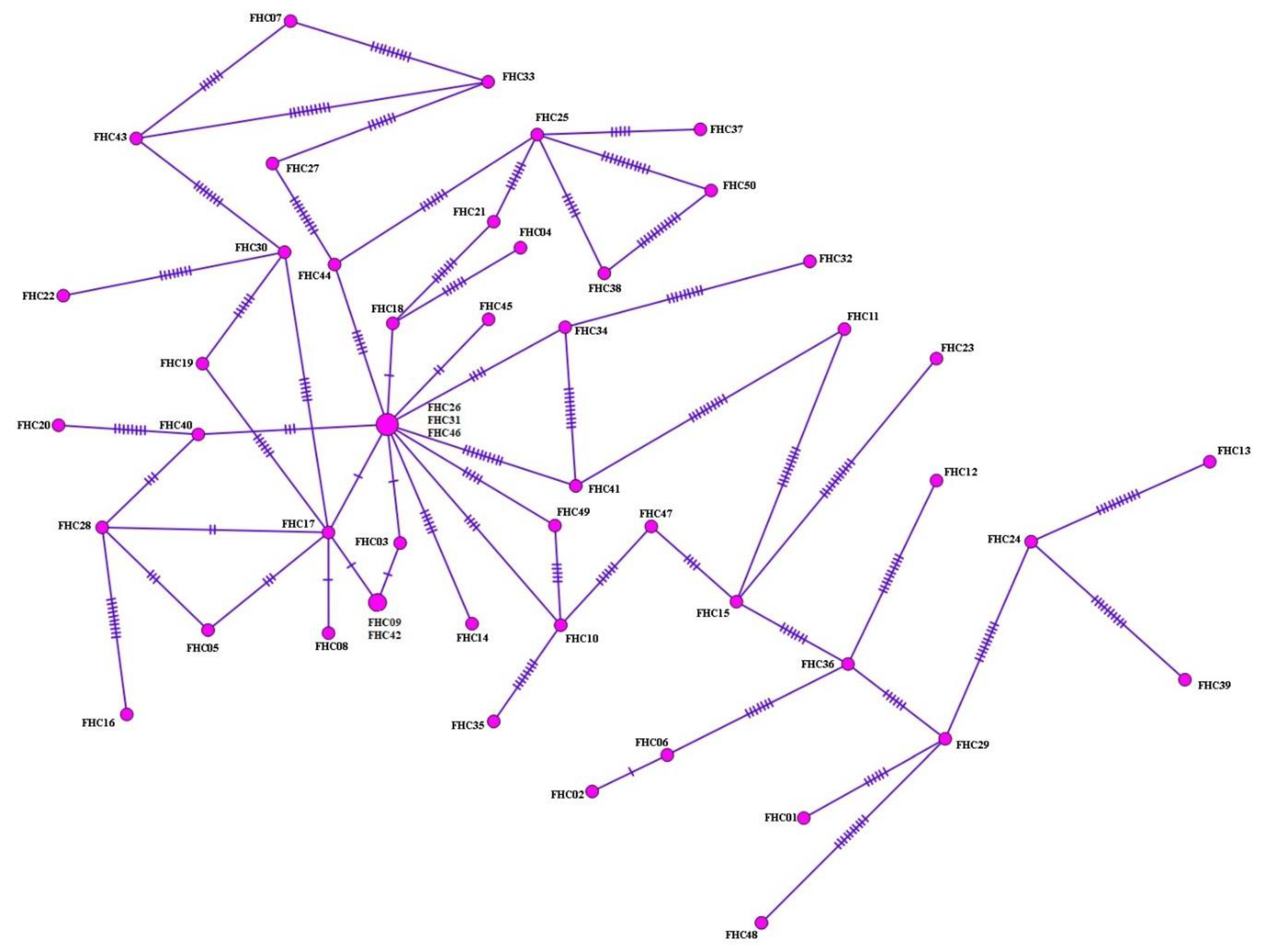
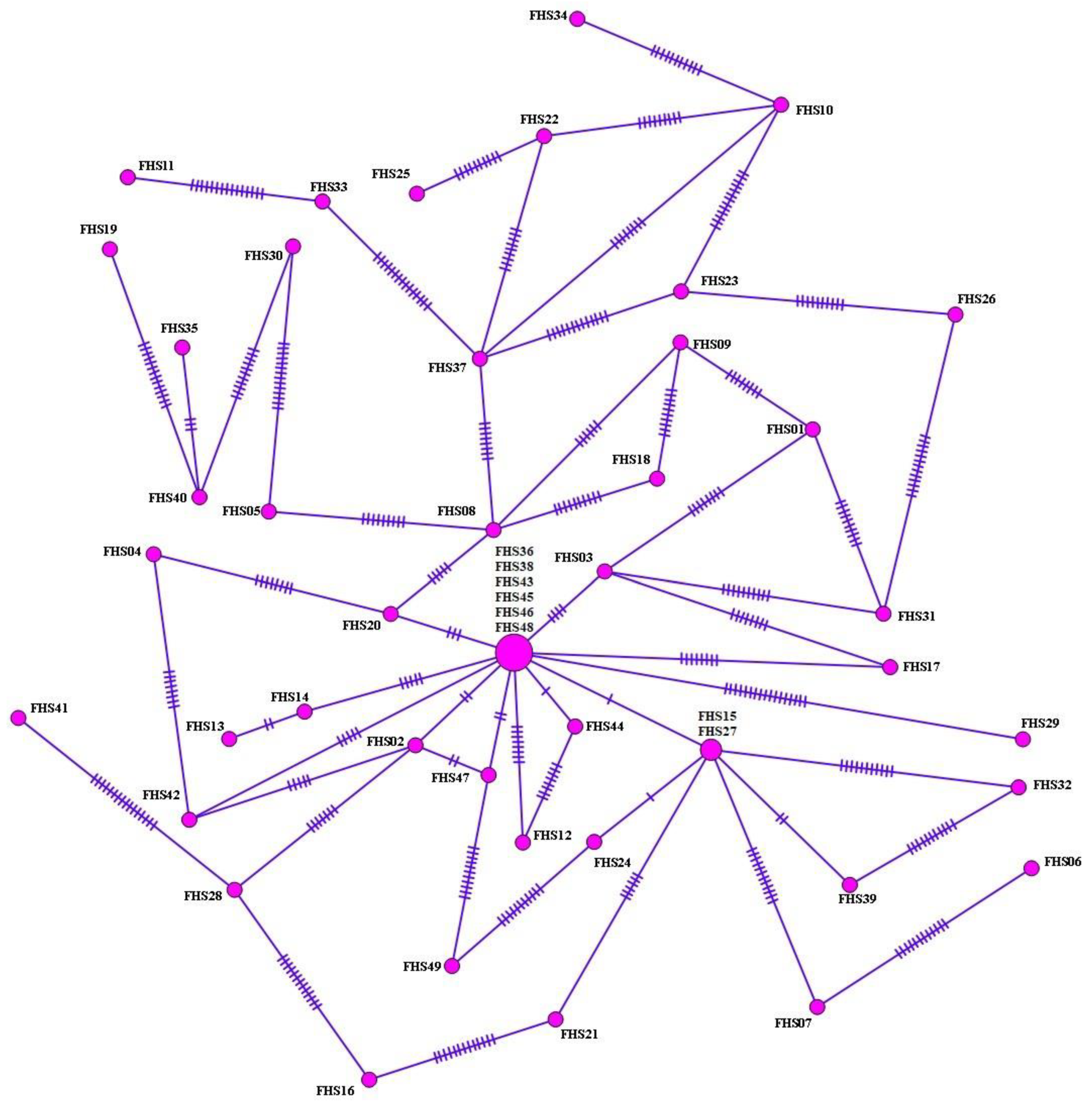
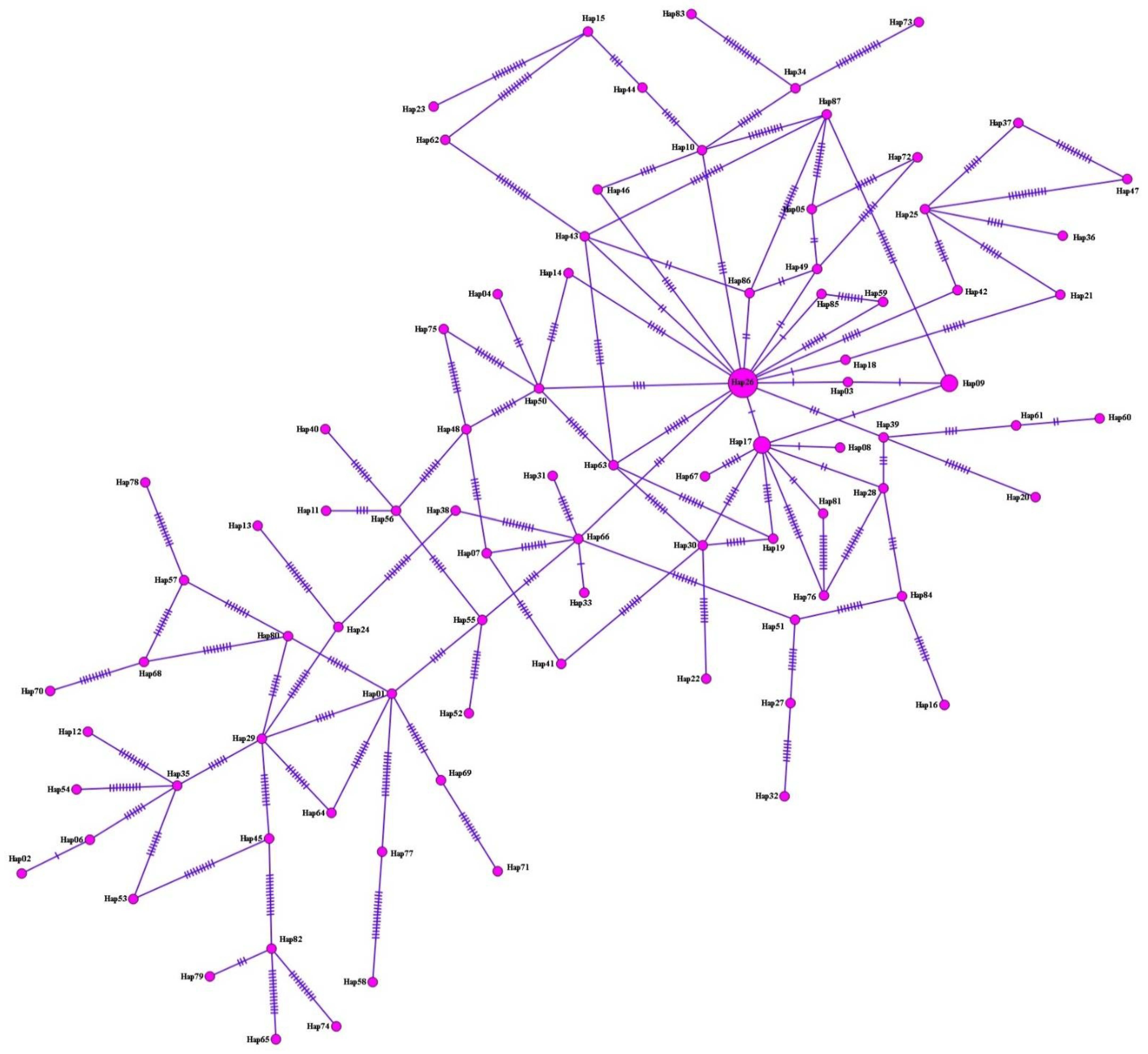
2.6. Haplotype Analysis

Sequencing data were exported to DnaSP 6 [21]. Population diversity indices (haplotype numbers (h), haplotype diversity (Hd), and nucleotide diversity (π)) and neutrality indices (Tajima’s D test [22], Fu’s statistics [23], and Fu and Li’s D and F tests [24]) were calculated using DnaSP 6 [21]. DnaSP 6 was used to create various output formats (e.g., NEXUS, which allows the user to add additional data for further analyses). A haplotype network was generated using PopART 1.7 (http://popart.otago.ac.nz) by applying the minimum spanning networks method, which contains all edges appearing in a minimum spanning tree [25].

3. Results

3.1. Multiplex PCR Data

pepck was subjected to multiplex PCR analysis with specific primers. Samples that generated a 510 bp band were considered to be F. gigantica, those that generated a 241 bp band were considered to be F. hepatica, and samples that generated both bands were considered to be hybrid forms (aspermic flukes). Of 102 Fasciola spp. isolates, only 4 (2 cattle and 2 sheep) generated a 510 bp band, and 84 (44 cattle and 40 sheep) generated a 241 bp band. Consequently, they were identified as F. gigantica and F. hepatica, respectively. Finally, 14 (five cattle and nine sheep) of 102 Fasciola spp. isolates generated both bands, and they were thus identified as hybrid forms.

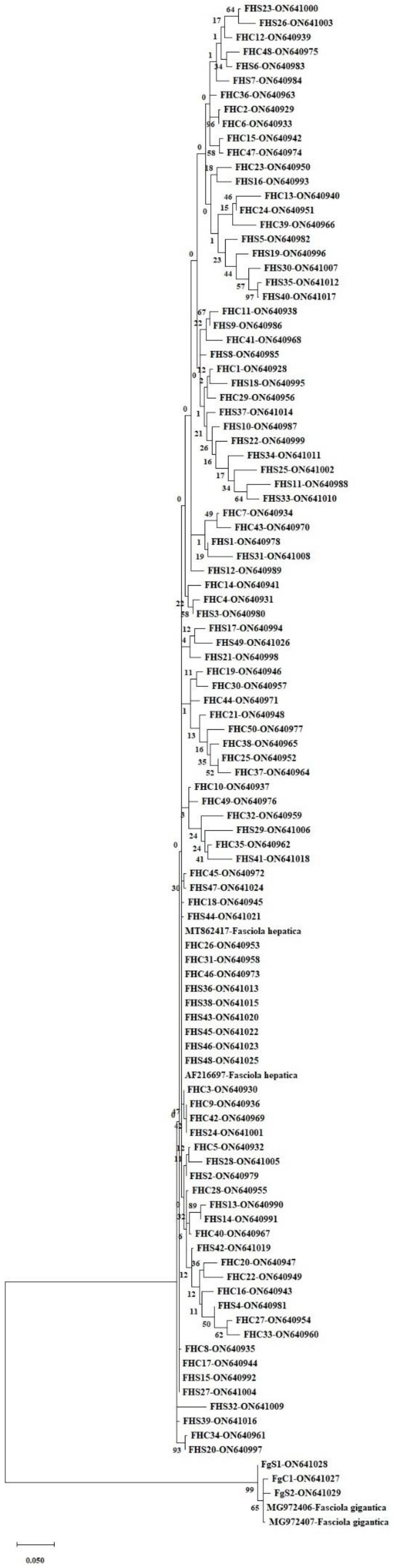
3.2. nad1 Sequence Analysis

Unidirectional DNA sequence analysis of nad1 (660 bp) of Fasciola spp. isolates (n = 102) was performed. Sequences were trimmed after being compared with published sequence data using BLAST, yielding sequences of 565 bp. Consequently, 3 sequences (encoding FGC01, FGS01, and FGS02), 2 from sheep and 1 from cattle, were identified as F. gigantica and 99 (FHC01–FHC50 and FHS01–FHS49), 50 from cattle and 49 from sheep, were identified as F. hepatica. All sequences were registered in GenBank. Figure 1 shows the phylogenetic tree constructed based on F. gigantica (n = 3) and F. hepatica (n = 99).
Table S1 shows the nuclear and mitochondrial DNA analysis findings of the flukes used in this study.

3.3. Haplotype Analysis, Nucleotide Polymorphism, and Diversity and Neutrality Indices

Haplotype analyses were performed on 50 samples identified as F. hepatica based upon nad1 sequence analysis of cattle isolates. From the haplotype network, 47 haplotypes were identified, arranged in a star-like configuration with the main haplotype, separated from other haplotypes by 1–22 mutation steps. They covered 6% (3/50) of total isolates (Figure 2). In addition, with regard to nad1 sequences, 68 polymorphic domains, 86.8% (59/68) of which were parsimony-informative sites, were detected. Additionally, high-haplotype and low-nucleotide diversities were observed (Table 1). Tajima’s D value was negative, expressing population expansion and/or selection purification. Significantly, negative Fu’s Fs values were obtained, indicating the presence of rare haplotypes, as would be expected from recent population expansion. Overall, 95.7% (45/47) of haplotype groups consisted of single haplotypes.
Similarly, haplotype analysis was performed in 49 samples identified as F. hepatica based upon nad1 sequence analysis of sheep isolates. From a haplotype network, 43 haplotypes were identified, arranged in a star-like configuration with the main haplotype, separated from other haplotypes by 1–26 mutation steps, and covering 12.2% (6/49) of total isolates (Figure 3). With regard to nad1 sequences, 81 polymorphic domains were detected, 95.1% (77/81) of which were parsimony-informative sites. High-haplotype and low-nucleotide diversities were observed. Tajima’s D value was negative, expressing population expansion and/or selection purification. Significantly negative Fu’s Fs values were obtained, indicating the presence of rare haplotypes as expected from recent population expansion (Table 2). In total, 95.3% (41/43) of haplotype groups consisted of single haplotypes.
With regard to F. hepatica, on performing haplotype analysis of both cattle and sheep isolates (n = 99) for nad1, a common main haplotype was identified, separated from other haplotypes by 1–26 mutation steps. Overall, 87 haplotypes were detected, covering 9.1% (9/99) of total isolates (Figure 4, Table 3). For nad1 sequences, 97 polymorphic domains were found, 92.8% (90/97) of which were parsimony-informative sites. High-haplotype and low-nucleotide diversities were observed. Tajima’s D value was negative, expressing population expansion and/or selection purification. Fu’s Fs values were significantly negative, indicating the presence of rare haplotypes, as expected from recent population expansion (Table 4). Additionally, 96.5% (84/87) of haplotype groups consisted of single haplotypes.
Of the 87 haplotypes detected for F. hepatica nad1 sequences (n = 99), only 4 have been previously reported by other researchers, while the remaining 83 have been reported for the first time in the current study. Hap26, a previously identified haplotype, was the main haplotype in this study. Nine sequences of this haplotype showed a 100% match with a previously registered F. hepatica sequence (AF216697) in GenBank. Hap17, another previously identified haplotype, was the second main haplotype in this study. Although the sequences of this haplotype showed a 100% match with a previously reported F. hepatica sequence (MK468850), they were different from Hap26 by a single nucleotide. Similarly, Hap18 (FHC18) sequence was 100% identical to MG972379, but it was different from Hap26 by one nucleotide. Hap66 (FHS20) sequences were 100% identical to MN594514 and differed from Hap26 by three nucleotides. The 83 haplotypes identified for the first time in this study matched with F. hepatica sequences in GenBank at rates ranging from 96.11% to 99.82%. Although Hap03 (FHC03) and Hap85 (FHS44) sequences showed a 99.82% match with F. hepatica sequences, a single nucleotide was different between them and Hap26. Finally, the Hap58 (FHS11) sequence, which differed from Hap26 by as many as 26 nucleotides, was 96.11% identical to F. hepatica sequences.
Table S2 shows nucleotide variation positions of nad1 (565 bp) among the 87 haplotypes detected in this study.

4. Discussion

Fasciolosis is a major and highly prevalent helminth infection, which is common in animals. The disease adversely affects the livestock sector, causing economic losses such as low fertility as well as meat, milk, and wool yield losses. Fasciolosis is mainly caused by F. hepatica and F. gigantica. F. hepatica is primarily distributed in the northern parts of Europe, America, Oceania, and Asia, while F. gigantica is mostly distributed in Africa and southern Asia [1]. In addition to these more recognized species, hybrid Fasciola flukes, resulting from cross-fertilization, have been found to be distributed in East, Southeast, and South Asian countries [10,11,12,26,27,28,29,30,31,32,33,34]. A few previous studies [19,34] have reported the differentiation of Fasciola isolates by molecular and morphometric analyses; however, these methods remain inadequate for the identification of hybrid species.
Ref. [19] performed PCR–RFLP analysis of partial mt-CO1 fragments using Turkish cattle isolates of F. hepatica (n = 16) and F. gigantica (n = 1). After digesting mt-CO1 PCR product with AluI, the RFLP profile of F. hepatica revealed two fragments (approximately 330 and 110 bp); however, the PCR product belonging to F. gigantica was not excised. Similarly, RsaI digestion generated two fragments (approximately 190 and 280 bp) from F. gigantica, but none from F. hepatica. Sequence analysis and alignment results revealed 100% and 98% identity for F. hepatica and F. gigantica isolates, respectively. As the existence of hybrid forms was unknown at the time, their status was not revealed [19].
New nuclear markers, in particular pepck and pold, have been successfully used to differentiate among F. hepatica, F. gigantica, and hybrid Fasciola flukes [10,11,12,26,27,28,29,30,31,32,33,34]. In addition, mt-CO1 and nad1 have been used to analyze intraspecies phylogenetic relationships among Fasciola spp. [11].
In a study, 92 Fasciola flukes collected from sheep in Kabul, Afghanistan were subjected to multiplex PCR. All were identified as F. hepatica based on pepck and pold screening. Although the pepck fragment pattern was unable to distinguish the species of seven Fasciola isolates, pepck nucleotide sequence data validated that they were F. hepatica [35]. Further, Tashiro et al. [36] analyzed 68 Fasciola isolates collected from high-plateau and steppe areas of Algeria using multiplex PCR and PCR–RFLP for pepck and pold, respectively. In their study, multiplex PCR identified 49 isolates as F. hepatica, one as F. gigantica, and 18 as hybrids.
Variations in pepck nucleotides can affect primer specificity, which may cause errors in fragment analysis of multiplex PCR products. Therefore, results must be validated by performing mt-CO1 or nad1 sequence analysis [35]. In this study, Fasciola isolates were analyzed by multiplex PCR and pepck-specific primers, and nad1 sequence analysis was performed to identify genetic differences.
In previous studies in Turkey, liver flukes (usually obtained after slaughter) were identified as F. hepatica, but species discrimination was not conducted [37,38,39,40,41]. A morphometric and molecular study of Fasciola spp. from ruminants was conducted in Iran, and found that 48 (88.9%) of 54 Fasciola isolates were F. hepatica and 4 (7.4%) were F. gigantica [42]. Further, another study investigated the genetic diversity of Fasciola spp. in Armenia, and on morphological identification and sequencing of nad1, 55 specimens were identified as F. hepatica and 7 as F. gigantica [43]. Furthermore, 4 of 87 haplotypes detected in the current study for F. hepatica nad1 sequences (n = 99) have been previously reported, while 83 were revealed for the first time in this study. One of the four previously reported haplotypes was from Australia, one was from Armenia, and two were from Iran. Hap26 was the main haplotype in this study; this haplotype was obtained using whole-genome sequencing of an F. hepatica isolate from Australia. Nine sequences of this haplotype showed a 100% match with a previously registered F. hepatica sequence (AF216697). Hap17, another previously determined haplotype, was the second main haplotype, and its sequence showed 100% match with a previously reported sequence of F. hepatica (MK468850), which was isolated from Iranian cattle. Hap17 and Hap26 sequences differed by a single nucleotide. Additionally, Hap18 (FHC18) sequence showed a 100% match with MG972379, which was obtained from a sheep isolate of Armenian origin. Hap18 and Hap26 sequences also differed by a single nucleotide. Furthermore, Hap66 (FHS20) sequence showed a 100% match with MN594514, which was obtained from a sheep isolate of Iranian origin. Hap66 and Hap26 sequences differed by three nucleotides [44]. The 83 haplotypes detected for the first time in this study matched with F. hepatica sequences in GenBank at rates ranging from 96.11% to 99.82%. One of the reasons for the high number of haplotypes detected in Turkey could be natural selection, which may lead to genetic differences between species. Diverse biological effects, such as different intermediate host preferences, tolerance to low or high temperatures, and sensitivity of parasites to anthelmintic drugs, could be responsible for genetic differences, resulting in haplotype diversity.

5. Conclusions

To summarize, the present study’s analyses led to the identification of hybrid Fasciola flukes, as well as 83 different haplotypes of F. hepatica, for the first time in Turkey. In the case of countries such as Turkey, where pasture farming is common and populations of cattle and sheep are large, the existence of hybrid F. gigantica and F. hepatica flukes is a major concern. Their presence may lead to the emergence of drug-resistant parasites and development of diseases that are more difficult and expensive to control. Consequently, not only animal but also human health can be endangered. Therefore, the implementation and maintenance of prevention and control strategies in fascioliasis-endemic regions can become a challenge.

Supplementary Materials

The following supporting information can be downloaded at: https://www.mdpi.com/article/10.3390/pathogens11111235/s1. Table S1. Profiles of Fasciola flukes; Table S2. Nucleotide variation positions of nad1 (565 bp) sequences among the 87 haplotypes analyzed in this study.

Author Contributions

Conceptualization, Investigation, Formal Analysis, V.U.; Conceptualization, Investigation, Formal Analysis, Writing—Original Draft, F.C.; Investigation, Writing—Review and Editing, Project Administration, Funding Acquisition, Supervision, S.S.; Formal Analysis, H.K.K.; Formal Analysis, S.G.K.; Writing—Review and Editing, X.Z.; Investigation, Writing—Review and Editing, H.A.; Funding Acquisition, Writing—Review & Editing, J.C. All authors have read and agreed to the published version of the manuscript.

Funding

This work was supported by the Scientific and Technological Research Council of Turkey (TÜBİTAK) (grant number 120O865 to S.S.), and the National Natural Science Foundation of China (grant numbers Nos. 81971969, 82272369, and 81772225 to J.C.).

Institutional Review Board Statement

Not applicable.

Informed Consent Statement

Not applicable.

Data Availability Statement

Not applicable.

Acknowledgments

We acknowledge all veterinarians for their excellent technical assistance in collecting parasite samples.

Conflicts of Interest

The authors declare no conflict of interest.

References

  1. Mas-Coma, S.; Valero, M.; Bargues, M. Fascioliasis. Adv. Exp. Med. Biol. 2019, 1154, 71–103. [Google Scholar] [PubMed]
  2. Amiri, S.; Shemshadi, B.; Shirali, S.; Kheirandish, F.; Fallahi, S. Accurate and rapid detection of Fasciola hepatica copro-DNA in sheep using loop-mediated isothermal amplification (LAMP) technique. Vet. Med. Sci. 2021, 7, 1316–1324. [Google Scholar] [CrossRef]
  3. Carnevale, S.; Malandrini, J.B.; Pantano, M.L.; Soria, C.C.; Rodrigues-Silva, R.; Machado-Silva, J.R.; Velásquez, J.N.; Kamenetzky, L. First genetic characterization of Fasciola hepatica in Argentina by nuclear and mitochondrial gene markers. Vet. Parasitol. 2017, 245, 34–38. [Google Scholar] [CrossRef] [PubMed]
  4. Bozorgomid, A.; Rouhani, S.; Harandi, M.F.; Ichikawa-Seki, M.; Raeghi, S. Genetic diversity and distribution of Fasciola hepatica haplotypes in Iran: Molecular and phylogenetic studies. Vet. Parasitol. Reg. Stud. Rep. 2020, 19, 100359. [Google Scholar] [CrossRef] [PubMed]
  5. Terasaki, K.; Itagaki, T.; Shibahara, T.; Noda, Y.; Moriyama-Gonda, N. Comparative study of the reproductive organs of Fasciola groups by optical microscope. J. Vet. Med. Sci. 2001, 63, 735–742. [Google Scholar] [CrossRef] [PubMed] [Green Version]
  6. Itagaki, T.; Ichinomiya, M.; Fukuda, K.; Fusyuku, S.; Carmona, C. Hybridization experiments indicate incomplete reproductive isolating mechanism between Fasciola hepatica and Fasciola gigantica. Parasitology 2011, 138, 1278–1284. [Google Scholar] [CrossRef] [PubMed]
  7. Amor, N.; Farjallah, S.; Merella, P.; Alagaili, A.N.; Mohammed, O.B. Multilocus approach reveals discordant molecular markers and corridors for gene flow between North African populations of Fasciola hepatica. Vet. Parasitol. 2020, 278, 109035. [Google Scholar] [CrossRef]
  8. Moazeni, M.; Sharifiyazdi, H.; Izadpanah, A. Characterization of Fasciola hepatica genotypes from cattle and sheep in Iran using cytochrome C oxidase gene (CO1). Parasitol. Res. 2012, 110, 2379–2384. [Google Scholar] [CrossRef]
  9. Ashrafi, K.; Bargues, M.D.; O’Neill, S.; Mas-Coma, S. Fascioliasis: A worldwide parasitic disease of importance in travel medicine. Travel Med. Infect. Dis. 2014, 12, 636–649. [Google Scholar] [CrossRef]
  10. Itagaki, T.; Kikawa, M.; Sakaguchi, K.; Shimo, J.; Terasaki, K.; Shibahara, T.; Fukuda, K. Genetic characterization of parthenogenic Fasciola sp. in Japan on the basis of the sequences of ribosomal and mitochondrial DNA. Parasitology 2005, 131, 679–685. [Google Scholar] [CrossRef]
  11. Itagaki, T.; Kikawa, M.; Terasaki, K.; Shibahara, T.; Fukuda, K. Molecular characterization of parthenogenic Fasciola sp. in Korea on the basis of DNA sequences of ribosomal ITS1 and mitochondrial NDI gene. J. Vet. Med. Sci. 2005, 67, 1115–1118. [Google Scholar] [CrossRef] [PubMed] [Green Version]
  12. Itagaki, T.; Sakaguchi, K.; Terasaki, K.; Sasaki, O.; Yoshihara, S.; Van Dung, T. Occurrence of spermic diploid and aspermic triploid forms of Fasciola in Vietnam and their molecular characterization based on nuclear and mitochondrial DNA. Parasitol. Int. 2009, 58, 81–85. [Google Scholar] [CrossRef]
  13. Peng, M.; Ichinomiya, M.; Ohtori, M.; Ichikawa, M.; Shibahara, T.; Itagaki, T. Molecular characterization of Fasciola hepatica, Fasciola gigantica, and aspermic Fasciola sp. in China based on nuclear and mitochondrial DNA. Parasitol. Res. 2009, 105, 809–815. [Google Scholar] [CrossRef] [PubMed]
  14. Miyazaki, T.; Kobayashi, T. Visualization of the dynamic behavior of ribosomal RNA gene repeats in living yeast cells. Gen. Cells 2011, 16, 491–502. [Google Scholar] [CrossRef] [PubMed]
  15. Shoriki, T.; Ichikawa-Seki, M.; Suganuma, K.; Naito, I.; Hayashi, K.; Nakao, M.; Aita, J.; Mohanta, U.K.; Inoue, N.; Murakami, K. Novel methods for the molecular discrimination of Fasciola spp. on the basis of nuclear protein-coding genes. Parasitol. Int. 2016, 65, 180–183. [Google Scholar] [CrossRef]
  16. Thang, T.N.; Vázquez-Prieto, S.; Vilas, R.; Paniagua, E.; Ubeira, F.M.; Ichikawa-Seki, M. Genetic diversity of Fasciola hepatica in Spain and Peru. Parasitol. Int. 2020, 76, 102100. [Google Scholar] [CrossRef]
  17. Giovanoli Evack, J.; Schmidt, R.S.; Boltryk, S.D.; Voss, T.S.; Batil, A.A.; Ngandolo, B.N.; Greter, H.; Utzinger, J.; Zinsstag, J.; Balmer, O. Molecular confirmation of a Fasciola gigantica× Fasciola hepatica hybrid in a Chadian bovine. J. Parasitol. 2020, 106, 316–322. [Google Scholar] [CrossRef] [Green Version]
  18. Javanmard, E.; Ohari, Y.; Sadeghi, A.; Cheraghipour, K.; Aghdaei, H.A.; Mirjalali, H.; Zali, M.R.; Itagaki, T. Multigene typing and phylogenetic analysis of Fasciola from endemic foci in Iran. Infect. Genet. Evol. 2020, 80, 104202. [Google Scholar] [CrossRef]
  19. Simsek, S.; Utuk, A.; Balkaya, I. Molecular differentiation of Turkey cattle isolates of Fasciola hepatica and Fasciola gigantica. Helminthologia 2011, 48, 3–7. [Google Scholar] [CrossRef] [Green Version]
  20. Kumar, S.; Stecher, G.; Li, M.; Knyaz, C.; Tamura, K. MEGA X: Molecular evolutionary genetics analysis across computing platforms. Mol. Biol. Evol. 2018, 35, 1547. [Google Scholar] [CrossRef]
  21. Rozas, J.; Ferrer-Mata, A.; Sánchez-DelBarrio, J.C.; Guirao-Rico, S.; Librado, P.; Ramos-Onsins, S.E.; Sánchez-Gracia, A. DnaSP 6: DNA sequence polymorphism analysis of large data sets. Mol. Biol. Evol. 2017, 34, 3299–3302. [Google Scholar] [CrossRef] [PubMed]
  22. Tajima, F. Statistical method for testing the neutral mutation hypothesis by DNA polymorphism. Genetics 1989, 123, 585–595. [Google Scholar] [CrossRef] [PubMed]
  23. Fu, Y.-X. Statistical tests of neutrality of mutations against population growth, hitchhiking and background selection. Genetics 1997, 147, 915–925. [Google Scholar] [CrossRef]
  24. Fu, Y.-X.; Li, W.-H. Statistical tests of neutrality of mutations. Genetics 1993, 133, 693–709. [Google Scholar] [CrossRef] [PubMed]
  25. Bandelt, H.-J.; Forster, P.; Röhl, A. Median-joining networks for inferring intraspecific phylogenies. Mol. Biol. Evol. 1999, 16, 37–48. [Google Scholar] [CrossRef]
  26. Hayashi, K.; Ichikawa-Seki, M.; Mohanta, U.K.; Shoriki, T.; Chaichanasak, P.; Itagaki, T. Hybrid origin of Asian aspermic Fasciola flukes is confirmed by analyzing two single-copy genes, pepck and pold. J. Vet. Med. Sci. 2017, 80, 98–102. [Google Scholar] [CrossRef] [Green Version]
  27. Hayashi, K.; Ichikawa-Seki, M.; Allamanda, P.; Wibowo, P.E.; Mohanta, U.K.; Guswanto, A.; Nishikawa, Y. Molecular characterization and phylogenetic analysis of Fasciola gigantica from western Java, Indonesia. Parasitol. Int. 2016, 65, 424–427. [Google Scholar] [CrossRef]
  28. Hayashi, K.; Mohanta, U.K.; Neeraja, T.; Itagaki, T. Molecular characterization of Fasciola gigantica in Delhi, India and its phylogenetic relation to the species from South Asian countries. J. Vet. Med. Sci. 2016, 78, 1529–1532. [Google Scholar] [CrossRef] [Green Version]
  29. Ichikawa, M.; Itagaki, T. Discrimination of the ITS1 types of Fasciola spp. based on a PCR–RFLP method. Parasitol. Res. 2010, 106, 757–761. [Google Scholar] [CrossRef]
  30. Ichikawa-Seki, M.; Peng, M.; Hayashi, K.; Shoriki, T.; Mohanta, U.K.; Shibahara, T.; Itagaki, T. Nuclear and mitochondrial DNA analysis reveals that hybridization between Fasciola hepatica and Fasciola gigantica occurred in China. Parasitology 2017, 144, 206–213. [Google Scholar] [CrossRef]
  31. Ichikawa, M.; Bawn, S.; Maw, N.N.; Htun, L.L.; Thein, M.; Gyi, A.; Sunn, K.; Katakura, K.; Itagaki, T. Characterization of Fasciola spp. in Myanmar on the basis of spermatogenesis status and nuclear and mitochondrial DNA markers. Parasitol. Int. 2011, 60, 474–479. [Google Scholar] [CrossRef] [PubMed]
  32. Chaichanasak, P.; Ichikawa, M.; Sobhon, P.; Itagaki, T. Identification of Fasciola flukes in Thailand based on their spermatogenesis and nuclear ribosomal DNA, and their intraspecific relationships based on mitochondrial DNA. Parasitol. Int. 2012, 61, 545–549. [Google Scholar] [CrossRef] [PubMed]
  33. Shoriki, T.; Ichikawa-Seki, M.; Devkota, B.; Rana, H.B.; Devkota, S.P.; Humagain, S.K.; Itagaki, T. Molecular phylogenetic identification of Fasciola flukes in Nepal. Parasitol. Int. 2014, 63, 758–762. [Google Scholar] [CrossRef] [PubMed]
  34. Mohanta, U.K.; Ichikawa-Seki, M.; Shoriki, T.; Katakura, K.; Itagaki, T. Characteristics and molecular phylogeny of Fasciola flukes from Bangladesh, determined based on spermatogenesis and nuclear and mitochondrial DNA analyses. Parasitol. Res. 2014, 113, 2493–2501. [Google Scholar] [CrossRef] [PubMed]
  35. Thang, T.N.; Hakim, H.; Rahimi, R.R.; Ichikawa-Seki, M. Molecular analysis reveals expansion of Fasciola hepatica distribution from Afghanistan to China. Parasitol. Int. 2019, 72, 101930. [Google Scholar] [CrossRef]
  36. Tashiro, M.; Zokbi, Z.; Chibout, Y.; Megrane, S.; Mebarka, F.; Ichikawa-Seki, M. Molecular characterization and phylogenetic analysis of Fasciola hepatica from high-plateau and steppe areas in Algeria. Parasitol. Int. 2021, 80, 102234. [Google Scholar]
  37. Aydenizöz, M.; Yildiz, K. Kırıkkale’de kesilen koyunlarda karaciğer trematodlarının yaygınlığı. Türkiye Parazitol. Derg. 2002, 26, 317–319. [Google Scholar]
  38. Toparlak, M. Van ili belediye mezbahasinda kesilen koyunlarda karaciğer trematod enfeksiyonlari üzerinde araştirmalar. Ankara Üniv. Vet. Fak. Derg. 1988, 35, 270–274. [Google Scholar]
  39. Değer, S.; Akgül, Y.; Ağaoğlu, Z.T.; Tasçi, S. Van ve yöresinde Fasciola gigantica’dan ileri gelen fascioliasis enfeksiyonlarinin epidemiyolojisi ve ekolojisiüzerinde araştirmalar. Yüzüncü Yıl Üniv. Vet. Fak. Derg. 1992, 3, 133–140. [Google Scholar]
  40. Akyol, Ç.V. Bursa ortak girişim tesislerinde (ETBA) kesilen koyunlarda Distomatozis’ in yayılışı. J. Fac. Vet. Med. 2001, 20, 23–27. [Google Scholar]
  41. Balkaya, İ.; Şimşek, S. Erzurum’da kesilen sığırlarda Hidatidosis ve Fasciolosis’ in yaygınlığı ve ekonomik önemi. Kafkas Univ. Vet. Fak. Derg. 2010, 16, 793–797. [Google Scholar]
  42. Akhlaghi, E.; Mohammadi, M.A.; Ziaali, N.; Baneshi, M.R.; Nasibi, S.; Kamyabi, H.; Rostami, S.; Harandi, M.F. Morphometric and molecular study of Fasciola isolates from ruminants in Iran. Türkiye Parazitol. Derg. 2017, 41, 192. [Google Scholar] [CrossRef] [PubMed] [Green Version]
  43. Aghayan, S.; Gevorgian, H.; Ebi, D.; Atoyan, H.A.; Addy, F.; Mackenstedt, U.; Romig, T.; Wassermann, M. Fasciola spp. in Armenia: Genetic diversity in a global context. Vet. Parasitol. 2019, 268, 21–31. [Google Scholar] [CrossRef] [PubMed]
  44. Cwiklinski, K.; Dalton, J.P.; Dufresne, P.J.; La Course, J.; Williams, D.J.; Hodgkinson, J.; Paterson, S. The Fasciola hepatica genome: Gene duplication and polymorphism reveals adaptation to the host environment and the capacity for rapid evolution. Genome Biol. 2015, 16, 71. [Google Scholar] [CrossRef] [PubMed]
Figure 1. Fasciola hepatica phylogenetic tree constructed using nad1 sequence of ON640928–ON640977 (FHC01-FHC50) and ON640978–ON641026 (FHS01-FHS49). F. gigantica phylogenetic tree constructed using nad1 sequences of ON641027 (FGC01), and ON641028-ON641029 (FGS01-FGS02) isolates. Reference sequences of F. hepatica were AF216697 and MT862417, and of F. gigantica were MG972406 and MG972407. The tree was created based on the GTR + G + I model with the maximum likelihood method in MEGA X, and reliability was assessed by performing 1000 bootstrap tests.
Pathogens 11 01235 g001
Figure 2. Haplotype network for nad1 of cattle isolates (565 bp) of F. hepatica. Circle size is proportional to the frequency of each haplotype. Numbers of mutations that distinguish haplotypes are indicated by dashes.
Pathogens 11 01235 g002
Figure 3. Haplotype network for nad1 of sheep isolates (565 bp) of F. hepatica. Circle size is proportional to the frequency of each haplotype. Number of mutations that distinguish haplotypes is indicated by dashes.
Pathogens 11 01235 g003
Figure 4. Haplotype network for nad1 of both cattle and sheep isolates (565 bp) of F. hepatica. Circle size is proportional to the frequency of each haplotype. Number of mutations that distinguish haplotypes is indicated by dashes.
Pathogens 11 01235 g004
Table 1. Diversity and neutrality indices obtained using nucleotide data of nad1 (565 bp) of F. hepatica in cattle isolates.
nhnhd ± SDπd ± SDTajima’s Dp ValueFu’s Fsp ValueFLDp ValueFLFp Value
50470.997 ± 0.0050.02423 ± 0.00159−0.43557p > 0.10−35.5370.0000.76537p > 0.100.38471p > 0.10
n: Isolate number, hn: haplotype number; hd: haplotype diversity; πd: nucleotide diversity; SD: standard deviation; FLD: Fu and Li’s D * statistic test; FLF: Fu and Li’s F * statistic test. p value: statistically not significant (p > 0.10).
Table 2. Diversity and neutrality indices obtained using nucleotide data of nad1 (565 bp) of F. hepatica sheep isolates.
nhnhd ± SDπd ± SDTajima’s Dp ValueFu’s Fsp ValueFLDp ValueFLFp Value
49430.986 ± 0.0110.02810 ± 0.00220−0.62751p > 0.10−22.7000.0001.23614p > 0.100.65299p > 0.10
n: Isolate number, hn: haplotype number; hd: haplotype diversity; πd: nucleotide diversity; SD: standard deviation; FLD: Fu and Li’s D * statistic test; FLF: Fu and Li’s F * statistic test. p value: statistically not significant (p > 0.10).
Table 3. Distribution of the haplotype network created for nad1 of F. hepatica for both cattle and sheep isolates. Number of isolates and their codes are shown.
NumberHaplotype NameIsolate No.Isolate Codes (Accession No.)
1Hap011[FHC01-ON640928]
2Hap021[FHC02-ON640929]
3Hap031[FHC03-ON640930]
4Hap041[FHC04-ON640931]
5Hap051[FHC05-ON640932]
6Hap061[FHC06-ON640933]
7Hap071[FHC07-ON640934]
8Hap081[FHC08-ON640935]
9Hap093[FHC09-ON640936 FHC42-ON640969 FHS24-ON641001]
10Hap101[FHC10-ON640937]
11Hap111[FHC11-ON640938]
12Hap121[FHC12-ON640939]
13Hap131[FHC13-ON640940]
14Hap141[FHC14-ON640941]
15Hap151[FHC15-ON640942]
16Hap161[FHC16-ON640943]
17Hap171[FHC17-ON640944 FHS15-ON640992 FHS27-ON641004]
18Hap181[FHC18-ON640945]
19Hap191[FHC19-ON640946]
20Hap201[FHC20-ON640947]
21Hap211[FHC21-ON640948]
22Hap221[FHC22-ON640949]
23Hap231[FHC23-ON640950]
24Hap241[FHC24-ON640951]
25Hap251FHC25-ON640952]
26Hap269[FHC26-ON640953 FHC31-ON640958 FHC46-ON640973 FHS36-ON641013 FHS38-ON641015 FHS43-ON641020 FHS45-ON641022 FHS46-ON641023 FHS48-ON641025]
27Hap271[FHC27-ON640954]
28Hap281[FHC28-ON640955]
29Hap291[FHC29-ON640956]
30Hap301[FHC30-ON640957]
31Hap311[FHC32-ON640959]
32Hap321[FHC33-ON640960]
33Hap331[FHC34-ON640961]
34Hap341[FHC35-ON640962]
35Hap351[FHC36-ON640963]
36Hap361[FHC37-ON640964]
37Hap371[FHC38-ON640965]
38Hap381[FHC39-ON640966]
39Hap391[FHC40-ON640967]
40Hap401[FHC41-ON640968]
41Hap411[FHC43-ON640970]
42Hap421[FHC44-ON640971]
43Hap431[FHC45-ON640972]
44Hap441[FHC47-ON640974]
45Hap451[FHC48-ON640975]
46Hap461[FHC49-ON640976]
47Hap471[FHC50-ON640977]
48Hap481[FHS01-ON640978]
49Hap491[FHS02-ON640979]
50Hap501[FHS03-ON640980]
51Hap511[FHS04-ON640981]
52Hap521[FHS05-ON640982]
53Hap531[FHS06-ON640983]
54Hap541[FHS7-ON640984]
55Hap551[FHS8-ON640985]
56Hap561[FHS9-ON640986]
57Hap571[FHS10-ON640987]
58Hap581[FHS11-ON640988]
59Hap591[FHS12-ON640989]
60Hap601[FHS13-ON640990]
61Hap611[FHS14-ON640991]
62Hap621[FHS16-ON640993]
63Hap631[FHS17-ON640994]
64Hap641[FHS18-ON640995]
65Hap651[FHS19-ON640996]
66Hap661[FHS20-ON640997]
67Hap671[FHS21-ON640998]
68Hap681[FHS22-ON640999]
69Hap691[FHS23-ON641000]
70Hap701[FHS25-ON641002]
71Hap711[FHS26-ON641003]
72Hap721[FHS28-ON641005]
73Hap731[FHS29-ON641006]
74Hap741[FHS30-ON641007]
75Hap751[FHS31-ON641008]
76Hap761[FHS32-ON641009]
77Hap771[FHS33-ON641010]
78Hap781[FHS34-ON641011]
79Hap791[FHS35-ON641012]
80Hap801[FHS37-ON641014]
81Hap811[FHS39-ON641016]
82Hap821[FHS40-ON641017]
83Hap831[FHS41-ON641018]
84Hap841[FHS42-ON641019]
85Hap851[FHS44-ON641021]
86Hap861[FHS47-ON641024]
87Hap871[FHS49-ON641026]
Table 4. Diversity and neutrality indices obtained using nucleotide data of both cattle and sheep isolates F. hepatica nad1 (565 bp).
nhnhd ± SDπd ± SDTajima’s Dp ValueFu’s Fsp ValueFLDp ValueFLFp Value
99870.991 ± 0.0050.02661 ± 0.00148−0.85844p > 0.10−33.4410.0001.00700p > 0.100.26059p > 0.10
n: Isolate number, hn: haplotype number; hd: haplotype diversity; πd: nucleotide diversity; SD: standard deviation; FLD: Fu and Li’s D * statistic test; FLF: Fu and Li’s F * statistic test. p value: statistically not significant (p > 0.10).
Publisher’s Note: MDPI stays neutral with regard to jurisdictional claims in published maps and institutional affiliations.

