Communication

A Comprehensive Evaluation of Enterobacteriaceae Primer Sets for Analysis of Host-Associated Microbiota

by
Carolina N. Resendiz-Nava
1,
Hilda V. Silva-Rojas
2,
Angel Rebollar-Alviter
3,
Dulce M. Rivera-Pastrana
1,
Edmundo M. Mercado-Silva
1 and
Gerardo M. Nava
1,*
1
Facultad de Quimica, Universidad Autonoma de Queretaro, Cerro de las Campanas S/N, Queretaro 76010, Mexico
2
Posgrado en Recursos Geneticos y Productividad, Produccion de Semillas, Colegio de Postgraduados, Km 36.5 Carretera Mexico-Texcoco, Texcoco 56230, Mexico
3
Centro Regional Morelia, Universidad Autonoma de Chapingo, Michoacan 58170, Mexico
*
Author to whom correspondence should be addressed.
Pathogens 2022, 11(1), 17; https://doi.org/10.3390/pathogens11010017
Submission received: 7 November 2021 / Revised: 17 December 2021 / Accepted: 20 December 2021 / Published: 23 December 2021
(This article belongs to the Special Issue Proteobacteria: A Common Factor in Human Diseases)

Abstract

:
Enterobacteriaceae is one of the most important bacterial groups within the Proteobacteria phylum. This bacterial group includes pathogens, commensal and beneficial populations. Numerous 16S rRNA gene PCR-based assays have been designed to analyze Enterobacteriaceae diversity and relative abundance, and, to the best of our knowledge, 16 primer pairs have been validated, published and used since 2003. Nonetheless, a comprehensive performance analysis of these primer sets has not yet been carried out. This information is of particular importance due to the recent taxonomic restructuration of Enterobacteriaceae into seven bacterial families. To overcome this lack of information, the identified collection of primer pairs (n = 16) was subjected to primer performance analysis using multiple bioinformatics tools. Herein it was revealed that, based on specificity and coverage of the 16S rRNA gene, these 16 primer sets could be divided into different categories: Enterobacterales-, multi-family-, multi-genus- and Enterobacteriaceae-specific primers. These results highlight the impact of taxonomy changes on performance of molecular assays and data interpretation. Moreover, they underline the urgent need to revise and update the molecular tools used for molecular microbial analyses.

1. Introduction

Family Enterobacteriaceae is an important member of the Proteobacteria phylum; this bacterial group comprises numerous genera known to colonize the small and large intestine of mammals, including humans [1]. Enterobacteriaceae includes numerous recognized pathogens and opportunistic bacteria associated with the occurrence of enteric illnesses, urinary tract infections, sepsis and meningitis in humans [2,3,4,5].
Furthermore, microbial molecular analyses have linked Enterobacteriaceae abundance to intestinal disorders such as ulcerative colitis, irritable bowel syndrome, diverticulitis, and Crohn’s disease [6,7,8,9,10]. Furthermore, increased levels of this bacterial group have been associated with the occurrence of non-alcoholic fatty liver disease and colorectal cancer [11,12,13].
Due to its biological importance, PCR-based approaches and massive 16S rRNA gene sequencing have been designed and implemented for analyses of diversity and abundance of the Enterobacteriaceae family in different environmental samples [14,15,16].
Molecular characterization of Enterobacteriaceae by PCR assays requires the use of taxon-specific primers; in the last eighteen years, to the best of our knowledge, 16 different primer sets have been designed, validated and published in scientific journals [15,17,18,19,20,21,22,23,24,25,26,27]. However, a comprehensive evaluation of their performance, specificity and coverage has not yet been carried out. This is of particular significance due to the taxonomic restructuration that the Enterobacteriaceae family underwent in 2016 [28].
After this taxonomic restructuration, the formerly Enterobacteriaceae group was divided into seven new families, Budviciaceae, Enterobacteriaceae, Erwiniaceae, Hafniaceae, Morganellaceae, Pectobacteriaceae and Yersiniaceae [28]. Importantly, this taxonomic upgrade has not been adopted in some of the recently published microbial studies [2,29,30]. Thus, the present study was designed to evaluate specificity and coverage of previously validated and published PCR primer pairs targeting specific amplification of Enterobacteriaceae 16S rRNA genes. The results of the present study provide a comprehensive performance analysis of different primer sets targeting Enterobacteriaceae for analysis of host-associated microbiota.

2. Results

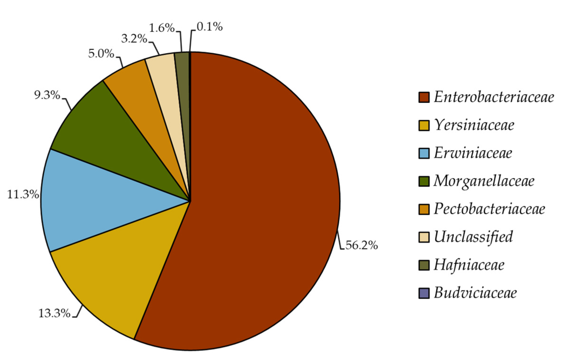
2.1. Reclassification of Formerly Enterobacteriaceae 16S rRNA Gene Sequences

The whole collection of formerly Enterobacteriaceae 16S rRNA gene sequences archived at the RDP database before its most recent taxonomic update (August 2020) are now classified as members of seven different families within Enterobacterales order. The majority (56.2%) of the sequences belong to the Enterobacteriaceae family, followed by Yersiniaceae (13.3%), Erwiniaceae (11.3%), Morganellaceae (9.3%), Pectobacteriaceae (5%), Hafniaceae (1.6%) and Budviciaceae (0.1%) families; interestingly, 3.2% of sequences remained unclassified at the family level (Figure 1).
To manage taxa-associated differences across databases (i.e., LPSN (List of Prokaryotic Names with Standing in Nomenclature [31]), NCBI (National Center for Biotechnology Information [32]), RDP (Ribosomal Database Project [33]) and SILVA [34])) and to drive more accurate comparisons, a multidatabase consensus taxon list comprising 26 genera within the Enterobacteriaceae family was obtained (Table S1) and considered for further evaluations.

2.2. Identification of Primer Sets

A total of 16 primer pairs targeting 16S rRNA genes from formerly Enterobacteriaceae were identified in the literature; these primers were published between 2003–2020 and were labeled Enterobacteriaceae-specific (Table 1). This collection of primer sets could generate PCR amplicons ranging from 49–1485 bp (based on E. coli numbering, accession A14565; Table 1), many of them targeting variable regions V3–V4 (18.8%), followed by V4–V5 (12.5%) (Table S2), based on 16S rRNA variable region numbering described elsewhere [35]. For 50 percent of the primers, the authors of the manuscripts provided a detailed validation process within the Materials and Methods of the publication. Moreover, 31 percent of the primers were validated using at least three Enterobacterales genera; whereas 19 percent of the primers used DNA extracted only from E. coli (Table S2).

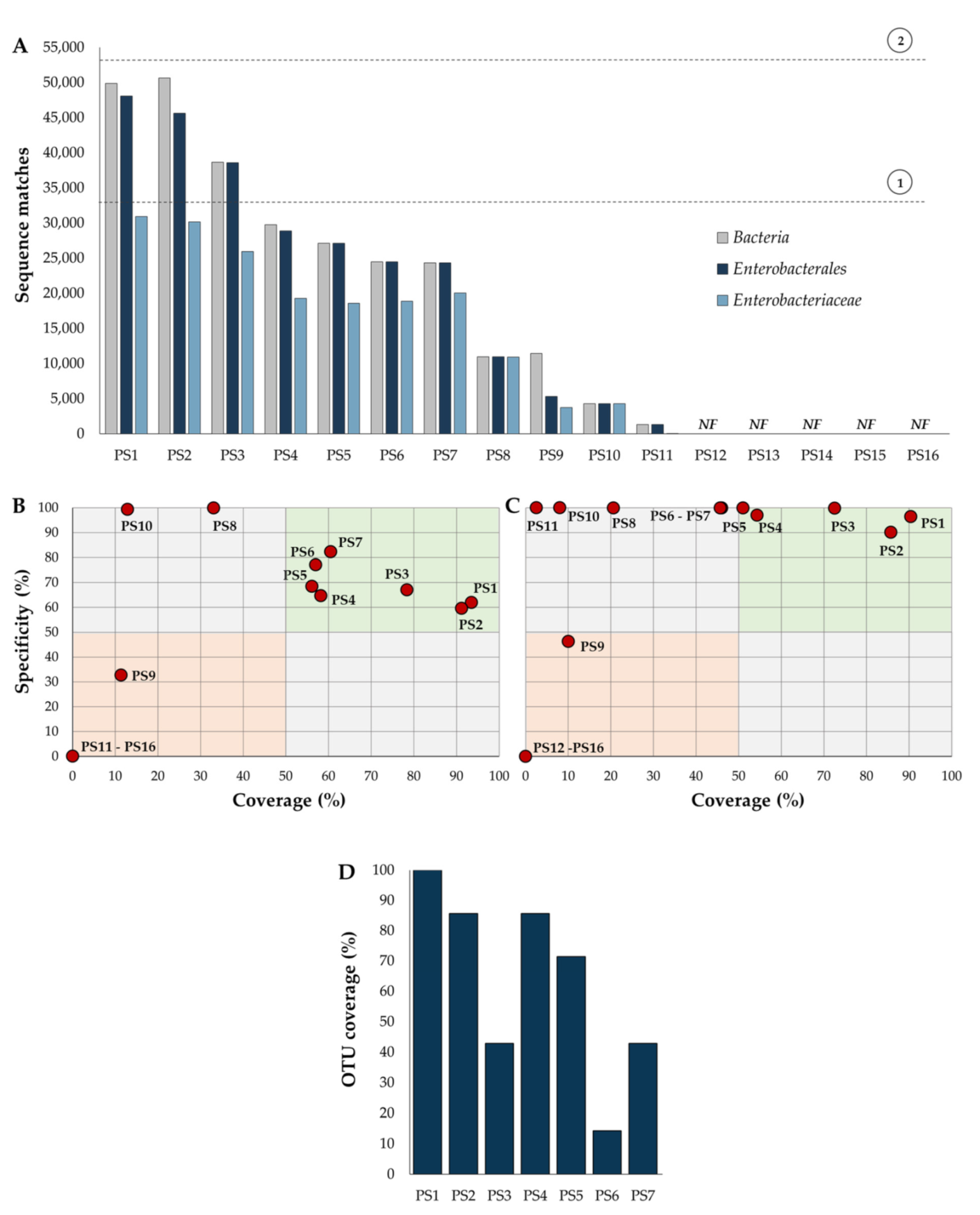
2.3. Primer Specificity and Coverage

Performance of 16 different primer sets (PS) was evaluated using the whole collection of 16S rRNA gene sequences annotated and archived at the RDP. This database was chosen and used because taxonomy of Enterobacterales was more in agreement with the LPSN, a database regulated by the International Code of Nomenclature of Prokaryotes [31], and because RDP was more compatible with the consensus taxon list identified in the present study (Table S1).
Overall, the present analysis revealed that selected primer pairs varied drastically in the number of 16S rRNA gene sequences targeted. For example, three primer pairs (PS1, PS2 and PS3) recognized >33,000 sequences, exceeding the total number (n = 33,092) of Enterobacteriaceae in the dataset, suggesting that these primers were out of target and cannot be considered specific for this bacterial group. Eight primers (PS4–PS11) recognized between 29,000–1300 sequences, suggesting a low coverage for Enterobacteriaceae. Remarkably, five primers (PS12–PS16) were unable to match sequences from the database (Figure 2A). Comparable results for all selected primer sets were obtained by using the TestPrime 1.0 software at the SILVA Database (Bremen, Germany) (Figure S1 and Table S3). Because primer pairs PS12–PS16 were unable to recognize 16S rRNA sequences from the RDP and SILVA databases, they were not considered for further analyses.
It is noteworthy that a preliminary assessment of potential problems associated with PS12–PS16 primer sets revealed two types of issues: (i) primer sets containing numerous sequence mismatches, and (ii) primer sets targeting the same strand of the DNA sequence (Table S4). Additional analyses are required to corroborate these observations and additional experiments should be performed to validate a corrected version of these PCR primers. The selected collection of primer pairs recognized a highly variable number of 16S rRNA gene sequences at the order and family level. For instance, only three primer sets (PS1–PS3) recognized >70% of the Enterobacterales sequences, and four primers (PS4–PS7) matched >55% of the Enterobacteriaceae genes (Figure 2A). These results suggest that the majority of the selected primer pairs have important differences in their performance.
To corroborate this idea, a comparison of primer specificity/coverage was performed. Only seven primer sets (PS1–PS7) showed >50% specificity and >50% coverage at the family level; however, none of them reached ≥75% specificity and coverage levels—the percentage considered as acceptable in molecular microbiology [36,37,38] (Figure 2B). These results suggest that currently available primer pairs underestimate diversity and relative abundance of the Enterobacteriaceae family.
As a result of the taxonomic restructuration of Enterobacterales [28], in the present analysis it was revealed that some of these primer sets were also unsuitable for characterization of the Enterobacterales order. For example, primer specificity of PS4–PS8, PS10 and PS11 were nearly 100%, but these primers had a coverage <60%. Importantly, it was identified that PS1, PS2 and PS3, had a specificity and coverage greater than 70%, suggesting that these primer pairs could be considered potential candidates for molecular analyses of the Enterobacterales order (Figure 2C).
To evaluate the number of families and genera targeted by these primer pairs, an OTU coverage analysis was carried out. It was revealed that PS1, PS2 and PS4 recognized at least 80% of the taxa belonging to Enterobacterales. The rest of the primers had an OTU coverage of <70% (Figure 2D and Table S5). On average, the estimated coverage at the genus level for these three primer sets was 80%, 67% and 55% for PS1, PS2 and PS4, respectively; the remaining primer pairs showed a genus coverage ranging from 3% to 49% (Figure 3). Moreover, if a minimum coverage threshold of 50% is considered, as suggested by some authors [36,37,38], PS6 could be categorized as a suitable primer pair for analysis of Enterobacteriaceae (Table 2 and Figure 3).

3. Discussion

After the most recent taxonomic restructuration of Enterobacteriaceae (December 2016, [28]), databases such as LPSN, NCBI, RDP and SILVA are still considering different genera assigned to Enterobacteriaceae (37, 29, 33 and 23 genera, respectively, as of October, 2021). Comparable issues were also observed in other families within the Enterobacterales order (Table S1). These taxonomy discrepancies highlight the need to review and update databases and bioinformatics tools used for 16S rRNA gene analyses. Moreover, researchers should revise and consider these taxonomic updates to drive more accurate conclusions when microbial analyses are performed. Unfortunately, various and recent studies are still using the formerly Enterobacteriaceae taxonomy removed in 2016 (for example [2,29,30,39]).
Recent studies describing the use of these 16 primers pairs (for example [40,41,42,43,44]) have not considered the latest taxonomic restructuration published in 2016. Thus, it becomes essential to review and update the specificity and coverage of these primer pairs. The limited performance of PS3 and PS5–PS11 [17,19,20,21,22,23,24], could be explained by the fact that most of these primer sets were designed and validated before the taxonomic restructuration of Enterobacteriaceae in 2016 [28]. Unfortunately, PS1 was published after this year [15] without considering the taxonomic restructuration [28]. These findings highlight the importance of performing frequent in-house evaluation of PCR primers.
Importantly, the present analysis provides a framework to review and update the specificity and coverage of primer pairs that had been previously validated, used and published in the literature. In the present study, it was identified that one primer set (PS1) could be considered Enterobacterales-specific, five primer pairs (PS2–PS5 and PS7) multi-family specific and four primers (PS8–PS11) multi-genus specific. Out of 16, only one primer set (PS6) could be considered suitable for analysis of Enterobacteriaceae.
Finally, because the formerly Enterobacteriaceae family is now restructured into seven families (Budviciaceae, Enterobacteriaceae, Erwiniaceae, Hafniaceae, Morganellaceae, Pectobacteriaceae and Yersiniaceae)—all of them members of the Enterobacterales order [28]. Primer sets PS1 and PS2 could be the most suitable option for analysis of this bacterial order. These two PCR primers could be an alternative for previously known-as (before 2016) Enterobacteriaceae-specific primers for analysis of host-associated microbiota.

4. Materials and Methods

4.1. Reclassification of Formerly Enterobacteriaceae 16S rRNA Gene Sequences

To evaluate the extent of taxonomic changes in previously characterized bacterial population data, a collection of 16S rRNA gene sequences formerly recognized as Enterobacteriaceae was subjected to reclassification using the current taxonomy for this bacterial group [28]. To accomplish this goal, a total of 23,824 full-length 16S rRNA gene sequences archived at the Ribosomal Database Project (RDP) [33] and previously identified as Enterobacteriaceae, before its latest update (RDP Taxonomy 18, August 2020), were used for the analysis. These sequences were subjected to sequence-clustering at 100% nucleotide identity (% ID) using the CD-HIT web-tool [44]. Representative sequences (n = 16,334) were then classified using the Naïve Bayesian Classifier tool (RDP Taxonomy 18) available at the RDP [45]. This classification tool was used for the analyses because taxonomy of Enterobacterales and Enterobacteriaceae was more in agreement with the LPSN (https://lpsn.dsmz.de/; accessed on 1 October 2021)—the most widely accepted taxonomy framework [31]—and with the consensus taxon list obtained from the RDP, SILVA, NCBI and LPSN (Table S1).

4.2. Identification of Primer Sets

A comprehensive literature review and evaluation was carried out to identify and integrate a collection of primer sets claiming specific amplification of formerly Enterobacteriaceae 16S rRNA genes. To accomplish this goal, publicly available search engines and databases such as Google (https://www.google.com/), PubMed (https://pubmed.ncbi.nlm.nih.gov/), ProbeBase (https://probebase.csb.univie.ac.at/;), ScienceDirect (https://www.sciencedirect.com/) and Scopus (https://www.scopus.com/) (all accessed on 1 December 2020) were used to identify previously validated and published primer sets. From this collection of articles forward and reverse PCR primer sequences (5′–3′), amplicon size, amplicon position, targeted 16S rRNA gene variable region [35] and genera used for PCR validation were identified and recorded.

4.3. Design of Primer Set #3 (PS3)

Enterobacteriaceae 16S rRNA gene sequences from type-strain isolates (n = 135) were retrieved from the RDP (Release 11, Update 5). These gene sequences were subjected to sequence clustering at 100% ID using the CD-HIT web-tool [45] as described above. Representative sequences were then used for primer design and in silico analysis using FastPCR software following recommendations described elsewhere [46]. The best primer set candidate (PS3) was synthesized by Integrated DNA Technologies (www.idtdna.com; Coralville, IA, USA;) and then validated in-house, using PCR assays targeting amplification of genomic DNA obtained from six Enterobacterales (Salmonella enterica, Escherichia coli, Citrobacter sp., Enterobacter sp., Klebsiella sp. and Serratia sp.) and six non-Enterobacterales strains (Aeromonas sp., Bacillus sp., Lactobacillus sp., Pseudomonas sp., Staphylococcus sp. and Stenotrophomonas sp.) from our laboratory strain collection. Genomic DNA extraction was carried out using the Quick-gDNA commercial kit (Zymo Research, Irvine, CA, USA). A PCR gradient assay (temperature range: 58–72 °C) was performed to identify the most adequate annealing temperature. PCR reactions were performed using Phire Hot Start II DNA Polymerase (Thermo Fisher Scientific, Waltham, MA, USA) and 10 ng of purified DNA. The optimized PCR protocol consisted of an initial denaturation at 94 °C for 1 min and 35 cycles of: denaturation at 94 °C for 30 s, annealing at 58–72 °C for 20 s, extension at 72 °C for 20 s and a final extension at 72 °C for 1 minute. Specificity of the PCR was analyzed via ethidium bromide/1.5% agarose gel electrophoresis.

4.4. Performance of Different Primer Sets Targeting Specific Amplification of Formerly Enterobacteriaceae 16S rRNA Gene

The total number of sequence matches (hits), target specificity and taxa coverage were estimated using Probe Match software, hosted at the RDP [33], for each primer pair. For these analyses, the whole 16S rRNA gene sequence database (n = 3,356,809 sequences, Release 11, Update 5 and Taxonomy 18; as of December 2020) was used with the following parameters: type and non-type strains, uncultured and isolate source, size (bp) ≥1200, good quality and zero differences allowed. Notably, this release of the RDP comprised 53,189 gene sequences of the Enterobacterales order. In the analyses, total sequence matches represent the number of hits obtained from the whole RDP database. Specificity denotes the number of targeted hits divided by the total number of Bacteria matches. Taxa coverage corresponds to the number of targeted hits divided by the available number of taxon-specific (Enterobacterales or Enterobacteriaceae) sequences. Operational Taxonomic Unit (OTU) coverage depicts the number of families recognized by the primer set. A family was considered covered when ≥50% of the sequences belonging to that taxon were targeted by the primer set [36. Performance of 16 primers was also corroborated using the TestPrime 1.0 software, hosted at the SILVA Database (Release 138.1, n = 2,224,740 sequences; as of December 2020) [34].

4.5. Identification of Bacterial Groups Targeted by Previously Validated and Published PCR Primer Sets

To define the specificity of previously validated primer sets after the taxonomic restructuration of the formerly Enterobacteriaceae family, an OTU coverage analysis was carried out as described above. Specificity of primer sets was defined at the order, family, or genus level. To portray a detailed description of OTU coverage, a heat map was constructed showing taxon coverage accomplished for each primer.

5. Conclusions

The most recent taxonomic restructuration of Enterobacterales has significantly impacted the ecological and epidemiological interpretation of results describing distribution and biology of this bacterial group. These taxonomic changes have also modified the specificity and coverage of previously published PCR assays designed for analysis of Enterobacteriaceae. Herein it is shown that only one of the currently published primer pairs could be considered suitable for an accurate and comprehensive analysis of the Enterobacteriaceae family. These findings highlight the imperative need to reevaluate the performance of PCR-based molecular assays designed to analyze microbial populations in human, animal and plant samples.

Supplementary Materials

The following are available online at https://www.mdpi.com/article/10.3390/pathogens11010017/s1: Table S1: Enterobacterales families and genera included in the LPSN, NCBI, RPD and SILVA databases (as of October 2021); Table S2: List of previously validated and published primer sets designed for PCR amplification of formerly Enterobacteriaceae 16S rRNA genes; Table S3: OTU coverage analysis, at the family level, using the SILVA database; Table S4: Primer sets PS12–PS16 and potential problems; Table S5: OTU coverage analysis, at the family level, using the RDP database; Figure S1: Performance of different primer sets (PS) designed for PCR amplification of formerly Enterobacteriaceae 16S rRNA genes using the TestPrime 1.0 software at the SILVA Database; Figure S2: Analysis of family and genus coverage of different primer sets (PS) targeting 16S rRNA genes using the TestPrime 1.0 software at the SILVA Database.

Author Contributions

C.N.R.-N., H.V.S.-R., A.R.-A., D.M.R.-P., E.M.M.-S. and G.M.N. performed conceptualization of the study and designed methodology; C.N.R.-N., H.V.S.-R., E.M.M.-S. and G.M.N. performed data analyses, validation and interpretation of results; C.N.R.-N., H.V.S.-R., A.R.-A., D.M.R.-P., E.M.M.-S. and G.M.N. performed the writing—original draft preparation and writing—review and editing. All authors have read and agreed to the published version of the manuscript.

Funding

C.N.R.N. was supported by a CONACYT graduate scholarship (692848) during the development of this work.

Institutional Review Board Statement

Not applicable.

Informed Consent Statement

Not applicable.

Data Availability Statement

CD-HIT: http://weizhongli-lab.org/cd-hit/; List of Prokaryotic Names with Standing in Nomenclature: https://lpsn.dsmz.de/; NCBI Taxonomy: https://www.ncbi.nlm.nih.gov/taxonomy; Ribosomal Database Project: https://rdp.cme.msu.edu/; SILVA: https://www.arb-silva.de/ (all accessed on 1 December 2020).

Conflicts of Interest

The authors declare no conflict of interest.

References

  1. Octavia, S.; Lan, R. The Family Enterobacteriaceae. In The Prokaryotes: Gammaproteobacteria; Rosenberg, E., DeLong, E.F., Lory, S., Stackebrandt, E., Thompson, F., Eds.; Springer: Berlin/Heidelberg, Germany, 2014; pp. 225–286. [Google Scholar]
  2. Guanghui, Z.; Jing, L.; Guojun, Z.; Hong, L. Epidemiology and Risk Factors of Neurosurgical Bacterial Meningitis/Encephalitis Induced by Carbapenem Resistant Enterobacteriaceae. J. Infect. Chemother. 2020, 26, 101–106. [Google Scholar] [CrossRef] [Green Version]
  3. Ding, Y.; Wang, Y.; Hsia, Y.; Sharland, M.; Heath, P.T. Systematic Review of Carbapenem-Resistant Enterobacteriaceae Causing Neonatal Sepsis in China. Ann. Clin. Microbiol. Antimicrob. 2019, 18, 36. [Google Scholar] [CrossRef]
  4. Flores-Mireles, A.L.; Walker, J.N.; Caparon, M.; Hultgren, S.J. Urinary Tract Infections: Epidemiology, Mechanisms of Infection and Treatment Options. Nat. Rev. Microbiol. 2015, 13, 269–284. [Google Scholar] [CrossRef]
  5. Wallace, M.J.; Fishbein, S.R.S.; Dantas, G. Antimicrobial Resistance in Enteric Bacteria: Current State and next-Generation Solutions. Gut Microbes 2020, 12, 1799654. [Google Scholar] [CrossRef]
  6. Xu, N.; Bai, X.; Cao, X.; Yue, W.; Jiang, W.; Yu, Z. Changes in Intestinal Microbiota and Correlation with TLRs in Ulcerative Colitis in the Coastal Area of Northern China. Microb. Pathog. 2021, 150, 104707. [Google Scholar] [CrossRef]
  7. Wright, E.K.; Kamm, M.A.; Teo, S.M.; Inouye, M.; Wagner, J.; Kirkwood, C.D. Recent Advances in Characterizing the Gastrointestinal Microbiome in Crohn’s Disease: A Systematic Review. Inflamm. Bowel. Dis. 2015, 21, 1219–1228. [Google Scholar] [CrossRef] [Green Version]
  8. Lopetuso, L.R.; Petito, V.; Graziani, C.; Schiavoni, E.; Paroni Sterbini, F.; Poscia, A.; Gaetani, E.; Franceschi, F.; Cammarota, G.; Sanguinetti, M.; et al. Gut Microbiota in Health, Diverticular Disease, Irritable Bowel Syndrome, and Inflammatory Bowel Diseases: Time for Microbial Marker of Gastrointestinal Disorders. Dig. Dis. 2018, 36, 56–65. [Google Scholar] [CrossRef]
  9. Carroll, I.M.; Ringel-Kulka, T.; Siddle, J.P.; Ringel, Y. Alterations in Composition and Diversity of the Intestinal Microbiota in Patients with Diarrhea-Predominant Irritable Bowel Syndrome. Neurogastroenterol. Motil. 2012, 24, 521-e248. [Google Scholar] [CrossRef] [Green Version]
  10. Linninge, C.; Roth, B.; Erlanson-Albertsson, C.; Molin, G.; Toth, E.; Ohlsson, B. Abundance of Enterobacteriaceae in the Colon Mucosa in Diverticular Disease. World J. Gastrointest. Pathophysiol. 2018, 9, 18–27. [Google Scholar] [CrossRef]
  11. Liu, X.; Cheng, Y.; Shao, L.; Ling, Z. Alterations of the Predominant Fecal Microbiota and Disruption of the Gut Mucosal Barrier in Patients with Early-Stage Colorectal Cancer. Biomed. Res. Int. 2020, 2020, 2948282. [Google Scholar] [CrossRef]
  12. Liu, C.-J.; Zhang, Y.-L.; Shang, Y.; Wu, B.; Yang, E.; Luo, Y.-Y.; Li, X.-R. Intestinal Bacteria Detected in Cancer and Adjacent Tissue from Patients with Colorectal Cancer. Oncol. Lett. 2019, 17, 1115–1127. [Google Scholar] [CrossRef] [Green Version]
  13. Shen, F.; Zheng, R.-D.; Sun, X.-Q.; Ding, W.-J.; Wang, X.-Y.; Fan, J.-G. Gut Microbiota Dysbiosis in Patients with Non-Alcoholic Fatty Liver Disease. Hepatobiliary Pancreat. Dis. Int. 2017, 16, 375–381. [Google Scholar] [CrossRef]
  14. Diskin, S.; Feygenberg, O.; Maurer, D.; Droby, S.; Prusky, D.; Alkan, N. Microbiome Alterations Are Correlated with Occurrence of Postharvest Stem-End Rot in Mango Fruit. Phytobiome 2017, 1, 117–127. [Google Scholar] [CrossRef] [Green Version]
  15. Kurina, I.; Popenko, A.; Klimenko, N.; Koshechkin, S.; Chuprikova, L.; Filipenko, M.; Tyakht, A.; Alexeev, D. Development of qPCR Platform with Probes for Quantifying Prevalent and Biomedically Relevant Human Gut Microbial Taxa. Mol. Cell. Probes 2020, 52, 101570. [Google Scholar] [CrossRef]
  16. Rossmann, B.; Müller, H.; Smalla, K.; Mpiira, S.; Tumuhairwe, J.B.; Staver, C.; Berg, G. Banana-Associated Microbial Communities in Uganda Are Highly Diverse but Dominated by Enterobacteriaceae. Appl. Environ. Microbiol. 2012, 78, 4933–4941. [Google Scholar] [CrossRef] [Green Version]
  17. Clavel, M.; Barraud, O.; Moucadel, V.; Meynier, F.; Karam, E.; Ploy, M.-C.; François, B. VALIBI study group Molecular Quantification of Bacteria from Respiratory Samples in Patients with Suspected Ventilator-Associated Pneumonia. Clin. Microbiol. Infect. 2016, 22, 812.e1–812.e7. [Google Scholar] [CrossRef] [Green Version]
  18. Castillo, M.; Martín-Orúe, S.M.; Manzanilla, E.G.; Badiola, I.; Martín, M.; Gasa, J. Quantification of Total Bacteria, Enterobacteria and Lactobacilli Populations in Pig Digesta by Real-Time PCR. Vet. Microbiol. 2006, 114, 165–170. [Google Scholar] [CrossRef]
  19. Hansen, R.; Russell, R.K.; Reiff, C.; Louis, P.; McIntosh, F.; Berry, S.H.; Mukhopadhya, I.; Bisset, W.M.; Barclay, A.R.; Bishop, J.; et al. Microbiota of De-Novo Pediatric IBD: Increased Faecalibacterium Prausnitzii and Reduced Bacterial Diversity in Crohn’s but Not in Ulcerative Colitis. Am. J. Gastroenterol. 2012, 107, 1913–1922. [Google Scholar] [CrossRef] [Green Version]
  20. Croswell, A.; Amir, E.; Teggatz, P.; Barman, M.; Salzman, N.H. Prolonged Impact of Antibiotics on Intestinal Microbial Ecology and Susceptibility to Enteric Salmonella Infection. Infect. Immun. 2009, 77, 2741–2753. [Google Scholar] [CrossRef] [Green Version]
  21. Binh, C.T.T.; Heuer, H.; Gomes, N.C.M.; Kaupenjohann, M.; Smalla, K. Similar Bacterial Community Structure and High Abundance of Sulfonamide Resistance Genes in Field-Scale Manures. In Manure: Management, Uses and Environmental Impacts; Dellaguardia, C.S., Ed.; Nova Science Publishers: Hauppauge, NY, USA, 2010; pp. 141–166. [Google Scholar]
  22. Bartosch, S.; Fite, A.; Macfarlane, G.T.; McMurdo, M.E.T. Characterization of Bacterial Communities in Feces from Healthy Elderly Volunteers and Hospitalized Elderly Patients by Using Real-Time PCR and Effects of Antibiotic Treatment on the Fecal Microbiota. Appl. Environ. Microbiol. 2004, 70, 3575–3581. [Google Scholar] [CrossRef] [Green Version]
  23. Handschur, M.; Pinar, G.; Gallist, B.; Lubitz, W.; Haslberger, A.G. Culture Free DGGE and Cloning Based Monitoring of Changes in Bacterial Communities of Salad Due to Processing. Food Chem. Toxicol. 2005, 43, 1595–1605. [Google Scholar] [CrossRef]
  24. Tuomisto, S.; Karhunen, P.J.; Pessi, T. Time-Dependent Post Mortem Changes in the Composition of Intestinal Bacteria Using Real-Time Quantitative PCR. Gut Pathog. 2013, 5, 35. [Google Scholar] [CrossRef] [Green Version]
  25. Nakano, S.; Kobayashi, T.; Funabiki, K.; Matsumura, A.; Nagao, Y.; Yamada, T. Development of a PCR Assay for Detection of Enterobacteriaceae in Foods. J. Food Prot. 2003, 66, 1798–1804. [Google Scholar] [CrossRef]
  26. Pélissier, M.-A.; Vasquez, N.; Balamurugan, R.; Pereira, E.; Dossou-Yovo, F.; Suau, A.; Pochart, P.; Magne, F. Metronidazole Effects on Microbiota and Mucus Layer Thickness in the Rat Gut. FEMS Microbiol. Ecol. 2010, 73, 601–610. [Google Scholar] [CrossRef] [Green Version]
  27. Martinon, A.; Cronin, U.P.; Wilkinson, M.G. Comparison of In-House and Commercial Real-Time PCR Systems for the Detection of Enterobacteriaceae and Their Evaluation Within an Interlaboratory Study Using Infant Formula Samples. Food Anal. Methods 2011, 4, 485–496. [Google Scholar] [CrossRef]
  28. Adeolu, M.; Alnajar, S.; Naushad, S.; Gupta, R.S. Genome-Based Phylogeny and Taxonomy of the ‘Enterobacteriales’: Proposal for Enterobacterales Ord. Nov. Divided into the Families Enterobacteriaceae, Erwiniaceae Fam. Nov., Pectobacteriaceae Fam. Nov., Yersiniaceae Fam. Nov., Hafniaceae Fam. Nov., Morganellaceae Fam. Nov., and Budviciaceae Fam. Nov. Int. J. Syst. Evol. Microbiol. 2016, 66, 5575–5599. [Google Scholar] [CrossRef]
  29. Zhang, H.; Zhang, Q.; Chen, S.; Zhang, Z.; Song, J.; Long, Z.; Yu, Y.; Fang, H. Enterobacteriaceae Predominate in the Endophytic Microbiome and Contribute to the Resistome of Strawberry. Sci. Total Environ. 2020, 727, 138708. [Google Scholar] [CrossRef]
  30. Saenz-García, C.E.; Castañeda-Serrano, P.; Mercado Silva, E.M.; Alvarado, C.Z.; Nava, G.M. Insights into the Identification of the Specific Spoilage Organisms in Chicken Meat. Foods 2020, 9, 225. [Google Scholar] [CrossRef] [Green Version]
  31. Parte, A.C. LPSN—List of Prokaryotic Names with Standing in Nomenclature. Nucleic Acids Res. 2014, 42, D613–D616. [Google Scholar] [CrossRef] [Green Version]
  32. Schoch, C.L.; Ciufo, S.; Domrachev, M.; Hotton, C.L.; Kannan, S.; Khovanskaya, R.; Leipe, D.; Mcveigh, R.; O’Neill, K.; Robbertse, B.; et al. NCBI Taxonomy: A Comprehensive Update on Curation, Resources and Tools. Database 2020, 2020, baaa062. [Google Scholar] [CrossRef]
  33. Cole, J.R.; Wang, Q.; Fish, J.A.; Chai, B.; McGarrell, D.M.; Sun, Y.; Brown, C.T.; Porras-Alfaro, A.; Kuske, C.R.; Tiedje, J.M. Ribosomal Database Project: Data and Tools for High Throughput RRNA Analysis. Nucleic Acids Res. 2014, 42, D633–D642. [Google Scholar] [CrossRef] [Green Version]
  34. Quast, C.; Pruesse, E.; Yilmaz, P.; Gerken, J.; Schweer, T.; Yarza, P.; Peplies, J.; Glöckner, F.O. The SILVA Ribosomal RNA Gene Database Project: Improved Data Processing and Web-Based Tools. Nucleic Acids Res. 2013, 41, D590–D596. [Google Scholar] [CrossRef]
  35. Yang, B.; Wang, Y.; Qian, P.-Y. Sensitivity and Correlation of Hypervariable Regions in 16S RRNA Genes in Phylogenetic Analysis. BMC Bioinform. 2016, 17, 135. [Google Scholar] [CrossRef] [Green Version]
  36. Klindworth, A.; Pruesse, E.; Schweer, T.; Peplies, J.; Quast, C.; Horn, M.; Glöckner, F.O. Evaluation of General 16S Ribosomal RNA Gene PCR Primers for Classical and Next-Generation Sequencing-Based Diversity Studies. Nucleic Acids Res. 2013, 41, e1. [Google Scholar] [CrossRef]
  37. Marquina, D.; Andersson, A.F.; Ronquist, F. New Mitochondrial Primers for Metabarcoding of Insects, Designed and Evaluated Using in Silico Methods. Mol. Ecol. Resour. 2019, 19, 90–104. [Google Scholar] [CrossRef] [Green Version]
  38. Toju, H.; Tanabe, A.S.; Yamamoto, S.; Sato, H. High-Coverage ITS Primers for the DNA-Based Identification of Ascomycetes and Basidiomycetes in Environmental Samples. PLoS ONE 2012, 7, e40863. [Google Scholar] [CrossRef] [Green Version]
  39. Bahlinger, E.; Dorn-In, S.; Beindorf, P.-M.; Mang, S.; Kaltner, F.; Gottschalk, C.; Gareis, M.; Schwaiger, K. Development of Two Specific Multiplex QPCRs to Determine Amounts of Pseudomonas, Enterobacteriaceae, Brochothrix Thermosphacta and Staphylococcus in Meat and Heat-Treated Meat Products. Int. J. Food Microbiol. 2021, 337, 108932. [Google Scholar] [CrossRef] [PubMed]
  40. Moura, I.B.; Normington, C.; Ewin, D.; Clark, E.; Wilcox, M.H.; Buckley, A.M.; Chilton, C.H. Method Comparison for the Direct Enumeration of Bacterial Species Using a Chemostat Model of the Human Colon. BMC Microbiol. 2020, 20, 2. [Google Scholar] [CrossRef]
  41. Štšepetova, J.; Baranova, J.; Simm, J.; Parm, Ü.; Rööp, T.; Sokmann, S.; Korrovits, P.; Jaagura, M.; Rosenstein, K.; Salumets, A.; et al. The Complex Microbiome from Native Semen to Embryo Culture Environment in Human in Vitro Fertilization Procedure. Reprod. Biol. Endocrinol. 2020, 18, 3. [Google Scholar] [CrossRef]
  42. Nel Van Zyl, K.; Whitelaw, A.C.; Newton-Foot, M. The Effect of Storage Conditions on Microbial Communities in Stool. PLoS ONE 2020, 15, e0227486. [Google Scholar] [CrossRef] [Green Version]
  43. Poeker, S.A.; Lacroix, C.; de Wouters, T.; Spalinger, M.R.; Scharl, M.; Geirnaert, A. Stepwise Development of an in Vitro Continuous Fermentation Model for the Murine Caecal Microbiota. Front. Microbiol. 2019, 10, 1166. [Google Scholar] [CrossRef] [PubMed]
  44. Huang, Y.; Niu, B.; Gao, Y.; Fu, L.; Li, W. CD-HIT Suite: A Web Server for Clustering and Comparing Biological Sequences. Bioinformatics 2010, 26, 680–682. [Google Scholar] [CrossRef] [PubMed]
  45. Wang, Q.; Garrity, G.M.; Tiedje, J.M.; Cole, J.R. Naïve Bayesian Classifier for Rapid Assignment of RRNA Sequences into the New Bacterial Taxonomy. Appl. Environ. Microbiol. 2007, 73, 5261–5267. [Google Scholar] [CrossRef] [PubMed] [Green Version]
  46. Kalendar, R.; Lee, D.; Schulman, A.H. Java Web Tools for PCR, in Silico PCR, and Oligonucleotide Assembly and Analysis. Genomics 2011, 98, 137–144. [Google Scholar] [CrossRef] [PubMed] [Green Version]
Figure 1. Reclassification of formerly Enterobacteriaceae into seven bacterial families. The 16S rRNA gene sequences (n = 23,824) previously belonging to the Enterobacteriaceae family (before 2016) were retrieved from the Ribosomal Database Project and subjected to reclassification analysis using the new Enterobacterales taxonomy published in December 2016 [28].
Pathogens 11 00017 g001
Figure 2. Performance of different primer sets (PS) designed for PCR amplification of formerly Enterobacteriaceae 16S rRNA genes. (A) Number of 16S rRNA gene sequences recognized by each primer pair using the Probe Match tool at the Ribosomal Database Project (RDP). Gray dotted lines represent the total number of sequences of (1) Enterobacteriaceae (n = 33,092) and (2) Enterobacterales (n = 53,189) in the RDP. NF: matches not found; primer sets PS12–PS16 were unable to match sequences from the RDP database. Analysis of specificity/coverage for each primer pair at (B) Enterobacteriaceae and (C) Enterobacterales level. (D) OTU coverage analysis at the family level for each primer set. The seven families included in the analysis are listed in Figure 3. Because primer sets PS8–PS16 covered <50% of the genera within each bacterial family, they were not included in panel D. A detailed description of the OTU coverage at the genus and family levels is depicted in Table S5. Comparable results (A, B, C and D) were obtained by using the TestPrime 1.0 software at the SILVA Database (Figure S1).
Pathogens 11 00017 g002
Figure 3. Analysis of family and genus coverage of different primer sets (PS) targeting 16S rRNA genes. Heat map depicts sequence coverage for each taxon belonging to the Enterobacterales order, using the Probe Match at the RDP Database.
Pathogens 11 00017 g003
Table 1. List of previously validated and published primer sets designed for PCR amplification of formerly Enterobacteriaceae 16S rRNA genes.
Primer SetOriginal NameSequence (5′-3′)Amplicon Size (bp) bPosition cReference
PS1 aForwardGGGGATAACYACTGGAAACGGTRGC236144–379[15]
ReverseGCATGGCTGCATCAGGSTTKC
PS2ForwardCGTTACYCGCAGAAGAAGCA259482–740[17]
ReverseCTGAGCGTCAGTCTTYGTCC
PS3Entero353-FGCAGTGGGGAATATTGCA474353–826This study
Entero809-RAAGGGCACAACCTCCAA
PS4F-entATGGCTGTCGTCAGCTCGT3631054–1416[18]
R-entCCTACTTCTTTTGCAACCCACTC
PS5Entero-F234GATGWRCCCRKATGGGA1198226–1423[19]
Entero-R1423AKCTAMCTRCTTCTTTTGCAA
PS6EnterobactDmod2FGACCTCGCGAGAGCA1611259–1419[20]
Enter1432modCCTACTTCTTTTGCAACCCA
PS7515FGTGCCAGCMGCCGCGGTAA312514–825[21]
826RGCCTCAAGGGCACAACCTCCAAG
PS8Eco1457FCATTGACGTTACCCGCAGAAGAAGC170476–645[22]
Eco1652RCTCTACGAGACTCAAGCTTGC
PS9fd2AGAGTTTGATCATGGCTCAG14857–1491[23]
rp1ACGGTTACCTTGTTACGACTT
PS10ForwardGCGGTAGCACAGAGAGCTT4965–113[24]
ReverseGGCAGTTTCCCAGACATTACTCA
PS11Forward 2CGTTACCGACAGAAGAAGCA259482–740[17]
ReverseCTGAGCGTCAGTCTTYGTCC
PS12ENT-FGTTGTAAAGCACTTTCAGTGGTGAGGAAGGNF dNF[25]
ENT-RGCCTCAAGGGCACAACCTCCAAG
PS13DG74fAGGAGGTGATCCAACCGCANFNF[23]
RW01rAACTGGAGGAGGGTGGGGAT
PS14Ent 1113TGGCAACAAAGGATAAGGNFNF[26]
Ent 1418CTTTTGCAACCCACT
PS15LUX—FCGGTGTACCCGCAGAAGAAGCACGNFNF[27]
LUX—RGCTTGCACCCTCCGTATTACC
PS16LUX—FCGGTGTACCCGCAGAAGAAGCACGNFNF[27]
ENT—RGCCTCAAGGGCACAACCTCCAAG
a Primer Set (PS) code assigned in the present study. b Amplicon size; c forward and reverse primer binding positions using the Escherichia coli 16S rRNA gene as reference (accession number A14565). d Not found.
Table 2. Revised list of bacterial targets accomplished by previously validated and published PCR primer sets.
Primer SetTargeted Taxa (Coverage) a
PS1Enterobacterales (90%)
PS2Budviciaceae (67%) + Enterobacteriaceae (89%) + Erwiniaceae (67%) + Hafniaceae (50%) + Pectobacteriaceae (80%) + Yersiniaceae (71%)
PS3Enterobacteriaceae (58%) + Hafniaceae (100%) + Yersiniaceae (71%)
PS4Budviciaceae (67%) + Enterobacteriaceae (65%) + Hafniaceae (100%) + Morganellaceae (63%) + Pectobacteriaceae (100%) + Yersiniaceae (71%)
PS5Budviciaceae (67%) + Enterobacteriaceae (65%) + Hafniaceae (100%) + Pectobacteriaceae (80%) + Yersiniaceae (71%)
PS6Enterobacteriaceae (62%)
PS7Enterobacteriaceae (50%) + Hafniaceae (50%) + Yersiniaceae (57%)
PS8Escherichia/Shigella (89%) + Pseudescherichia (78%)
PS9Pragia (87%) + Salmonella (65%) + Chania (100%) + Ewingella (50%)
PS10Klebsiella (53%) + Lelliottia (71%) + Raoultella (55%)
PS11Morganella (89%) + Providencia (87%)
a Primer set covered the seven families assigned to the Enterobacterales order. A taxon was included in this table when ≥50% of the sequences belonging to that taxon were targeted by the primer set.
Publisher’s Note: MDPI stays neutral with regard to jurisdictional claims in published maps and institutional affiliations.

