
How Bacteria Change after Exposure to Silver Nanoformulations: Analysis of the Genome and Outer Membrane Proteome

 , , , ,
Abstract
1. Introduction
2. Results
2.1. Genome Analysis
2.2. Proteome Analysis
3. Discussion
4. Materials and Methods
4.1. Materials
4.1.1. Strains
4.1.2. Reagents
Genomic DNA Isolation
OMP Isolation
Two-Dimensional Gel Electrophoresis
In-Gel Protein Digestion and MS Protein Identification
4.2. Methods
4.2.1. Genome Analysis
4.2.2. Proteome Analysis
5. Conclusions
Supplementary Materials
Author Contributions
Funding
Acknowledgments
Conflicts of Interest
References
- Kędziora, A.; Wernecki, M.; Korzekwa, K.; Speruda, M.; Gerasymchuk, Y.; Łukowiak, A.; Bugla-Płoskońska, G. Consequences of Long-Term Bacteria’s Exposure To Silver Nanoformulations With Different PhysicoChemical Properties. Int. J. Nanomed. 2020, 15, 199–213. [Google Scholar] [CrossRef] [PubMed]
- Kędziora, A.; Speruda, M.; Krzyżewska, E.; Rybka, J.; Łukowiak, A.; Bugla-Płoskońska, G. Similarities and Differences between Silver Ions and Silver in Nanoforms as Antibacterial Agents. Int. J. Mol. Sci. 2018, 19, 444. [Google Scholar] [CrossRef] [PubMed]
- Pareek, V.; Gupta, R.; Panwar, J. Do physico-chemical properties of silver nanoparticles decide their interaction with biological media and bactericidal action? Mater. Sci. Eng. C Mater. Biol. Appl. 2018, 90, 739–749. [Google Scholar] [CrossRef]
- Kędziora, A.; Wieczorek, R.; Speruda, M.; Matolínová, I.; Goszczyñski, T.M.; Litwin, I.; Matolín, V.; Bugla-łoskonska, G. Comparison of Antibacterial Mode of Action of Silver Ions and Silver Nanoformulations With Different Physico-Chemical Properties: Experimental and Computational Studies. Front. Microbiol. 2021, 12, 659614. [Google Scholar] [CrossRef]
- Liu, Y.F.; Yan, J.J.; Lei, H.Y. Loss of outer membrane protein C in Escherichia coli contributes to both antibiotic resistance and escaping antibody-dependent bactericidal activity. Infect. Immun. 2012, 80, 1815–1822. [Google Scholar] [CrossRef] [PubMed]
- Li, X.Z.; Nikaido, H.; Williams, K.E. Silver resistant mutants of Escherichia coli display active efflux of Ag+ and are deficient in porins. J. Bacteriol. 1997, 179, 6127–6132. [Google Scholar] [CrossRef]
- Radzig, M.; Nadtochenko, V.; Koksharova, O.; Kiwi, J.; Lipasova, V.; Khmel, I. Antibacterial effects of silver nanoparticles on gram negative bacteria: Influence on the growth and biofilms, mechanisms of action. Colloids Surf. B Biointerfaces 2013, 102, 300–306. [Google Scholar] [CrossRef] [PubMed]
- Randall, C.; Gupta, A.; Jackson, N.; Busse, D.; O’Neill, A.J. Silver resistance in Gram-negative bacteria: A dissection of endogenous and exogenous mechanisms. J. Antimicrob. Chemother. 2015, 70, 1037–1046. [Google Scholar] [CrossRef]
- Ansari, M.A.; Khan, H.M.; Khan, A.A.; Ahmad, M.K.; Mahdi, A.A.; Pal, R.; Cameotra, S.S. Interaction of silver nanoparticles with Escherichia coli and their cell envelope biomolecules. J. Basic Microbiol. 2014, 54, 905–915. [Google Scholar] [CrossRef]
- Panáček, A.; Kvítek, L.; Smékalová, M.; Večeřová, R.; Kolář, M.; Röderová, M.; Dyčka, F.; Šebela, M.; Prucek, R.; Tomanec, O.; et al. Bacterial resistance to silver nanoparticles and how to overcome it. Nat. Nanotechnol. 2018, 13, 65–71. [Google Scholar] [CrossRef]
- Henikoff, S.; Henikoff, J.G. Amino acid substitution matrices from protein blocks. Proc. Natl. Acad. Sci. USA 1992, 89, 10915–10919. [Google Scholar] [CrossRef] [PubMed]
- Lee, H.; Popodi, E.; Tang, H.; Foster, P.L. Rate and molecular spectrum of spontaneous mutations in the bacterium Escherichia coli as determined by whole-genome sequencing. Proc. Natl. Acad. Sci. USA 2012, 109, E2774–E2783. [Google Scholar] [CrossRef]
- Nazir, R.; Rehman, S.; Nisa, M.; Baba, U.A. Exploring Bacterial Diversity: From Cell to Sequence; Freshwater Microbiology Elsevier Inc.: London, UK, 2019; ISBN 978-0-12-817495-1. [Google Scholar] [CrossRef]
- Li, W.; Sun, T.; Zhou, S.; Ma, Y.; Shi, Q.; Xie, X.; Huang, X. A comparative analysis of antibacterial activity, dynamics, and effects of silver ions and silver nanoparticles against four bacterial strains. Int. Biodeterior. Biodegrad. 2017, 123, 304–310. [Google Scholar] [CrossRef]
- Yan, X.; He, B.; Liu, L.; Qu, G.; Shi, J.; Hu, L.; Jiang, G. Antibacterial mechanism of silver nanoparticles in Pseudomonas aeruginosa: Proteomics approach. Metallomics 2018, 10, 557. [Google Scholar] [CrossRef] [PubMed]
- Anuj, S.A.; Gajera, H.; Hirpara, D.G.; Golakiya, B.A. Bacterial membrane destabilization with cationic particles of nano-silver to combat efflux-mediated antibiotic resistance in Gram-negative bacteria. Life Sci. 2019, 230, 178–187. [Google Scholar] [CrossRef]
- Lok, C.; Ho, C.; Chen, R.; He, Q.I.; Yu, W.; Sun, H.; Tam, P.K.; Chiu, J.; Che, C. Proteomic Analysis of the Mode of Antibacterial Action of Silver Nanoparticles. Proteome Res. 2006, 5, 916–924. [Google Scholar] [CrossRef] [PubMed]
- Kedziora, A.; Korzekwa, K.; Strek, W.; Pawlak, A.; Doroszkiewicz, W.; Bugla-Ploskonska, G. Silver Nanofilms as a Therapeutic Agent for Killing Escherichia coli and Certain ESKAPE Pathogens. Curr. Microbiol. 2016, 73, 139–147. [Google Scholar] [CrossRef] [PubMed][Green Version]
- Kedziora, A.; Strek, W.; Kȩpiński, L.; Bugla-Ploskonska, G.; Doroszkiewicz, W. Synthesis and antibacterial activity of novel titanium dioxide doped with silver. J. Sol-Gel Sci. Technol. 2012, 62, 79–86. [Google Scholar] [CrossRef]
- Martin, M. Cutadapt removes adapter sequences from high-throughput sequencing reads. EMB Net. J. 2011, 17, 10. [Google Scholar] [CrossRef]
- Bankevich, A.; Nurk, S.; Antipov, D.; Gurevich, A.; Dvorkin, M.; Kulikov, A.; Lesin, V.M.; Nikolenko, S.; Pham, S.; Prjibelski, A.D.; et al. SPAdes: A new genome assembly algorithm and its applications to single-cell sequencing. J. Comput. Biol. 2012, 19, 455–477. [Google Scholar] [CrossRef]
- Darling, A.C.E.; Mau, B.; Blattner, F.R.; Perna, N.T. Mauve: Multiple alignment of conserved genomic sequence with rearrangements. Genome Res. 2004, 14, 1394–1403. [Google Scholar] [CrossRef] [PubMed]
- Darling, A.E.; Mau, B.; Perna, N.T. Progressive Mauve: Multiple genome alignment with gene gain, loss and rearrangement. PLoS ONE 2010, 5, e11147. [Google Scholar] [CrossRef]
- Seemann, T. Prokka: Rapid prokaryotic genome annotation. Bioinformatics 2014, 30, 2068–2069. [Google Scholar] [CrossRef] [PubMed]
- Sueki, A.; Stein, F.; Savitski, M.M.; Selkrig, J.; Typas, A. Systematic Localization of Escherichia coli Membrane Proteins. Msystems 2020, 5, 2. [Google Scholar] [CrossRef] [PubMed]
- Wagih, O.; Galardini, M.; Busby, B.P.; Memon, D.; Typas, A.; Beltrao, P. A resource of variant effect predictions of single nucleotide variants in model organisms. Mol. Syst. Biol. 2018, 14, e8430. [Google Scholar] [CrossRef]
- Murphy, T.F.; Bartos, L.C. Surface-exposed and antigenically conserved determinants of outer membrane proteins of Branhamella catarrhalis. Infect. Immun. 1989, 57, 2938–2941. [Google Scholar] [CrossRef]
- Bugla-Płoskońska, G.; Korzeniowska-Kowal, A.; Guz-Regner, K. Reptiles as a source of Salmonella O48-clinically important bacteria for children: The relationship between resistance to normal cord serum and outer membrane protein patterns. Microb. Ecol. 2011, 61, 41–51. [Google Scholar] [CrossRef]
- Farrell, P.H. High resolution two-dimensional electrophoresis of proteins. J. Biol. Chem. 1975, 250, 4007–4021. [Google Scholar] [CrossRef]
- Bednarz-Misa, I.; Serek, P.; Dudek, B.; Pawlak, A.; Bugla-Płoskońska, G.; Gamian, A. Application of zwitterionic detergent to the solubilization of Klebsiella pneumoniae outer membrane proteins for two-dimensional gel electrophoresis. J. Microbiol. Methods 2014, 107, 74–79. [Google Scholar] [CrossRef]
- Shevchenko, A.; Tomas, H.; Havlis, J.; Olsen, J.V.; Mann, M.J. In-gel digestion for mass spectrometric characterization of proteins and proteomes. Nat. Protoc. 2006, 1, 2856–2860. [Google Scholar] [CrossRef]
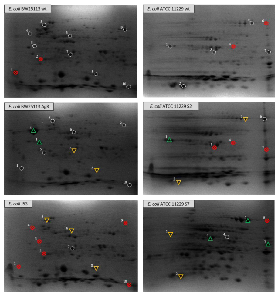
 reference spot;
  selected spots not found in tested sample;
  spot with increased optical density;
  spot with decreased optical density.
reference spot; selected spots not found in tested sample; spot with increased optical density; spot with decreased optical density.
spot with decreased optical density.
reference spot; selected spots not found in tested sample; spot with increased optical density; spot with decreased optical density.

| Function of the Gene Product | Gene | Product Description | Mutations | Predicted Effects | |
|---|---|---|---|---|---|
| Transmembrane transporter activity | Channel | cusC | Cation efflux system protein CusC | Thr81Ala, Asn117Asp, Thr118Ser | - |
| fimD | Outer membrane usher protein | Ser397Thr, Ile401Val, Gly403Ala, Glu410Lys | Alteration in a conserved region, destabilizing mutation | ||
| ompC | Outer membrane porin C | Ile306Val Thr305Val | - | ||
| ompF | Outer membrane porin F | Asp48Gly, Val51Glu, Lys60Met, | - | ||
| ompG | Outer membrane porin G | Ala67Ser | - | ||
| ompN | Outer membrane porin N | Lys90Thr | Destabilizing mutation | ||
| tsx | Nucleoside-specific channel-forming protein | Ile53Leu | - | ||
| uidC | Membrane-associated protein | Asn285Ile | - | ||
| yehB | Outer membrane usher protein | Thr377Ser, Gln540His, Ile699Val | Alteration in a conserved region | ||
| yfaL | Probable autotransporter | Thr879Ala, Pro920Pro, Pro921Thr, Ser922Pro, Thr919Asn, Ser1091Asn Glu1098Asp Thr1101Val | - | ||
| Siderophore transporter | cirA | Colicin I receptor | Ile174Val | - | |
| fecA | Fe(3+) dicitrate transport protein | Ile56Phe, Val350Leu, Ile397Val, Gly600Asp, Val605Ile, Leu651Phe, Ser684Ala, Met693Ile | - | ||
| fepA | Ferrienterobactin receptor | Thr356Ala | - | ||
| fhuA | Ferrichrome outer membrane transporter/phage receptor | Ser110Ala | - | ||
| fhuE | Outer-membrane receptor for Fe(III)-coprogen; Fe(III)-ferrioxamine B and Fe(III)-rhodotorulic acid | Ser61Asn | destabilizing mutation | ||
| fiu | Catecholate siderophore receptor | Thr493Ala | - | ||
| Others | gspD | Putative type II secretion system protein D | Gln617Leu, Val627Ile | - | |
| pgaA | Poly-beta-1;6-N-acetyl-D-glucosamine (PGA) export protein | Gln11Lys, Ile18Leu, Val26Ala, Ile87Val, Ser90Arg, Ile106Val, Pro129Ser, Thr150Ser, Pro451His, Phe599Val | Alteration in conserved regions | ||
| Peptidoglycan-related | lpoA | Penicillin-binding protein activator | Ala96Thr, Val106Ala, Ala292Val | - | |
| lpoB | Penicillin-binding protein activator | Pro71Leu | - | ||
| lpp | Major outer membrane lipoprotein (Braun lipoprotein) | Val26Ile | - | ||
| mliC | Membrane-bound lysozyme inhibitor of C-type lysozyme | His25Arg, Ala29Asp | - | ||
| mltB | Membrane-bound lytic murein transglycosylase B | Val49Met, Asp64Glu, Lys324Arg | - | ||
| nlpD | Murein hydrolase activator | Ala228Thr | - | ||
| Lipid-related | blc | Outer membrane lipoprotein | Gly84Glu | - | |
| lolB | Outer membrane lipoprotein | Ala115Ser | - | ||
| mlaA | Intermembrane phospholipid transport system lipoprotein | Gly168Ser | - | ||
| ycaL | Metalloprotease | Ser158Arg | - | ||
| Various | Bam | bamB | Outer membrane protein assembly factor | Ser96Asn, Ser335Gly | - |
| bamC | Outer membrane protein assembly factor | Asp287Glu, Gln289His | - | ||
| Others | bcsC | Cellulose synthase operon protein C | Val65Ile, Pro110Ser, Ala558Gly, Ala775Thr | - | |
| nfrA | Bacteriophage adsorption protein A | Ala98Asp, Ala115Asp, Ile784Leu | - | ||
| pgaB | Poly-beta-1;6-N-acetyl-D-glucosamine N-deacetylase (PGA N-deacetylase) | Leu575Met | - | ||
| slyB | Outer membrane lipoprotein | Val78Ile | - | ||
| tam | Trans-aconitate 2-methyltransferase | Asn121Ser, Gln123Leu, Ser158Pro, Ile162Val, Ala210Thr Leu216His | - | ||
| yajI | Uncharacterized lipoprotein | Glu164Asp, Asp167Gly | - | ||
| yceB | Uncharacterized lipoprotein | Glu126Gly | Alteration in a conserved region | ||
| yfeY | Uncharacterized protein | Gly142Ser, Arg167Ser | - | ||
| Spots In Figure 1 | Identified Proteins (Encoding Gene) | Data | Strains | Function |
|---|---|---|---|---|
| 1–4 | Flagellin FliC (flyC) | MW = 51,265 pI = 4.5 | E. coli BW25113 AgR only | Subunit protein which polymerizes to form the filaments of bacterial flagellum (extracellular component) |
| 5 | Chaperone protein DnaK (dnaK) | MW = 69,072 pI = 4.83 | E. coli BW25113 wt | Essential role in the initiation of phage lambda DNA replication, involved in chromosomal DNA replication, participates actively in the response to hyperosmotic shock |
| 6 | Isocitrate lyase AceA (aceA) | MW = 47,782 pI = 5.16 | E. coli BW25113 AgR only | Involved in the metabolic adaptation in response to environmental changes; catalyzes the reversible formation of succinate and glyoxylate from isocitrate, a key step of the glyoxylate cycle, which operates as an anaplerotic route for replenishing the tricarboxylic acid cycle during growth on fatty acid substrates, metal binding |
| 7 | D-galactose binding protein MglB (mglB) | MW = 35,690 pI = 5.68 | E. coli BW25113 AgR only | Protein involved in the active transport of galactose and glucose. It plays a role in the chemotaxis towards the two sugars by interacting with the trg chemoreceptor |
| 8 | Superoxide dismutase SodF, SODF, SOD2, FeSOD I (sodF) | MW = 21,311 pI = 5.58 | E. coli BW25113 wt | Destroys superoxide anion radicals which are normally produced within the cells and which are toxic to biological systems |
| 9 | Aldehyde reductase YahK (yahK) | MW = 37,954 pI = 5.80 | E. coli BW25113 wt | Uncharacterized zinc-type alcohol dehydrogenase |
| 10 | Copper homeostasis protein CutC (cutC) | MW = 26,623 pI = 5.75 | E. coli BW25113 wt and AgR | Control of copper homeostasis, copper ions binding |
| Spots in Figure 1 | Identified Protein (Encoding Gene) | Data | Strains | Function (https://www.uniprot.org/ accessed on 12 November 2020) |
|---|---|---|---|---|
| 1 | OmpC (ompC) | M = 40,343, pI = 4.58 | E. coli ATCC 11229 wt only | Forms pores that allow passive diffusion of small molecules across the outer membrane. Microbial infection: supports colicin E5 entry in the absence of its major receptor OmpF, A mixed OmpC-OmpF heterotrimer is the outer membrane receptor for toxin CdiA-EC536; polymorphisms in extracellular loops 4 and 5 of OmpC confer susceptibility to CdiA-EC536-mediated toxicity. |
| 2 | Glutaredoxin-4 GrxD (grxD) | M = 13,044, pI = 4.75 | E. coli ATCC 11229 wt only | Monothiol glutaredoxin involved in the biogenesis of iron-sulfur clusters. |
| 3 | D-galactose-binding periplasmic protein MglB (mglB) | M = 35,690, pI = 5.68 | E. coli ATCC 11229 var. S7 | Protein involved in the active transport of galactose and glucose. It plays a role in the chemotaxis towards the two sugars by interacting with the trg chemoreceptor. |
| 4 | Malate dehydrogenase Mgh (mdh) | M = 32,317, pI = 5.61 | E. coli ATCC 11229 var. S7 | Catalyzes the reversible oxidation of malate to oxaloacetate. |
| 5–6 | Periplasmic oligopeptide-binding protein OppA (oppA) | M = 60,977, pI = 6.05 | E. coli ATCC 11229 var. S2 and S7 | Component of the oligopeptide permease, a binding protein-dependent transport system, it binds peptides up to five amino acids long with high affinity. |
| 7 | Thiosulfate-binding protein CysP (cysP) | M = 37,591, pI = 6.81 | E. coli ATCC 11229 wt only | Part of the ABC transporter complex CysAWTP involved in sulfate/thiosulfate import. This protein specifically binds thiosulfate and is involved in its transmembrane transport. |
| Bacteria Strains | Genome Analysis | Proteome Analysis | |
|---|---|---|---|
| Wild Type Strains and Its Variants | Description | ||
| E. coli BW25113 wt | Wild type strain | [8] (Reference strain) | This work |
| E. coli BW25113 AgR (full name: E. coli BW25113 ompRG596AcusSG1130A) | Variant of E. coli BW25113 wt resistant to silver ions (Ag+) due to mutations, in ompR and cusS, respectively, conferred loss of the OmpC/F porins and derepression of the CusCFBA efflux transporter | [8] | This work |
| E. coli J53 | Model organism with pMG101 plasmid encoding sil genes determining the resistance to silver ions | [8] (Reference strain with exogenous resistance to silver ions) | This work |
| E. coli ATCC 11229 wt | Wild type strain treated with different silver (ions and nanoformulations) | This work (reference strain) | This work |
| E. coli ATCC 11229 var. S2 | Variant of E. coli ATCC 11229 wt with decreased sensitivity to SNF S2 * (after S2 treatment) | This work | This work |
| E. coli ATCC 11229 var. S7 | Variant of E. coli ATCC 11229 wt with decreased sensitivity to SNF S7 * (after S7 treatment) | Small number of mutations analyzed in previous work [1] | This work |
Publisher’s Note: MDPI stays neutral with regard to jurisdictional claims in published maps and institutional affiliations. |
© 2021 by the authors. Licensee MDPI, Basel, Switzerland. This article is an open access article distributed under the terms and conditions of the Creative Commons Attribution (CC BY) license (https://creativecommons.org/licenses/by/4.0/).
Share and Cite
Kędziora, A.; Speruda, M.; Wernecki, M.; Dudek, B.; Kapczynska, K.; Krzyżewska, E.; Rybka, J.; Bugla-Płoskońska, G. How Bacteria Change after Exposure to Silver Nanoformulations: Analysis of the Genome and Outer Membrane Proteome. Pathogens 2021, 10, 817. https://doi.org/10.3390/pathogens10070817
Kędziora A, Speruda M, Wernecki M, Dudek B, Kapczynska K, Krzyżewska E, Rybka J, Bugla-Płoskońska G. How Bacteria Change after Exposure to Silver Nanoformulations: Analysis of the Genome and Outer Membrane Proteome. Pathogens. 2021; 10(7):817. https://doi.org/10.3390/pathogens10070817
Chicago/Turabian StyleKędziora, Anna, Mateusz Speruda, Maciej Wernecki, Bartłomiej Dudek, Katarzyna Kapczynska, Eva Krzyżewska, Jacek Rybka, and Gabriela Bugla-Płoskońska. 2021. "How Bacteria Change after Exposure to Silver Nanoformulations: Analysis of the Genome and Outer Membrane Proteome" Pathogens 10, no. 7: 817. https://doi.org/10.3390/pathogens10070817
APA StyleKędziora, A., Speruda, M., Wernecki, M., Dudek, B., Kapczynska, K., Krzyżewska, E., Rybka, J., & Bugla-Płoskońska, G. (2021). How Bacteria Change after Exposure to Silver Nanoformulations: Analysis of the Genome and Outer Membrane Proteome. Pathogens, 10(7), 817. https://doi.org/10.3390/pathogens10070817