
Molecular Characterization of Associated Pathogens in Febrile Patients during Inter-Epidemic Periods of Urban Arboviral Diseases in Tapachula Southern Mexico

 , ,
, ,  , and
, and
Abstract
1. Introduction
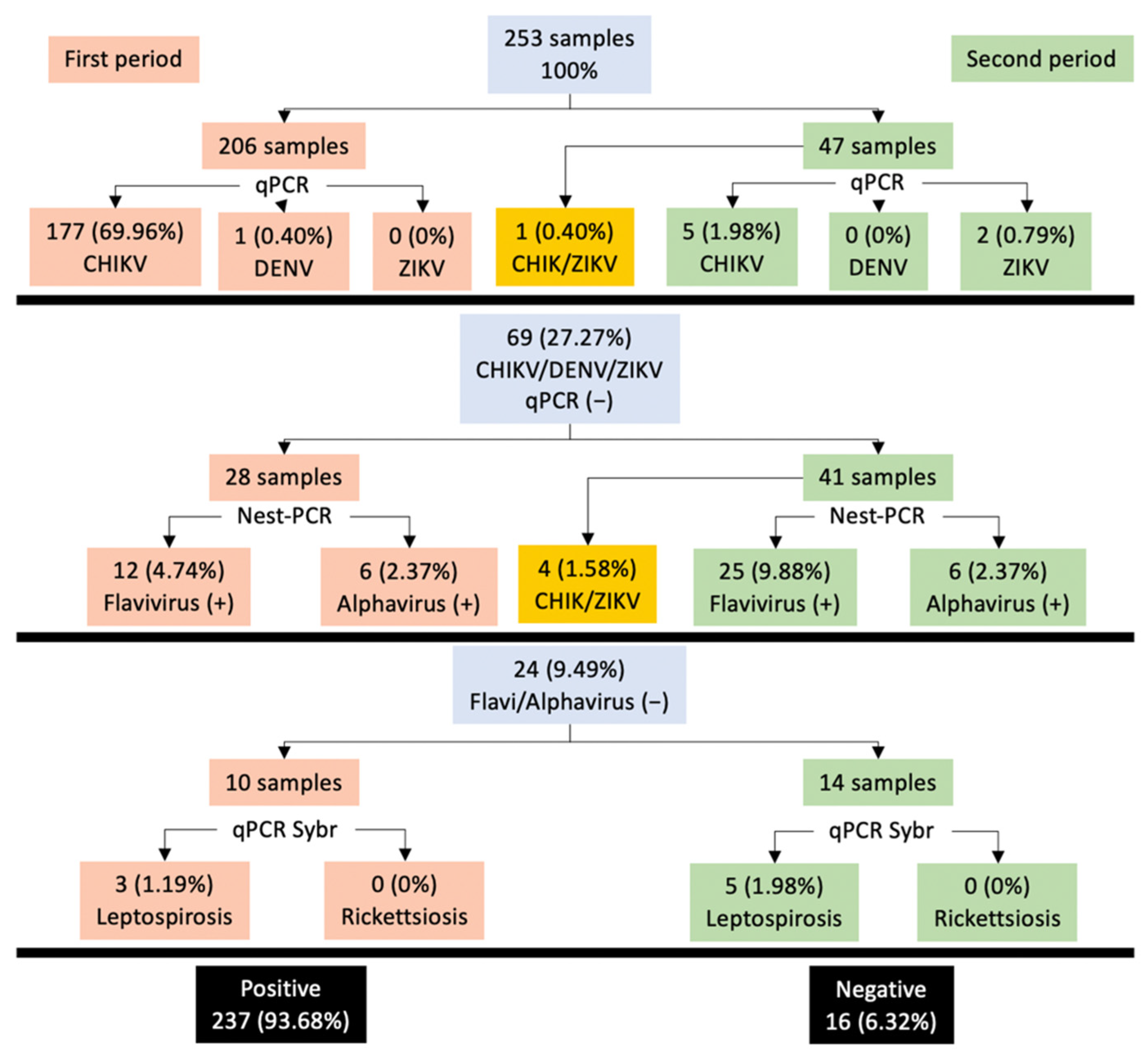
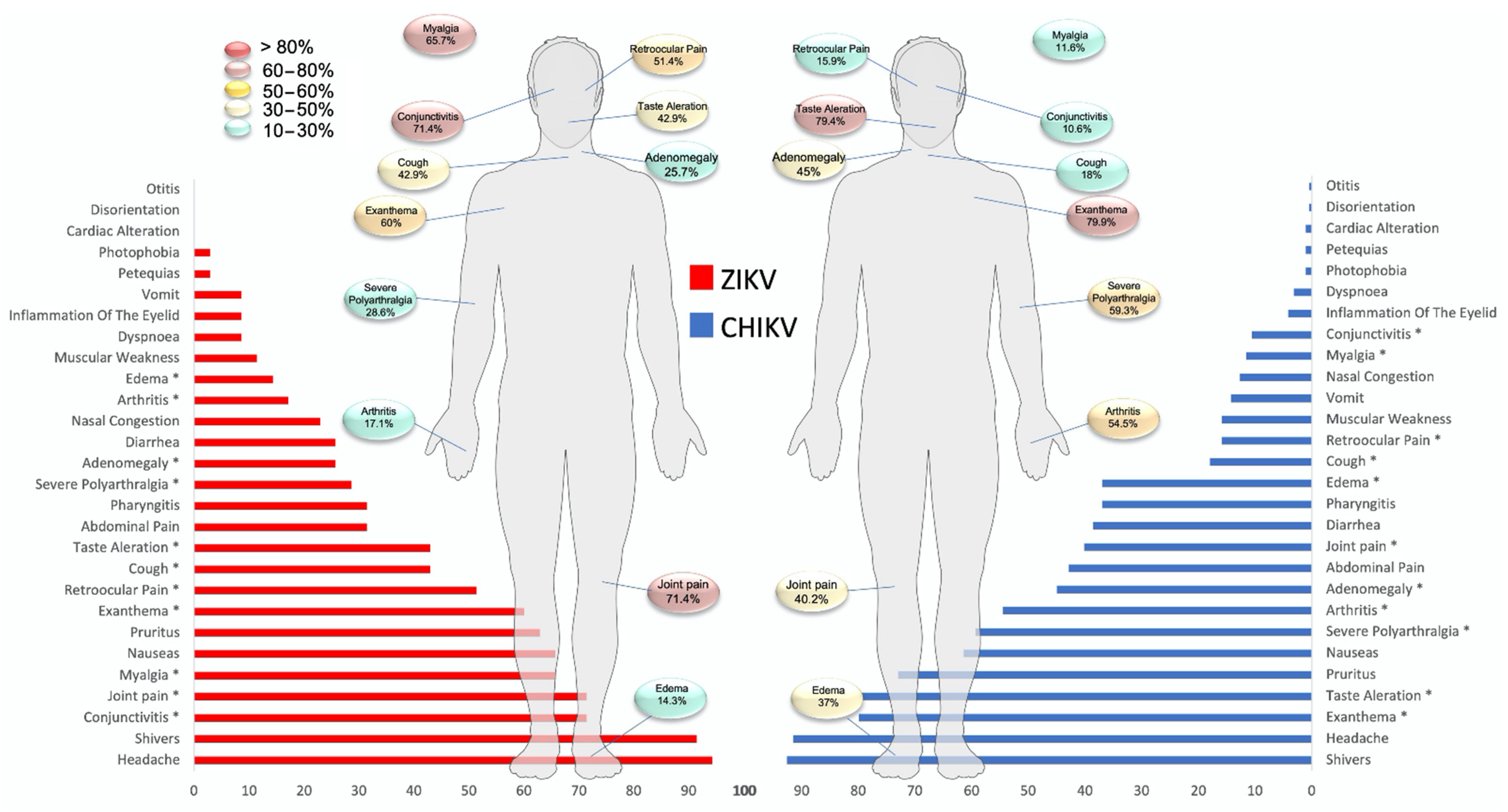
2. Results
3. Discussion
4. Materials and Methods
4.1. Biological Material
4.2. Nucleic Acid Extraction
4.3. Arbovirus Detection Performed by One-Step RT-qPCR
4.4. Nested-PCR
4.5. Identification of Leptospirosis and Rickettsiosis
4.6. Sequencing and BLAST
4.7. Statistical Analysis
5. Conclusions
Supplementary Materials
Author Contributions
Funding
Institutional Review Board Statement
Informed Consent Statement
Data Availability Statement
Conflicts of Interest
References
- Ananth, S.; Shrestha, N.; Treviño, J.A.T.; Nguyen, U.-S.; Haque, U.; Angulo-Molina, A.; Lopez-Lemus, U.A.; Lubinda, J.; Sharif, R.M.; Zaki, R.A.; et al. Clinical Symptoms of Arboviruses in Mexico. Pathogens 2020, 9, 964. [Google Scholar] [CrossRef]
- Carteaux, G.; Maquart, M.; Bedet, A.; Contou, D.; Brugières, P.; Fourati, S.; Cleret de Langavant, L.; De Broucker, T.; Brun-Buisson, C.; Leparc-Goffart, I.; et al. Zika Virus Associated with Meningoencephalitis. N. Engl. J. Med. 2016, 374, 1595–1596. [Google Scholar] [CrossRef] [PubMed]
- Danis-Lozano, R.; Díaz-González, E.E.; Trujillo-Murillo, K.D.C.; Caballero-Sosa, S.; Sepúlveda-Delgado, J.; Malo-García, I.R.; Canseco-Ávila, L.M.; Salgado-Corsantes, L.M.; Domínguez-Arrevillaga, S.; Torres-Zapata, R.; et al. Clinical characterization of acute and convalescent illness of confirmed chikungunya cases from Chiapas, S. Mexico: A cross sectional study. PLoS ONE 2017, 12, e0186923. [Google Scholar] [CrossRef] [PubMed]
- Galán-Huerta, K.A.; Martínez-Landeros, E.; Delgado-Gallegos, J.L.; Caballero-Sosa, S.; Malo-García, I.R.; Fernández-Salas, I.; Ramos-Jiménez, J.; Rivas-Estilla, A.M. Molecular and Clinical Characterization of Chikungunya Virus Infections in Southeast Mexico. Viruses 2018, 10, 248. [Google Scholar] [CrossRef]
- Garza-González, E.; Mendoza-Olazarán, S.; Roman-Campos, R.; Téllez-Marroquín, R.; Saldívar-Rodríguez, D.; Soria-López, J.A.; Guzman, A.; Flores-Treviño, S.; Camacho-Ortiz, A. Rapid Spread of an Ongoing Outbreak of Zika Virus Disease in Pregnant Women in a Mexican Hospital. Braz. J. Infect. Dis. 2017, 21, 554–556. [Google Scholar] [CrossRef]
- Guerbois, M.; Fernandez-Salas, I.; Azar, S.R.; Danis-Lozano, R.; Alpuche-Aranda, C.M.; Leal, G.; Garcia-Malo, I.R.; Diaz-Gonzalez, E.E.; Casas-Martinez, M.; Rossi, S.L.; et al. Outbreak of Zika Virus Infection, Chiapas State, Mexico, 2015, and First Confirmed Transmission by Aedes Aegypti Mosquitoes in the Americas. J. Infect. Dis. 2016, 214, 1349–1356. [Google Scholar] [CrossRef]
- Muller, D.A.; Depelsenaire, A.C.; Young, P.R. Clinical and Laboratory Diagnosis of Dengue Virus Infection. J. Infect. Dis. 2017, 215, S89–S95. [Google Scholar] [CrossRef] [PubMed]
- Hunsberger, S.; Ortega-Villa, A.M.; Powers, J.H., 3rd; Rincón León, H.A.; Caballero Sosa, S.; Ruiz Hernández, E.; Nájera Cancino, J.G.; Nason, M.; Lumbard, K.; Sepulveda, J.; et al. Patterns of Signs, Symptoms, and Laboratory Values Associated with Zika, Dengue, and Undefined Acute Illnesses in a Dengue Endemic Region: Secondary Analysis of a Prospective Cohort Study in Southern Mexico. Int. J. Infect. Dis. 2020, 98, 241–249. [Google Scholar] [CrossRef] [PubMed]
- Mwachui, M.A.; Crump, L.; Hartskeerl, R.; Zinsstag, J.; Hattendorf, J. Environmental and Behavioural Determinants of Lep-tospirosis Transmission: A Systematic Review. PLoS Negl. Trop. Dis. 2015, 9, e0003843. [Google Scholar] [CrossRef] [PubMed]
- Moreira, J.; Barros, J.; Lapouble, O.; Lacerda, M.V.G.; Felger, I.; Brasil, P.; Dittrich, S.; Siqueira, A.M. When Fever Is Not Malaria in Latin America: A Systematic Review. BMC Med. 2020, 18, 294. [Google Scholar] [CrossRef]
- Rodriguez-Morales, A.J.; Villamil-Gómez, W.E.; Franco-Paredes, C. The arboviral burden of disease caused by co-circulation and co-infection of dengue, chikungunya and Zika in the Americas. Travel Med. Infect. Dis. 2016, 14, 177–179. [Google Scholar] [CrossRef]
- Roundy, C.; Azar, S.; Rossi, S.; Huang, J.; Leal, G.; Fernandez-Salas, I.; Vitek, C.; Paploski, I.; Kitron, U.; Ribeiro, G.; et al. Variation in Aedes aegypti Mosquito Competence for Zika Virus Transmission. Emerg. Infect Dis. 2017, 23, 625–632. [Google Scholar] [CrossRef] [PubMed]
- Díaz-González, E.; Kautz, T.; Dorantes-Delgado, A.; Malo-García, I.; Laguna-Aguilar, M.; Langsjoen, R.; Chen, R.; Auguste, D.; Sánchez-Casas, R.; Danis-Lozano, R.; et al. First Report of Aedes aegypti Transmission of Chikungunya Virus in the Americas. Am. J. Trop Med. Hyg. 2015, 93, 1325–1329. [Google Scholar] [CrossRef]
- Danis-Lozano, R.; Díaz-González, E.; Malo-García, I.; Rodríguez, M.; Ramos-Castañeda, J.; Juárez-Palma, L.; Ramos, C.; López-Ordóñez, T.; Mosso-González, C.; Fernández-Salas, I. Vertical transmission of dengue virus in Aedes aegypti and its role in the epidemiological persistence of dengue in Central and Southern Mexico. Trop Med. Int. Health 2019, 24, 1311–1319. [Google Scholar] [CrossRef] [PubMed]
- Karpagam, K.B.; Ganesh, B. Leptospirosis: A Neglected Tropical Zoonotic Infection of Public Health Importance—An Updated Review. Eur. J. Clin. Microbiol. Infect. Diseases 2020, 39, 835–846. [Google Scholar] [CrossRef] [PubMed]
- da Silva, M.A.L.; Soares, C.R.P.; Medeiros, R.A.; Medeiros, Z.; de Melo, F.L. Optimization of single-tube nested PCR for the diagnosis of visceral leishmaniasis. Exp. Parasitol. 2013, 134, 206–210. [Google Scholar] [CrossRef] [PubMed]
- Burchill, S.; Lewis, I.J.; Selby, P. Improved methods using the reverse transcriptase polymerase chain reaction to detect tumour cells. Br. J. Cancer 1999, 79, 971–977. [Google Scholar] [CrossRef][Green Version]
- Keilholz, U.; Willhauck, M.; Rimoldi, D.; Brasseur, F.; Dummer, W.; Rass, K.; de Vries, T.; Blaheta, J.; Voit, C.; Lethé, B.; et al. Reliability of Reverse Transcription-Polymerase Chain Reaction (RT-PCR)-Based Assays for the Detection of Circulating Tu-mour Cells: A Quality-Assurance Initiative of the EORTC Melanoma Cooperative Group. Eur. J. Cancer. 1998, 34, 750–753. [Google Scholar] [CrossRef]
- Kamau, E.; Agoti, C.N.; Lewa, C.S.; Oketch, J.; Owor, B.E.; Otieno, G.P.; Bett, A.; Cane, P.A.; Nokes, D.J. Recent sequence variation in probe binding site affected detection of respiratory syncytial virus group B by real-time RT-PCR. J. Clin. Virol. 2017, 88, 21–25. [Google Scholar] [CrossRef]
- Kindhauser, M.K.; Allen, T.; Frank, V.; Santhana, R.S.; Dye, C. Zika: The origin and spread of a mosquito-borne virus. Bull. World Health Organ. 2016, 94, 675–686C. [Google Scholar] [CrossRef] [PubMed]
- Díaz-Quiñonez, J.A.; López-Martínez, I.; Torres-Longoria, B.; Vázquez-Pichardo, M.; Cruz-Ramírez, E.; Ramírez-González, J.E.; Ruiz-Matus, C.; Kuri-Morales, P. Evidence of the presence of the Zika virus in Mexico since early 2015. Virus Genes 2016, 52, 855–857. [Google Scholar] [CrossRef] [PubMed]
- Metsky, H.C.; Matranga, C.B.; Wohl, S.; Schaffner, S.F.; Freije, C.A.; Winnicki, S.M.; West, K.; Quigley, J.E.; Baniecki, M.L.; Gladden-Young, A.; et al. Zika virus evolution and spread in the Americas. Nature 2017, 546, 411–415. [Google Scholar] [CrossRef]
- Thézé, J.; Li, T.; du Plessis, L.; Bouquet, J.; Kraemer, M.U.; Somasekar, S.; Yu, G.; de Cesare, M.; Balmaseda, A.; Kuan, G.; et al. Genomic Epidemiology Reconstructs the Introduction and Spread of Zika Virus in Central America and Mexico. Cell Host Microbe 2018, 23, 855–864.e7. [Google Scholar] [CrossRef]
- Costa, F.; Hagan, J.; Calcagno, J.; Kane, M.; Torgerson, P.; Martinez-Silveira, M.S.; Stein, C.; Abela-Ridder, B.; Ko, A.I. Global Morbidity and Mortality of Leptospirosis: A Systematic Review. PLoS Negl. Trop. Dis. 2015, 9, e0003898. [Google Scholar] [CrossRef]
- Guernier, V.; Goarant, C.; Benschop, J.; Lau, C.L. A systematic review of human and animal leptospirosis in the Pacific Islands reveals pathogen and reservoir diversity. PLoS Negl. Trop. Dis. 2018, 12, e0006503. [Google Scholar] [CrossRef]
- Yescas-Benítez, J.E.; Perez, N.R.; Montiel-Díaz, H.; Valladares-Carranza, B.; Peláez-Acero, A.; Morales-Ubaldo, A.L.; Bastida, A.Z. Comportamiento Epidemiológico de La Leptospirosis En México Durante El Periodo 2013–2019. Rev. Salud Pública 2020, 22, e202. [Google Scholar] [CrossRef]
- Sandoval-Carrillo, A.A.; Salas-Pacheco, J.M.; Antuna-Salcido, E.I.; Castro-Martínez, K.S.; Ortiz-Montaño, D.S.; Beristain-Garcia, I.; Alvarado-Retana, H.M.; Ramos-Nevarez, A.; Salas-Pacheco, S.M.; Sifuentes-Alvarez, A.; et al. Leptospira infection in people in the city of Durango, Mexico: A cross sectional study. J. Int. Med. Res. 2021, 49, 3000605211004020. [Google Scholar] [CrossRef]
- Galarde-López, M.; Bobadilla-Del Valle, M.; Sánchez-Zamorano, L.M.; Ordaz-Vázquez, A.; Velazquez-Meza, M.E.; So-beranis-Ramos, O. High Exposure to Pathogenic Leptospires by the Population Residing in Dairy Farms in Hidalgo, Mexico. Braz. J. Microbiol. 2021, 52, 1013–1019. [Google Scholar] [CrossRef]
- Sánchez-Montes, S.; Espinosa-Martínez, D.V.; Ríos-Muñoz, C.A.; Berzunza-Cruz, M.; Becker, I. Leptospirosis in Mexico: Epidemiology and Potential Distribution of Human Cases. PLoS ONE 2015, 10, e0133720. [Google Scholar] [CrossRef]
- Ali, M.R.M.; Safiee, A.W.M.; Yusof, N.Y.; Fauzi, M.H.; Yean, C.Y.; Ismail, N. Isolation of Leptospira kmetyi from residential areas of patients with leptospirosis in Kelantan, Malaysia. J. Infect. Public Health 2018, 11, 578–580. [Google Scholar] [CrossRef]
- Saito, M.; Miyahara, S.; Villanueva, S.Y.A.M.; Aramaki, N.; Ikejiri, M.; Kobayashi, Y.; Guevarra, J.P.; Masuzawa, T.; Gloriani, N.G.; Yanagihara, Y.; et al. PCR and Culture Identification of Pathogenic Leptospira spp. from Coastal Soil in Leyte, Philippines, after a Storm Surge during Super Typhoon Haiyan (Yolanda). Appl. Environ. Microbiol. 2014, 80, 6926–6932. [Google Scholar] [CrossRef]
- Zaki, A.M.; Hod, R.; Shamsusah, N.A.; Isa, Z.M.; Bejo, S.K.; Agustar, H.K. Detection of Leptospira kmetyi at recreational areas in Peninsular Malaysia. Environ. Monit. Assess. 2020, 192, 703. [Google Scholar] [CrossRef] [PubMed]
- Esteves, L.; Bulhões, S.M.; Branco, C.C.; Carreira, T.; Vieira, M.L.; Gomes-Solecki, M.; Mota-Vieira, L. Diagnosis of Human Leptospirosis in a Clinical Setting: Real-Time PCR High Resolution Melting Analysis for Detection of Leptospira at the Onset of Disease. Sci. Rep. 2018, 8, 9213. [Google Scholar] [CrossRef] [PubMed]
- Picardeau, M. Virulence of the zoonotic agent of leptospirosis: Still terra incognita? Nat. Rev. Microbiol. 2017, 15, 297–307. [Google Scholar] [CrossRef]
- Bourhy, P.; Storck, C.H.; Theodose, R.; Olive, C.; Nicolas, M.; Hochedez, P.; Lamaury, I.; Zinini, F.; Brémont, S.; Landier, A.; et al. Serovar Diversity of Pathogenic Leptospira Circulating in the French West Indies. PLoS Negl. Trop. Dis. 2013, 7, e2114. [Google Scholar] [CrossRef]
- Meny, P.; Menéndez, C.; Quintero, J.; Hernández, E.; Ríos, C.; Balassiano, I.T.; Trindade, C.N.D.R.; Vital-Brazil, J.M.; Ramos, T.M.V.; Ashfield, N.; et al. Characterization of Leptospira isolates from humans and the environment in Uruguay. Rev. Inst. Med. Trop. São Paulo 2017, 59, e79. [Google Scholar] [CrossRef]
- Staples, J.E.; Breiman, R.F.; Powers, A.M. Chikungunya Fever: An Epidemiological Review of a Re-Emerging Infectious Disease. Clin. Infect. Dis. 2009, 49, 942–948. [Google Scholar] [CrossRef] [PubMed]
- Musso, D.; Gubler, D.J. Zika Virus. Clin. Microbiol. Rev. 2016, 29, 487–524. [Google Scholar] [CrossRef]
- Duffy, M.R.; Chen, T.-H.; Hancock, W.T.; Powers, A.M.; Kool, J.L.; Lanciotti, R.S.; Pretrick, M.; Marfel, M.; Holzbauer, S.; DuBray, C.; et al. Zika Virus Outbreak on Yap Island, Federated States of Micronesia. N. Engl. J. Med. 2009, 360, 2536–2543. [Google Scholar] [CrossRef]
- Belaunzarán-Zamudio, P.F.; Mateja, A.; Guerra-De-Blas, P.D.C.; Rincón-León, H.A.; Navarro-Fuentes, K.; Ruiz-Hernández, E.; Caballero-Sosa, S.; Camas-Durán, F.; Priego-Smith, Z.; Nájera-Cancino, J.G.; et al. Comparison of clinical characteristics of Zika and dengue symptomatic infections and other acute illnesses of unidentified origin in Mexico. PLoS Negl. Trop. Dis. 2021, 15, e0009133. [Google Scholar] [CrossRef]
- Liu, J.; Ochieng, C.; Wiersma, S.; Ströher, U.; Towner, J.S.; Whitmer, S.; Nichol, S.T.; Moore, C.C.; Kersh, G.J.; Kato, C.; et al. Development of a TaqMan Array Card for Acute-Febrile-Illness Outbreak Investigation and Surveillance of Emerging Pathogens, Including Ebola Virus. J. Clin. Microbiol. 2016, 54, 49–58. [Google Scholar] [CrossRef] [PubMed]
- Corman, V.M.; Rasche, A.; Baronti, C.; Aldabbagh, S.; Cadar, D.; Reusken, C.B.; Pas, S.D.; Goorhuis, A.; Schinkel, J.; Molenkamp, R.; et al. Assay optimization for molecular detection of Zika virus. Bull. World Health Organ. 2016, 94, 880–892. [Google Scholar] [CrossRef] [PubMed]
- Lanciotti, R.S.; Kosoy, O.L.; Laven, J.J.; Velez, J.O.; Lambert, A.J.; Johnson, A.J.; Stanfield, S.M.; Duffy, M.R. Genetic and Serologic Properties of Zika Virus Associated with an Epidemic, Yap State, Micronesia, 2007. Emerg. Infect. Dis. 2008, 14, 1232–1239. [Google Scholar] [CrossRef]
- Gurukumar, K.R.; Priyadarshini, D.; Patil, J.A.; Bhagat, A.; Singh, A.; Shah, P.S.; Cecilia, D. Development of real time PCR for detection and quantitation of Dengue Viruses. Virol. J. 2009, 6, 10. [Google Scholar] [CrossRef]
- Scaramozzino, N.; Crance, J.-M.; Jouan, A.; DeBriel, D.A.; Stoll, F.; Garin, D. Comparison of Flavivirus Universal Primer Pairs and Development of a Rapid, Highly Sensitive Heminested Reverse Transcription-PCR Assay for Detection of Flaviviruses Targeted to a Conserved Region of the NS5 Gene Sequences. J. Clin. Microbiol. 2001, 39, 1922–1927. [Google Scholar] [CrossRef]
- Grywna, K.; Kupfer, B.; Panning, M.; Drexler, J.F.; Emmerich, P.; Drosten, C.; Kümmerer, B.M. Detection of All Species of the Genus Alphavirus by Reverse Transcription-PCR with Diagnostic Sensitivity. J. Clin. Microbiol. 2010, 48, 3386–3387. [Google Scholar] [CrossRef] [PubMed]
- Garcia-Ruiz, D.; Martínez-Guzmán, M.A.; Cárdenas-Vargas, A.; Marino-Marmolejo, E.; Gutiérrez-Ortega, A.; González-Díaz, E.; Morfin-Otero, R.; Rodríguez-Noriega, E.; Pérez-Gómez, H.; Elizondo-Quiroga, D. Detection of dengue, west Nile virus, rickettsiosis and leptospirosis by a new real-time PCR strategy. SpringerPlus 2016, 5, 671. [Google Scholar] [CrossRef] [PubMed][Green Version]


| Primer and Probe | Sequence (5′–3′) | Target | Reference |
|---|---|---|---|
| Probe | |||
| ZIKV 1086 | CCGCTGCCCAACACAAG | E | [43] |
| ZIKV 1162c | CCACTAACGTTCTTTTGCAGACAT | E | [43] |
| ZIKV 1107-FAM | FAM-AGCCTACCTTGACAAGCAGTCAGACACTCAA-TAMRA | E | [43] |
| CHIK856 | ACCATCGGTGTTCCATCTAAAG | nsP1 | [3] |
| CHIK962c | GCCTGGGCTCATCGTTATT | nsP1 | [3] |
| CHIK908-FAM | FAM-ACAGTGGTTTCGTGTGAGGGCTAC-TAMRA | nsP1 | [3] |
| DENV 10635 | GARAGACCAGAGATCCTGCTGTCT | 3′UTR | [44] |
| DENV 10682 | ACCATTCCATTTTCTGGCGTT | 3′UTR | [44] |
| DENV123-10663-FAM | FAM-AGCATCATTCCAGGCAC-MGB | 3′UTR | [44] |
| DENV4-10663-FAM | FAM-AACATCAATCCAGGCAC-MGB | 3′UTR | This study |
| Nested PCR | |||
| cFD2 | GTGTCCCAGCCGGCGGTGTCATCAGC | ns5 | [45] |
| MAMD | AACATGATGGGRAARAGRGARAA | ns5 | [45] |
| FS 778 | AARGGHAGYMCDGCHATHTGGT | ns5 | [45] |
| ALPHA-1-Fod | TTTAAGTTTGGTGCGATGATGAAGTC | nsP4 | [46] |
| ALPHA-1-Rev | GCATCTATGATATTGACTTCCATGTT | nsP4 | [46] |
| ALPHA-2-Fod | GGTGCGATGATGAAGTCTGGGATGT | nsP4 | [46] |
| ALPHA-2-Rev | CTATGATATTGACTTCCATGTTCAKCCA | nsP4 | [46] |
| Sybr-Green | |||
| RICK-ADN-For | TATGCTTGCGGCTGTCGGTTCTC | gltA | [47] |
| RICK-ADN-Rev | TTGCGGTAAGTTCGTAGTCTGCTTCTT | gltA | [47] |
| LEP-ADN-For | AGCAGCCGCGGTAATACGTATGG | 16S rRNA | [47] |
| LEP-ADN-Rev | TTTAGGGCGTGGATTACTGGGG | 16S rRNA | [47] |
Publisher’s Note: MDPI stays neutral with regard to jurisdictional claims in published maps and institutional affiliations. |
© 2021 by the authors. Licensee MDPI, Basel, Switzerland. This article is an open access article distributed under the terms and conditions of the Creative Commons Attribution (CC BY) license (https://creativecommons.org/licenses/by/4.0/).
Share and Cite
Calvo-Anguiano, G.; Lugo-Trampe, J.d.J.; Ponce-García, G.; Lugo-Trampe, A.; Martinez-Garza, L.E.; Ibarra-Ramirez, M.; Campos-Acevedo, L.D.; Caballero-Sosa, S.; Juache-Villagrana, A.E.; Fernández-Salas, I.; et al. Molecular Characterization of Associated Pathogens in Febrile Patients during Inter-Epidemic Periods of Urban Arboviral Diseases in Tapachula Southern Mexico. Pathogens 2021, 10, 1450. https://doi.org/10.3390/pathogens10111450
Calvo-Anguiano G, Lugo-Trampe JdJ, Ponce-García G, Lugo-Trampe A, Martinez-Garza LE, Ibarra-Ramirez M, Campos-Acevedo LD, Caballero-Sosa S, Juache-Villagrana AE, Fernández-Salas I, et al. Molecular Characterization of Associated Pathogens in Febrile Patients during Inter-Epidemic Periods of Urban Arboviral Diseases in Tapachula Southern Mexico. Pathogens. 2021; 10(11):1450. https://doi.org/10.3390/pathogens10111450
Chicago/Turabian StyleCalvo-Anguiano, Geovana, José de Jesús Lugo-Trampe, Gustavo Ponce-García, Angel Lugo-Trampe, Laura Elia Martinez-Garza, Marisol Ibarra-Ramirez, Luis Daniel Campos-Acevedo, Sandra Caballero-Sosa, Alan Esteban Juache-Villagrana, Ildefonso Fernández-Salas, and et al. 2021. "Molecular Characterization of Associated Pathogens in Febrile Patients during Inter-Epidemic Periods of Urban Arboviral Diseases in Tapachula Southern Mexico" Pathogens 10, no. 11: 1450. https://doi.org/10.3390/pathogens10111450
APA StyleCalvo-Anguiano, G., Lugo-Trampe, J. d. J., Ponce-García, G., Lugo-Trampe, A., Martinez-Garza, L. E., Ibarra-Ramirez, M., Campos-Acevedo, L. D., Caballero-Sosa, S., Juache-Villagrana, A. E., Fernández-Salas, I., Flores-Suarez, A. E., Rodriguez-Sanchez, I. P., & Trujillo-Murillo, K. d. C. (2021). Molecular Characterization of Associated Pathogens in Febrile Patients during Inter-Epidemic Periods of Urban Arboviral Diseases in Tapachula Southern Mexico. Pathogens, 10(11), 1450. https://doi.org/10.3390/pathogens10111450