Article

Rickettsia lusitaniae in Ornithodoros Porcinus Ticks, Zambia

1
Department of Paraclinical Studies, Faculty of Health Sciences and Veterinary Medicine, School of Veterinary Medicine, University of Namibia, Windhoek Private Bag 13301, Namibia
2
Department of Biomedical Sciences, School of Health Sciences, University of Zambia, Lusaka P.O. Box 50110, Zambia
3
School of Life Sciences, College of Agriculture, Engineering and Sciences, University of KwaZulu—Natal, Private Bag X54001, Durban 4000, South Africa
4
International Institute for Zoonosis Control, Hokkaido University, N 20 W10, Kita-ku, Sapporo 001-0020, Japan
5
Central Veterinary Research Institute (CVRI), Ministry of Fisheries and Livestock, Lusaka P.O. Box 33980, Zambia
6
Department of Biomedical Sciences, School of Veterinary Medicine, University of Zambia, Lusaka P.O. Box 32379, Zambia
7
Department of Paraclinical Studies, School of Veterinary Medicine, University of Zambia, Lusaka P.O. Box 32379, Zambia
8
Department of Biomedical Sciences, Michael Chilufya Sata School of Medicine, Copperbelt University, Kitwe P.O. Box 21692, Zambia
9
Department of Wildlife Sciences, School of Natural Resources, Copperbelt University, Kitwe P.O. Box 21692, Zambia
10
Department of Disease Control, School of Veterinary Medicine, University of Zambia, Lusaka P.O. Box 32379, Zambia
11
Wildlife Diseases Unit, Ministry of Fisheries and Livestock, Lusaka P.O. Box 50060, Zambia
12
Department of Clinical Studies, School of Veterinary Medicine, University of Zambia, Lusaka P.O. Box 32379, Zambia
13
International Collaboration Unit, International Institute for Zoonosis Control, Hokkaido University, N 20 W10, Kita-ku, Sapporo 001-0020, Japan
14
Global Virus Network, 725 W Lombard St., Baltimore, MD 21201, USA
15
Macha Research Trust, Choma P.O. Box 630166, Zambia
*
Authors to whom correspondence should be addressed.
These authors contributed equally to this work.
Pathogens 2021, 10(10), 1306; https://doi.org/10.3390/pathogens10101306
Submission received: 14 September 2021 / Revised: 1 October 2021 / Accepted: 5 October 2021 / Published: 12 October 2021
(This article belongs to the Special Issue Advances in the Epidemiological Surveillance of Tick-Borne Pathogens)

Abstract

:
Rickettsial pathogens are amongst the emerging and re-emerging vector-borne zoonoses of public health importance. Though traditionally considered to be transmitted by ixodid ticks, the role of argasid ticks as vectors of these pathogens is increasingly being recognized. While bat-feeding (Ornithodoros faini) and chicken-feeding (Argas walkerae) argasid ticks have been shown to harbor Rickettsia pathogens in Zambia, there are currently no reports of Rickettsia infection in southern Africa from warthog-feeding (Phacochoerus africanus) soft ticks, particularly Ornithodoros moubata and Ornithodoros porcinus. Our study sought to expand on the existing knowledge on the role of soft ticks in the epidemiology of Rickettsia species through screening for Rickettsia pathogens in warthog burrow-dwelling soft ticks from two national parks in Zambia. The tick species from which Rickettsia were detected in this study were identified as Ornithodoros porcinus, and an overall minimal Rickettsia infection rate of 19.8% (32/162) was observed. All of the sequenced Rickettsia were identified as Rickettsia lusitaniae based on nucleotide sequence similarity and phylogenetic analysis of the citrate synthase (gltA) and 17kDa common antigen (htrA) genes. Utilizing all of the gltA (n = 10) and htrA (n = 12) nucleotide sequences obtained in this study, BLAST analysis showed 100% nucleotide similarity to Rickettsia lusitaniae. Phylogenetic analysis revealed that all of the Zambian gltA and htrA gene sequences could be grouped with those of Rickettsia lusitaniae obtained in various parts of the world. Our data suggest that Rickettsia lusitaniae has a wider geographic and vector range, enhancing to our understanding of Rickettsia lusitaniae epidemiology in sub-Saharan Africa.

1. Introduction

The Rickettsia genus is composed of pathogens considered to be amongst the emerging zoonotic vector-borne pathogens [1,2] and can be divided into three primary groups based on genotype: the ancestral, typhus and spotted fever group (SFG) [3]. The common vectors for this genus include ticks, fleas, mites and lice [4]. Whilst the role of hard ticks (Family Ixodidae) as important vectors and reservoirs of Rickettsia species is well established [4,5,6], recent evidence indicates that soft ticks (Family Argasidae) also play a significant role as vectors [7,8,9,10,11,12].
Within the tropical areas of Africa, 10 Argasidae species have been defined, with Ornithodoros moubata complex (vectors of African swine fever and human relapsing fever), Ornithodoros porcinus (vectors of African swine fever) and Ornithodoros savignyi (cause of sand tampan toxicosis) being considered to have the greatest veterinary and public health significance [13]. In Africa, the role of soft ticks as vectors of zoonotic pathogens is very well established in the transmission of tick-borne relapsing fever causative agents (Ornithodoros moubata complex—Borrelia duttonii, Ornithodoros sonraiBorrelia crocidurae and Ornithodoros erraticusBorrelia hispanica) [14]. However, investigations into the role of soft ticks in the transmission of Rickettsia species have been limited, but interest in this line of research is currently gaining momentum.
The circulation of Rickettsia species in African soft ticks has been reported in the genera Ornithodoros, Argas and Carios, with the reported Rickettsia including R. hoogstraalii, R. lusitaniae, Candidatus Rickettsia africaseptentrionalis, Candidatus R. mauretanica, as well as some unidentified Rickettsia species, with these reports emerging from Morocco, Algeria, Ethiopia, Tunisia, Namibia, South Africa and Zambia [7,8,9,10,11,12]. The soft ticks analyzed across Africa have been collected from bat caves, rodent burrows, bird nests and crevices in human and livestock dwellings.
In Zambia, Qiu et al. [10] reported the presence of R. hoogstraalii and R. lusitaniae in Argas walkerae and Ornithodoros faini ticks, respectively, with R. lusitaniae being reported for the first time in Africa. R. lusitaniae is a newly emergent Rickettsia species whose pathogenicity to humans, geographical spread and vector and/or host range is yet to be fully elucidated. Considering the reports of Rickettsia species in soft ticks in Zambia [10], our study sought to screen for these pathogens in ticks collected from warthog burrows in selected national parks of Zambia, expanding our knowledge on their role in the epidemiology of Rickettsia pathogens in the country.

2. Results

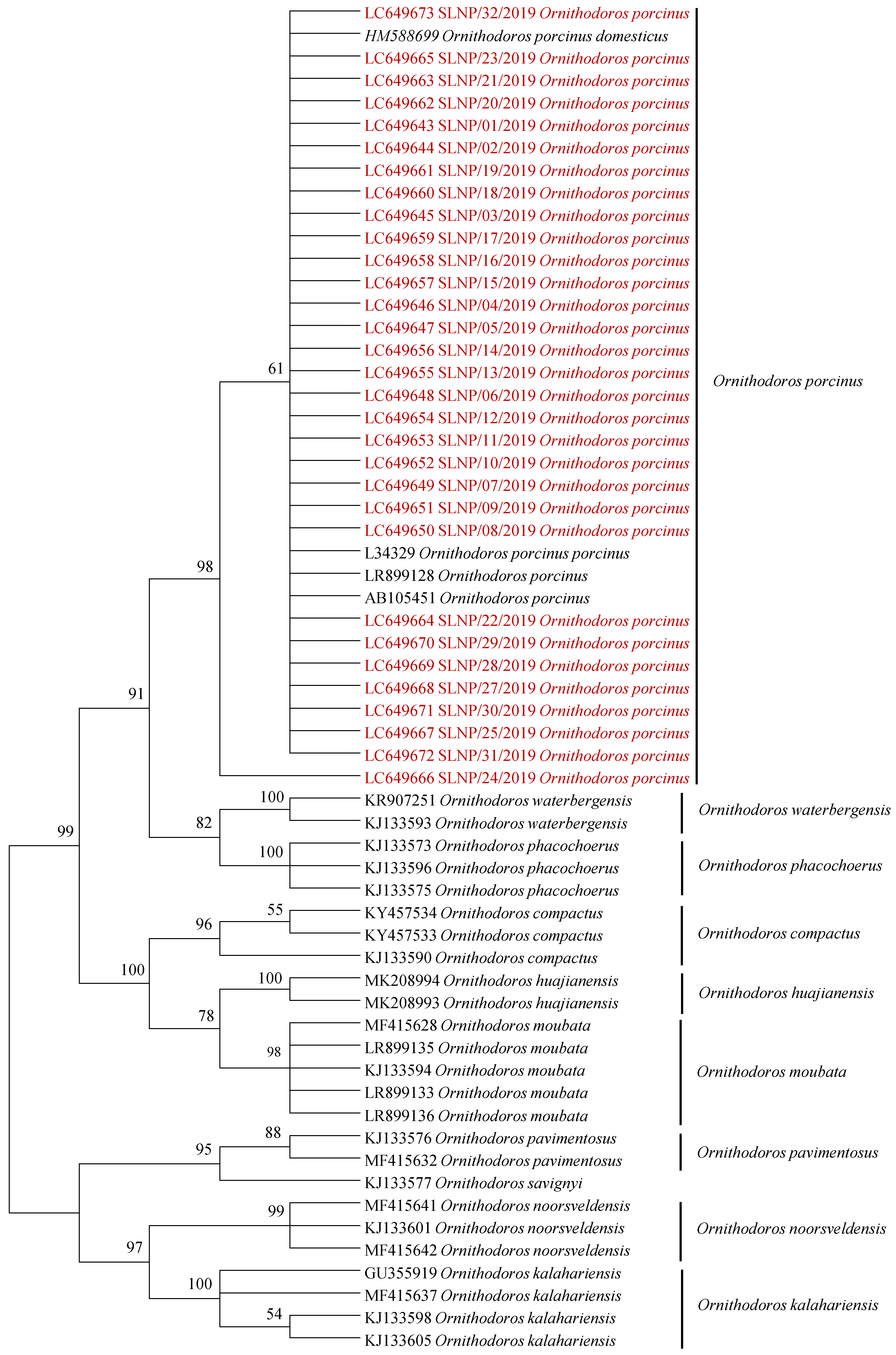
2.1. Tick Sampling and Identification

A total of 162 ticks, comprising 75 from Kafue National Park (KNP) and 87 from South Luangwa National Park (SLNP), were analyzed in this study. All the ticks were morphologically identified as Ornithodoros species and were subsequently pooled into a total of 58 pools (KNP—24, SLNP—34). Further confirmation of the species’ identity was obtained by phylogenetic analysis of the mitochondrial 16S rRNA gene sequences in those pools that tested positive for Rickettsia species. Phylogenetic analysis revealed that the 31 mitochondrial 16S rRNA gene sequences obtained in this study clustered within the O. porcinus complex group (Figure 1).

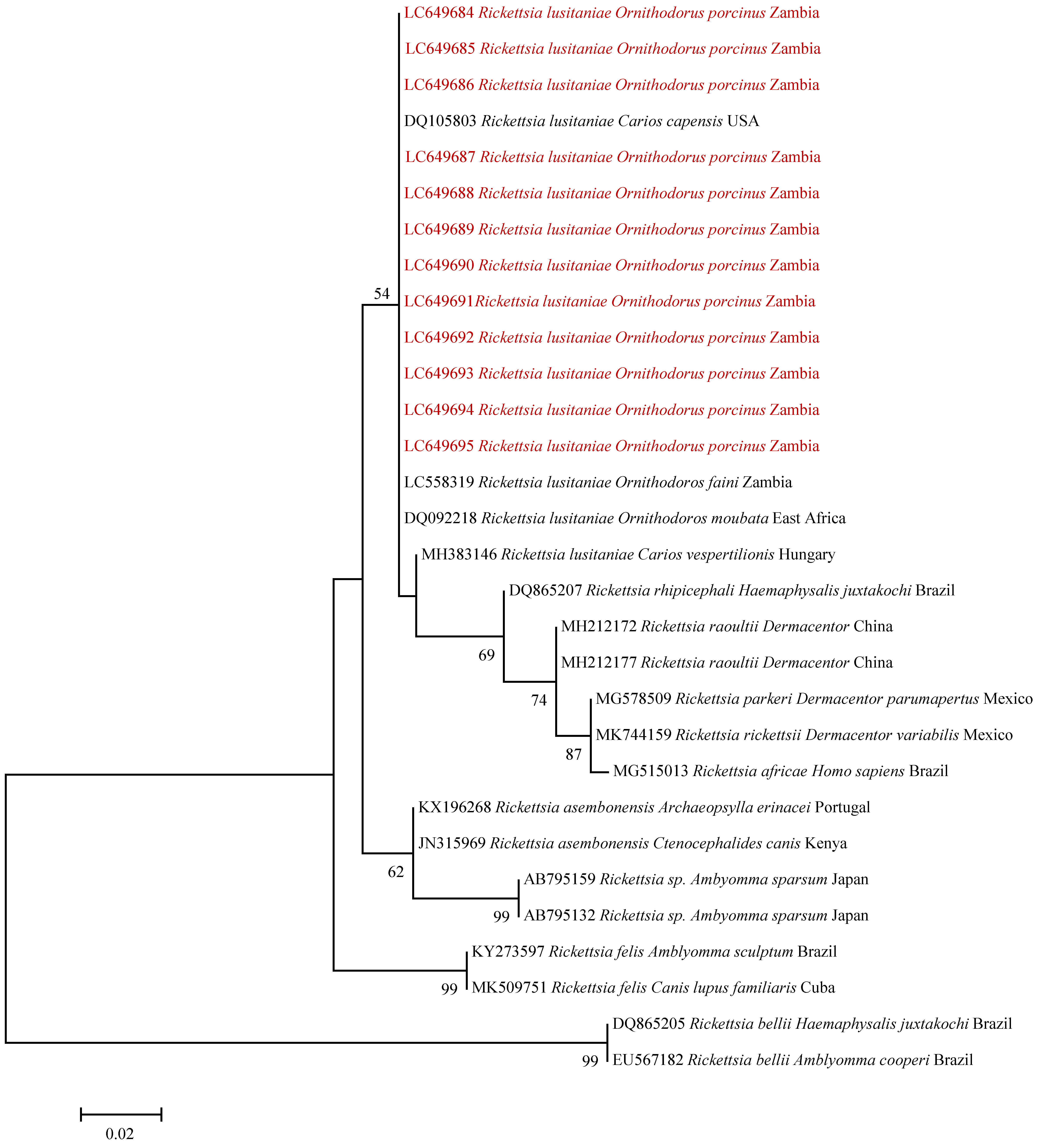
2.2. Rickettsia Screening and Identification

On initial screening for Rickettsia using the loop-mediated isothermal amplification (LAMP) technique, an overall minimum infection rate (MIR) of 19.8% (32/162) was observed. When separated by area of sampling, all of the positive pools were from SLNP, while no Rickettsia genome was detected in ticks from KNP. The MIR for SNLP was determined as 36.8% (32/87). On statistical analysis, the overall pooled prevalence was determined as 23% [95% CI (16.4–31.8)], with the pooled prevalence for SLNP determined as 61.1% [95% CI (41.8–80.7)].
The sequences obtained from the positive pools, based on the gltA gene, showed 100% identity to the R. lusitaniae strain ZS13 that had been detected in the heart of the common pipistrelle (Pipistrellus pipistrellus) in China (GenBank accession: MN388795). Further sequence comparison based on the htrA gene showed 100% nucleotide sequence identity to the R. lusitaniae strain OnF11 detected in O. faini ticks in Zambia (GenBank accession: LC558319) [10].
On phylogenetic analysis of the gltA gene, the samples in our study clustered with Rickettsia lusitaniae were detected in various parts of the world (Figure 2).
A similar clustering pattern was observed upon phylogenetic analysis of the htrA gene (Figure 3).

3. Discussion

In this study, we sought to investigate the presence of Rickettsia species in argasid ticks collected from warthog burrows in two national parks in Zambia. To the best of our knowledge, this is the second study that has sought to examine the role of soft ticks in the epidemiology of Rickettsia species in the country. The previous study screened for Rickettsia and Anaplasma species in ticks collected from warthog burrows (O. moubata), chicken coops (Ar. walkerae) and a bat cave (O. faini), with Rickettsia species being detected in both Ar. walkerae (R. hoogstraalii) and O. faini (R. lusitaniae) species [10]. As such, our study adds to the growing body of knowledge concerning the presence and distribution of Rickettsia lusitaniae in argasid ticks, with O. porcinus now being added as potential vectors of this group of pathogens. Rickettsia species have previously been reported in Argas [8,9], Ornithodoros [7,11] and Carios [11] tick species, which are associated with birds, bats and rodents across the African continent. Our study adds warthogs to the list of possible hosts of soft-tick-associated various Rickettsia species, further improving our understanding of the circulation of Rickettsia species and their potential vectors in Zambia [10,15,16,17,18].
The observed prevalence in our study was higher than that observed by Qiu et al. [10] [23% vs. 10%]. Whilst the difference in study design (pooling vs. single samples) cannot be discounted as a cause of the observed differences in prevalence, it is also possible that a difference in the tick species sampled, a difference in sampling locations and the hosts on which the ticks were feeding, could also have contributed to the observed difference, especially as these are known to influence the tick microbiome [19,20,21,22,23]. Geographic variation could also explain the absence of Rickettsia infection in ticks collected from KNP. It has been suggested that differences in microbial composition of soil can influence the tick microbiome [21], resulting in a difference in the microbial composition in ticks collected from different geographical locations.
The present study is the first to report the presence of bacteria in O. porcinus ticks, presenting an expanded geographic range of the bacteria in the country. Our study also reports the presence of bacteria in the eastern part of the country, adding to the findings that Lusaka previously reported [10]. In contrast, the study conducted by Qiu et al. [10] did not detect R. lusitaniae in O. moubata collected from warthog burrows. The O. porcinus in our study was shown to carry this pathogen, thus indicating that warthogs could also play a role in the epidemiology of the pathogen, in addition to the bats previously reported. We also observed a very high infection rate of R. lusitaniae in the screened samples. This observation was not surprising considering the fact that Rickettsia species are transovarially and transstadially transmitted in ticks [24].

4. Materials and Methods

4.1. Study Sites

The study was conducted in KNP and SLNP. The KNP, located in south-central Zambia, is the biggest National Park in the country and is approximately 22,400 km2 in size. The vegetation is mainly Miombo woodland, Kalahari woodland and swamps. In contrast, the SLNP, located in the Luangwa Valley, eastern Zambia, is roughly 9050 km2 in size. The vegetation in SLNP is a combination of Mopane and Miombo woodlands with mountainous Muchinga escarpment. Soft ticks were collected from warthog (Phacochoerus africanus) burrows and culverts in the two National Parks as previously described [25,26]. The loose soil and litter were manually collected using a shovel and placed on black polythene bags, after which they were exposed to sunlight to elicit tick movement. The ticks were collected using entomological forceps and kept alive in tick bottles until identification using morphological and molecular techniques [27,28,29]. From each infested burrow, 2–3 adult ticks were picked at random and pooled. DNA was extracted from the pooled ticks using the DNeasy® Blood and Tissue Kit (Qiagen, Hilden, Germany), according to the manufacturer’s guidelines.

4.2. Molecular Identification of Soft Ticks

Soft tick pools from SLNP that tested positive for Rickettsia were analyzed further using primers 16S+1 (5′-CTGCTCAATGATTTTTTAAATTGCTGTGG-3′) and 16S-1 (5′-CCGGTCTGAACTCAGATCAAGT-3′) to target the mitochondrial 16S rRNA gene [27]. The PCR was performed using Ampdirect plus buffer (Shimadzu, Tokyo, Japan) with BioTaq Polymerase (Bioline, London, UK) as described by the manufacturers. The amplification reaction was conducted at one-step enzyme activation at 95 °C for 5 mins and 35 cycles of 94 °C DNA denaturation—1 min, 54 °C annealing—35 s and 72 °C extensions—1.5 min with a final extension at 72 °C—7 min. Amplicons of the expected size were purified from PCR products using AMPure XP beads (Beckman Coulter, High Wycombe, UK) and sequenced directly using the BigDye terminator cycle sequencing ready reaction kit version 3.1 (Applied Biosystems) on a 3500 Genetic Analyzer (Applied Biosystems, Foster City, CA, USA). The obtained sequences were assembled and edited using GENETYX ATGC software version 7.5.1 (GENETYX Corporation, Tokyo, Japan). Sequences were deposited in the DDBJ GenBank under the accession numbers LC649643–LC649673.
Phylogenetic analysis was used to confirm the tick species. Evolutionary history was inferred in MEGA6 using the maximum likelihood method based on the Hasegawa–Kishino–Yano model. Model selection was conducted using MEGA6. Discrete Gamma distribution was used to model the evolutionary rate differences between sites (5 categories (+G, parameter = 0.4299)). The analysis involved 60 nucleotide sequences, and all positions containing gaps and missing data were eliminated. The bootstrap method (with 1000 replicates) was used to assess the phylogenetic tree topological reliability.

4.3. Molecular Screening and Identification of Rickettsia Species

Tick genomic DNA samples were subjected to loop-mediated isothermal amplification (LAMP) for detection of rickettsial DNA by targeting the 17-kDa fragment, using the following primers: Rr17F3 (5’-TGTTACAAGCCTGTAACGG-3’), Rr17B3 (5′-TCCTGTTCATCCATACCTG-3′), Rr17FIP (5′-GAGAACCAAGTAATGCGCCGGGCGGTATGAATAAACAAGG-3′), Rr17BIP (5′-AATTCGGTAAGGGCAAAGGACCACCGATTTGTCCACCAA-3′), Rr17LoopF (5′-F1-X-CCGCCAAGAAGTGTTCCTGTA-3′) and Rr17LoopB (5′-Biotin-AGCTTGTTGGAGTAGGTGTAGGTG-3′) [30]. The LAMP reactions were performed at 62 °C for 60 min and monitored using the Rotor-Gene 3000 thermocycler (Corbett Research, Sydney, Australia), after which melting temperature analysis was performed. For the purpose of sequencing using the Sanger method, samples that were positive on the LAMP analysis were randomly selected for amplification using conventional PCR targeting of the 581-bp fragment of the citrate synthase gene [31] or the 550-bp fragment of the 17-kDa gene [32]. The PCR was performed as described under amplification of soft tick DNA.
PCR product purification, sequencing, assembly and editing was conducted as described under molecular identification of ticks. Sequences were deposited in the DDBJ GenBank under the accession numbers LC649674–LC649695. For phylogenetic analysis, maximum likelihood trees for the gltA and htrA gene sequences were generated in MEGA6 using the Tamura 3-parameter and Kimura 2-parameter evolutionary models as determined in MEGA6, respectively. Topological support was assessed using the bootstrap method with 1000 replicates.

4.4. Statistical Analysis

The results from the LAMP analysis were used to determine the MIR, which was calculated as the proportion of tick pools that showed amplification of the target gene, out of the total number of ticks tested, multiplied by 100 [33]. Pooled prevalence estimates for perfect tests with exact confidence limits were calculated using EpiTools epidemiological calculators [34], assuming 100% test sensitivity and specificity for a fixed pool size. Exact confidence (95% CI) limits were calculated based on binomial theory [35].

5. Conclusions

We report the detection of R. lusitaniae in O. porcinus ticks in Zambia. Our study adds to the growing knowledge of this newly emergent Rickettsia species as well as tick species involved in the epidemiology of Rickettsia species in Zambia, with previous reports concerning hard ticks indicating the circulation of a number of Rickettsia species, some of which are of known zoonotic potential. Considering the apparent widespread presence of Rickettsia species in hard and soft ticks in Zambia, there is a need for further studies in order to explore various tick–host associations and to gain a more comprehensive understanding of the epidemiology of these pathogens in the country.

Author Contributions

Conceptualization, S.C., H.M.C., L.C.M., K.C. and E.S.; sample collection, H.M.C., M.S., P.F., D.S. and Y.Q.; development of laboratory methods, S.C., H.M.C., L.C.M., K.H., J.Y., B.M., K.C., W.M. and E.S.; laboratory analysis, H.M.C., L.C.M., K.H. and J.Y.; supervision, S.C., K.S.N., S.H. and E.S.; writing—original draft, S.C., H.M.C., L.C.M., B.M. and E.S.; data analysis, all authors; Writing—review and editing, all authors. All authors have read and agreed to the published version of the manuscript.

Funding

This study was supported in part by: The Department of Veterinary Services under the Ministry of Fisheries and Livestock of the Government of the Republic of Zambia; Japan Program for Infectious Diseases Research and Infrastructure (JP21wm0125008) from Japan Agency for Medical Research and Development (AMED); and grants for the AMED and Japan International Cooperation Agency (JICA) within the framework of the Science and Technology Research Partnership for Sustainable Development (SATREPS) (JP21jm0110019); Grants-in-Aid for Scientific Research on Innovative Areas from the Ministry of Education, Culture, Sports, Science and Technology (MEXT) of Japan (16H06429, 16H06431, 16K21723); Japan Society for the Promotion of Science (JSPS) KAKENHI (16H05805); National Institute of Allergy and Infectious Diseases of the National Institutes of Health ‘Spatial eco-epidemiology of tick-borne rickettsial pathogens’ under award number R01AI136035 [PI – Gaff H], through a sub-award to the University of Zambia [PI – Chitanga S]. The Funders had no role in the study design, data collection and interpretation.

Institutional Review Board Statement

The study was approved by the Department of Veterinary Services under the Ministry of Fisheries and Livestock of the Government of the Republic of Zambia. Permission (DNPW/8/27/1) to collect soft ticks was obtained from the Department of National Parks and Wildlife under the Ministry of Tourism and Arts, Republic of Zambia.

Informed Consent Statement

Not applicable.

Data Availability Statement

All sequences generated from this study have been uploaded on the public database, DDBJ GenBank under accession numbers LC649643–LC649695.

Acknowledgments

We would like to acknowledge the support from the Department of National Parks and Wildlife for permission and assistance with the sampling.

Conflicts of Interest

The authors declare no conflict of interest.

References

  1. Angelakis, E.; Richet, H.; Raoult, D. Rickettsia sibirica mongolitimonaeInfection, France, 2010–2014. Emerg. Infect. Dis. 2016, 22, 880–882. [Google Scholar] [CrossRef] [Green Version]
  2. Mediannikov, O.; Diatta, G.; Fenollar, F.; Sokhna, C.; Trape, J.-F.; Raoult, D. Tick-Borne Rickettsioses, Neglected Emerging Diseases in Rural Senegal. PLoS Negl. Trop. Dis. 2010, 4, e821. [Google Scholar] [CrossRef] [PubMed] [Green Version]
  3. Fournier, P.-E.; Dumler, J.S.; Greub, G.; Zhang, J.; Wu, Y.; Raoult, D. Gene Sequence-Based Criteria for Identification of New Rickettsia Isolates and Description of Rickettsia heilongjiangensis sp. nov. J. Clin. Microbiol. 2003, 41, 5456–5465. [Google Scholar] [CrossRef] [Green Version]
  4. Adem, P.V. Emerging and re-emerging rickettsial infections. Semin. Diagn. Pathol. 2019, 36, 146–151. [Google Scholar] [CrossRef] [PubMed]
  5. Anderson, J.F.; Magnarelli, L.A. Biology of Ticks. Infect. Dis. Clin. N. Am. 2008, 22, 195–215. [Google Scholar] [CrossRef] [PubMed]
  6. Raoult, D.; Roux, V. Rickettsioses as paradigms of new or emerging infectious diseases. Clin. Microbiol. Rev. 1997, 10, 694–719. [Google Scholar] [CrossRef] [PubMed]
  7. Buysse, M.; Duron, O. Two novel Rickettsia species of soft ticks in North Africa: ‘Candidatus Rickettsia africaseptentrionalis’ and ‘Candidatus Rickettsia mauretanica’. Ticks Tick-Borne Dis. 2020, 11, 101376. [Google Scholar] [CrossRef] [PubMed]
  8. Hornok, S.; Szőke, K.; Meli, M.L.; Sándor, A.D.; Görföl, T.; Estók, P.; Wang, Y.; Tu, V.T.; Kováts, D.; Boldogh, S.A.; et al. Molecular detection of vector-borne bacteria in bat ticks (Acari: Ixodidae, Argasidae) from eight countries of the Old and New Worlds. Parasites Vectors 2019, 12, 1–7. [Google Scholar] [CrossRef] [Green Version]
  9. Reeves, W.K.; Mans, B.J.; Durden, L.A.; Miller, M.M.; Gratton, E.M.; Laverty, T.M. Rickettsia hoogstraalii and a Rickettsiella from the Bat Tick Argas transgariepinus, in Namibia. J. Parasitol. 2020, 106, 663–669. [Google Scholar] [CrossRef]
  10. Qiu, Y.; Simuunza, M.; Kajihara, M.; Chambaro, H.; Harima, H.; Eto, Y.; Simulundu, E.; Squarre, D.; Torii, S.; Takada, A.; et al. Screening of tick-borne pathogens in argasid ticks in Zambia: Expansion of the geographic distribution of Rickettsia lusitaniae and Rickettsia hoogstraalii and detection of putative novel Anaplasma species. Ticks Tick Borne Dis. 2021, 12, 101720. [Google Scholar] [CrossRef]
  11. Lafri, I.; Leulmi, H.; Baziz-Neffah, F.; Lalout, R.; Mohamed, C.; Mohamed, K.; Parola, P.; Bitam, I. Detection of a novel Rickettsia sp. in soft ticks (Acari: Argasidae) in Algeria. Microbes Infect. 2015, 17, 859–861. [Google Scholar] [CrossRef] [PubMed]
  12. Pader, V.; Nikitorowicz-Buniak, J.; Abdissa, A.; Adamu, H.; Tolosa, T.; Gashaw, A.; Cutler, R.R.; Cutler, S.J. Candidatus Rickettsia hoogstraalii in Ethiopian Argas persicus ticks. Ticks Tick-Borne Dis. 2012, 3, 338–345. [Google Scholar] [CrossRef] [PubMed]
  13. Bakkes, D.K.; De Klerk, D.; Latif, A.A.; Mans, B.J. Integrative taxonomy of Afrotropical Ornithodoros (Ornithodoros) (Acari: Ixodida: Argasidae). Ticks Tick-Borne Dis. 2018, 9, 1006–1037. [Google Scholar] [CrossRef] [PubMed]
  14. Cutler, S.J.; Browning, P.; Scott, J.C. Ornithodoros moubata, a Soft Tick Vector for Rickettsia in East Africa? Ann. N. Y. Acad. Sci. 2006, 1078, 373–377. [Google Scholar] [CrossRef]
  15. Chitanga, S.; Chibesa, K.; Sichibalo, K.; Mubemba, B.; Nalubamba, K.S.; Muleya, W.; Changula, K.; Simulundu, E. Molecular Detection and Characterization of Rickettsia Species in Ixodid Ticks Collected From Cattle in Southern Zambia. Front. Vet. Sci. 2021, 8, 684487. [Google Scholar] [CrossRef]
  16. Moonga, L.C.; Hayashida, K.; Nakao, R.; Lisulo, M.; Kaneko, C.; Nakamura, I.; Eshita, Y.; Mweene, A.S.; Namangala, B.; Sugimoto, C.; et al. Molecular detection of Rickettsia felis in dogs, rodents and cat fleas in Zambia. Parasites Vectors 2019, 12, 1–9. [Google Scholar] [CrossRef] [Green Version]
  17. Chitimia-Dobler, L.; Dobler, G.; Schaper, S.; Kattner, S.; Wölfel, S.; Küpper, T. First detection of Rickettsia conorii ssp. caspia in Rhipicephalus sanguineus in Zambia. Parasitol. Res. 2017, 116, 3249–3251. [Google Scholar] [CrossRef]
  18. Andoh, M.; Sakata, A.; Takano, A.; Kawabata, H.; Fujita, H.; Une, Y.; Goka, K.; Kishimoto, T.; Ando, S. Detection of Rickettsia and Ehrlichia spp. in Ticks Associated with Exotic Reptiles and Amphibians Imported into Japan. PLoS ONE 2015, 10, e0133700. [Google Scholar] [CrossRef]
  19. Brinkerhoff, R.J.; Clark, C.; Ocasio, K.; Gauthier, D.T.; Hynes, W.L. Factors affecting the microbiome of Ixodes scapularis and Amblyomma americanum. PLoS ONE 2020, 15, e0232398. [Google Scholar] [CrossRef]
  20. Swei, A.; Kwan, J.Y. Tick microbiome and pathogen acquisition altered by host blood meal. ISME J. 2016, 11, 813–816. [Google Scholar] [CrossRef]
  21. Fryxell, R.T.T.; DeBruyn, J. The Microbiome of Ehrlichia-Infected and Uninfected Lone Star Ticks (Amblyomma americanum). PLoS ONE 2016, 11, e0146651. [Google Scholar] [CrossRef] [Green Version]
  22. Van Treuren, W.; Ponnusamy, L.; Brinkerhoff, R.J.; Gonzalez, A.; Parobek, C.M.; Juliano, J.J.; Andreadis, T.G.; Falco, R.C.; Ziegler, L.B.; Hathaway, N.; et al. Variation in the Microbiota of Ixodes Ticks with Regard to Geography, Species, and Sex. Appl. Environ. Microbiol. 2015, 81, 6200–6209. [Google Scholar] [CrossRef] [PubMed] [Green Version]
  23. Hawlena, H.; Rynkiewicz, E.C.; Toh, E.; Alfred, A.; Durden, L.A.; Hastriter, M.W.; Nelson, D.E.; Rong, R.; Munro, D.; Dong, Q.; et al. The arthropod, but not the vertebrate host or its environment, dictates bacterial community composition of fleas and ticks. ISME J. 2013, 7, 221–223. [Google Scholar] [CrossRef] [PubMed] [Green Version]
  24. Moore, T.C.; Pulscher, L.A.; Caddell, L.; Von Fricken, M.E.; Anderson, B.D.; Gonchigoo, B.; Gray, G.C. Evidence for transovarial transmission of tick-borne rickettsiae circulating in Northern Mongolia. PLoS Negl. Trop. Dis. 2018, 12, e0006696. [Google Scholar] [CrossRef] [Green Version]
  25. Chambaro, H.M.; Sasaki, M.; Sinkala, Y.; Gonzalez, G.; Squarre, D.; Fandamu, P.; Lubaba, C.; Mataa, L.; Shawa, M.; Mwape, K.E.; et al. Evidence for exposure of asymptomatic domestic pigs to African swine fever virus during an inter-epidemic period in Zambia. Transbound. Emerg. Dis. 2020, 67, 2741–2752. [Google Scholar] [CrossRef] [PubMed]
  26. Jori, F.; Vial, L.; Penrith, M.; Sánchez, R.P.; Etter, E.; Albina, E.; Michaud, V.; Roger, F. Review of the sylvatic cycle of African swine fever in sub-Saharan Africa and the Indian ocean. Virus Res. 2013, 173, 212–227. [Google Scholar] [CrossRef]
  27. Black, W.C.; Piesman, J. Phylogeny of hard- and soft-tick taxa (Acari: Ixodida) based on mitochondrial 16S rDNA sequences. Proc. Natl. Acad. Sci. USA 1994, 91, 10034–10038. [Google Scholar] [CrossRef] [Green Version]
  28. Walton, G.A. A taxonomic review of the Ornithodoros moubata (murray) 1877 (sensu walton, 1962) species group in africa. Recent Adv. Acarol. 1979, 2, 491–500. [Google Scholar] [CrossRef]
  29. Walton, G.A. The Ornithodorus moubata super species problem in relation to human relapsing fever epidemiology. Symp. Zool. Soc. Lond. 1962, 6, 83–156. [Google Scholar]
  30. Moonga, L.; Hayashida, K.; Kawai, N.; Nakao, R.; Sugimoto, C.; Namangala, B.; Yamagishi, J. Development of a Multiplex Loop-Mediated Isothermal Amplification (LAMP) Method for Simultaneous Detection of Spotted Fever Group Rickettsiae and Malaria Parasites by Dipstick DNA Chromatography. Diagnostics 2020, 10, 897. [Google Scholar] [CrossRef]
  31. Gaowa; Ohashi, N.; Aochi, M.; Wuritu, D.; Wu, D.; Yoshikawa, F.; Kawamori, F.; Honda, T.; Fujita, H.; Takada, N.; et al. Rickettsiae in Ticks, Japan, 2007–2011. Emerg. Infect. Dis. 2013, 19, 338–340. [Google Scholar] [CrossRef]
  32. Labruna, M.B.; Whitworth, T.; Bouyer, D.H.; McBride, J.; Camargo, L.M.A.; Camargo, E.P.; Popov, V.; Walker, D.H. Rickettsia bellii and Rickettsia amblyommii in Amblyomma Ticks from the State of Rondônia, Western Amazon, Brazil. J. Med. Èntomol. 2004, 41, 1073–1081. [Google Scholar] [CrossRef]
  33. Burket, C.T.; Vann, C.N.; Pinger, R.R.; Chatot, C.L.; Steiner, F.E. Minimum Infection Rate of Ambylomma americanum (Acari: Ixodidae) by Ehrlichia chaffeensis (Rickettsiales: Ehrlichieae in Southern Indiana. J. Med. Èntomol. 1998, 35, 653–659. [Google Scholar] [CrossRef] [PubMed]
  34. Sergeant, E.S.G. EpiTools Epidemiological Calculators. Ausvet. 2018. Available online: http://epitools.ausvet.com.au (accessed on 4 June 2021).
  35. Cowling, D.W.; Gardner, I.A.; Johnson, W.O. Comparison of methods for estimation of individual-level prevalence based on pooled samples. Prev. Vet. Med. 1999, 39, 211–225. [Google Scholar] [CrossRef]
Figure 1. Phylogenetic analysis of the mitochondrial 16S rRNA gene of soft ticks collected in South Luangwa National Park in Zambia. The phylogenetic tree was generated using the maximum likelihood method. The analysis involved 60 nucleotide sequences with a total of 431 positions in the final dataset. The reference sequences included in the analysis are shown with their GenBank accession numbers and species names, while sequences obtained in this study are in red text. Bootstrap values ≥50% are shown at branch nodes.
Pathogens 10 01306 g001
Figure 2. Phylogenetic tree of Rickettsia spp. based on gltA gene sequences and those detected from O. porcinus ticks collected in Zambia (red text). The genetic tree was generated using the maximum likelihood method. The analysis involved 28 nucleotide sequences with a total of 320 positions in the final dataset. The Rickettsia spp. are divided into transitional group (TRG), spotted fever group (SFG) and ancestral group (AG). Bootstrap values ≥ 50% are shown at branch nodes. The scale bar shows the number of substitutions per site.
Pathogens 10 01306 g002
Figure 3. Phylogenetic tree of Rickettsia spp. based on htrA gene sequences. The maximum likelihood method was used to produce the phylogenetic tree. The analysis involved 30 nucleotide sequences and a total of 233 positions in the final dataset. The sequences detected in O. porcinus collected from Zambia are shown in red text. Reference sequences are shown with their accession numbers, Rickettsia species, host and country of origin. Bootstrap values ≥ 50% are shown at branch nodes. The scale bar shows the number of substitutions per site.
Pathogens 10 01306 g003
Publisher’s Note: MDPI stays neutral with regard to jurisdictional claims in published maps and institutional affiliations.

